Asian Journal of Pharmaceutical Research and Development
(An International Peer-Reviewed Journal of Pharmaceutical Research and Development)www.ajprd.com
ISSN 2320-4850
Review Article
DIFFERENT BODY FLUIDS: AN OVERVIEW
Narendra Gauttam*, Surya Pratap Singh, M.P.Khinchi, Nitin Nama,
Saloni Jain
Department of Pharmaceutics, Kota College of Pharmacy, Kota, Rajasthan, India
ABSTRACT
The focus of this article is to review the recent advances in proteome analysis of human body fluids, including plasma/serum, urine, cerebrospinal fluid, saliva, bronchoalveolar lavage fluid, synovial fluid, nipple aspirate fluid, tear fluid, and amniotic fluid, as well as its applications to human disease biomarker discovery. As a consequence the focus of the present review is to recognize the different body fluid compartments, to clinically assess the degree of dehydration, to know how the equilibrium between extracellular fluid and intracellular fluid is maintained, to calculate the effective blood osmolality and discuss both parenteral fluid requirments and repair.
Keywords : Extracellular fluid, Intracellular fluid, Human body fluids, Requirments
INTRODUCTION
ody fluid, bodily fluids or bio fluids are liquids originating from inside the bodies of living people. They include fluids that are excreted or secreted from the body, and body water that normally is not. The dominating content of body fluids is body water.
Approximately 60-65% of body water is contained within the cells (in intracellular fluid) with the other 35-40% of body water contained outside the cells (in extracellular fluid). This fluid component outside the cells includes the fluid between the cells (interstitial fluid), lymph and blood. There are approximately 6 to 10 liters of lymph in the body, compared to 3.5to5liters of blood. Body fluids are the fluids origination inside the body of living beings. They fluids are originating from inside the bodies . As approximately 60-65% of body water is consisted in cells (intracellular fluid) and other 35-40% of body water outside the cells (is extracellular fluid). There are 6 to 10 liter of lymph in the body, compared to 3.5 to 5 liters of blood.
Corresponding author: *
Narendra Gauttam
B.Pharm Kota College of Pharmacy, Kota, Rajasthan, India
E mail: [email protected]
Mobile. - 9587449729
These are some of the functions that the body fluids inside the body do to maintain the water content. Body fluids help regulate body temperature, help in blood circulation, helps in digestion and keeping the skin moist. Let’s explore what are body fluids and see what are the different types of fluids present in our body. The intracellular fluid of the cytosol or intracellular fluid (or cytoplasmic matrix) is the liquid found inside cells. The cytosol is a complex mixture of substances including proteins, ions, and organelles dissolved in water. Extracellular fluid (ECF) or extracellular fluid volume (ECFV) usually denotes all body fluid outside of cells, and consists of plasma,
interstitial, and trancellular fluid. An
extracellular matrix is an extracellular fluid space containing cell-excreted molecules, and varies in their type and function. Plasma also serves as an ECM for the cells and molecules of the blood Interstitial fluid (or tissue fluid) is a solution that bathes and surrounds the cells of multicellular animals Transcellular fluid is the portion of total body water contained within epithelial lined spaces. Total amount of fluid in the human body is approximately 70% of body weight.
Body fluid has been divided into two compartments –
Intracellular fluid (ICF): Inside the cells 55% of total body water
Extracellular fluid (ECF): Outside the cells 45% of total body water
Body Fluid Compartments:-
Extracellular fluid Interstitial fluid
Present between the cells approximately 80% of ECF.
Present in blood approximately 20% of ECF Also includes-
Plasma Lymph Synovial fluid Aqueous humor Cerebrospinal fluid
Anatomy & Physiology Of Body Fluids Body Water Content:-
Factors which determine the overall water weight of a human being include sex, age, mass and body fat percentage. Infants, with their low bone mass and low body fat, are 73% water! Due to the high concentration of water, an infant’s skin appears ―dewy‖ and soft. Total body water declines after infancy, and by the team one reaches old age, total body water is only about 45%. The average young man is around 60% water, while a healthy young woman is about 50%. This is because women typically have less skeletal muscle and more fat than males. Adipose (fat) tissue is the least hydrated tissue in the body (20% hydrated), even bone contains more water than fat. In contrast, skeletal muscle contains 75% water. So, the more muscles one has, the higher the total body water % will be.
Fluid Compartments:-
There are two main fluid compartments water occupies in the body. About two-thirds is in the intracellular fluid compartment (ICF). The intracellular fluid is the fluid within the cells of
the body. The remaining one-third of body water is outside cells, in the extracellular fluid compartment (ECF). The ECF is the body’s internal environment and the cell external environment. Exchange of gases, nutrients, water, and wastes between the three fluid compartments of the body. In the image above, the ECF compartment is divisible in two compartments: (1) Plasma, the fluid portion of blood, and (2) interstitial fluid (IF), the fluid in the spaces between tissue cells.
Composition of body fluids:-
• Electrolytes and Non electrolytes:-
Non electrolytes have bonds (usually covalent bonds) that prevent them from disassociating in a solution. Because of this, no electrically charged species are created when non electrolytes dissolve in water. Most non electrolytes are organic molecules — lipids, glucose, urea, creatinine, for example.
In contrast, electrolytes are chemical
compounds that do disassociate into ions in water. Since ions are charged particles, they can conduct an electrical current — that’s why they’re called electrolytes! For the most part, electrolytes include organic salts, some proteins, and both organic and inorganic acids and bases.
Electrolytes have much greater osmotic power than non electrolytes because each electrolyte molecule disassociates into at least two ions. For instance, a molecule of sodium chloride (NaCl) contributes twice as many solute particles as glucose, and a molecule of magnesium chloride (MgCl2) contributes three times as many.
Figure 1- Exchange of gases, nutrients, water, and wastes between the three fluid compartments of the body.
Electrolyte concentrations of body fluids are usually expressed in milli equivalents per liter (mEq/L), a measure of the number of electrical charges in one liter of solution. We can compute the concentration of any solution using the following equation: mEq/L = ion concentration (mg/L) divided by the atomic weight of the ion (mg/mmol) X the number of electrical charges
on the ion. For instance to calculate the mEq/L of sodium we would determine the normal concentration of the ion in plasma, look up its atomic weight in the periodic table and plug the values into the equation: Na+ (sodium) = 3300 mg/L divided by 23 mg/mmol X 1 = 143 mEq/L. We could do the same thing for calcium.
Figure 2-The major fluid compartments of the body
Comparison of Extracellular and Intracellular Fluids:-
If you look at the bar graph above you can see that each fluid compartment has a distinctive pattern of electrolytes. Beside the relatively high protein content in plasma, the extracellular fluids are very similar. The chief cation is sodium and the major anion is chloride. However, plasma contains fewer chloride molecules than interstitial fluid, because
concentrations high. Renal mechanisms can enforce ion distribution by secreting potassium into the filtrate as sodium is reabsorbed from the filtrate. Electrolytes are the most abundant solutes in body fluids and determine most of their chemical and physical reactions, but they do not constitute the bulk of dissolved solutes in these fluids. Proteins and non electrolytes (phospholipids, cholesterol, and triglyceride) found in the ECF are large molecules. They account for around 90% of the mass of dissolved solutes in plasma and 60% in the IF, and 97% in the ICF.
TYPES OF BODY FLUIDS
• Amniotic fluid
• Aqueous humour and vitreous humour • Bile
• Blood serum • Breast milk
• Cerebrospinal fluid • Cerumen (earwax) • Chyle
• Chyme
• Endolymph and perilymph • Exudates
• Feces - see diarrhea • Female ejaculate • Gastric acid • Gastric juice
• Mucus (including nasal drainage and phlegm)
• Pericardial fluid
• Peritoneal fluid • Pleural fluid • Pus
• Rheum • Saliva
• Sebum (skin oil) • Serous fluid • Semen • Smegma • Sputum • Synovial fluid • Sweat
• Tears • Urine
• Vaginal secretion • Vomit
By type:
Intracellular fluid Extracellular fluid
Intravascular fluid (blood plasma) Interstitial fluid
Lymphatic fluid (sometimes included in interstitial fluid)
Transcellular fluid
AMNIOTIC FLUID:-
The amniotic fluid, commonly called a pregnant
woman's water or waters (Latin liquor amnii), is the protective liquid contained by the
amniotic sac of a pregnant female.
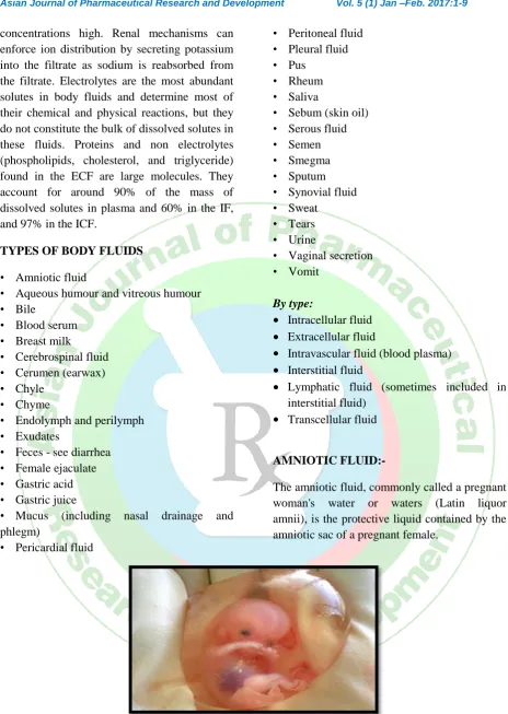
Figure 3 : Week old human fetus surrounded by amniotic fluid within the amniotic sac
Development:-
Amniotic fluid is present from the formation of the gestational sac. Amniotic fluid is present in the amniotic sac. It is generated from maternal
plasma, and passes through the foetal
membranes by osmotic and hydrostatic forces.
Function:-
Amniotic fluid swallowed by the fetus helps in the formation of the gastrointestinal tract. Contrary to popular belief, amniotic fluid has not been conclusively shown to be inhaled and exhaled by the foetus.
AQUEOUS HUMOUR:-
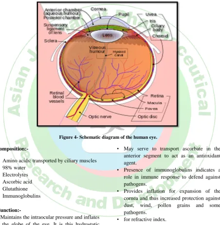
The aqueous humour is a transparent, watery fluid similar to plasma, but containing low protein concentrations. It is secreted from the ciliary epithelium, a structure supporting the lens. It fills both the anterior and the posterior chambers of the eye, and is not to be confused with the vitreous humour, which is located in the space between the lens and the retina, also known as the posterior cavity or vitreous chamber.
Figure 4- Schematic diagram of the human eye.
Composition:-
• Amino acids: transported by ciliary muscles • 98% water
• Electrolytes • Ascorbic acid • Glutathione • Immunoglobulins
Function:-
• Maintains the intraocular pressure and inflates the globe of the eye. It is this hydrostatic pressure
which keeps the eyeball in a roughly spherical shape and keeps the walls of the eyeball taut.
• Provides nutrition (e.g. amino acids and glucose) for the avascular ocular tissues; posterior cornea, trabecular meshwork, lens, and anterior vitreous.
• May serve to transport ascorbate in the anterior segment to act as an antioxidant agent.
• Presence of immunoglobulins indicates a role in immune response to defend against pathogens.
• Provides inflation for expansion of the cornea and thus increased protection against dust, wind, pollen grains and some pathogens.
• for refractive index.
Production and drainage:-
Schlemm's canal by one of two ways: directly, via aqueous vein to the episcleral vein, or indirectly, through collector channels to the episcleral vein by intrascleral plexus and eventually into the veins of the orbit. 5 alpha-dihydrocortisol, an enzyme inhibited by 5-alpha reductase inhibitors, may be involved in production of aqueous humor.
Drainage:-
Aqueous humor is continually produced by the ciliary processes and this rate of production must be balanced by an equal rate of aqueous humor drainage. Small variations in the production or outflow of aqueous humour will have a large influence on the intraocular pressure.
BREAST MILK :-
Breast milk is the milk produced by the breasts (or mammary glands) of a human female for her baby. Milk is the primary source of nutrition for newborns before they are able to eat and digest other foods; older infants and toddlers may continue to be breastfed, either exclusively or in combination with other foods from around six months of age when solid foods may be introduced.
Production:-
Under the influence of the hormones prolactin and oxytocin, women produce milk after childbirth to feed the baby. The initial milk produced is referred to as colostrum, which is high in the immunoglobulin IgA, which coats the gastrointestinal tract. This helps to protect the newborn until its own immune system is functioning properly. It also creates a mild laxative effect, expelling meconium and helping to prevent the build-up of bilirubin (a contributory factor in jaundice). Actual inability to produce enough milk is rare, with studies showing that mothers from developing countries experiencing nutritional hardship still produce amounts of milk of similar quality to that of mothers in developed countries. There are many reasons a mother may not produce enough breast milk. Some of the most common reasons are an improper latch (i.e., the baby does not connect efficiently with the nipple), not nursing or pumping enough to meet supply, certain
medications (including estrogen-containing
hormonal contraceptives), illness, and
dehydration. A rarer reason is Sheehan's
syndrome, also known as postpartum
hypopituitarism, which is associated with prolactin deficiency and may require hormone replacement. The amount of milk produced depends on how often the mother is nursing and/or pumping: the more the mother nurses her baby or pumps, the more milk is produced. It is beneficial to nurse when the baby wants to nurse rather than on a schedule. A Cochrane review came to the conclusion that a greater volume of milk is expressed whilst listening to relaxing audio during breastfeeding, along with warming and massaging of the breast prior to and during feeding. A greater volume of milk expressed can also be attributed to instances where the mother starts pumping milk sooner, even if the infant is unable to breastfeed.
Sodium concentration is higher in hand-expressed milk, when compared with the use of manual and electric pumps, and fat content is higher when the breast has been massaged, in conjunction with listening to relaxing audio. This may be important for low birthweight infants. If pumping, it is helpful to have an electric, high-grade pump so that all of the milk ducts are stimulated. Galactagogues increase milk supply, although even herbal variants carry risks; therefore non-pharmaceutical methods should be tried first.
CEREBROSPINAL FLUID:-
Cerebrospinal fluid (CSF) is a clear, colorless body fluid found in the brain and spine. It is produced in the choroid plexuses of the ventricles of the brain.
Production:-
into the CSF space. Chloride, with a negative charge, moves with the positively charged sodium and a neutral charge is maintained. As a result, CSF contains a higher concentration of sodium and chloride than blood plasma, but less potassium, calcium and glucose and protein. The Orešković and Klarica hypothesis suggests that the CSF is not primarily produced by the choroid plexus, but is being permanently produced inside the entire CSF system, as a consequence of water filtration through the capillary walls into the interstitial fluid (ISF) of surrounding brain tissue, regulated by AQP-4.
Function:-
CSF serves several purposes
Buoyancy: The actual mass of the human brain is about 1400 grams; however, the net weight of the brain suspended in the CSF is equivalent to a mass of 25 grams.
Protection: CSF protects the brain tissue from injury when jolted or hit.
Chemical stability: CSF flows throughout the inner ventricular system in the brain and is absorbed back into the bloodstream, rinsing the metabolic waste from the central nervous system through the blood–brain barrier Prevention of brain ischemia: The prevention of brain ischemia is made by decreasing the amount of CSF in the limited space inside the skull.
Clearing waste: CSF has been shown by the research group of Maiken Nedergaard to be critical in the brain's glymphatic system, which plays an important role in flushing metabolic toxins or waste from the brain's tissues' cellular interstitial fluid (ISF).
CERUMEN (EARWAX):-
Earwax, also known as cerumen, is a
yellowish waxy substance secreted in the ear canal of humans and other mammals. It protects the skin of the human ear canal, assists in cleaning and lubrication, and also provides
some protection
from bacteria, fungi, insects and water.
Uses:-
In medieval times, earwax and other substances such as urine were used to
prepare pigments used by scribes to
illustrate illuminated manuscripts.
The first lip balm may have been based on earwax.
Before waxed thread was commonly available seamstresses would use their own earwax to stop the cut ends of threads from fraying.
ENDOLYMPH & PERILYMPH:-
Endolymph:-
Endolymph is the fluid contained in the membranous labyrinth of the inner ear. It is also called Scarpa's fluid, after Antonio Scarpa.
Function:-
Fluid waves occur in the endolymph in the various parts of the membranous labyrinth in response to fluid waves in the perilymph. Hearing: Cochlear duct: fluid waves in the endolymph of the cochlear duct stimulate the receptor cells, which in turn translate their movement into nerve impulses that the brain perceives as sound.
Balance: Semicircular canals: angular
acceleration of the endolymph in the semicircular canals stimulate the vestibular
receptors of the endolymph. The
semicircular canals of both inner ears act in concert to coordinate balance.
Clinical significance:-
Disruption of the endolymph due to jerky movements (like spinning around or driving over bumps while riding in a car) can cause motion sickness. A condition where the volume of the endolymph is greatly enlarged is called endolymphatic hydrops and has been linked to Ménière's disease.
Perilymph:-
Perilymph (also known as Cotunnius' liquid, and liquor cotunnii) is an extracellular fluid located within the cochlea (part of the inner ear) in two of its three compartments: the scala tympani and scala vestibuli.
Clinical significance:-
EXUDATES:-
An exudate is a fluid emitted by an organism through pores or a wound, a process known as exuding. Exudate is derived from exude, "to ooze, from the Latinexsūdāre, "to (ooze) out like sweat" (ex- "out" and sūdāre "to sweat").
Types:-
Purulent or suppurative exudate consists of
plasma with both active and dead
neutrophils, fibrinogen, and necrotic
parenchymal cells. This kind of exudate is consistent with more severe infections, and is commonly referred to as pus.
Fibrinous exudate is composed mainly of fibrinogen and fibrin. It is characteristic of rheumatic carditis, but is seen in all severe injuries such as strep throat and bacterial pneumonia.
Catarrhal exudate is seen in the nose and throat and is characterized by a high content of mucus.
Serous exudate (sometimes classified as serous transudate) is usually seen in mild inflammation, with relatively low protein. Its consistency resembles that of serum, and can usually be seen in certain disease states like tuberculosis. (See below for difference between transudate and exudate)
Malignant (or cancerous) pleural effusion is effusion where cancer cells are present . It is usually classified as exudate.
FEMALE EJACULATE:-
Female ejaculation is the expulsion of fluid by
the paraurethral ducts through and
around the human female urethra during or befo re an orgasm. It is also known colloquially as sq
uirting or gushing, although these are
considered to be different phenomena in some research publications. The exact source and nature of the fluid continue to be a topic of debate among medical professionals, which is also related to doubts over the existence of the G-spot.
Function:-
The physiological function of the purported liquid is unknown. A 2009 paper in Medical Hypotheses suggests that it may have an anti-microbial function, protecting from urinary tract infections.
GASTRIC ACID :-
Gastric acid, gastric juice or stomach acid, is a digestive fluid, formed in the stomach and is composed of hydrochloric acid (HCl) .05–
0.1 M (roughly 5,000–10,000 parts per
million or 0.5-1% potassium chloride (KCl) and sodium chloride (NaCl).
Figure 5- Diagram depicting the major determinants of gastric acid secretion, with inclusion of drug targets for peptic ulcer disease (PUD) and gastroesophageal reflux disease (GERD).
Role in disease:-
potentially leading to problems as the disinfectant properties of the gastric lumen are decreased.
PLEURAL FLUID :-
A pleural effusion is excess fluid that
accumulates in the pleural cavity, the fluid-filled space that surrounds the lungs. This excess can impair breathing by limiting the expansion of the lungs.
Types of pleural fluid :-
Various methods can be used to classify pleural fluid.
By the origin of the fluid: Serous fluid (hydrothorax) Blood (haemothorax) Chyle (chylothorax)
Pus (pyothorax or empyema) Urine (urinothorax)
By pathophysiology:
Transudative pleural effusion
Exudativepleural
Pathophysiology:-
Pleural fluid is secreted by the parietal layer of the pleura and reabsorbed by the lymphatics in the most dependent parts of the parietal pleura, primarily the diaphragmatic and mediastinal regions. Exudative pleural effuisions occur when the pleura is damaged, e.g., by trauma, infection or malignancy, and transudative pleural effusions develop when there is either excessive production of pleural fluid or the resorption capacity is exceeded.
Treatment:-
Treatment depends on the underlying cause of the pleural effusion. Therapeutic aspiration may be sufficient; larger effusions may require insertion of an intercostal drain (either pigtail or surgical).
REFERENCES
1. Lymphatic Congestion - Symptoms, Diagnosis, Treatment and Information". Diagnose- me.com. Retrieved 2012-11-14.
2. Packaging Guidelines for Clinical Samples - Retrieved 7 August 2014.
3. .specimen - www.thefreedictionary.com. Retrieved 7 August 2014
4. "Semen & Blood II". Artnet.com. Retrieved 2010-11-13.
5. A B Mann, T (1954). "The Biochemistry of Semen". London: Methuen & Co; New York: John Wiley & Sons. Retrieved November 9, 2013.
6. Guyton, Arthur C. (1991). Textbook of Medical Physiology (8th ed.). Philadelphia: W.B. Saunders. pp. 890–891. ISBN 0-7216-3994-1.
7. Harvey, Clare (1948). "Relation between the Volume
and Fructose Content of Human Semen". Nature. 162 (4125): 812.
doi:10.1038/162812a0. PMID 18121921.
8. Canale, D.; Bartelloni, M.; Negroni, A.; Meschini, P.; Izzo, P. L.; Bianchi, B.; Menchini-Fabris, G. F. (1986). "Zinc in human semen". International Journal of Andrology. 9 (6): 477–80. doi:10.1111/j.1365-2605.1986.tb00909.x. PMID 3570537.
9. World Health Organization (2003). Laboratory Manual for the Examination of Human Semen and Semen–Cervical Mucus Interaction, 4th edition. Cambridge, UK: Cambridge University Press. p. 60. ISBN 0-521-64599-9. Retrieved November 9, 2013.