R E S E A R C H
Open Access
Function of ankle ligaments for subtalar
and talocrural joint stability during an
inversion movement
–
an in vitro study
Lu Li
1*, Albert Gollhofer
1, Heinz Lohrer
1,2,3, Nadja Dorn-Lange
4, Guiseppe Bonsignore
4and Dominic Gehring
1Abstract
Background:The lateral ankle ligament complex consisting of the anterior talofibular ligament (ATFL), the calcaneofibular ligament (CFL) and the posterior talofibular ligament (PTFL) is known to provide stability against ankle joint inversion. As injuries of the ankle joint have been reported at a wide range of plantarflexion/dorsiflexion angles, the aim of the present study was to evaluate the stabilizing function of these ligaments depending on the sagittal plane positioning of the ankle joint.
Methods:Eight fresh-frozen specimens were tested on a custom-built ankle deflection tester allowing the application of inversion torques in various plantarflexion/dorsiflexion positions. A motion capture system recorded kinematic data from the talus, calcaneus and fibula with bone-pin markers during inversion movements at 10° of dorsiflexion, at neutral position and at plantarflexion 10°. ATFL, CFL and PTFL were separately but sequentially sectioned in order to assess the contribution of the individual ligament with regard to ankle joint stability.
Results:Joint- and position-specific modulations could be observed when the ligaments were cut. Cutting the ATFL did not lead to any observable alterations in ankle inversion angle at a given torque. But subsequently cutting the CFL increased the inversion angle of the talocrural joint in the 10° plantarflexed position, and significantly increased the inversion angle of the subtalar joint in the 10° dorsiflexed position. Sectioning of the PTFL led to minor increases of inversion angles in both joints.
Conclusions:The CFL is the primary ligamentous stabilizer of the ankle joint against a forced inversion. Its functioning depends greatly on the plantar−/dorsiflexion position of the ankle joint complex, as it provides the stability of the talocrural joint primarily during plantarflexion and the stability of the subtalar joint primarily during dorsiflexion.
Keywords:Lateral ankle ligament complex, Ankle stability, Ankle inversion restriction, Biomechanics
Introduction
Epidemiological data indicate that ankle injuries repre-sent the most frequent type of sport-related injuries [1]. There exists overwhelming evidence that most of the ankle injuries affect the ligaments [2] and that approxi-mately 85% of those injuries are lateral ankle sprains [3]. The aetiology for injuries of the lateral ankle joint typic-ally comprises excessive inversion, often combined with a pronounced plantarflexion and internal rotation of the ankle joint complex [4]. It has been shown that an initial
sprain can impair the function of the ankle joint com-plex in the long term, which is referred to as chronic ankle instability [5].
From an anatomical perspective, the lateral ankle ment complex consists of the anterior talofibular liga-ment (ATFL), the calcaneofibular ligaliga-ment (CFL) and the posterior talofibular ligament (PTFL). Epidemiologic surveys indicate that the ATFL is the ligament that is in-jured in 85% of all ankle sprain ligament injuries, while the CFL is involved in 35% and the PTFL in 12% [6]. In trying to identify why the individual ligaments are affected during an ankle sprain with different rates, their functional roles and their mechanical properties must be taken into consideration. Most of the current information about the
© The Author(s). 2019Open AccessThis article is distributed under the terms of the Creative Commons Attribution 4.0 International License (http://creativecommons.org/licenses/by/4.0/), which permits unrestricted use, distribution, and reproduction in any medium, provided you give appropriate credit to the original author(s) and the source, provide a link to the Creative Commons license, and indicate if changes were made. The Creative Commons Public Domain Dedication waiver (http://creativecommons.org/publicdomain/zero/1.0/) applies to the data made available in this article, unless otherwise stated.
* Correspondence:[email protected]
1Department of Sport and Sport Science, University of Freiburg, Schwarzwaldstraße 175, 79117 Freiburg, Germany
to restrict both plantarflexion and inversion movements [14] and because the maximal tension until failure is quite low [8]. Moreover it has been shown that a certain inversion torque, applied at the fixed foot, increases the posterior tibial displacement by a factor of 2 when the ATFL is dissected [15]. In addition to the ATFL, the CFL seems to resist ankle inversion, as cutting this liga-ment led to an increased range of inversion [9]. Applying an inversion loading following a dissection of both ATFL and CFL respectively increased the range of motion and decreased the end-range stiffness when compared with the intact and solely ATFL-sectioned ankles [16]. Fur-thermore, evidence exists showing that a dissection of the CFL causes significant range of motion changes re-garding all three planes in the subtalar joint [10]. Thus, it seems that the CFL plays a key role in the lateral stabilization of both the ankle and the subtalar joint. The functional properties of the PTFL were analysed by Ozeki and Kitaoka, who concluded that the PTFL is an important stabilizer especially when the ankle was in a dorsiflexed postion [17]. Previous research has also shown that the strain of the PTFL increases in dorsiflex-ion (DF) and plantarflexdorsiflex-ion (PF), but is only minimally affected by ankle inversion [18]. In summary, the PTFL seems to play a supplementary role in lateral ankle sta-bility, especially when the ATFL and CFL are intact [19]. Although it is possible to draw important conclusions about the ligament function from the above mentioned in-vitro studies, specific methodological restrictions have to be considered. First, some of the papers selectively fo-cused on only one specific ligament of the ankle and its defined functional role [9,14,15,18,19] while others in-vestigated multiple ligaments but considered only
cor-onal movements [7, 16]. Secondly, most of the studies
tested the lateral ankle ligament complex with a distinct torque but only either in sagittal or in coronal plane [7, 10,15,18,20,21]. Yet, investigations of the injury mech-anism have revealed that ankle sprains can occur at a
wide range of plantarflexion/dorsiflexion angles [4, 22].
It therefore appears important to evaluate the functional properties of the involved ligaments under different plantarflexion/dorsiflexion positions. Third, most of the
This may be problematic because it is known that liga-ment stiffness is related to the speed of its elongation [23] and that typically the ligaments are loaded at high angular velocities during ankle sprains [13].
Therefore, the major purpose of the present study was to evaluate the biomechanics of the talocrural and subta-lar joints when the ligaments of the lateral ankle joint complex are sequentially resected. Specifically, we aimed to assess how the sagittal positions would influence the relative contributions of the individual ligaments to joint stability during dynamic inversion.
Methods Specimens
Fourteen fresh-frozen human anatomic lower limb spec-imens, amputated above the knee joints, were selected for the experiments. Already during the experiments, it became evident that fixation of bone-screws was loos-ened during dynamic movements in six specimens. Fi-nally, eight cadavers could be included in the study (age: 84.0 ± 5.6 years; donors were two men and two women). Before preparation and measurement, the specimens were kept at room temperature for 24 h in order to en-sure conditional physical properties. An experienced anatomist inspected the ankles by manual examination for normal function and confirmed that there was no sign of pathological limitations on any of the cadaver legs. Thereafter, all ankles were exposed layer by layer creating a skin and soft tissue window (5 × 5 cm) distal to the lateral malleolus and ankle. The integrity of the anterior and posterior talofibular ligaments (ATFL and PTFL) and the calcaneofibular ligament (CFL) was veri-fied by visual inspection. During the entire measure-ments the specimen were regularly irrigated with saline, preventing them from drying-out. The procedures were in accordance with the Declaration of Helsinki and ap-proved by the local ethics committee (#10006/18).
Experimental equipment setup
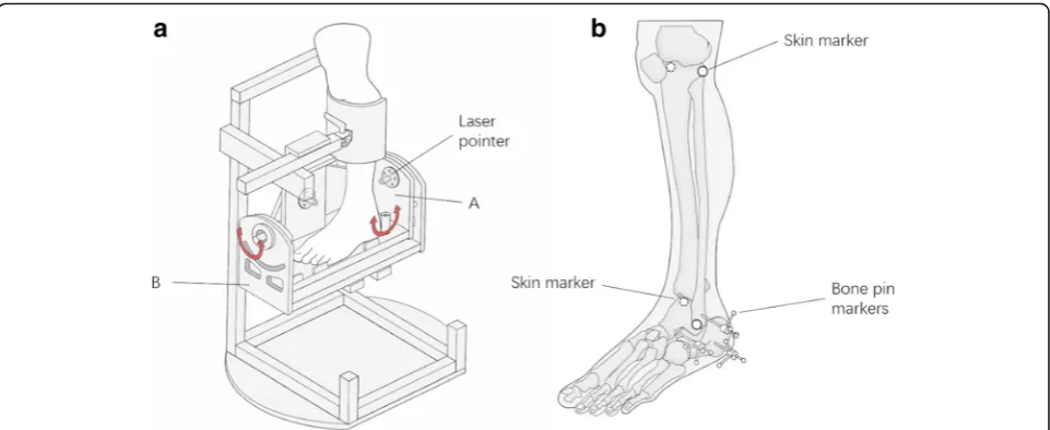
A custom-built ankle deflection tester (ADT) allowed application of inversion torques in various
ADT consisted of a leg fixation and a foot plate that allowed for independent rotations around the ankle joint in the sagittal (up to ±30°) and frontal planes (up to
±40°). Fixation of the specimen’s foot on the foot plate
was secured by applying two non-elastic belts covering
the mid-foot and the forefoot (Fig.1a). Additionally, the
leg was fixed to a height-adjustable clamp with straps to ensure stabilization of this segment during inversion
movements. To adapt the ADT to the specimen’s
indi-vidual morphological characteristic, the foot plate was adjusted in height and anterior-posterior position set-ting, allowing the mechanical rotation axis of the ADT
to coincide with the specimen’s talocrural joint. Two
goniometer sensors (MODEL 157, Vishay, USA) re-corded the angular information of each component along the two mechanical axes of the ADT. A torque
meter (TA 125–350, ME-Meßsysteme GmbH, Germany)
with a handle was connected with the anterior-posterior axis of the ADT to manually apply and measure the in-version torque.
To record the skeletal motion of the calcaneus, talus and fibula, three Kirschner wires (1.5 mm) were drilled into the lateral aspect of each bone segment, each of them being equipped at its ends with 4 reflective markers (6 mm), building a three-dimensional Cartesian
coordinate system (see Fig. 1b). By means of a motion
capture system with 11 cameras, the data of each seg-ment in three-dimensional space were tracked at 200 Hz (Vicon Motion System, UK). Additionally, skin markers were placed at the medial and lateral epicondyle of the knee as well as at the medial and lateral malleolus in
order to define the anatomical segments in accordance with the recommendation of the ISB (International Society of Biomechanics) [24]. Synchronized with the motion data, the goniometer signals of the ADT were captured with Vicon Nexus 2.5 (Vicon Motion System, UK) at 1000 Hz.
Experimental procedure
To assess the contribution of each individual liga-ment on ankle stability an experiliga-mental setup was selected in which ligaments were successively cut and the remaining resistance of the ankle joint against external force application was measured. We started with the intact condition. Then, we per-formed a fixed order of sequentially cutting the liga-ments: we first cut the ATFL, then the CFL and finally the PTFL as it is known that the ATFL is typically the first or even only ligament being injured [12], followed by the CFL, and only in rare cases by the PTFL [25]. During each session, specimens were tested in three different positions in the sagittal plane: neutral position, 10° dorsiflexion and 10° plan-tarflexion. Manually, torque was applied at the ADT
in the coronal plane resulting in a sustained
inversion-eversion movement. Each simulation trial required 10 inversion-eversion cycles at approxi-mately 1 Hz, in which an inversion angle of 30° had to be reached. The signal of the torque meter and goniometer sensors from the ADT as well as kinematical data of the markers were recorded synchronously.
and talus (representing the subtalar joint), a Joint Coord-inate System approach was used allowing the calculation of the intersegmental movement in 6 degrees of free-dom. The inversion/eversion rotation occurs about a floating axis, while the rotation axes for plantar-/dorsi-flexion and internal/external rotation occur around body-fixed axes of the proximal or distal segment, re-spectively. As the ISB recommendation for the ankle joint complex does not differentiate between the talo-crural and the subtalar joint, the anatomical coordinate system of the talus was set to correspond to the coordin-ate system of the calcaneus. Before performing the cal-culations, all marker trajectories were filtered through a low-pass 2nd order Butterworth filter at 10 Hz.
Aiming to assess the joint stiffness properties in the different measurement conditions, the amount of inver-sion rotation at a given torque value was extracted. As pre-analysis of the data indicated that irrespective of ligament sectioning the joint stiffness was highly specimen-specific, different torques were needed to in-duce comparable inversion rotations of the ankle joints between specimens. In order to account for these
specimen-specific differences, individualized torque
thresholds were defined after pre-analysis using the fol-lowing approach: For each specimen and throughout all measurement conditions the lowest observable torque value to induce 30° of inversion was extracted. In order to stay always in the linear region of the torque-angle re-lationship, 90% of this observed torque was set to assess the associated inversion of the ankle joint complex, the talocrural joint and the subtalar joint for all measure-ment conditions in each specimen. The data extraction was performed with a custom-written script in Matlab (The Mathworks, Natick, MA, USA). For every condi-tion, five continuous inversion-eversion cycles were used for further statistical analysis.
Statistics
The inversion angles of the ankle joint complex, the talocrural joint and the subtalar joint were considered for statistical analyses. The results of different ligament sectioning conditions were compared using a Friedman
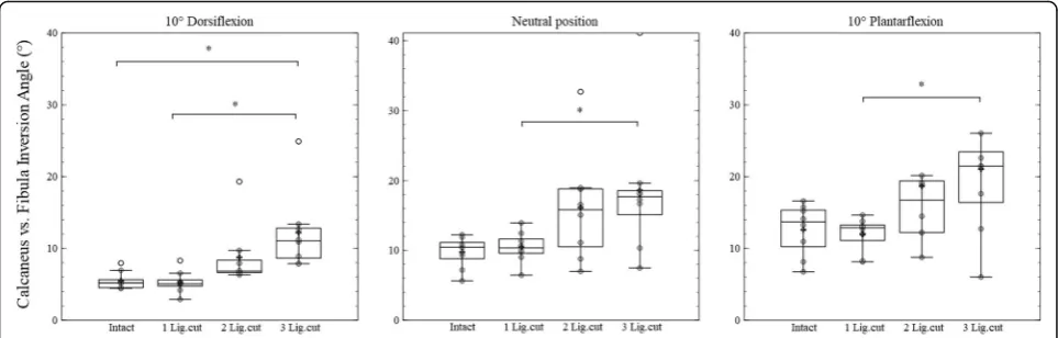
Ankle joint complex
For the inversion angle of the ankle joint complex, main condition effects were observed for all plantarflexion/ dorsiflexion positions (at 10° dorsiflexion: p= 0.001, ef-fect size = 0.631; in neutral position:p= 0.009, effect size
= 0.382; in 10° plantarflexion: p= 0.013, effect size =
0.362). Post-hoc analysis revealed that at the 10° dorsi-flexed angle, cutting all three ligaments significantly in-creased the inversion angle compared to the conditions
when all ligaments were intact (p= 0.015) and when the
ATFL was cut (p= 0.015). In the neutral and in the 10°
plantarflexed position a significantly increased inversion was observed when all ligaments were cut compared to
the condition when only the ATFL was cut (p= 0.015
andp= 0.045; Fig.2).
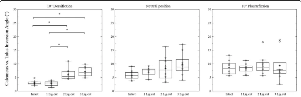
Talocrural joint
Regarding the talocrural joint, main ligament condition effects were detected for all three positions (at 10° dorsi-flexion: p= 0.005, effect size = 0.475; in neutral position:
p= 0.004, effect size = 0.425; at 10° plantarflexion: p= 0.001, effect size = 0.598). Positioned at 10° dorsiflexion, cutting all three ligaments led to a significantly increased inversion angle (p= 0.015). In the neutral position, dis-secting all ligaments was different from the condition
when only the ATFL was cut (p= 0.015). At 10° of
plan-tarflexion, cutting all the ligaments significantly in-creased the inversion angle compared to the condition
when no ligament was sectioned (p= 0.015). Moreover,
dissecting the CFL and the PTFL caused a significant increase of the inversion angle compared to the
con-dition with a section of the AFTL alone (p= 0.026
and p= 0.015, see Fig.3).
Subtalar joint
0.048). Moreover, compared to the ATFL dissection ses-sion, cutting the CFL alone or in combination with the PTFL led to a significant increase of the inversion
angle (both p = 0.015). Furthermore, cutting all three
lateral ankle ligaments leads to a significant increase
of the inversion angle (p = 0.039). Detailed data of
bone-to-bone kinematics can be found in addtional
tables in Additional file 1 and Additional file 2.
Discussion
Using an experimental cadaver approach, our data indi-cate that the lateral ankle ligaments have different bio-mechanical functions in stabilizing bony structures during an inversion movement. The motion task of our study (the mean angular velocity is 64.1°/s ± 11.8°/s) was comparable with the previously reported basic move-ments such as running (maximum ankle inversion
vel-ocity: 85.1°/s), cutting (maximum ankle inversion
velocity: 37.2°/s) and jumping (maximum ankle inversion velocity: 22.5°/s) [26]. However, our motion task design
only reached 25% of an ankle ligamentous sprain simula-tion, reporting ankle inversion velocities from 200°/s to 300°/s [27]. Depending on the sagittal plane position, the stabilizing function of ligaments changed, especially when not the entire ankle joint complex but rather the sub-structures (talocrural and subtalar joint) were con-sidered. The results support the assumption that when the ATFL is ruptured the main function of the CFL is to stabilize the talocrural joint against inversion in a plan-tarlexed position, while its primary function during dorsiflexion is most likely to resist the inversion of the subtalar joint. By imitating a lateral ankle sprain and sim-ultaneously investigating the bone-to-bone stabilization directly, this study extends the current knowledge of the function of the lateral ankle ligament complex, providing the following important details:
Stability of the global ankle joint complex
From a biomechanical perspective, the ankle joint com-plex consists of the talocrural joint and the subtalar joint
Fig. 2Box-plots including arithmetic mean values (indicated by +) of the ankle joint complex inversion in 10° dorsiflexion, neutral and 10° plantarflexion position. The results of an intact session (Intact), ATFL cut session (1 Lig.cut), ATFL and CFL cut session (2 Lig.cut), ATFL, CFL and PTFL cut session (3 Lig.cut) are shown in each figure. The significant differences between sessions are marked by *
and thus can be interpreted as a connection between the tibia/fibula segment and the calcaneus [24]. Regarding the ligamentous structures, the ATFL and CFL should have functional importance in resisting the ankle joint
complex both in sagittal and coronal plane [14, 25].
However, the results of the present study reveal that only in situations when all three ligaments were cut, the in-version stability of the entire ankle joint complex is compromised. Our results therefore at least partly con-firm earlier experimental data showing that following sectioning the ATFL alone, the maximum inversion motion between calcaneus and tibia in dorsiflexion position was not influenced, whereas sectioning both the ATFL and the CFL significantly increased the in-version motion [28].
Nevertheless, it still remains difficult to understand the function of ligaments in resisting external rotational torques applied to the ankle joint complex if considering the results from the ankle joint complex only. Thus, it seems necessary to separately evaluate the ankle joint complex composing sub-joints, namely the talocrural joint and subtalar joint.
Stability of the talocrural joint
According to our results, the CFL had a substantial re-strictive function for the talocrural joint when the ankle is in 10° plantarflexion position, while at 10°dorsiflexion and in the neutral position, there is no clear evidence for a stabilizing function of the CFL. Specifically, in the plantarflexed position, cutting the CFL additionally to the ATFL increased the inversion angle by 3.3° com-pared to the situation where solely the ATFL was sec-tioned. The geometry of the talocrural joint, with its oblique rotation axis and the specific shape of the talus, causes that when the talus is moved in its largest anterior position, the joint was more unstable in
plantarflexion [29]. This explains why the stability of the talocrural joint was sensitive to a destruction of the CFL specifically in a plantarflexion positon. Functionally, the CFL constrains the talus through the calcaneus, stabilizing both the talocrural joint and the subtalar joint. The results showed a phenomenon comparable with findings from pre-vious studies observing significantly increased inversion an-gles after sectioning the CFL significantly during ATFL ruptured conditions [30]. However, based on present data, the stabilizing function of the CFL on the talocrural joint was prominent preferentially in plantarflexion.
In addition, the PTFL could have a potential function in constraining the talocrural joint. When the ATFL and the CFL were already cut, sectioning the PTFL increased the mean inversion angle by 39% (2.1°) in the neutral position and by 75% (2.2°) in dorsiflexion. Even though there was no clear statistical evidence about the contribution of the PTFL, in 10° plantarflexion positions the three ligaments cut condition differed significantly from the other conditions and in 10° dorsiflexion position the same phenomenon was observed. Even in the neutral position, a difference of inversion angle was found between the ATFL cut condition and the three ligaments cut condition. This means that sectioning the complex of lateral ankle liga-ments compromises the stabilization of the talocrural joint. In summary, it is concluded that the PTFL contributed to stabilizing the talocrural joint in all three sagittal plane posi-tions. Yet, when interpreting the data carefully, it has to be considered that a specific sequence of sectioning the liga-ments was used and the PTFL was always the last ligament that was cut. Thus, our results are comparable to an earlier investigation that the PTFL could not restrict the ankle joint in the inversion movement independently from the other ligaments. This indicates that in an otherwise intact ligamentous complex, the PTFL plays only a supplementary role in ankle stability [19].
Stability of the subtalar joint
In the subtalar joint, all statistical effects of the liga-ments were discovered only in the dorsiflexed position and in general no significant effects were observed in neutral and plantarflexion positions. Referring to the dis-cussion above, one reason for these findings could be that the talocrual joint seems to be stiffened by bony constraints in the dorsiflexed positionand therefore the subtalar joint takes over to lead the motion. In addition, the ATFL was observed loose during a dorsiflexed pos-ition in a previous study. This explains why no effects were detected after sectioning the ATFL in dorsiflexion condition [18]. In consequence, the importance of the CFL and the PTFL in contributing to subtalar joint sta-bility is increased in dorsiflexion. Another reason for this observation could be the differences in the orientation of the axes of the subtalar and talocrural joints. Previous studies showed that the inclination angle of the subtalar joint axis was different from that of the ankle joint
com-plex and the talocrural joint [31, 32]. The axes of the
sub-joints both differ from the biomechanical axis of the ankle joint complex and this indicates that during an in-version motion the calcaneus might not only rotate in the coronal plane but also internally rotate at dorsiflex-ion conditdorsiflex-ion.
A previous study reported that specifically a rupture of the CFL would lead to a laxity of the subtalar joint [10]. Furthermore, the CFL was strained and stretched in a dorsiflexion position during an inversion movement [11,18]. Kobayashi also showed that the CFL tension in-creased when moving from plantarflexion to dorsiflexion during inversion [30]. Our results support these previous findings. Compared to the ATFL cut condition, section-ing the CFL led to a significant increase of the inversion angle (2.2°) in the dorsiflexion position. This indicated that the dorsiflexion caused a larger stretch between the tibia and calcaneus. Thus, the CFL seems to have a more stabilizing effect when the ankle joint is in dorsiflexion. It is important to remark, that the present study does not allow drawing a direct conclusion about the function of the CFL solely, as a fixed sectioning order was chosen and the ATFL was already sectioned before. Yet, espe-cially in the dorsiflexed position, no changes in ankle joint stability were observed when the ATFL alone was cut. This provides evidence for the important role of the CFL in lateral stabilization of the ankle joint.
Limitations
Some limitations have to be considered when interpret-ing the results of the present study. First, the number of specimens might influence the systemic significance of our results. Second, due to the manually induced exter-nal torque, minor fluctuations in amplitude and fre-quency were apparent. In order to minimize this
potential error, always the same researcher was re-quested to rotate the foot plate to the maximum pos-ition in every trial. Third, based on the prevalence of ligament rupture, we sectioned the ligaments always in the same sequence and therefore isolated effects of the CFL or the PTFL could not be investigated properly in every positioning condition. A random sectioning se-quence could be applied in a bigger sample size in the future to detect the specific stabilizing functions of these structures in isolated conditions. Fourth, the results might be influenced by the age of the specimens and fu-ture studies with younger samples are desired.
Conclusions
The present study suggested that the CFL is the primary ligamentous stabilizer of the ankle joint against a forced inversion. Its functioning depends greatly on the plan-tar-/dorsiflexion position of the ankle joint complex, as it provides the stability of the talocrural joint primarily during plantarflexion and the stability of the subtalar joint primarily during dorsiflexion.
Additional files
Additional file 1:Supplementary data 1. (XLSX 9 kb) Additional file 2:Supplementary data 2. (XLSX 11 kb)
Abbreviations
ADT:Ankle deflection tester; ATFL: Anterior talofibular ligament; CFL: Calcaneofibular ligament; ISB: International Society of Biomechanics; PTFL: Posterior talofibular ligament
Acknowledgments
The authors would like to acknowledge and thank those anonymous individuals who generously donated their bodies so that this study could be performed. The author Lu Li’s PhD program was supported by the China Scholarship Council (CSC). The Fig.1was provided by Stephan Schur.
Funding
No funding.
Availability of data and materials
The datasets used during the current study are available from the corresponding author on reasonable request.
Authors’contributions
All authors have contributed substantially to the conception, design, analysis and interpretation of the data in this manuscript and will take public responsibility for the content. All authors read and approved the final manuscript.
Ethics approval and consent to participate
The procedures were in accordance with the Declaration of Helsinki and approved by the local ethics committee (#10006/18).
Consent for publication
Not applicable.
Competing interests
1. Waterman BR, Owens BD, Davey S, Zacchilli MA, Belmont PJ. The epidemiology of ankle sprains in the United States. J Bone Jt Surg - Ser A. 2010;92:2279–84.
https://doi.org/10.2106/JBJS.I.01537.
2. Fong DT-P, Hong Y, Chan L-K, Yung PS-H, Chan K-M. A systematic review on ankle injury and ankle sprain in sports. Sport Med. 2007;37:73–94.https:// doi.org/10.2165/00007256-200737010-00006.
3. Ferran NA, Maffulli N. Epidemiology of sprains of the lateral ankle ligament complex. Foot Ankle Clin. 2006;11:659–62.https://doi.org/10.1016/j.fcl.2006. 07.002.
4. Fong DTP, Ha SCW, Mok KM, Chan CWL, Chan KM. Kinematics analysis of ankle inversion ligamentous sprain injuries in sports: five cases from televised tennis competitions. Am J Sports Med. 2012;40:2627–32.
https://doi.org/10.1177/0363546512458259.
5. Attenborough AS, Hiller CE, Smith RM, Stuelcken M, Greene A, Sinclair PJ. Chronic ankle instability in sporting populations. Sport Med. 2014;44:1545–56.
https://doi.org/10.1007/s40279-014-0218-2.
6. Swenson DM, Collins CL, Fields SK, Comstock RD. Epidemiology of US high school sports-related ligamentous ankle injuries, 2005/06-2010/11. Clin J Sport Med. 2013;23:190–6.https://doi.org/10.1097/JSM.0b013e31827d21fe. 7. Kovaleski JE, Hollis JM, Heitman RJ, Gurchiek LR, Pearsall AW IV. Assessment
of ankle-subtalar-joint-complex laxity using an instrumented ankle arthrometer: An experimental cadaveric investigation. J Athl Train. 2002;37:467–74. 8. Bahr R, Pena F, Shine J, Lew WD, Engebretsen L. Ligament force and joint
motion in the intact ankle: a cadaveric study. Knee Surgery, Sport Traumatol Arthrosc. 1998;6:115–21.https://doi.org/10.1007/s001670050083.
9. Hollis JM, Blasier RD, Flahiff CM. Simulated lateral ankle ligamentous injury. Change in ankle stability. Am J Sports Med. 1995;23:672–7.https://doi.org/ 10.1177/036354659502300606.
10. Weindel S, Schmidt R, Rammelt S, Claes L, Campe AV, Rein S. Subtalar instability: a biomechanical cadaver study. Arch Orthop Trauma Surg. 2010; 130:313–9.https://doi.org/10.1007/s00402-008-0743-2.
11. Edama M, Kageyama I, Kikumoto T, Nakamura M, Ito W, Nakamura E, et al. The effects on calcaneofibular ligament function of differences in the angle of the calcaneofibular ligament with respect to the long axis of the fibula: a simulation study. J Foot Ankle Res. 2017;10:60.https://doi.org/10.1186/ s13047-017-0242-1.
12. Kumai T, Takakura Y, Rufai A, Milz S, Benjamin M. The functional anatomy of the human anterior talofibular ligament in relation to ankle sprains. J Anat. 2002;200:457–65.https://doi.org/10.1046/j.1469-7580.2002.00050.x. 13. Fong DT, Chan Y-Y, Mok K-M, Yung PS, Chan K-M. Understanding acute
ankle ligamentous sprain injury in sports. BMC Sports Sci Med Rehabil. 2009;1:14.https://doi.org/10.1186/1758-2555-1-14.
14. Khawaji B, Soames R. The anterior talofibular ligament: a detailed morphological study. Foot. 2015;25:141–7.https://doi.org/10.1016/j.foot.2015.05.004.
15. Markolf KL, Jackson S, McAllister DR. Force and displacement measurements of the distal fibula during simulated ankle loading tests for high ankle sprains. Foot Ankle Int. 2012;33:779–86.https://doi.org/10.3113/FAI.2012. 0779.
16. Kovaleski JE, Heitman RJ, Gurchiek LR, Hollis JM, Liu W, Pearsall AW IV. Joint stability characteristics of the ankle complex after lateral ligamentous injury, part I: a laboratory comparison using arthrometric measurement. J Athl Train. 2014;49:192–7.https://doi.org/10.4085/1062-6050-49.2.07.
17. Ozeki S, Kitaoka H, Uchiyama E, Luo Z-P, Kaufman K, An K-N. Ankle ligament tensile forces at the end points of passive circumferential rotating motion of the ankle and subtalar joint complex. Foot Ankle Int. 2006;27:965–9.
https://doi.org/10.1177/107110070602701117.
understanding when , how and why ligament failure occurs. J Sci Med Sport. 2017;20:1057–61.https://doi.org/10.1016/j.jsams.2017.05.006. 23. Choisne J, Ringleb SI, Samaan MA, Bawab SY, Naik D, Anderson CD.
Influence of kinematic analysis methods on detecting ankle and subtalar joint instability. J Biomech. 2012;45:46–52.https://doi.org/10. 1016/j.jbiomech.2011.10.015.
24. Wu G, Siegler S, Allard P, Kirtley C, Leardini A, Rosenbaum D, et al. ISB recommendation on definitions of joint coordinate system of various joints for the reporting of human joint motion—part I: ankle, hip, and spine. J Biomech. 2002;35:543–8.https://doi.org/10.1016/S0021-9290(01)00222-6. 25. Van Den Bekerom MPJ, Oostra RJ, Alvarez PG, Van Dijk CN. The anatomy in
relation to injury of the lateral collateral ligaments of the ankle: a current concepts review. Clin Anat. 2008;21:619–26.https://doi.org/10.1002/ca.20703. 26. Chu VWS, Fong DTP, Chan YY, Yung PSH, Fung KY, Chan KM. Differentiation
of ankle sprain motion and common sporting motion by ankle inversion velocity. J Biomech. 2010;43:2035–8.https://doi.org/10.1016/j.jbiomech.2010. 03.029.
27. Gehring D, Wissler S, Lohrer H, Nauck T, Gollhofer A. Expecting ankle tilts and wearing an ankle brace influence joint control in an imitated ankle sprain mechanism during walking. Gait Posture. 2014;39:894–8.https://doi. org/10.1016/j.gaitpost.2013.11.016.
28. Ringleb SI, Dhakal A, Anderson CD, Bawab S, Paranjape R. Effects of lateral ligament sectioning on the stability of the ankle and subtalar joint. J Orthop Res. 2011;29:1459–64.https://doi.org/10.1002/jor.21407.
29. Brockett CL, Chapman GJ. Biomechanics of the ankle. Orthop Trauma. 2016; 30:232–8.https://doi.org/10.1016/j.mporth.2016.04.015.
30. Kobayashi T, Yamakawa S, Watanabe K, Kimura K, Suzuki D, Otsubo H, et al. The in situ force in the calcaneofibular ligament and the contribution of this ligament to ankle joint stability. Clin Biomech. 2016;40:8–13.https://doi. org/10.1016/j.clinbiomech.2016.10.009.
31. Lewis GS, Kirby KA, Piazza SJ. Determination of subtalar joint axis location by restriction of talocrural joint motion. Gait Posture. 2007;25:63–9.https://doi. org/10.1016/j.gaitpost.2006.01.001.