Mice expressing low levels of CalDAG-GEFI exhibit markedly
impaired platelet activation with minor impact on hemostasis
Raymond Piatt1, David S. Paul1, Robert H. Lee1, Steven E. McKenzie2, Leslie V. Parise3,
Dale O. Cowley4, Brian C. Cooley5, and Wolfgang Bergmeier1,3
1McAllister Heart Institute, University of North Carolina, Chapel Hill, NC
2Cardeza Foundation for Hematological Research, Department of Medicine, Thomas Jefferson
University, Philadelphia, PA
3Department of Biochemistry and Biophysics, University of North Carolina at Chapel Hill
4Animal Models Core, University of North Carolina at Chapel Hill
5Rodent Advanced Surgical Core, University of North Carolina at Chapel Hill
Abstract
Objective—The tight regulation of platelet adhesiveness, mediated by the αIIbβ3 integrin, is critical for hemostasis and prevention of thrombosis. We recently demonstrated that integrin affinity in platelets is controlled by the guanine nucleotide exchange factor, CalDAG-GEFI (GEFI), and its target, RAP1. In this study, we investigated whether low-level expression of CD-GEFI leads to protection from thrombosis without pathological bleeding in mice.
Approach and Results—Cdg1low mice were generated by knock-in of human CD-GEFI cDNA into the mouse Cdg1 locus. CD-GEFI expression in platelets from Cdg1low mice was reduced by ~90% when compared to controls. Activation of RAP1 and αIIbβ3 was abolished at low agonist concentrations and partially inhibited at high agonist concentrations in Cdg1low platelets. Consistently, the aggregation response of Cdg1low platelets was weaker than that of wild-type (WT) platelets, but more efficient than that observed in Cdg1−/− platelets. Importantly, Cdg1low mice were strongly protected from arterial and immune complex-mediated thrombosis, with only minimal impact on primary hemostasis.
Corresponding author: Wolfgang Bergmeier, Department of Biochemistry and Biophysics, 120 Mason Farm Rd., 3113 GMB bldg., University of North Carolina at Chapel Hill, Chapel Hill, NC 27599-7260, [email protected], phone: 919.962.7331, fax: 919.966.2852.
DISCLOSURES
D.O.C. is employed by, has equity ownership in and serves on the board of directors of TransViragen, the company which has been contracted by UNC-Chapel Hill to manage its Animal Models Core Facility. The other authors declare no competing financial interests.
AUTHORSHIP
Contribution: R.P., and D.S.P, designed and performed experiments, analyzed and interpreted data, and wrote the paper; R.H.L. and
HHS Public Access
Author manuscript
Arterioscler Thromb Vasc Biol
. Author manuscript; available in PMC 2017 September 01.Published in final edited form as:
Arterioscler Thromb Vasc Biol. 2016 September ; 36(9): 1838–1846. doi:10.1161/ATVBAHA. 116.307874.
A
uthor Man
uscr
ipt
A
uthor Man
uscr
ipt
A
uthor Man
uscr
ipt
A
uthor Man
uscr
Conclusion—Together, our studies suggest the partial inhibition of CD-GEFI function as a powerful new approach to safely prevent thrombotic complications.
GRAPHICAL ABSTRACT
Keywords
Platelet; Hemostasis; Thrombosis; Mouse
INTRODUCTION
Platelet aggregate formation, mediated by activated integrin αIIbβ3, is critical for hemostasis upon tissue injury. Excessive platelet activation and aggregation, however, can lead to thrombotic complications, such as heart attack and stroke1, 2. The signaling events required for platelet integrin activation are initiated by engagement of surface-expressed receptors for agonists such as thrombin and/or collagen3. These agonists trigger a first wave of platelet signaling that includes a rapid increase in the cytosolic calcium concentration ([Ca2+]i). This increase in [Ca2+]i leads to activation of the calcium-binding guanine nucleotide exchange factor, CalDAG-GEFI (CD-GEFI; RASGRP2), a key regulator of the small GTPase RAP14. RAP1 controls various platelet responses important for hemostatic plug formation, including the inside-out activation of integrin receptors5, 6. In the absence of additional signals, CD-GEFI-mediated RAP1 signaling is terminated by the GTPase-activating protein (GAP), RASA37. The signal for RASA3 inactivation and sustained RAP1 signaling is provided by stimulation via P2Y12, the receptor for the second-wave mediator ADP and the target for various clinically used anti-platelet drugs8, 9.
Work by us and others provided important mechanistic information on how interference with RAP1 signaling affects platelet adhesion at sites of vascular injury. Mice deficient in the main RAP1 isoform, RAP1B, exhibit marked defects in platelet aggregation to various agonists in vitro and impaired hemostasis and thrombosis in vivo10, 11. As outlined above,
the activity state of RAP1 in platelets is controlled by CD-GEFI and RASA34, 7. Active Rasa3 is required to keep circulating platelets in a quiescent state7. During hemostatic plug formation, its activity must be down-modulated as part of the P2Y12/PI3 kinase signaling pathway in order for stable platelet adhesion to occur. Consistently, mice lacking RASA3 are characterized by severe thrombocytopenia due to platelet pre-activation and clearance7, while both hemostatic plugs and pathological thrombi in mice lacking P2Y12 function are
A
uthor Man
uscr
ipt
A
uthor Man
uscr
ipt
A
uthor Man
uscr
ipt
A
uthor Man
uscr
highly unstable9, 12. In contrast, RAP1 activation in platelets from mice lacking CD-GEFI occurs with a delay and requires higher doses of strong agonists, such as thrombin and collagen. Under flow conditions in vitro, platelets lacking CD-GEFI (Cdg1−/−) are markedly impaired in their ability to form three-dimensional thrombi, especially under conditions of high shear stress12. Consistent with the in vitro phenotype, Cdg1−/− mice are strongly protected from both immune-mediated thrombocytopenia and thrombosis (ITT) and arterial thrombosis, but they also show a marked defect in hemostatic plug formation12, 13.
Importantly, the main findings in Cdg1−/− mice were recently confirmed in three patients with a loss-of-function mutation in CD-GEFI. In their studies, Canault et al., found that platelets from heterozygous patients, who did not show defects in hemostasis, exhibited a significant adhesive defect under flow conditions14. Thus, the various studies in knockout mice and patients suggest partial inhibition of CD-GEFI as a powerful yet safe strategy to prevent thrombosis. In the present study, we describe a hypomorphic mouse strain
expressing low levels of human CD-GEFI (Cdg1low) instead of the endogenous mouse CD-GEFI. Platelets from Cdg1low mice showed decreased platelet activation when compared to WT controls. Importantly, however, their integrin activation response was significantly stronger than that of Cdg1−/− mice. Consistent with the in vitro integrin activation phenotype, Cdg1low mice exhibited only a mild defect in primary hemostasis while they were strongly protected from experimental thrombosis.
MATERIALS AND METHODS
Materials and Methods are available in the online-only Data Supplement
RESULTS
Generation of hypomorphic Cdg1 mutant mice
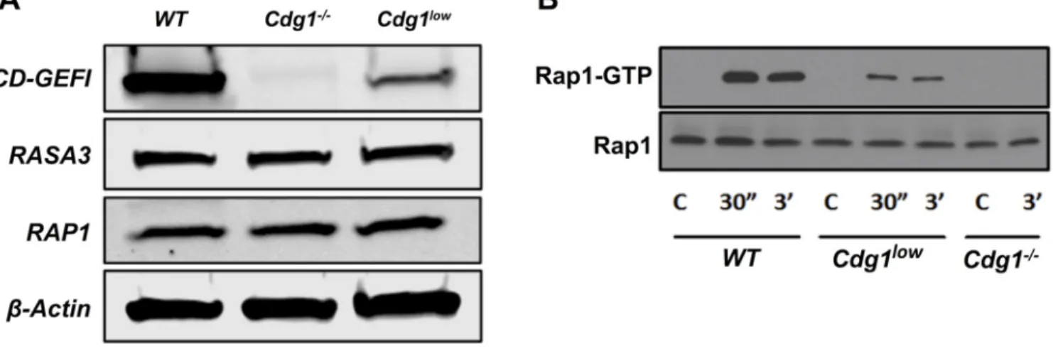
The platelet and hemostasis phenotype of Cdg1−/− mice has been characterized extensively. To investigate the regulation of CD-GEFI function in platelets by genetic means, we aimed to establish a cDNA knock-in model where WT or mutant human Cdg1 cDNA is knocked into the murine Cdg1 locus (Figure 1). We selected to express human CD-GEFI variants in mice, as (1) there is 96% and 100% amino acid sequence identity between human and murine CD-GEFI and RAP1B, respectively, and (2) this approach would allow us to evaluate the ability of human CD-GEFI variants to support platelet activation and plug formation in mice. We noticed, however, that the cDNA knock-in of human Cdg1 led to markedly reduced expression of CD-GEFI protein in platelets isolated from these mice (~90% reduction compared to controls) (Figure 2A), while the expression of RASA3, RAP1, or β -ACTIN was not affected. Just like Cdg1−/− mice4, these Cdg1low mice exhibited a small but significant increase in circulating neutrophils, but they did not exhibit changes in the peripheral platelet count or the platelet size (Table 1). However, compared to WT controls, platelets from Cdg1low mice showed marked defects in RAP1 activation in response to agonist stimulation (Figure 2B). Thus, cDNA knock-in of human Cdg1 strongly reduced platelet CD-GEFI expression and thus led to the generation of hypomorphic Cdg1 mutant mice.
A
uthor Man
uscr
ipt
A
uthor Man
uscr
ipt
A
uthor Man
uscr
ipt
A
uthor Man
uscr
Impaired integrin activation and aggregation of Cdg1low platelets
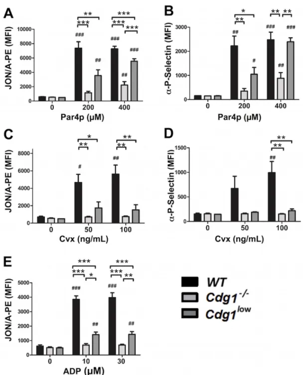
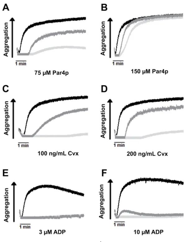
Consistent with the defect in RAP1 activation, platelets from Cdg1low mice were markedly impaired in their ability to activate αIIbβ3 integrin (Figure 3A, C, E) and to aggregate when stimulated with various doses of Par4 activating peptide (Par4p, Figure 4 A, B), the collagen mimetic convulxin (Cvx, Figure 4 C, D), or ADP (Figure 4, E, F). Cdg1low platelets also showed a significant defect in α-granule release in response to activation with Par4p (Figure 3B) or Cvx (Figure 3D). Importantly, however, integrin activation, granule release and aggregation were significantly increased in Cdg1low platelets when compared to platelets from Cdg1−/− mice. Thus, expression of low levels of human CD-GEFI partially rescued integrin activation in platelets from mice lacking endogenous CD-GEFI. We next investigated whether this increase in integrin function translated to improved platelet adhesion to collagen under flow conditions. As expected, platelets in Cdg1low blood exhibited markedly impaired adhesive function when compared to WT controls (Figure 5). However, both platelet accumulation and platelet coverage of the collagen surface were significantly higher in Cdg1low blood when compared to the Cdg1−/− sample, both under low (Figures 5 A,B,C) and high (Figures 5 D,E,F) shear stress conditions. As shown in Figures 5C and F, Cdg1low platelets only formed three-dimensional thrombi when perfused over collagen at low shear conditions, confirming our previous observations that CD-GEFI is particularly important for platelet adhesion under arterial shear stress conditions12.
Cdg1low mice show mild defect in hemostasis
A loss-of-function mutation or genetic knockout of Cdg1 leads to moderate to severe bleeding upon challenge in humans14 and mice4, 12, respectively. It is important to note, however, that, unlike mice deficient in the integrin adapters talin-1 or kindlin-315, 16, complete deficiency in CD-GEFI does not lead to perinatal bleeding and increased mortality. Consistent with these findings, Cdg1low mice showed no signs of perinatal bleeding or reduced viability (not shown). To evaluate hemostasis, we subjected Cdg1low mice to a model of precise laser-induced injury to the saphenous vein17 as well as the more widely used tail bleeding time assay. Injuries in the saphenous vein bleeding model are small (<100µm in diameter) and thus can be imaged and analyzed by intravital microscopy for platelet adhesion (Figures 6A,B) and time to hemostatic plug formation (Figures 6A,C) (also see supplemental videos 1,2, and 3). Compared to WT controls, both platelet adhesion and hemostasis were markedly impaired in Cdg1−/− mice. A significant reduction in platelet adhesion and a delay in hemostatic plug formation were also observed in Cdg1low mice. However, while Cdg1−/− mice bled for the entire observation period (300 sec), hemostasis was achieved within about 80 seconds in Cdg1low mice. The delay in hemostatic plug formation observed in Cdg1low mice correlated well with a delay in platelet adhesion at the site of laser injury. Consistent with the findings in this small injury hemostasis model, we also observed markedly decreased blood loss from severed tails of Cdg1low mice when compared to Cdg1−/− mice (Figure 6D). In fact, blood loss in Cdg1low mice was not significantly higher than that in WT control mice, even though we observed continuous “oozing” of very small amounts of blood from the transected tails of about 60% of Cdg1low mice (Figure 6E). Together, these studies suggest that expression of ~10% of CD-GEFI in platelets is sufficient to maintain hemostasis in mice.
Cdg1low mice are strongly protected from experimental thrombosis
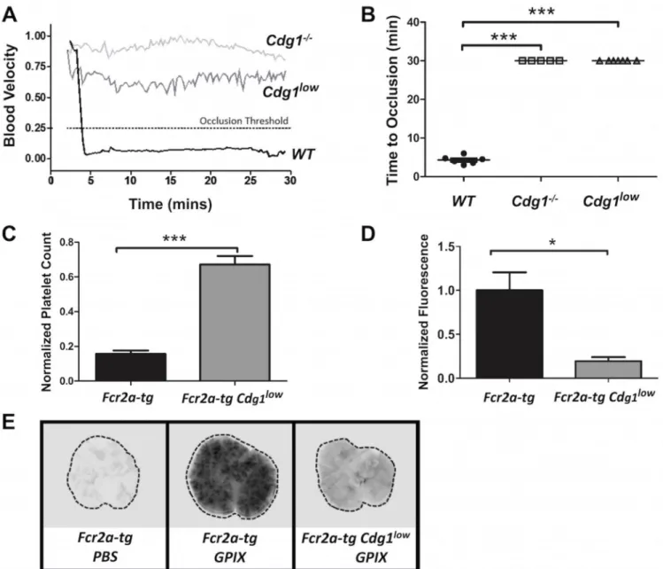
Mice lacking CD-GEFI are strongly protected from arterial thrombosis and ITT12, 13. Given their minor hemostatic defect, we were curious to see whether Cdg1low mice were still protected from experimental thrombosis. Similar to Cdg1−/− mice, Cdg1low mice did not form occlusive thrombi in a model of FeCl3-induced injury to the carotid artery (Figure
7A,B). In this model, vascular occlusion is defined as a reduction of blood flow by >75%, caused by the formation of a large thrombus. Since thrombus formation was not directly visualized in this model, we were not able to compare platelet adhesion to sites of FeCl3
injury between WT, Cdg1−/− and Cdg1low mice. We did, however, visualize platelet
adhesion in the laser injury hemostasis model (Figure 6A, supplemental videos 1–3). In WT mice, hemostatic plug formation was characterized by a first phase of rapid growth that was followed by “pacification” of the thrombus, i.e. the shrinking of the thrombus down to a core region. The observed kinetics of platelet adhesion to sites of laser injury in WT mice are consistent with studies by Brass and colleagues, who demonstrated that thrombus
pacification is due to the reversible binding of weakly activated platelets in the shell region of the thrombus18. Interestingly, thrombi in Cdg1low mice were markedly smaller and did not seem to contain a shell region. Thus, it is likely that Cdg1low mice formed very small platelet thrombi at sites of FeCl3 injury, but that these thrombi cannot grow big enough to
impede the blood flow in this high shear stress environment.
We also subjected Cdg1low mice to a recently established model of ITT13. In this model, platelet activation and pulmonary embolism is triggered via FcγRIIA, a receptor that is expressed on human but not murine platelets. To circumvent this limitation, these studies were performed with transgenic mice expressing human FcγRIIA19. Fcr2a-tg and
Cdg1lowFcr2a-tg mice were injected with an antibody against GPIX, a subunit of the platelet Von Willebrand receptor complex. As shown recently, anti-GPIX antibody treatment of mice leads to platelet activation and pulmonary embolism in an FcγRIIA-dependent fashion13. Consistent with previous results, injection of anti-GPIX antibody into Fcr2a-tg mice led to a >80% decrease in the peripheral platelet count (Figure 7C) and the formation of large pulmonary emboli (Figures 7D,E). In contrast, the peripheral platelet count in
Cdg1lowFcr2a-tg mice injected with anti-GPIX antibody dropped by only ~30% and platelet accumulation in the lungs was dramatically decreased compared to Fcr2a-tg mice.
DISCUSSION
Current strategies to prevent excessive platelet activation and arterial thrombosis include inhibitors of the main platelet integrin, αIIbβ3, and drugs targeting feedback signaling by ADP and TxA2 (P2Y12 antagonists and aspirin; also known as dual anti-platelet therapy –
DAPT20, 21). αIIbβ3 antagonists provide very powerful protection from thrombosis but also markedly increase the patients’ risk for pathological bleeding. In comparison, DAPT provides weaker protection from thrombosis but is safer with regard to unwanted bleeding. To provide a significant improvement over existing therapies, the next generation of anti-platelet drugs should be as effective as αIIbβ3 inhibitors in preventing thrombosis, but with a safety (bleeding) profile similar or better than that of DAPT.
Here we report that mice expressing small amounts of CD-GEFI are strongly protected from arterial and immune complex-mediated thrombosis. Importantly, as compared to Cdg1−/− mice, this protection in Cdg1low mice does not come at the expense of a deficient hemostatic response. Thus, small amounts of functional CD-GEFI are sufficient to preserve most of the hemostatic function of platelets, but not sufficient for the formation of pathological thrombi. This phenotype is similar to that of mice with impaired, but not abolished, expression/ function of the integrin adapter proteins, TALIN-122 or KINDLIN-323. In all cases αIIbβ 3-mediated platelet aggregation is delayed, allowing for the formation of small hemostatic plugs. If confirmed in humans, these findings would suggest TALIN-1, KINDLIN-3, and CD-GEFI as promising new targets for anti-platelet therapy. Compared to TALIN and KINDLIN, however, CD-GEFI may be a preferred target as (1) its expression is largely confined to platelets and neutrophils, and (2) mice deficient in talin-115 or kindlin-316, but not Cdg1−/− mice exhibit spontaneous bleeding and high perinatal mortality. Interestingly, impaired platelet function and bleeding upon challenge are the main phenotypes of dogs24 and humans14, 25 lacking functional CD-GEFI, suggesting that neutrophil function is less dependent on the CD-GEFI/RAP1 signaling pathway. Our work in Cdg1−/− mice identified significant defects in neutrophil integrin activation and adhesion26, but these defects were mild when compared to those observed in knockout platelets or those in neutrophils lacking kindlin-327. As neutrophils are known modulators of experimental thrombosis28, however, studies in mice lacking CD-GEFI in platelets or neutrophils only will be required to determine whether impaired neutrophil function contributes to the anti-thrombotic phenotype observed in CD-GEFI mutant mice.
Based on our studies, we propose that targeting CD-GEFI could provide certain advantages over existing antiplatelet therapies. While P2Y12 inhibitors affect the sustained activation of RAP1 and thus protect from thrombosis by destabilizing existing thrombi12, lack of
functional CD-GEFI delays platelet activation and impairs thrombus formation in mice4, 12, 29 and humans14, 25. CD-GEFI also plays a critical role for ITAM-dependent platelet activation29, and both knockout12, 13, 30 and Cdg1low mice are strongly protected from arterial and IC-mediated thrombosis. Thus, inhibitors of CD-GEFI are expected to provide significantly better protection from thrombosis than drugs targeting P2Y12. Obviously, CD-GEFI inhibitors would have to be carefully monitored in patients, as
complete lack of function in this protein is associated with a marked bleeding risk in humans and mice8, 9, 12, 14, 25. Alternatively, smarter strategies to inhibit CD-GEFI function could be developed. For example, our recent work demonstrated that deletion of the C1 regulatory domain in CD-GEFI leads to an ~70% reduction in GEF activity12. If we succeed in identifying how the C1 domain contributes to CD-GEFI function, inhibitors could be developed that specifically target this regulatory domain. Such inhibitors would not need to be titrated in patients as complete inhibition of CD-GEFI could not be achieved with such an approach. Lastly, inhibitors to CD-GEFI could be used as a safer alternative to αIIbβ3 inhibitors currently used in high-risk patients. The studies reported here combined with our previous work suggest that the antithrombotic effect of a putative CD-GEFI inhibitor would be comparable to that of αIIbβ3 inhibitors – but at a lower risk for bleeding.
In summary, we provide evidence that low-level expression of CD-GEFI leads to protection from thrombosis, but not to marked bleeding, in mice. Based on these findings, we propose
that specific targeting of CD-GEFI would provide a significant improvement over clinically used anti-platelet therapies, such as αIIbβ3 inhibitors and DAPT.
Supplementary Material
Refer to Web version on PubMed Central for supplementary material.
Acknowledgments
We thank Katie Poe for providing excellent animal husbandry services and the Animal Models Core facility staff for excellent technical work in generating the Cdg1low mice.
SOURCES OF FUNDING
This work was supported by the American Heart Association (14EIA18910004, W.B.) and NIH grants R01HL106009 (W.B. and S.E.M.) R01 HL121650 (W.B.), P01 HL120846 (W.B.), R01 HL126124 (L.V.P), and P01HL110860 (S.E.M.).
Nonstandard Abbreviations and Acronyms
Cdg1 CalDAG-GEFI
Cvx Convulxin
Par4p Par4 activating peptide
DAPT Dual antiplatelet therapy
Fcr2a-tg FcγRIIa transgenic mice
ITT Immune-mediated thrombocytopenia and thrombosis
REFERENCES
1. Ruggeri ZM. Platelets in atherothrombosis. Nat Med. 2002; 8:1227–1234. [PubMed: 12411949] 2. Jackson SP. Arterial thrombosis--insidious, unpredictable and deadly. Nat Med. 2011; 17:1423–
1436. [PubMed: 22064432]
3. Broos K, Feys HB, De Meyer SF, Vanhoorelbeke K, Deckmyn H. Platelets at work in primary hemostasis. Blood Rev. 2011; 25:155–167. [PubMed: 21496978]
4. Crittenden JR, Bergmeier W, Zhang Y, Piffath CL, Liang Y, Wagner DD, Housman DE, Graybiel AM. CalDAG-GEFI integrates signaling for platelet aggregation and thrombus formation. Nat Med. 2004; 10:982–986. [PubMed: 15334074]
5. Stefanini L, Bergmeier W. Rap1-GTPase signaling and platelet function. J Mol Med (Berl). 2016; 94:13–19. [PubMed: 26423530]
6. Guidetti GF, Torti M. The small GTPase Rap1b: A bidirectional regulator of platelet adhesion receptors. J Signal Transduct. 2012; 2012:412089. [PubMed: 22745904]
7. Stefanini L, Paul DS, Robledo RF, et al. RASA3 is a critical inhibitor of Rap1-dependent platelet activation. J Clin Invest. 2015; 125:1419–1432. [PubMed: 25705885]
8. Dorsam RT, Kunapuli SP. Central role of the P2Y12 receptor in platelet activation. J Clin Invest. 2004; 113:340–345. [PubMed: 14755328]
9. Andre P, Delaney SM, LaRocca T, Vincent D, DeGuzman F, Jurek M, Koller B, Phillips DR, Conley PB. P2Y12 regulates platelet adhesion/activation, thrombus growth, and thrombus stability in injured arteries. J Clin Invest. 2003; 112:398–406. [PubMed: 12897207]
A
uthor Man
uscr
ipt
A
uthor Man
uscr
ipt
A
uthor Man
uscr
ipt
A
uthor Man
uscr
10. Chrzanowska-Wodnicka M, Smyth SS, Schoenwaelder SM, Fischer TH, White GC 2nd. Rap1b is required for normal platelet function and hemostasis in mice. J Clin Invest. 2005; 115:680–687. [PubMed: 15696195]
11. Zhang G, Xiang B, Ye S, Chrzanowska-Wodnicka M, Morris AJ, Gartner TK, Whiteheart SW, White GC 2nd, Smyth SS, Li Z. Distinct roles for Rap1b protein in platelet secretion and integrin αIIbβ3 outside-in signaling. J Biol Chem. 2011; 286:39466–39477. [PubMed: 21940635] 12. Stolla M, Stefanini L, Roden RC, Chavez M, Hirsch J, Greene T, Ouellette TD, Maloney SF,
Diamond SL, Poncz M, Woulfe DS, Bergmeier W. The kinetics of αIIbβ3 activation determines the size and stability of thrombi in mice: Implications for antiplatelet therapy. Blood. 2011; 117:1005–1013. [PubMed: 20971951]
13. Stolla M, Stefanini L, Andre P, Ouellette TD, Reilly MP, McKenzie SE, Bergmeier W. CalDAG-GEFI deficiency protects mice in a novel model of FCϒRIIa-mediated thrombosis and thrombocytopenia. Blood. 2011; 118:1113–1120. [PubMed: 21652673]
14. Canault M, Ghalloussi D, Grosdidier C, et al. Human CalDAG-GEFI gene (RASGRP2) mutation affects platelet function and causes severe bleeding. J Exp Med. 2014; 211:1349–1362. [PubMed: 24958846]
15. Petrich BG, Marchese P, Ruggeri ZM, Spiess S, Weichert RA, Ye F, Tiedt R, Skoda RC, Monkley SJ, Critchley DR, Ginsberg MH. Talin is required for integrin-mediated platelet function in hemostasis and thrombosis. J Exp Med. 2007; 204:3103–3111. [PubMed: 18086863] 16. Moser M, Nieswandt B, Ussar S, Pozgajova M, Fassler R. Kindlin-3 is essential for integrin
αIIbβ3 activation and platelet aggregation. Nat Med. 2008; 14:325–330. [PubMed: 18278053] 17. Getz TM, Piatt R, Petrich BG, Monroe D, Mackman N, Bergmeier W. Novel mouse hemostasis
model for real-time determination of bleeding time and hemostatic plug composition. J Thromb Haemost. 2015; 13:417–425. [PubMed: 25442192]
18. Stalker TJ, Traxler EA, Wu J, Wannemacher KM, Cermignano SL, Voronov R, Diamond SL, Brass LF. Hierarchical organization in the hemostatic response and its relationship to the platelet-signaling network. Blood. 2013; 121:1875–1885. [PubMed: 23303817]
19. McKenzie SE, Taylor SM, Malladi P, Yuhan H, Cassel DL, Chien P, Schwartz E, Schreiber AD, Surrey S, Reilly MP. The role of the human FC receptor FCϒRIIa in the immune clearance of platelets: A transgenic mouse model. J Immunol. 1999; 162:4311–4318. [PubMed: 10201963] 20. Michelson AD. Antiplatelet therapies for the treatment of cardiovascular disease. Nat Rev Drug
Discov. 2010; 9:154–169. [PubMed: 20118963]
21. Park SJ, Park DW, Kim YH, et al. Duration of dual antiplatelet therapy after implantation of drug-eluting stents. N Engl J Med. 2010; 362:1374–1382. [PubMed: 20231231]
22. Stefanini L, Ye F, Snider AK, Sarabakhsh K, Piatt R, Paul DS, Bergmeier W, Petrich BG. A talin mutant that impairs talin-integrin binding in platelets decelerates αIIbβ3 activation without pathological bleeding. Blood. 2014; 123:2722–2731. [PubMed: 24585775]
23. Klapproth S, Moretti FA, Zeiler M, Ruppert R, Breithaupt U, Mueller S, Haas R, Mann M, Sperandio M, Fassler R, Moser M. Minimal amounts of kindlin-3 suffice for basal platelet and leukocyte functions in mice. Blood. 2015; 126:2592–2600. [PubMed: 26438512]
24. Boudreaux MK. Characteristics, diagnosis, and treatment of inherited platelet disorders in mammals. J Am Vet Med Assoc. 2008; 233:1251–1259. [PubMed: 18922051]
25. Lozano ML, Cook A, Bastida JM, et al. Novel mutations in RASGRP2 encoding for CalDAG-GEFI abrogate Rap1 activation causing platelet dysfunction. Blood. in press.
26. Bergmeier W, Goerge T, Wang H-W, Crittenden JR, Baldwin ACW, Cifuni SM, Housman DE, Graybiel AM, Wagner DD. Mice lacking the signaling molecule CalDAG-GEFI represent a model for leukocyte adhesion deficiency type III. The Journal of Clinical Investigation. 2007; 117:1699– 1707. [PubMed: 17492052]
27. Svensson L, Howarth K, McDowall A, Patzak I, Evans R, Ussar S, Moser M, Metin A, Fried M, Tomlinson I, Hogg N. Leukocyte adhesion deficiency-III is caused by mutations in Kindlin3 affecting integrin activation. Nat Med. 2009; 15:306–312. [PubMed: 19234463]
28. Mócsai A. Diverse novel functions of neutrophils in immunity, inflammation, and beyond. The Journal of Experimental Medicine. 2013; 210:1283–1299. [PubMed: 23825232]
29. Stefanini L, Roden RC, Bergmeier W. CalDAG-GEFI is at the nexus of calcium-dependent platelet activation. Blood. 2009; 114:2506–2514. [PubMed: 19628710]
30. Amirkhosravi A, Boulaftali Y, Robles-Carrillo L, Meyer T, McKenzie SE, Francis JL, Bergmeier W. CalDAG-GEFI deficiency protects mice from FCϒRIIa-mediated thrombotic thrombocytopenia induced by CD40L and β2GPI immune complexes. J Thromb Haemost. 2014; 12:2113–2119. [PubMed: 25287077]
A
uthor Man
uscr
ipt
A
uthor Man
uscr
ipt
A
uthor Man
uscr
ipt
A
uthor Man
uscr
HIGHLIGHTS
• Low level expression of CalDAG-GEFI is sufficient for the hemostatic function of platelets
• Mice expressing low levels of CalDAG-GEFI are fully protected from experimental thrombosis
• Partial inhibition of CalDAG-GEFI may provide a powerful yet safe approach to prevent thrombosis
A
uthor Man
uscr
ipt
A
uthor Man
uscr
ipt
A
uthor Man
uscr
ipt
A
uthor Man
uscr
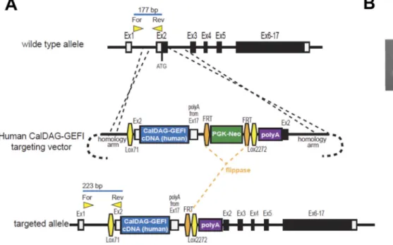
Figure 1. Generation of a CalDAG-GEFI humanized mouse model
(A) Schematic of human CalDAG-GEFI knock-in strategy. A gene targeting vector was constructed to insert the human CalDAG-GEFI cDNA at the start codon of the mouse CalDAG-GEFI locus with a copy of the mouse 3’UTR/polyadenylation sequence placed immediately downstream of the human cDNA. A FRT-flanked PGK-Neo resistance cassette was placed downstream of the expression cassette, followed by an additional
polyadenylation signal. Lox71 and Lox2272 sites were inserted flanking the cDNA cassette and selectable marker to allow replacement of the cassette by recombinase-mediated cassette exchange strategy. Positions of forward and reverse genotyping primers are indicated along with the size of PCR products to be obtained from the wild-type and knock-in alleles. (B) PCR genotyping of animals. Primers shown in panel A were used to amplify DNA from wild-type (wt/wt), heterozygous knock-in (wt/tg) and homozygous knock-in (tg/tg) mice.
A
uthor Man
uscr
ipt
A
uthor Man
uscr
ipt
A
uthor Man
uscr
ipt
A
uthor Man
uscr
Figure 2. Markedly reduced expression of CD-GEFI in hypomorphic mice (Cdg1low)
A) Representative Western blots for CD-GEFI, RASA3, RAP1, and β-ACTIN in platelet lysates from WT, Cdg1−/−, and Cdg1low mice. B) RAP1-GTP levels (top panel) in WT, Cdg1−/−, and Cdg1low platelets left unstimulated (C) or activated with PAR4 peptide for 30” or 3’. Total RAP1 is provided as a loading control (bottom panel). Results are representative of 3 independent experiments.
A
uthor Man
uscr
ipt
A
uthor Man
uscr
ipt
A
uthor Man
uscr
ipt
A
uthor Man
uscr
Figure 3. Improved integrin activation in Cdg1low compared to Cdg1−/− platelets
(A-E) αIIbβ3 integrin activation and alpha granule release. JON/A-PE (A,C,E) and anti-P-selectin Alexa Fluor 488 (B,D) binding to platelets from the indicated mice, activated with various concentrations of PAR4p (A,B), convulxin (Cvx; C,D), or ADP (E). Data are shown as mean fluorescence intensity (MFI) ± SEM; n=4–6, *p<0.05, **p<0.01, ***p<0.001; # p<0.05, # #p<0.01, ### p<0.001 relative to resting controls, analyzed by two-way ANOVA with a Bonferonni post test.
A
uthor Man
uscr
ipt
A
uthor Man
uscr
ipt
A
uthor Man
uscr
ipt
A
uthor Man
uscr
Figure 4. Intermediate aggregation response of Cdg1low platelets compared to controls (A-F) Representative aggregometry traces for WT (black line), Cdg1−/− (light grey), and Cdg1low (dark grey) platelets activated with the indicated agonists. Aggregometric responses to 75 (A) and 150 (B) µM Par4p, 100 (C) and 200 (D) ng/mL convulxin, and 3 (E) and 10 (F) µM ADP.
A
uthor Man
uscr
ipt
A
uthor Man
uscr
ipt
A
uthor Man
uscr
ipt
A
uthor Man
uscr
Figure 5. Improved platelet adhesion to collagen of Cdg1low compared to Cdg1−/− platelets Whole blood from WT (black bars), Cdg1low (dark grey), and Cdg1−/− mice (light grey bars) was perfused for 5 minutes over fibrillar collagen (200 µg/mL) at venous (400 s−1, A-C) or arterial (1600 s−1, D-F) shear rates. Platelets were labeled with Alexa Fluor 488-labeled antibodies to GPIX before perfusion. At the end of the perfusion period, fluorescence images (C,F) were taken and the sum intensity (A,D) and the surface area coverage (B,E) were determined. Fluorescence intensities were normalized to the maximum intensity measured in WT control samples (Relative Fluorescence). Area coverage represents the area
A
uthor Man
uscr
ipt
A
uthor Man
uscr
ipt
A
uthor Man
uscr
ipt
A
uthor Man
uscr
in the field of view covered by fluorescently labeled platelets. Data are shown as mean ± SEM, (n = 5) *p<0.05, **p<0.01, ***p<0.001. Images taken on a Nikon TE300 equipped with a QImaging Retiga Exi CCD camera, 20x/0.5 magnification, Alexa Fluor 488-labeled GPIX, Slidebook 5.0 Software at room temperature.
A
uthor Man
uscr
ipt
A
uthor Man
uscr
ipt
A
uthor Man
uscr
ipt
A
uthor Man
uscr
Figure 6. Improved hemostasis in Cdg1low compared to Cdg1−/− mice
(A-C) Intravital microscopy studies to monitor hemostatic plug formation after laser injury to the saphenous vein in WT (black; also see Supplemental Video 1), Cdg1−/− (light grey;
also see Supplemental Video 2), and Cdg1low mice (dark grey; also see Supplemental Video
3). Prior to laser injury, animals were injected with Alexa Fluor 488–labeled antibodies to GPIX. (A) Representative images taken 90 seconds after laser injury. Arrow highlights blood loss at the site of injury in Cdg1−/− mice. Scale bar: 100 µm. (B) Sum fluorescence intensity
± SEM recorded at the site of injury over time in the indicated mice (n = 5–11). (C) Time to stable occlusion (no leakage of blood for more than 60 seconds) of the vascular lesion in the indicated mice. Recordings were stopped 300 seconds after laser injury. (D,E) Blood loss (D) and bleeding times (E) in WT (black circle), Cdg1−/− (light gray square), and Cdg1low (dark gray triangle) mice after tail transection. ** P<0.01 ***P < 0.001, Chi-square test was performed between groups (n= 10 (WT), 10 (Cdg1−/−), 10 (Cdg1low)). Images were taken on
A
uthor Man
uscr
ipt
A
uthor Man
uscr
ipt
A
uthor Man
uscr
ipt
A
uthor Man
uscr
a Zeiss Examiner Z1 equipped with a Hamamatsu C9300 camera, 20x/1 magnification, Alexa Fluor 488-labeled GPIX, Slidebook 5.0 Software at room temperature.
A
uthor Man
uscr
ipt
A
uthor Man
uscr
ipt
A
uthor Man
uscr
ipt
A
uthor Man
uscr
Figure 7. Cdg1low mice are protected from FeCl3- and immune mediated thrombosis
(A) Representative blood flow velocity traces recorded after exposure of the carotid artery of a WT (black line), Cdg1−/− (light grey), or Cdg1low (dark grey) mouse to 20% FeCl
3 for 1
minute. A decrease in blood flow by > 75% (dotted line) was considered as complete vessel occlusion. (B) Time to occlusion recorded in individual WT (black circle, n=6), Cdg1−/− (light gray square, n=5), and Cdg1low mice (dark gray triangle, n=5). (C) Platelet counts in whole blood of Fcr2a-tg control and Cdg1lowFcr2a-tg mice 4 hours after administration of 1 µg/g body weight of α-GPIX-IRDye800 antibody. Platelet counts are expressed as
percentage of baseline value. (D,E) Four hours after antibody infusion, lungs were extracted and scanned on a Li-COR Odyssey at 800 nm. (D) Quantitative analysis of the integrated fluorescence intensity (ImageStudio Lite 4.0). Results are shown as arbitrary fluorescence intensity (a.u.) ± SEM normalized to Fcr2a-tg controls; n = 5. Fcr2a-tg (black bar), Cdg1lowFcr2a-tg (gray bar). (E) Representative images. *p<0.05, ***p<0.001.
A
uthor Man
uscr
ipt
A
uthor Man
uscr
ipt
A
uthor Man
uscr
ipt
A
uthor Man
uscr
A
uthor Man
uscr
ipt
A
uthor Man
uscr
ipt
A
uthor Man
uscr
ipt
A
uthor Man
uscr
ipt
Table 1
Blood Cell Analysis
Wild Type Cdg1−/− Cdg1low
Platelet Count, K/μL 956 ± 304 906 ± 258 1198 ± 356
Mean Platelet Volume, fL 4.33 ± 0.122 4.189 ± 0.127 4.411 ± 0.136
Neutrophil Count, K/μL 0.70 ± 0.41 2.30 ± 0.757*** 1.67 ± 0.7443*
Monocyte Count, K/μL 0.96 ± 0.35 1.584 ± 0.51* 1.231 ± 0.55
Lymphocyte Count, K/μL 7.807 ± 2.92 10.06 ± 1.82 9.033 ± 2.35
Erythrocyte Count, M/μL 12.92 ± 2.26 12.87 ± 1.95 14.43 ± 4.00
Hemoglobin, g/dL 17.89 ± 2.74 19.04 ± 2.79 18.84 ± 3.26
Circulating blood cell analysis. Data are reported as mean ± SD.
* P < 0.05,
***