ABSTRACT
Stefanie Ann Ward Mortimer
Methods for High‐Throughput Analysis of RNA Structure and Dynamics
(Under the direction of Kevin M Weeks)
conformational changes on the order of ~10‐4 sec‐1. Third, I extend SHAPE chemistry to a benzoyl cyanide scaffold to make possible facile time‐resolved kinetic studies of RNA in ~1 s snapshots. I then use SHAPE chemistry to follow the time‐dependent folding of an RNase P specificity domain RNA and identify a slow folding region in the RNA. I am able to attribute this slow folding step to the conformational dynamics of a single nucleotide. Finally, I show that show that N,N‐(dimethylamino)dimethylchlorosilane (DMAS‐Cl) reacts selectively at the guanosine N2 position. Critically, DMAS‐Cl reactivity yields a near‐perfect measure (r ≥ 0.82) of solvent accessibility for this position in a folded RNA. This silane‐based chemistry represents a significant improvement over classical approaches that employ carbon electrophiles for probing solvent accessibility at the base pairing face of guanosine in RNA.
AKNOWLEDGEMENT
I first and foremost want to thank Kevin Weeks. Not only did he give me the opportunity to work on this great project, but also he is an outstanding mentor. I am the scientist I am today because of his guidance and insight.
There are so many people who have helped me along the way and mean so much to me. Jeremy, this past year has been one of the greatest in my life because you have been in it; thank you for being so supportive of me and my aspirations. My family, thanks for being a constant source of support even though I have been far from home for a long time. I know I can do anything with you behind me. I want to thank my friends, those whom I have known and those whom I have met along the way in graduate school, for all the good times we shared and will hopefully continue to share in the future.
Lastly, I want to thank all the members of the lab for making it such a great place to work. I couldn’t ask to be among a better group of scientists and friends, I will truly miss you all.
TABLE OF CONTENTS
LIST OF TABLES……….xi
LIST OF FIGURES………..xii
LIST OF ABBREVIATIONS………xv
CHAPTER 1. Towards Chemical Methods for the Efficient and Accurate Analysis of RNA Structure and Dynamics……….1
1.1 Introduction………...2
1.1.1 RNA structure and function………..2
1.1.2 RNA SHAPE chemistry and secondary structure prediction……….2
1.1.3 RNA tertiary structure and solvent accessibility…….6
1.1.4 RNA folding and dynamics………7
1.1.5 Research overview……….8
1.1.6 Perspective………..9
2. A Fast‐Acting Reagent for Accurate Anaylsis of RNA Secondary and Tertiary
Structure by SHAPE Chemistry………13
2.1 Introduction………14
2.2 Results………16
2.2.1 Comparative reactivity of 1M7 and NMIA………16
2.2.2 Accurate and quantitative RNA structure analysis using 1M7………....……..18
2.2.3 Prediction of highly accurate secondary structure models using 1M7……….24
2.3 Discussion……….26
2.4 Experimental………..26
2.4.1 Synthesis of [32P]‐labeled pAp‐ethyl………26
2.4.2 Synthesis of 1‐methyl‐7‐nitroisatoic anhydride (1M7)……...26
2.4.3 NMIA and 1M7 hydrolysis and 2′‐O‐adduct Formation……...27
2.4.4 Synthesis of Bacillus subtilis RNase P RNA………...27
2.4.5 Structure‐Selective RNA Modification………28
2.4.6 Primer Extension……….28
2.4.7 Data Analysis………..29
References………31
3. Slow Conformational Dyanmics at C2´‐Endo Nucleotides in RNA……….33
3.1 Introduction………34
3.2 Results………36
3.2.1 SHAPE analysis of C2´‐endo nucleotides………...36
3.3. Discussion………44
3.4 Experimental………..46
3.4.1 Derivation of equation 1………..46
3.4.2 IA, NMIA, 4NIA, and 1M7 hydrolysis………47
3.4.3 RNA constructs………..48
3.4.4 SHAPE anaylsis………..48
3.4.5 Primer extension………..49
3.4.6 Data analysis………...49
3.4.7 Refinement of RNase P structure………...50
References………52
4. Time Resolved RNA SHAPE Chemistry………..55
4.1 Introduction………56
4.2 Results………58
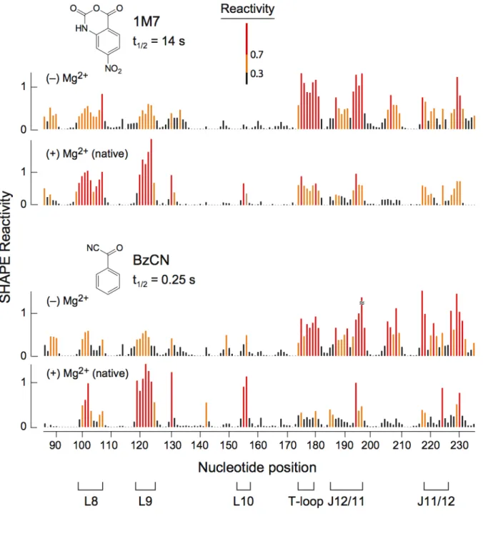
4.2.1 BzCN reacts 2′‐hydroxyl groups in a structure‐selective manner….58 4.2.2 BzCN undergoes rapid inactivation by hydrolysis with water………59
4.2.3 Time‐resolved SHAPE with BzCN yields a nucleotide‐resolution view of RNase P tertiary folding……….63
4.3 Discussion……….68
4.4 Experimental………..68
4.4.1 Benzoyl cyanide 2′‐O‐adduct formation and hydrolysis……….68
4.4.2 Structure‐Selective RNA Modification………69
4.4.3 Primer extension………..70
References………72
5. C2´‐Endo Nucleotides as Molecular Timers in RNA Folding………75
5.1 Introduction………76
5.2 Results………77
5.2.1 A130 has distinct local dynamics and a critical role in RNase P function………..77
5.2.2 The ∆A130 RNA has the same global fold as the native RNA………...82
5.2.3 Fast Folding Kinetics of the ∆A130 RNA………84
5.3 Discussion………....90
5.4 Experimental………..93
5.4.1 Synthesis of Bacillus subtilis RNase P RNAs……….93
5.4.2 SHAPE Analysis……….94
5.4.3 Hydroxy Radical Probing………95
5.4.4 Primer Extension and Data Analysis of Modified RNA……….95
5.4.5 Folding Kinetics Monitored by Fluorescence Spectroscopy………..96
References………97
6. Structure Selective N‐Silylation of Guanosine Residues in RNA………100
6.1 Introduction……….101
6.2. Results………104
6.2.1 Silylation at the Guanosine N2 Position………..104
6.2.2 Structure‐selective Reaction of DMAS‐Cl with RNA…………106
6.2.3 Comparison of DMAS‐Cl and Kethoxal Reactivity……….108
6.4 Experimental………...114 6.4.1 Synthesis of Wild Type Bacillus subtilis mgtE aptamer
domain (M‐box) and inosine M‐box RNAs……….114
6.4.2 Structure selective N‐silylation of RNA………...115 6.4.3 RNA modification with kethoxal……….116 6.4.4 Primer extension………...116
6.4.5 Reaction of DMAS‐Cl with 2´‐
deoxyguanosinemonophosphate (2´‐dGMP)………...117
6.4.6 Data Analysis………...117 References……….118
LIST OF FIGURES Figure 1.1 The hierarchical nature of RNA structure………3 Figure 1.2 High‐throughput RNA SHAPE chemistry……….5 Figure 2.1 Mechanism of RNA SHAPE Chemistry……….15 Figure 2.2 Comparative reactivity of 1M7 and NMIA via hydrolysis and 2´‐O‐adduct formation with pAp‐ethyl ………..17 Figure 2.3 Comparison of processed capillary electrophoresis traces for SHAPE experiments performed using 1M7 and NMIA………..19 Figure 2.4 Histograms and difference plot of absolute reactivities for SHAPE experiments performed with 1M7 in the presence and absence of
6 mM Mg2+………20
Figure 2.5 Base pairing and tertiary interactions for the specificity domain of
Bacillus subtilis RNase P………...22
Figure 2.6 Reaction between the model nucleotide, pAp‐ethyl, and 1M7 is
Figure 3.5 Absence of a dependence of fraction adduct formed as a function of khydrolysis for reaction of the (unconstrained) model nucleotide pAp‐ethyl and for nucleotide 45 in the loop of the C2´‐endo construct….42 Figure 3.6 SHAPE reactivities at C2´‐endo nucleotides in the specificity domain of RNase P……….43 Figure 3.7 Base stacking and hydrogen bonding interactions at C2´‐endo nucleotides that undergo slow conformational changes……….45 Figure 4.1 Mechanism of RNA SHAPE chemistry with BzCN………56 Figure 4.2 Histograms comparing absolute SHAPE reactivities for the RNase P specificity domain in the presence and absence of Mg2+ obtained using 1M7 and BzCN………59
Figure 4.3 Correlation between SHAPE reactivities obtained with 1M7 and BzCN in the presence and absence of Mg2+………...60 Figure 4.4 Reaction half‐life of BzCN………61 Figure 4.5 Formation of tertiary interactions in the RNase P specificity domain……64 Figure 4.6 Mehcanism for folding of the RNase P specificity domain………..66 Figure 5.1 Structural context and local nucleotide dynamics of A130………77 Figure 5.2 Binding of mature tRNA to the Bacillus subtilis ribonuclease P enzyme…79 Figure 5.3 A130 and A374 experience slow conformational dynamics in the complete Bacillus subtilis RNase P RNA………80 Figure 5.4 Two‐step mechanism for tertiary folding in the RNase P specificity domain………82 Figure 5.5 Native sequence and the ∆A130 mutant RNase P specificity domain RNAs form similar tertairy structures………84 Figure 5.6 Deletion of A130 accelerates RNA folding………85 Figure 5.7 Comparison of emission spectra of free Oregon green versus dye‐labeled native and mutant RNAs in the presence and absence
of Mg2+……….87
LIST OF ABBREVIATIONS
1M7 1‐methyl‐7‐nitroisatoic anhydride
4NIA 4‐nitro isatoic anhydride
A adenine
BzCN benzoyl cyanide
C cytosine
Ci curie
°C degree Celsius DMSO dimethylsulfoxide
DMAS‐Cl N,N‐(dimethylamino)dimethylchlorosilane DNA deoxyribonucleic acid
DTT dithiotreitol
EDTA ethylenediaminetetraacetic acid EtOH ethanol
G guanosine
GMPS guanosine monophosphorothioate
h hour
H2O water
HEPES N‐2‐hydroxyethylpiperazine‐N´‐2‐ethanesulfonic acid IA isatoic anhydride
kopen opening rate kclose closing rate khydrolysis hydrolysis rate kadduct adduct rate
L liter
Mg2+ magnesium ion MgCl2 magnesium chloride
min minute
mg milligram mRNA messenger RNA ms millisecond
µg microgram
µL microliter
µM micromolar
NMIA N‐methyl isatoic anhydride NaCl sodium chloride
nM nanomolar
NMR nuclear magnetic resonance
ns nanosecond
nt nucleotide
NTP nucleotide triphosphate
Phe phenylalanine pmol picomol ps picosecond RNA ribonucleic acid RNase Ribonuclease
s second
SHAPE selective 2´‐hydroxyl acylation analyzed by primer extension TBE 90 mM Tris‐borate, 2 mM EDTA
TE 10 mM Tris (pH 7.5), 1 mM EDTA Tris tris(hydroxymethyl)aminomethane tRNA transfer RNA
U uridine
v volume
V volt
w weight
W watt
1. Introduction
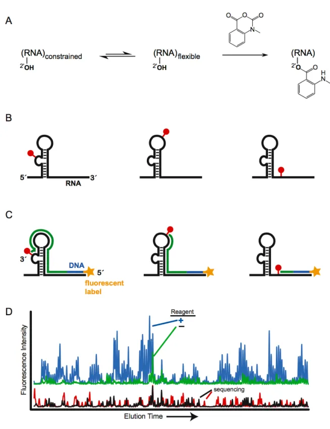
1.1.1. RNA structure and function. RNA molecules carry out many important
roles in the cell besides being a passive carrier of genetic information (mRNAs) [1]: RNA is essential for protein translation (tRNAs and ribosomes), the splicing of mRNA (group I and II introns and the splicosome), and in genetic regulation (riboswitches, siRNAs, and miRNAs). RNA molecules often form complex three‐dimensional structures in order carry out important cellular mechanisms. Higher‐order RNA structures are built up from base‐paired secondary and long‐range tertiary interactions (Figure 1.1) [2, 3]. Accurate and comprehensive knowledge of these interactions is essential to understanding the function of almost all RNAs.
development of SHAPE (selective 2´‐hydroxyl acylation analyzed by primer extension) chemistry, has radically simplified and improved analysis of RNA secondary structure [11].
SHAPE is an alternate high‐throughput approach to chemically map RNA structure that exploits the discovery that the nucleophilic reactivity of the ribose 2´‐ hydroxyl group is strongly gated by the underlying nucleotide flexibility (Figure 1.2A) [11‐13]. Flexible nucleotides preferentially adopt conformations that react with a hydroxyl‐selective electrophile to form a 2´‐O‐adduct, while base paired or otherwise constrained nucleotides are unreactive (Figure 1.2B). Sites of 2´‐O‐adduct formation cause reverse transcriptase to stop exactly one nucleotide prior to the modified base. The primers extended in the reverse transcriptase reaction are labeled with a fluorophore to allow the cDNA transcripts to be visualized when they are separated on a capillary DNA sequencer (Figure 1.2C). The length and amount of a cDNA transcript correlates with the position and degree of modification at each position in the RNA. The length of each cDNA can be assigned by comparison with a primer extension reaction in which a dideoxy nucleotide is incorporated, which causes the termination of extension at specific nucleotides (Figure 1.2D).
secondary structure prediction program, called RNAstructure, to constrain or limit the structures predicted. Current algorithms correctly predict 50‐70% of known base pairs on average [17]. Incorporating SHAPE reactivity information dramatically improves the prediction of RNA secondary structures, especially for large RNAs, with accuracies usually reaching 90% or greater [18].
The initial electrophile developed for SHAPE, NMIA, is not very reactive and therefore RNA structures must be probed over periods of tens of minutes (Figure 1.2A). This represented a major disadvantage for the initial version of SHAPE chemistry. Much of my research work focused on creating superior reagents for SHAPE chemistry including, those that are faster reacting and capable of interrogating RNA structure on the second timescale.
1.1.3. RNA tertiary structure and solvent accessibility. The analysis of RNA tertiary structure in solution is a necessary step to understand the function of an RNA. An important experimental objective is the ability to map solvent accessibility in RNA at single nucleotide resolution. At present there are very few ways to do this in a direct and quantitative way.
between hydroxyl radical reactivity and solvent accessibility are good, but imperfect [21, 22].
I am interested in developing new chemical probes of RNA structure that would react with RNA to form stable covalent adducts at solvent accessible positions. These reagents would provide a means to define RNA solvent accessibility in solution in a way that is not convoluted by electrostatic and other factors; an experimental objective that to date has not been accomplished.
1.1.5. Research overview. The overarching goal of my research has been to create efficient and information–rich experiments that allow one to understand as completely as possible the structure of any RNA. My focus is to develop new chemical methods that make it possible to go directly and accurately from the primary sequence to the secondary structure and eventually from the secondary structure to the tertiary structure.
It is becoming increasingly evident that SHAPE represents a major advance relative to other ways of looking at RNA structure. SHAPE allows accurate and comprehensive RNA structural information to be achieved in a single experiment. The initial electrophile used for SHAPE, NMIA, is not very reactive and therefore RNA structures must be probed over periods of tens of minutes. In Chapter 2, I design and synthesize a significantly faster and more useful SHAPE reagent, 1‐methyl‐7‐ nitroisatoic anhydride (1M7). My “second generation” reagent, 1M7, is capable of taking snapshots of RNA structure over 70 s periods, is insensitive to solution conditions like Mg2+, and provides highly accurate secondary structure models. In Chapter 3, Costin Gherghe and I apply SHAPE reagents of varying electrophilicity to monitor conformational dynamics over distinct time domains. We observed that select C2´‐endo nucleotides in both a model RNA and a large RNA with a complex tertiary structure undergo extraordinarily slow conformational dynamics. This discovery is the first observation of extremely slow motions in RNA.
straightforward and very accessible technologies for following RNA structure in a time‐ dependent manner. I use this chemistry to study the time‐dependent folding of the ribonuclease P specificity domain. Specifically, I identify a slow folding region in the RNA and, in Chapter 5, I attribute this slow folding step to the slow conformational dynamics at a single C2´‐endo nucleotide in the RNA. This discovery was the first evidence that extremely slow conformational dynamics at certain C2´‐endo nucleotides can play a central role in RNA folding.
Finally, in Chapter 6, I create a new way to assess RNA tertiary structure in an experimentally straightforward way by developing a reagent, N,N‐ (dimethylamino)dimethylsilyl chloride (DMAS‐Cl), that reacts with the N2 position of guanosine in a manner that is rigorously proportional to solvent accessibility. DMAS‐Cl yields near‐perfect measures of solvent accessibility in RNA and we believe that this may now be the best reagent choice for analyzing the solvent accessibility of the base‐ pairing face of guanosine in RNA.
1.1.6. Perspective. In this work I utilize principles of physical organic chemistry
1.2 References
1. Gesteland, R.F., Cech, T.R., and Atkins, J.F., The RNA world. 2006, Cold Spring Harbor: Cold Spring Harbor Laboratory Press.
2. Brion, P. and Westhof, E., Hierarchy and Dynamics of RNA folding. Annu. Rev. Biophys. Biomol. Struct., 1997. 26: p. 113-137.
3. Tinoco, I. and Bustamante, C., How RNA folds. J. Mol. Biol., 1999. 293: p. 271-281.
4. Peattie, D.A. and Gilbert, W., Chemical Probes for higher-order structure in RNA. Proc. Natl. Acad. Sci. U.S.A., 1980. 77: p. 4679-4682.
5. Ehresmann, C., et al., Probing the structure of RNAs in solution. Nucleic Acids Res., 1987. 15: p. 9109-9128.
6. Lavery, R. and Pullman, A., A New Theoretical Index of Biochemical Reactivity Combining Steric and Electrostatic Factors - an Application to Yeast Transfer Rnaphe. Biophys. Chem., 1984. 19(2): p. 171-181.
7. Mathews, D.H., et al., Incorporating chemical modification constraints into a dynamic programming algorithm for prediction of RNA secondary structure. Proc. Natl. Acad. Sci. U. S. A., 2004. 101(19): p. 7287-92.
8. Krasilnikov, A.S., et al., Crystal structure of the specificity domain of ribonuclease P. Nature, 2003. 421(760-764).
9. Brown, J.W., et al., Comparative analysis of ribonuclease P RNA using gene sequences from natural microbial populations reveals tertiary structural elements. Proc. Natl. Acad. Sci. U. S. A., 1996. 93(7): p. 3001-6.
10. Massire, C., Jaeger, L., and Westhof, E., Derivation of the three-dimensional architecture of bacterial ribonuclease P RNAs from comparative sequence analysis. J. Mol. Biol., 1998. 279(4): p. 773-93.
12. Wilkinson, K.A., et al., High-throughput SHAPE analysis reveals structures in HIV-1 genomic RNA strongly conserved across distinct biological states. PLoS Biol., 2008. 6(4): p. e96.
13. Wilkinson, K.A., Merino, E.J., and Weeks, K.M., Selective 2'-hydroxyl acylation analyzed by primer extension (SHAPE): Quantitative RNA structure analysis at single nucleotide resolution. Nat. Protoc., 2006. 1: p. 1610-1616.
14. Wilkinson, K.A., Merino, E.J., and Weeks, K.M., RNA SHAPE chemistry reveals non-hierarchical interactions dominate equilibrium structural transitions in tRNAAsp transcripts. J. Am. Chem. Soc., 2005. 127: p. 4659-4667.
15. Wang, B., Wilkinson, K.A., and Weeks, K.M., Complex Ligand-Induced Conformational Changes in tRNAAsp
Revealed by Single-Nucleotide Resolution SHAPE Chemistry. Biochemistry, 2008. 47: p. 3454-3461.
16. Duncan, C.D.S. and Weeks, K.M., SHAPE analysis of long-range interactions reveals extensive and thermodynamically preferred misfolding in a fragile group I intron RNA. Biochemistry, 2008. 47: p. 8504-8513.
17. Mathews, D.H. and Turner, D.H., Prediction of RNA secondary structure by free energy minimization. Curr. Opin. Struct. Biol., 2006. 16: p. 270-278.
18. Deigan, K.E., et al., Accurate SHAPE-Directed RNA Structure Determination Proc. Natl. Acad. Sci. U. S. A., 2008. submitted.
19. Tullius, T.D. and Dombroski, B.A., Hydroxyl radical "footprinting": high-resolution information about DNA-protein contacts and application to lambda repressor and Cro protein. Proc. Natl. Acad. Sci. U. S. A., 1986. 83(15): p. 5469-73.
20. Balasubramanian, B., Pogozelski, W.K., and Tullius, T.D., DNA strand breaking by the hydroxyl radical is governed by the accessible surface areas of the hydrogen atoms of the DNA backbone. Proc. Natl. Acad. Sci. U. S. A., 1998. 95(17): p. 9738-43.
22. Adams, P.L., et al., Crystal Structure of a Group I Intron Splicing Intermediate. RNA, 2004. 10: p. 1867-1887.
23. Chen, C., et al., Structural energetics and base-pair opening dynamics in sarcin-ricin domain RNA. Biochemistry, 2006. 45(45): p. 13606-13.
24. Shajani, Z. and Varani, G., NMR studies of dynamics in RNA and DNA by 13C relaxation. Biopolymers, 2007. 86(5-6): p. 348-59.
25. Hennelly, S.P., et al., A time-resolved investigation of ribosomal subunit association. J. Mol. Biol., 2005. 346(5): p. 1243-58.
26. Tijerina, P., Mohr, S., and Russell, R., DMS footprinting of structured RNAs and RNA-protein complexes. Nat. Protoc., 2007. 2(10): p. 2608-2623.
27. Sclavi, B., et al., RNA folding at millisecond intervals by synchrotron hydroxyl radical footprinting. Science, 1998. 279(5358): p. 1940-1943.
28. Shcherbakova, I. and Brenowitz, M., Monitoring structural changes in nucleic acids with single residue spatial and millisecond time resolution by quantitative hydroxyl radical footprinting. Nat. Protoc., 2008. 3(2): p. 288-302.
CHAPTER 2
A FastActing Reagent for Accurate Analysis of RNA Secondary and
Tertiary Structure by SHAPE Chemistry
2.1 Introduction
RNA sequences fold back on themselves to form structures that are difficult to predict, especially if only a single sequence is known [1‐3]. Current algorithms correctly predict 50‐70% of known base pairs on average [4, 5]. Predicted secondary structure models achieving 50‐70% accuracy tend to have regions in which the overall topology differs significantly from the correct one, making it difficult or impossible to develop robust biological hypotheses. Knowledge of which nucleotides are likely to be paired or single‐stranded can significantly improve prediction accuracies [6‐10].
The ideal technology for chemically‐assisted RNA structure analysis would (1) be experimentally straightforward, (2) use a single reagent that reacts generically with all four nucleotides, (3) employ a self‐quenching reagent, (4) involve short reaction times, and (5) be proven to yield accurate results with complex RNAs of known structure.
Selective 2′‐hydroxyl acylation analyzed by primer extension (SHAPE) chemistry
[11‐13] takes advantage of the discovery that the nucleophilic reactivity of a ribose 2′‐
hydroxyl group is gated by local nucleotide flexibility. At nucleotides constrained by base pairing or tertiary interactions, the 3′‐phosphodiester anion and other interactions
reduce reactivity of the 2′‐hydroxyl [11]. In contrast, flexible positions preferentially adopt conformations (Figure 2.1A) that react with N‐methlyisatoic anhydride (1, NMIA) to form a 2′‐O‐adduct (Figure 2.1B). NMIA reacts generically with all four nucleotides and the reagent undergoes a parallel, self‐inactivating, hydrolysis reaction (Figure 2.1B). Thus SHAPE chemistry meets the first three of the five criteria outlined above.
However, NMIA is relatively unreactive and requires tens of minutes to react to completion. To address the final two criteria for a near‐ideal RNA structure interrogation technology, we design a significantly more useful, fast acting, reagent for SHAPE chemistry. We then show that structural constraints obtained using this reagent allow the secondary and tertiary structure of a large RNA to be assessed with high accuracy.
2.2 Results
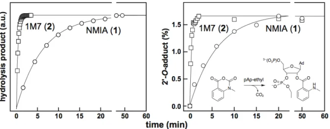
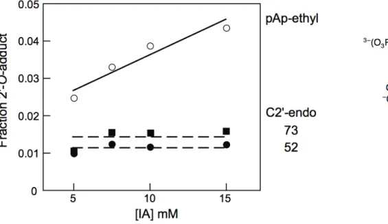
2.2.1 Comparative Reactivity of 1M7 and NMIA. The reactive carbonyl of NMIA is in the benzylic position relative to the aromatic ring system and should be sensitive to substituents in the meta or the para positions, due to a direct resonance effect. We therefore evaluated the 2′‐O‐adduct‐forming and hydrolysis activities of 1‐ methyl‐7‐nitroisatoic anhydride (2, 1M7) (Figure 2.1B). The para nitro substituent is strongly electron‐withdrawing (σp = 0.81) [14] and should increase adduct formation and hydrolysis rates in two ways: via a ground state effect by increasing the electrophilicity of the reactive carbonyl and via a transition state effect by stabilizing the negative charge in the developing tetrahedral reaction intermediate. We first monitored reagent hydrolysis as the increase in UV absorbance of the aminobenzoate products. 1M7 is significantly more labile towards hydrolysis than NMIA. 1M7 undergoes hydrolysis with a half‐life of 14 sec and therefore the reaction is complete in ~70 sec; whereas, NMIA requires over 20 minutes to react to completion (left panel, Figure 2.2).
We next evaluated the ability of each compound to react with 3′‐phosphoethyl‐ 5′‐adenosine monophosphate (pAp‐ethyl). pAp‐ethyl contains a 2′‐hydroxyl and 3′‐
phosphodiester monoanion and is a good analogue for an unstructured RNA nucleotide [11]. 1M7 reacts significantly more rapidly with pAp‐ethyl than does NMIA; however, the final extent of 2'‐O‐adduct formation for the two compounds is identical, within error (right panel,Figure 2.2).
Identical extents of reaction for NMIA and 1M7, despite the much faster reactivity of 1M7, indicate that the rates of hydrolysis and of 2'‐hydroxyl acylation have increased by precisely the same 20‐fold increment. These experiments indicate that 1M7 has the ideal chemical characteristics for a fast acting and self‐quenching reagent for RNA SHAPE chemistry.
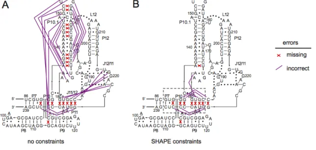
2.2.2 Accurate and quantitative RNA structure analysis using 1M7. We next evaluated the extent to which 1M7 provides accurate and quantitative information regarding RNA structure using the specificity domain of the Bacillus subtilis RNase P enzyme. This domain was chosen because it is a large (154 nt) RNA with a known structure [15] that does not contain pseudoknots, which are not well predicted by current algorithms. This RNA spans numerous typical base‐pairing and stacking interactions, a tetraloop‐receptor tertiary interaction (involving L12 and P10.1) common to many large RNAs, and two large internal loops (J11/12 and J12/11) stabilized by an extensive series of non‐canonical interactions [15].
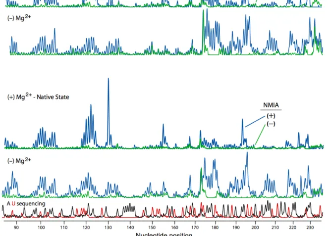
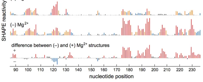
primer extension, using fluorescently labeled DNA primers, resolved by capillary electrophoresis [16] (Figure 2.3). Absolute SHAPE reactivities were calculated by subtracting the background observed in no‐reagent control experiments that omitted 1M7. Reactivity at each nucleotide was classified as high, medium, low, or near‐zero (red, orange, blue, and black columns, respectively, in Figure 2.4).
Superposition of the quantitative reactivity information on a secondary structure diagram [15] for the RNase P specificity domain shows that a 70 sec reaction with 1M7 accurately reports the known secondary and tertiary structure for this RNA (Figure 2.5A). Essentially all nucleotides involved in Watson‐Crick base‐pairs are unreactive; moreover, many non‐canonical, but stable, U•G, A•A, and A•G pairs are
unreactive. Nucleotides in P10.1 and in L12 that form the tetraloop‐receptor tertiary structure motif are also unreactive. In contrast, nucleotides in loops or adjacent to bulges or other irregularities are reactive. Nucleotides in the structurally idiosyncratic module involving J11/12 and J12/11 show a wide range of reactivities. Strikingly, the most highly conserved nucleotides in this module (A187, A191, G219‐G220, A222), which participate in stabilizing tertiary interactions [15], also show the lowest SHAPE reactivities using 1M7.
reflect changes in RNA secondary and tertiary structure and not Mg2+‐induced differences in reagent properties.
We quantified the effect of Mg2+ on the structure of the RNA using a difference plot in which nucleotide reactivities in the (+) Mg2+ experiment were subtracted from the (–) Mg2+ experiment. Positive and negative peaks thus indicate an increase or decrease in local nucleotide flexibility in the absence of Mg2+, respectively (lower panel, Figure 2.4). Many sites in the (–) Mg2+ experiment show increased SHAPE reactivity. Strikingly, increased reactivity occurs precisely at nucleotides that participate in tertiary interactions in the RNase SHAPE reactivity also shows that the irregularly stacked P7‐P10‐P11 helical domain unfolds when Mg2+ is removed (Figure 2.5B).
prediction features an overall topology that closely resembles the correct structure. 2.3 Discussion
SHAPE chemistry performed with 1M7 accurately reports the known structure of the RNase P specificity domain under native conditions. 1M7 reactivity detects nucleotides constrained both by base pairing and by idiosyncratic, non‐canonical tertiary interactions (Figure 2.5). SHAPE chemistry enables very precise analysis of the differences between two structures, such as Mg2+‐dependent tertiary interactions. 1M7 is easily handled in the laboratory and enables analysis of large RNA structures at single nucleotide resolution in less than 70 seconds.
2.4 Experimental
2.4.1. Synthesis of [32P]labeled pApethyl. Adenosine‐3′‐(O‐ethyl)‐phosphate prescursor (10 µM final) was 5′‐[32P]‐labeled using T4 polynucleotide kinase [10 µL;
containing 70 mM Tris‐HCl, 10 mM MgCl2, 5 mM dithiothreitol, 1 µL T4 PNK (10,000
units/mL), 60 µCi [γ‐32P]ATP; 37 °C for 1 hr]; purified by gel electrophoresis (30%
polyacrylamide, 29:1 acrylamide:bisacrylamide, 0.4 mm × 28.5 cm × 23 cm; 30 W; 1 hr);
excised from the gel; passively eluted into 300 µL HE (10 mM Hepes pH 8.0, 1 mM
EDTA; overnight at 4 °C); and separated from solid acrylamide by microfiltration (EZ
spin columns, Millipore).
mixture was stirred at room temperature for 4 hours. The reaction was poured into 50 mL of cold 1 N HCl, and the resulting bright orange precipitate was filtered and washed sequentially with water and then ether to give 608.3 mg (86%) of product. 1H NMR (CO(CD3)2, 400 MHz, ) δ 3.69 (s, 3 H, ‐NCH3‐), 8.12 (dd, J = 8.8 Hz, 2 Hz, 1 H, ArH), 8.2 (d, J = 2 Hz, 1H, ArH), 8.34 (d, J = 8.4 Hz, 1 H, ArH).
2.4.3. NMIA and 1M7 hydrolysis and 2′Oadduct Formation. Hydrolysis was
followed by adding (1.5 mM NMIA or 2.0 mM 1M7 in 300 µL DMSO) reagent to 1.1×
buffer [2.7 mL, 6.7 mM MgCl2, 111 mM NaCl, 111 mM HEPES (pH 8.0)] equilibrated at 37 °C in a cuvette. Pseudo‐first‐order rates were obtained by monitoring the
absorbance of the hydrolysis product (at 360 nm for 2‐methylaminobenzoate and 430 nm for 2‐methylamino‐4‐nitrobenzoate). Rates of adduct formation for [32P]‐labeled pAp‐ethyl (10,000 cpm/µL) were obtained by adding 10% (v/v) reagent (5 mM final
NMIA or 1M7 in DMSO) to 1.1× reaction buffer; quenching the reaction with 1 vol 250
mM dithiothreitol; resolution by gel electrophoresis (30% polyacrylamide; 29:1 acrylamide:bisacrylamide; 0.4 mm × 28.5 cm × 23 cm; 30 W; 45 min); and quantifying by phosphorimaging. Reaction rates were obtained using an equation that accounts for parallel reaction of NMIA or 1M7 by 2′‐O‐adduct formation (kadduct) and by hydrolysis (khydrolysis): fraction product = 1 – exp[(kadduct[reagent]0/(khydrolysis))(e‐(khydrolysis)t – 1)] [17].
2.4.4. Synthesis of Bacillus subtilis RNase P RNA. A DNA template for transcription of the specificity domain of the B. subtilis RNase P, inserted in the context of a 5′ and 3′ flanking structure cassette [12, 13], was generated by PCR [1 mL;
each forward and reverse primer, 5 pM template, and 0.025 units/µL Taq polymerase;
denaturation at 94 °C, 45 s; annealing 55 °C, 30 s; and elongation 72 °C, 1 min; 38
cycles]. The PCR product was recovered by ethanol precipitation and resuspended in 150 µL of TE [10 mM Tris (pH 8.0), 1 mM EDTA]. Transcription reactions (1.5 mL, 37 °C,
4 h) contained 40 mM Tris (pH 8.0), 10 mM MgCl2, 10 mM DTT, 2 mM spermidine, 0.01% (v/v) Triton X‐100, 4% (w/v) poly(ethylene) glycol 8000, 2 mM each NTP, 50 µL
of PCR‐generated template, and 0.1 mg/mL of T7 RNA polymerase. The RNA product was purified by denaturing polyacrylamide gel electrophoresis (8% polyacrylamide, 7 M urea, 29:1 acrylamide:bisacrylamide, 32 W, 2 h), excised from the gel, and recovered by electroelution and ethanol precipitation. The purified RNA (~4 nmol) was resuspended in 100 µL TE.
2.4.5. StructureSelective RNA Modification. RNA (2 pmol) in 5 µL 1/2× TE
was heated at 95 °C for 2 min, cooled on ice, treated with 3 µL of 3× folding buffer [333
mM NaCl, 333 mM Hepes (pH 8.0), 33.3 mM MgCl2 (or no MgCl2)], and incubated at 37
°C for 20 min. The RNA solution was treated with 1M7 or NMIA (1 µL, 65 mM in
anhydrous DMSO), allowed to react for 70 sec (equal to five 1M7 hydrolysis half‐lives, accompanied by a colorimetric change from pale yellow‐orange to deep orange‐brown upon completion) or 25 min (five NMIA hydrolysis half‐lives). No‐reagent control reactions contained 1 µL of DMSO. Modified RNA was recovered by ethanol
precipitation [90 µL sterile H2O, 5 µL NaCl (5 M), 1 µL Glycogen (20 mg/mL), 400 µL
ethanol; 30 min at –80 °C] and resuspended in 10 µL of TE.
AGC CCG; 3 µL, 0.4 µM) was added to the RNA (10 µL, from the previous step) by
heating to 65 °C (6 min) and 35 °C (20 min). Reverse transcription buffer [6 µL; 167
mM Tris (pH 8.3), 250 mM KCl, 10 mM MgCl2, 1.67 mM each dNTP] was added; the RNA was heated to 52 °C; Superscript III (1 µL, 200 units) was added and reactions were
incubated at 52 °C for 30 min. Primer extension reactions were quenched by addition of
4 µL of an equal mixture of EDTA (100 mM) and sodium acetate (3 M, pH 5.2). The
resulting cDNAs were recovered by ethanol precipitation, washed twice with 70% ethanol, dried in a SpeedVac for 10 min, and resuspended in 40 µL de‐ionized
formamide. Dideoxy sequencing markers were generated using unmodified RNA and primers labeled with unique fluorophores (D2 or IR800, 1 µM), and by adding 1 µL of
3′‐deoxythymidine (10 mM) or 3′‐dideoxyadenosine (2 mM) triphosphate after
addition of reverse transcription buffer. The cDNA extension products were separated by capillary electrophoresis using a Beckman Coulter CEQ 2000XL DNA Analysis System.
nucleotides (3 nts), averaging the next 8% of reactive nucleotides (12 nts), and then dividing all intensities by this average high value. This gives intensities from 0 to slightly greater than 2. In RNAstructure [9], nucleotides with reactivities greater than 0.75 were required to be single stranded and positions with reactivities greater than 0.35 were prohibited from forming internal Watson‐Crick pairs.
2.5 References
1. Tinoco, I. and Bustamante, C., How RNA folds. J. Mol. Biol., 1999. 293: p. 271-281.
2. Eddy, S.R., How do RNA folding algorithms work? Nature Biotechnol., 2004. 22(11): p. 1457-1458.
3. Doshi, K.J., et al., Evaluation of the suitability of free-energy minimization using nearest-neighbor energy parameters for RNA secondary structure prediction. BMC Bioinf., 2004. 5: p. 105.
4. Dowell, R.D. and Eddy, S.R., Evaluation of several lightweight stochastic context-free grammars for RNA secondary structure prediction. BMC Bioinf., 2004. 5: p. 71.
5. Mathews, D.H. and Turner, D.H., Prediction of RNA secondary structure by free energy minimization. Curr. Opin. Struct. Biol., 2006. 16: p. 270-278.
6. Peattie, D.A. and Gilbert, W., Chemical Probes for higher-order structure in RNA. Proc. Natl. Acad. Sci. U.S.A., 1980. 77: p. 4679-4682.
7. Ehresmann, C., et al., Probing the structure of RNAs in solution. Nucleic Acids Res., 1987. 15: p. 9109-9128.
8. Knapp, G., Enzymic approaches to probing of RNA secondary and tertiary structure. Methods Enzymol., 1989. 180: p. 192-212.
9. Mathews, D., et al., Incorporating chemical modification constraints into a dynamic programming algorithm for prediction of RNA secondary structure. Proc. Natl. Acad. Sci. U. S. A., 2004. 101(19): p. 7287-7292.
10. Badorrek, C.S. and Weeks, K.M., RNA flexibility in the dimerization domain of a gamma retrovirus. Nat. Chem. Biol., 2005. 1(2): p. 104-11.
12. Wilkinson, K.A., Merino, E.J., and Weeks, K.M., RNA SHAPE chemistry reveals non-hierarchical interactions dominate equilibrium structural transitions in tRNAAsp
transcripts. J. Am. Chem. Soc., 2005. 127: p. 4659-4667.
13. Wilkinson, K.A., Merino, E.J., and Weeks, K.M., Selective 2'-hydroxyl acylation analyzed by primer extension (SHAPE): quantitative RNA structure analysis at single nucleotide resolution. Nat. Protoc., 2006. 1(3): p. 1610-6.
14. Exner, O., Correlation Analysis in Chemistry, ed. Chapmen, N.B. and J.Shorter. 1978, New York: Plenum Press.
15. Krasilnikov, A.S., et al., Crystal structure of the specificity domain of ribonuclease P. Nature, 2003. 421: p. 760-764.
16. Wilkinson, K.A., et al., High-throughput SHAPE analysis reveals structures in HIV-1 genomic RNA strongly conserved across distinct biological states. PLoS Biol., 2008.
6(4): p. e96.
17. Chamberlin, S.I. and Weeks, K.M., Mapping local nucleotide flexibility by selective acylation of 2 '-amine substituted RNA. J. Am. Chem. Soc., 2000. 122(2): p. 216-224.
18. Vasa, S.M., et al., ShapeFinder: a software system for high-throughput quantitative analysis of nucleic acid reactivity information resolved by capillary electrophoresis. RNA, 2008. 14(10): p. 1979-90.
3.1 Introduction
Local and global dynamics in folded RNAs occur over broad timescales spanning
picoseconds to minutes [1, 2]. Slow motions likely play predominant roles in governing
RNA folding and ribonucleoprotein assembly reactions. However, slow local motions
are extremely difficult to detect in a routine way, especially for large RNAs with
complex structures.
The local environment and degree of flexibility can be evaluated at nucleotide
resolution for RNAs of any size using selective 2'‐hydroxyl acylation analyzed by primer
extension (SHAPE) chemistry [3‐5]. RNA nucleotides exist in equilibrium between
constrained (closed) and flexible (open) states. The 2'‐OH group in flexible nucleotides
preferentially adopts an open, reactive, conformation that facilitates reaction with
electrophilic reagents to form a 2'‐O‐adduct (Figure 3.1). SHAPE experiments work well
using electrophiles based on the isatoic anhydride (IA) scaffold [3, 6]. Positions that
form 2'‐O‐adducts are detected by primer extension [3‐5].
IA derivatives both react with the RNA 2'‐OH group and also undergo concurrent
degradation by hydrolysis (Figure 3.1). 2'‐hydroxyl reactivity is thus conveniently
monitored by allowing a reaction to proceed until the reagent has been consumed,
either by hydrolysis or by reaction with RNA. At this endpoint, the fraction adduct [7] at
any nucleotide (ƒ) is:
f ≈ 1 – e–(kobs/khydrolysis) (1) where
€
kobs=
kopenkadduct[reagent] kopen+kclose+kadduct[reagent]
(2)
And the rate of hydrolysis has been shown to be proportional to the rate of adduct formation [4, 6],
kadduct/khydrolysis = β (3)
These relationships lead to two limits. In Limit 1, kopen + kclose >> kadduct[reagent],
f = 1 –
€
e
− kopen
kopen+kclose
β[reagent]
(4)
For Limit 2, kopen + kclose << kadduct[reagent],
f = 1 – e–kopen/khydrolysis + A (5),
where A represents a plateau term that corresponds to a small fraction of RNAs that are
unfolded, therefore reactive towards all reagents.
In this chapter, Costin Gherghe and I show that is possible to monitor local
nucleotide dynamics in RNA under conditions where Limit 2 applies by varying the
reactivity (or khydrolysis) of the hydroxyl‐selective electrophile. IA has a hydrolysis half‐ life (t1/2) of 430 sec at 37 °C (table, Figure 3.1). Electron withdrawing substituents at the cyclic amine (R1) or in the benzene ring (R2) enhance reagent reactivity. Compared to IA, N‐methyl isatoic anhydride (NMIA), 4‐nitro‐isatioc anhydride (4NIA) and 1‐
methyl 7‐nitroisatoic anhydride (1M7) [6] have progressively shorter hydrolysis half‐
lives (Figure 3.1).
3.2 Results
3.2.1 SHAPE analysis of C2´endo nucleotides. To investigate if distinct local
local nucleotide dynamics at an important variation in RNA structure: the C2'‐endo
conformation. Although C2'‐endo nucleotides are relatively rare, they are highly
overrepresented in important RNA tertiary interactions and in catalytic active sites [8].
Local structure at tandem G•A mismatches depends on the local sequence context [9‐
11]. Guanosine nucleotides in G•A pairs adopt the C2'‐endo conformation in the
sequences (UGAA)2 [10] and (GGAU)2 [11], the C3'‐endo conformation typical of standard A‐form helix geometry in (CGAG)2 [9], and a mixture of C2'‐endo/C3'‐endo conformations in (UGAG)2 [11]. Costin Gherghe constructed a simple hairpin RNA (termed the C2'‐endo RNA) containing each of these sequences. Because these G•A
mismatch‐containing sequences are palindromic, there are two equivalent examples of
each G•A pair in the RNA, including four total C2'‐endo nucleotides (in red, Figure 3.2A).
When the C2'‐endo RNA was subjected to SHAPE analysis using the fastest
reagent, 1M7, flexible nucleotides in the apical loop (nts 45‐48) are reactive, while
positions constrained by base pairing are unreactive, regardless of the sugar pucker
(top panel, Figure 3.2B). When an otherwise identical experiment was performed with
IA, which reacts 30‐fold more slowly, nucleotides in the apical loop were again reactive
while most of the base paired nucleotides were unreactive, similar to their reactivities
with 1M7. In strong contrast, the four G nucleotides that adopt the C2'‐endo
conformation were highly reactive, even more so than some nucleotides in the flexible
loop (Figure 3.2B, bottom panel; red bars at positions 19, 40, 52, 73). For the two
reagents with intermediate reactivities, 4NIA and NMIA, the C2'‐endo positions are
moderately reactive (Figure 3.2B). Nucleotides constrained in these C2'‐endo
reactive towards the slower reagents.
Limit 1 (Eqn. 4) predicts that adduct formation is proportional to the
equilibrium constant for formation of the open state [kopen/(kopen + kclose)] and is independent of the reagent hydrolysis rate. Most nucleotides thus far analyzed by
SHAPE, including in tRNA [3, 4] and in an RNase P RNA [6], are characterized by limit 1.
In contrast, the observation of a strong dependence of adduct formation on
khydrolysis suggests that Limit 2 applies to the C2'‐endo nucleotides in Figure 2A. Limit 2 also implies (1) that the extent of reaction at C2'‐endo nucleotides will be independent
of reagent concentration and (2) that kobs reports kopen (compare Eqns. 4 & 5). Costin Gherghe analyzed the concentration dependence for reaction at positions 52 and 73
using isatoic anhydride and found, as predicted by Limit 2, that adduct formation is
independent of reagent concentration under conditions where reaction of the
unconstrained model nucleotide, pAp‐ethyl, showed a clear concentration dependence
(Figure 3.3).
Costin Gherghe estimated the magnitude of kopen at the C2'‐endo nucleotides at positions 19/73 and 40/52 by fitting the extent of 2'‐O‐adduct formation as a function
of khydrolysis to Eqn. 5 (Figure 3.4). In both cases, kopen is 4 × 10⁻⁵ s–1. In contrast,
reactivities for both pAp‐ethyl and flexible loop nucleotides are independent of khydrolysis (Figure 3.5).
Critically, some C2'‐endo nucleotides thus experience extraordinarily slow local
dynamics to form conformations reactive towards isatoic anhydride‐based
electrophiles.
Figure 3.4. Determination of kopen for C2´‐endo position 19/73 and 40/52. Lines represent a fit to Eqn. 5; rate constants are ~ 4 × 10‐5 s‐1. Error bars indicate standard
Figure 3.5. Absence of a dependence of fraction adduct formed as a function of khydrolysis for reaction of the (unconstrained) model nucleotide pAp‐ethyl and for nucleotide 45 in the loop of the C2´‐endo construct.
3.2.2. Identification of slowmoving nucleotides in RNase P. We next explored
whether the differential reactivity between the 1M7 and IA can be used to identify
nucleotides that undergo slow conformational dynamics in an RNA with a complex
structure, the specificity domain of the B. subtilis ribonuclease P enzyme (RNase P) [12].
After excluding nucleotides where the electron density was not well defined or that
participate in crystal contacts, we identified 10 C2'‐endo nucleotides in the RNase P
RNA (in color, Figure 3.6).
For the vast majority of RNase P nucleotides, including all positions with C3'‐
endo conformations, SHAPE reactivities were identical for both the fast (1M7) and slow
(IA) reagents (Figure 3.6). These nucleotides reflect limit 1. The 10 well defined C2'‐
endonucleotides fell into three categories: (i) most C2'‐endo nucleotides are highly
constrained and, as expected [3], unreactive towards both reagents (blue nucleotides,
Figure 3.6A), (ii) one nucleotide is not constrained and is reactive towards both
electrophiles (red nucleotide, Figure 3.6A), and (iii) two C2'‐endo positions show large
changes in reactivity (nts A130 and G168, circled nucleotides and red bars, Figure 3.6).
Two other nucleotides showed smaller changes in reactivity but were in regions of the
structure where experimental electron density was poorly defined (gray columns,
Figure 3.6B). A similar distribution of reactive and unreactive C2'‐endo nucleotides
occurs in the Tetrahymena P5‐P4‐P6 domain using NMIA [13].
3.3. Discussion
While the C2'‐endo conformation by itself clearly does not govern SHAPE
dynamics. These nucleotides in both the simple C2'‐endo RNA (Figure 3.2B) and in the
RNase P RNA (Figure 3.6B) share key characteristics: (1) the ribose group has the C2'‐
endo conformation and (2) the nucleotide conformation is partially constrained by base
stacking and hydrogen bonding interactions (Figure 3.7). These C2'‐endo dynamics are
orders of magnitude slower than for other local RNA conformational changes like base
opening reactions [14, 15] and are also slower than folding processes that involve
assembly of whole domains in large RNAs [2].
In our view, the best precedent for slow conformational changes at a single
residue in a biopolymer is the cis‐trans isomerization of prolyl residues in proteins.
Proline cis‐trans conformations interconvert on the order of 10–2–10–5 s–1 [16‐18] and are thus comparable to the rates measured here for local dynamics at some C2'‐endo
nucleotides in RNA. Cis‐trans isomerization can function as a molecular switch in
biology [16].
We postulate that slow conformational dynamics at C2'‐endo nucleotides also
have the potential to function as rate‐determining molecular switches and will play
important, but currently unexplored, roles in RNA folding, ligand recognition, and
catalysis.
3.4 Experimental
3.4.1. Derivation of Eqn. 1. The electrophile‐dependent reaction of RNA to form a
2'‐O‐adduct involves the following mechanism and four relevant rate constants:
Scheme 1
The observed rate of 2'‐O‐adduct formation is given by: €
kobs = kopenkadduct[reagent]
kopen +kclose +kadduct[reagent]
then [3], ƒ
€
≈
1
−
e
− kobs
khydrolysis(1−e
−khydrolysist)
where ƒ is the fraction 2'‐O‐adduct formed at any given nucleotide.
If the reaction is allowed to proceed until reagent hydrolysis is complete (t→∞), this
equation simplifies to:
ƒ
€
≈
1
−
e
− kobs
khydrolysis as given in Eqn. 1. 3.4.2. IA, NMIA, 4NIA and 1M7 hydrolysis. Hydrolysis was followed by adding
reagent (2.0 mM IA, 1.5 mM NMIA, 2.5 mM 4NIA, or 2.0 mM 1M7 in 300 µL DMSO) to
absorbance of the hydrolysis product (at 345 nm for 2‐aminobenzoate, 360 nm for 2‐
methylaminobenzoate, 440 nm for 2‐amino‐4‐nitrobenzoate, and 430 nm for 2‐
methylamino‐4‐nitrobenzoate).
3.4.3. RNA constructs. The C2'‐endo hairpin RNA (Figure 3.2A) and the
specificity domain of the RNase P RNA were synthesized by in vitro transcription using
a single stranded DNA (IDT) or a PCR‐generated template [6], respectively. In both
cases, the RNAs were embedded in the context of 5' and 3' structure cassette [5]
sequences. RNAs were purified by denaturing polyacrylamide gel electrophoresis,
excised from the gel, and recovered by electroelution and ethanol precipitation. Purified
RNAs were resuspended in TE [10 mM Tris (pH 8.0), 1 mM EDTA] at concentrations of
about 30 µM and stored at ‐20 °C.
3.4.4. SHAPE analysis. pAp‐ethyl was 5'‐end radiolabeled using γ‐[32P]‐ATP,
purified by denaturing gel electrophoresis, and resuspended in 1/2× TE. The pAp‐ethyl
(1 µL, 10000 cpm) was heated to 95 °C for 2 min, cooled on ice, mixed with 3 µL of 3.3×
folding buffer [264 mM NaCl, 66 mM Hepes (pH 8.0), 16.5 mM MgCl2], and incubated at 37 °C for 20 min. The pAp‐ethyl solution was treated with reagent (1 µL; 100 mM; in
anhydrous DMSO), allowed to react for 36 min (equal to five IA hydrolysis half‐lives[3]).
The no‐reagent control contained 1 µL neat DMSO. The C2'‐endo RNA (4 pmol) SHAPE
experiments were performed as described for pAp‐ethyl with the addition of a primer
extension step. Modified RNA was recovered by ethanol precipitation [90 µL sterile
H2O, 5 µL NaCl (4 M), 1 µL glycogen (20 mg/mL), 400 µL ethanol; 30 min at ‐80 °C] and resuspended in 10 µL of TE. Analysis of the RNase P RNA was performed similarly