COMPARISON OF TWO GINGIVAL DISPLACMENT PROCEDURES; A PILOT STUDY
Anthony P. Prudenti
A thesis submitted to the faculty at the University of North Carolina at Chapel Hill in partial fulfillment of the requirements for the degree of Master of Science in the Department of
Prosthodontics in the School of Dentistry.
Chapel Hill 2017
Approved by:
Sumitha N. Ahmed
Terence E. Donovan
Ryan D. Cook
ABSTRACT
Anthony P. Prudenti: Comparison of Two Gingival Displacement Procedures; a Pilot Study (Under the direction of Sumitha N. Ahmed)
Objective: The primary objective was to examine if a cordless gingival displacement
procedure displaces sulcular tissue to facilitate acceptable impressions for fixed prosthodontic
restorations.
Materials and Methods: Fifteen (15) patients were recruited; cordless impressions
(n=7) and conventional corded impression (n=8) were made during routine treatment for fixed
dental prostheses.
Results: Fisher’s exact tests, Wilcoxon rank sum tests, and an unpaired t-test were used
to compare variables between (CD) and (CL) groups, and to compare variables between
acceptable and unacceptable impressions. Level of significance was set at 0.05 for all analysis.
Within this small sample size, the 2 groups (CD and CL) are significantly similar in relation to
most variables. Only TEAR and EVAL were significantly different between CD and CL, and
VOID was significantly different between acceptable and not acceptable impression groups.
Conclusions: Within the limitations of this study, marginal tearing statistically affected
ACKNOWLEDGEMENTS
I would like to express my sincere appreciation to my mentors in the Department of
Prosthodontics: Dr. Lyndon Cooper, Dr. Ryan Cook, Dr. Kent Healey, and Dr. Glenn Minsley
for their invaluable guidance, patience, time and effort through these 3 years.
Thank you to all of the prosthodontics faculty and staff who have helped me thorough
this incredible learning experience here at UNC.
I would like to thank Dr. Terry Donovan and Dr. Sumitha Ahmed in the Department of
Operative Dentistry for inspiring this research objective, for their willingness to be part of this
committee, and for their contributions with this project. I would also like to thank former
Operative Dentistry resident, Dr. Clayton Rau, for his in-depth research on this subject matter;
much of this project was based on his project’s defined criteria.
I would like to thank Dr. Ceib Phillips, her staff, and graduate student Pooja Tanya Saha,
for their extensive help with the data collection forms, randomization, and statistical analysis.
I would like to thank Teresa Etscovitz for her detailed dedication and significant efforts
during the IRB approval process.
I would also like to thank my co-residents who made the effort to participate in this study
PREFACE
I would like to acknowledge the previous work completed in 2015 by Clayton T. Rau,
for his master’s degree thesis titled, “THE QUALITY OF FIXED PROSTHODONTIC
IMPRESSIONS: AN ASSESSMENT OF CROWN AND BRIDGE IMPRESSIONS
RECEIVED AT COMMERCIAL LABORATORIES.” It was much of his work, and that of his
mentor Dr. Terry Donovan, that outlined, defined, and guided this project.
TABLE OF CONTENTS
LIST OF TABLES ... ix
LIST OF FIGURES ...x
LIST OF ABREVIATIONS ... xi
1. CHAPTER 1: LITERATURE REVIEW ...1
1.1 A History of Impression Quality ...2
1.2 Impression Materials ...3
1.2.1 Effects Of Moisture ...4
1.2.2 Interactions With Other Materials ...6
1.3 Impression Trays ...7
1.3.1 Stock Trays ...7
1.3.2 Custom Trays ...8
1.3.3 Dual Arch Trays ...8
1.4 Margin Design and Placement ...9
1.4.1 Subgingival Margins ...10
1.4.2 Biologic Width ...10
1.5 Gingival Displacement ...11
1.5.1 Gingival Retraction Cords and Medicaments ...12
1.5.2 Classical Displacement Methods ...13
1.5.3 Alternative Methods ...14
2. CHAPTER 2: Comparison of Two Gingival Displacement Procedures;
a Pilot Study ...25
2.1 Introduction ...25
2.2 Material and Methods ...28
2.3 Statistical Analysis ...32
2.4 Results ...32
2.5 Discussion ...33
2.6 Limitations ...38
2.7 Conclusions ...40
3. TABLES...41
4. FIGURES ...44
LIST OF TABLES
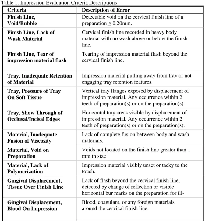
Table 1. Impression Evaluation Criteria Descriptions ...41
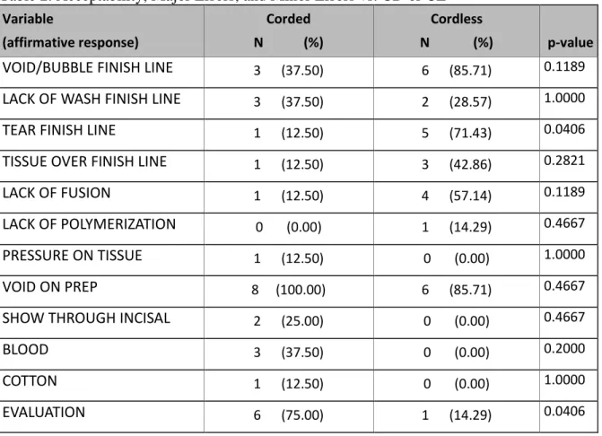
Table 2. Acceptability, Major Errors, and Minor Errors vs. CD or CL ...42
Table 3. Major Errors vs. Acceptability...42
Table 4. Age ...42
LIST OF FIGURES
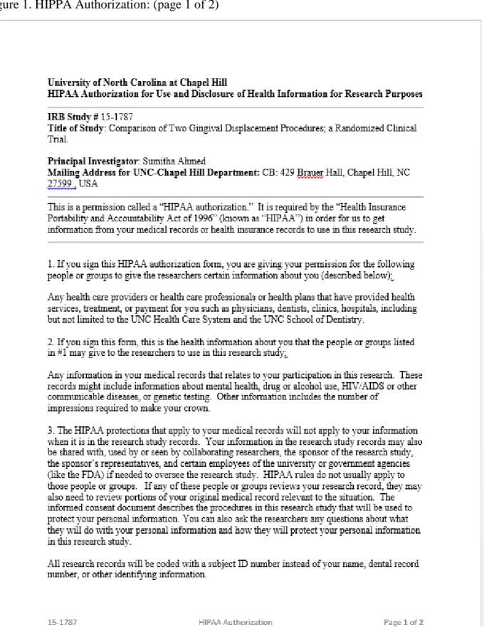
Figure 1. HIPPA Authorization ...44
Figure 2. Adult Consent to Participate in Research Study...46
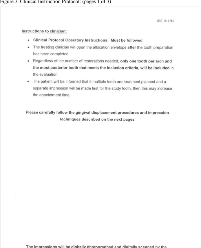
Figure 3. Clinical Instruction Protocol ...50
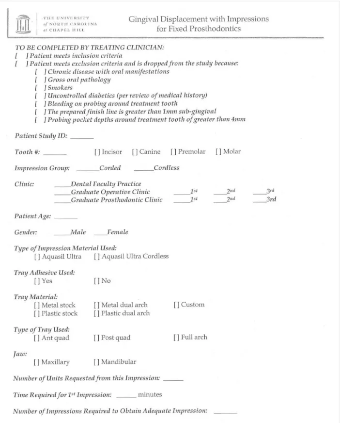
Figure 4. Clinical Impression Form ...53
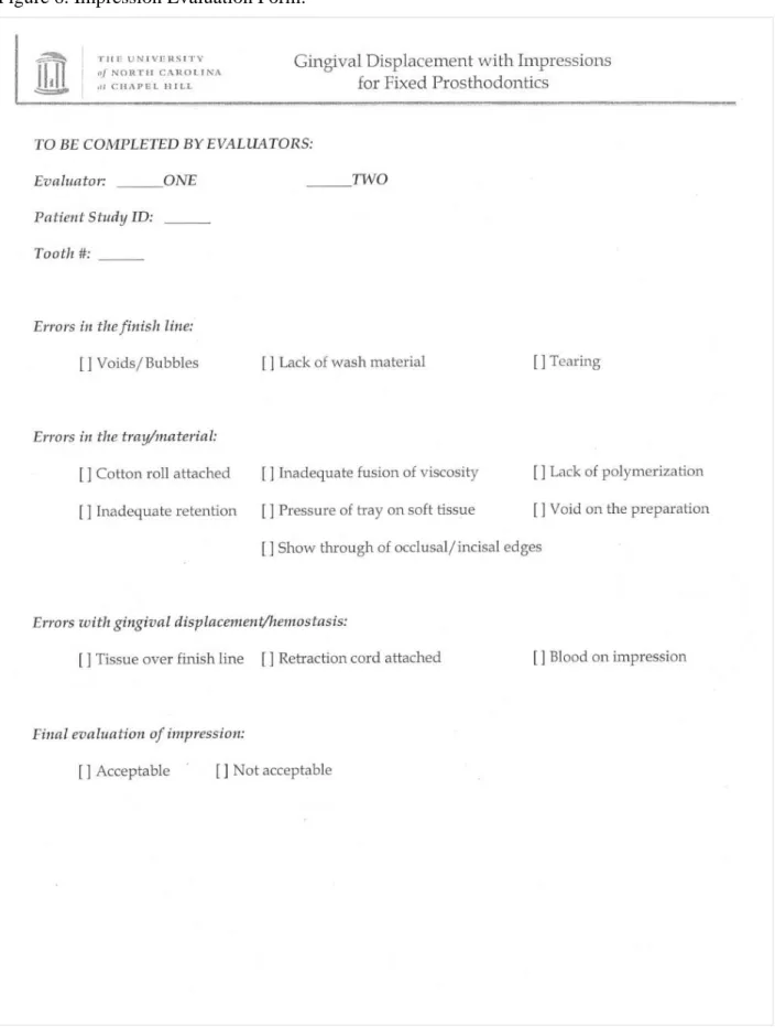
Figure 5. Impression Evaluation Form ...54
LIST OF ABBREVIATIONS
PE Polyether
PVS Poly-vinyl siloxane
VPS Vinyl polysiloxane
UNC University of North Carolina
SOD School of Dentistry
CL Cordless Impression Group
CD Corded Impression Group
VOID Voids or bubbles on the finish line
WASH Lack of wash material on the finish line
TEAR Tearing of material at the finish line
TISSUE Tissue over the finish line
CHAPTER 1: LITERATURE REVIEW
Introduction
The success of any fixed prosthesis starts with the accuracy of the impression. Obtaining
an impression that accurately captures the prepared margin and cervical finish line is paramount
in the fabrication of well-fitting indirect restorations. A vital component in impression making is
retraction of gingiva. Atraumatic gingival displacement allows access for impression material to
accurately record the finish line and provides sufficient thickness of impression material in the
gingival sulcus to prevent tearing during removal.1 Making an optimal impression for indirect
restorations remains one of dentistry’s most challenging procedures.2,3 Clinicians must be able to
properly select gingival displacement procedures and impression materials, as well as evaluate the
quality of their impressions.4-7 These play a critical role in the success or failure of the final
restoration.4,5
Modern impression materials have improved the accuracy of impression making.8,9 Despite
these improvements, many studies have reported that impressions sent to dental laboratories for
fabrication of indirect restorations still remain inadequate.4,5,10,11,12 To date all impression materials
require control of the gingival tissues adjacent to the preparation, adequate placement of the
material around the finish line, and the use of an appropriate impression tray.3 Stewardson in 2005
recognized that a lack of impression making principles is one of the major causes of unacceptable
1.1.A History of Impression Quality
Historically, studies have shown that clinicians consistently make inadequate
impressions.14 In 1984, Aquilino and Taylor11 recognized the discrepancy between dental
education, private practice, and what was being sent to dental laboratories. The study expresses
concerns that recent graduates are gaining less laboratory experience and exposure in school, and
that they quickly abandon the sound principles they were taught in school once they get out into
private practice.
Winstanely et al.10 evaluated 290 impressions from four commercial dental laboratories.
They reported that an acceptable restoration could be fabricated on 57% of the impressions
evaluated, and that 20% of the impressions would be impossible or doubtful to fabricate an
acceptable final restoration. In this study, the major cause of defective impressions was
indiscernible recording of the finish line. Irreversible hydrocolloid was the material used for most
all of the impressions evaluated in this study.
Albashaireh et al.15 evaluated 136 impressions sent to commercial laboratories for
fabrication of fixed restorations. They studied the quality of impressions made and found that 50%
of impressions/dies to be unsatisfactory or unusable.
Samet et al.4evaluated 193 impressions from 11 different laboratories. Using a more
detailed evaluation criterion they found that 89% of all impressions evaluated had at least one
detectable error. This study also found that 51% of the defects involved the cervical finish line.
In 2007 Beier et al.2evaluated 1,466 impressions and found a remarkably low
unacceptable rate of 3%. An explanation for this low unacceptable rate may be due to the strict
protocol the clinicians followed, using retraction cord and controlling for moisture. Findings in
most practices.
1.2.Impression Materials
Today most impressions are made with polyether (PE) and polyvinyl siloxane (PVS).
Digital impression techniques (optical scanning) have become popular and are promoted as the
future of dentistry. This review focuses on PVS impression material because it is most applicable
to the study design.
Poly-vinyl siloxane, also known as vinyl polysiloxane, polyvinyl, and addition silicone
have been used as an impression material since the mid-1970s. The major advantages to the
material is that its superior reproduction of fine details and elastic recovery.8 PVS materials set by
way of an addition reaction, which involves the linking of a vinylsiloxane with a hydrogen siloxane
via a platinum catalyst. Hydrogen is produced as a byproduct of this reaction, and is then scavenged
by platinum. Silica fillers are used to control viscosity and rigidity of the material.9,16,17 Another
major advantage of PVS impression materials are that their dimension stability over time. This
material can be stored for weeks without losing accuracy.8 The major disadvantages of PVS
impression material is its hydrophobic nature. This quality makes it sensitive to blood, saliva, and
crevicular fluids in the unset phase.8,9,16,17
Chai et al. describes three mechanical properties of impression materials that are clinically
relevant: yield strength determines the ability of a material to withstand stress without permanent
deformation, strain at yield point indicates the amount of undercut that the impression material
can overcome without permanent elastic deformation, and tear energy indicates the resistance to
tear of impression material.18 Perakis et al. suggest that the ideal impression material will absorb
the most energy prior to the point of permanent deformation, without tearing.19 Laufer et al. in
when the sulcular space allows a sufficient thickness of material of 0.2 mm or greater.20
1.2.1. Effects of Moisture
The way impression materials interact with moisture is described as the material’s
hydrophilic or hydrophobic nature. The hydrophilicity of a material is measured by the angle a
standardized droplet of water makes with the material. Materials forming angles less than 90
degrees are defined as hydrophilic and those forming angles greater than 90 degrees as
hydrophobic.15 PVS materials are principally hydrophobic because they contain hydrophobic
aliphatic hydrocarbon groups around the siloxane bond.9,16,21,22
Peutzfeldt and Asmussen22evaluated how the hydrophilicity and viscosity affect the
ability of impression materials to displace water and replicate surface detail. Their results found
that materials with contact angles below 70 degrees performed better at displacing water as the
hydrophilicity increased. Materials that were more hydrophobic (contact angles over 70 degrees)
showed a propensity to displace water more readily with increases in viscosity. When water was
omitted all impression materials achieved 100% reproduction of detail.
Johnson et al.23 evaluated the ability of PE and PVS materials to replicate surface detail
of a standard pattern metal plate. They evaluated three variables: material (PE and PVS), surface
conditions (wet and dry), and technique (mono- and dual-phase). They found that PE was
superior to PVS, monophase was superior to a dual viscosity, and dry conditions were superior to
wet conditions in replication of preparation details. It was noted that the pattern used in the study
contained ridge heights of 10µm, while ISO specification for fine detail reproduction is 20µm.
Considering this, all of the PE and PVS samples except one dual-phase PVS, produced
Petrie et al.24 published similar findings also in 2003. This study created dry, wet, and
“moist” surfaces to evaluate the detail reproduction of two PVS materials. Stainless steel dies
fabricated to ADA specification no. 19 were used in to evaluate these impression materials. This
study found that as the moisture level increased the ability to reproduce surface detail for PVS
materials significantly decreased. In 2005, Walker et al.25repeated the study with PVS and PE
materials under dry and moist conditions. The PE materials achieved complete reproduction of
surface details on the dies in both dry and moist conditions, while the PVS materials were not able
to reproduce the details in moist condition.
Rupp et al.26,27 showed that improved PVS materials with the addition of surfactants failed
to achieve a similar hydrophilicity to PE materials. It was shown that the surface tension improved
over 60 minutes, and that the addition of surfactants may be beneficial during the pouring of the
impression, and not during the impression making process itself.28
Nagrath et al.29 evaluated four hydrophilic VPS impression materials. They found that the
dimensional stability remained intact in all conditions (wet, moist, and dry), but the best surface
detail results were obtained only under dry conditions for all the four materials.
Basapogu et al.30 compared hydrophilic and hydrophobic VPS impression materials in a
moist environment. Using stainless steel die prescribed to ADA specification no. 19 for
elastomeric impression materials they concluded that hydrophilic vinyl polysiloxane was more
dimensionally accurate than hydrophobic vinyl polysiloxane in monophase, one step and two step
putty wash impression techniques under moist conditions.
Manufacturers have attempted to address problems with material wettability by the addition
of surfactants. These products are labeled as "improved,” “hydrophilic,” or “smart wetting” vinyl
polysiloxane. Despite the addition of surfactants, the above studies have shown that PVS materials
control is still paramount on the final quality of the impression.
1.2.2. Interactions With Other Materials
It has been reported that PVS impression materials can have interactions with many items
commonly used during restorative procedures.8,9,13,17,31,32,33 Interactions with sulfur or sulfur
containing compounds and the PVS catalyst can inhibit the contaminated surface from completing
the setting reaction. This can occur by direct or by indirect contact with the PVS materials.9
It has been reported that polymerization inhibition of PVS materials can be caused by direct
contact with 96% of latex products like gloves and rubber dams, and be indirect contact by hands
that had previously been wearing latex gloves or intraoral tissues that had come in contact with
latex products.8,17,33 It is hypothesized that the chloroplatinic acid catalyst reacts with unreacted
sulfur in these latex products.34It has been a belief that latex-free vinyl products do not cause this
inhibition reaction,8,17 however, Amaya-Pajaras recently showed in 2014 that two light body PVS
materials can be inhibited by direct contact with several latex and latex-free products.35
It has been suggested that compounds in hemostatic agents may interfere with the setting
of PVS. In 1993 the research by Camargo et al.36evaluated 3 latex samples, 5 retraction cords and
4 medicaments during PVS setting. The retract ion cords nor the medicam ent s i nhi bit ed
the setti ng reaction , as opposed to latex control samples. It was concluded that the
medicaments and retraction cords were not the cause of the polymerization inhibition reported in
previous studies, but that handling of the cords with latex gloves caused the contamination effect.
In 2011, Machado and Guedes37 found no inhibitory affect with any combination of gloves or
hemostatic agents they evaluated. It is possible that improvements in materials have made them
less or non-reactive to excess sulfur in latex products.
When used around a preparation that has been restored with fresh composites, or a veneer prep
with immediate dentin sealing, unset material may result.9,38This interaction can be avoided if the
inhibition layer is removed by surface polishing with instruments or flour of pumice, air-particle
abrasion, or by curing through a glycerin gel.8,9,17,38
1.3.Impression Trays
Impression tray selection is often overlooked as a criterion for successful impression
making. Gordon et al.39stated that dentists are regularly using less expensive prefabricated plastic
trays because of the time and cost associated with fabricating custom impression trays. Research
studies from 1980-2009 show a trend in tray selection,4,10,11,40,41where the use of stock trays has
increased from 75%41to nearly 100%,11,40and the use of quadrant trays has increased from 35%41
to 88%.40
1.3.1. Stock Trays
Rigid trays are preferred in order to resist deformation from pressure during the impression,
after removal from the mouth, and when pouring. A difference in rigidity exists between
commercially available disposable plastic trays, custom trays, and metal stock trays. Cho and Chee
in 200442 found a statistically significant difference between the mean cross arch change of metal
and plastic stock trays, and raised concerns for the use of plastic impression trays with high
viscosity materials potentially leading to discrepancies in the final restorations.
Carrotte et al.43 evaluated rigid, semi-rigid, and flexible tray systems based on the
approximate thickness of the plastic tray material and presence of a reinforcing border. They found
that with a high viscosity putty wash impression, the rigid metal and rigid plastic trays were
identical, but the semi-rigid and flexible trays produced castings with greater marginal openings.
trays were still preformed lower than the semi-rigid and flexible trays.
1.3.2. Custom Trays
The major advantages of custom trays are rigidity, ability to resist deformation, and the
ability to provide a uniform thickness of impression material.44 The uniform bulk of material for
optimal PVS impressions has been demonstrated to be 2 mm.16,45,46 The ideal characteristics of a
custom tray should include: 1) good adhesion to the impression material, 2) dimensional stability,
3) allowing even thickness of impression material, and 4) sufficient rigidity to resist
deformation.8,32,42,45,47 Christensen (1994)47 recognizes that many dentists think custom trays are
too expensive, but he points out that stock trays require three to four times more material than
proper custom trays, and the savings in material will offset the cost. Many researchers and
clinicians still recommend the use of custom trays3,8,13,18,32,43,45,46,47,49, while some others believe
there is no clinical difference between stock and custom.39,50,51
1.3.3. Dual Arch Trays
Dual arch, “closed bite” impressions have been in use in dentistry since the early 1980’s
when they were described by Wilson and Werrin.52 They are designed to efficiently obtain
impressions of the prepared teeth, opposing dentition, and intercuspal relationship simultaneously,
while using less material than full arch impressions.53 The indications and requirements for their
accurate use are limited to the following: 1) a maximum of two prepared teeth, 2) unprepared stops
both anterior and posterior to the preparations, 3) stable, reproducible intercuspal position, 4) the
patient must be able to close into maximum intercuspal position with the tray in place, 5) existing
anterior guidance, 6) the canine must be recorded in the impression, 7) the tray must not impinge
on any teeth or soft tissue, and 8) the provider must be familiar with the procedures being
occlusal pattern, 2) unstable maximum intercuspal position, and 3) a planned alteration of the
vertical dimension of occlusion.57
A series of studies from 2002-200958-62 showed no clinically significant difference in dies
from dual arch trays compared with those made in custom trays, and Parker57 showed dual arch
impressions had less horizontal contact error than custom full arch trays. Metal dual arch trays
were shown to be superior to plastic dual arch trays by Cox et al.61 and Wostmann et al.63
Wostmann points out that impression distortion from the impression tray is due to the elastic
recovery from how the tray resets when it is removed from the mouth.63 Small et al.56 recommends
that the trays have sidewalls that extend just to the gingival margin of the preparation to maintain
material at this level, but it is advised to avoid large sidewalls that can cause soft tissue
impingement, risking tray distortion.
Johnson et al.64studied 116 dual arch impressions in 2010 and showed that 64% of
impressions were successful, but that PVS produced significantly more successful impressions
compared with PE, 70% and 58% respectively. The most common errors pertained to the finish
line and inadequate gingival displacement, and is consistent with previous impression studies.4,10,11
In 2003, Lane et al.53 showed that the double arch impression technique is faster, more
comfortable, uses less material, and is preferred by 80 percent of patients.
1.4.Margin Design and Placement
Although clinicians should make decisions for margin design and margin location based
on factors such as material, access, and esthetics, it was noted by Hunter et al. in 1990 that most
dentists probably have a “preferred” design they feel comfortable preparing.65 No matter what
margin is chosen, the advantages of improved control of contours, esthetics, structural rigidity,
ease of evaluating preparations, and clearer impressions allowed by wider margins must be
should be considered: 1) the selected margin must provide a predictable level of integrity, 2) to
minimize plaque accumulation, the selected margin must present smooth materials to the gingival
sulcus, and 3) in some situations, the margin also must provide acceptable esthetics.
1.4.1. Subgingival Margins
It is crucial to consider the proper placement of the gingival margin in relation to the free
gingival margin, the epithelial attachment, and the alveolar crest.67,68 It has been shown that a
supragingival position is best to place a margin, however, clinical practice recognizes that
subgingival margins are sometimes needed. Retention and resistance form must be obtained, and
this sometimes requires extending preparations subgingivally.69 Caries, extent of previous
restorative margins, root sensitivity, cervical defects, and esthetics are factors that sometimes
dictate subgingival placement of a margin.69,70,71
When a subgingival margin is indicated, current recommendations indicate placing margins
0.5 mm apical to the free gingival margin, or sounding of the alveolar crest to make sure the
biologic width is not violated.67,72,73Kois in 1994 mentions the relationship of the margin location
to the bone as being more critical than the distance below the free gingival margin.73
1.4.2. Biologic Width
In 1961, Gargiulo et al.74first described the concept of biologic width when he measured
the average length of the gingival attachment to the root, the junctional epithelium, and the sulcus
depth in human cadavers. When Loe75 published his article in 1968 on the reaction of gingival
tissues to restorative procedures, the iatrogenic biologic response to the periodontium was
revealed.72Most consider the total biologic width to be approximately 2-3 mm to maintain normal
gingival and osseous health, with 1 mm of gingival attachment, 1 mm of junctional epithelium,
measurements vary.72,74Sounding the osseous crest has been recommended as the most accurate
way to determinant how far subgingival margins can be placed without violating the biologic
width.73
Newcomb,76in 1974, showed increasing levels of inflammation in anterior teeth with direct
correlation to the distance between the crown margin and the base of the sulcus. Felton et al.77in
1991 showed a strong correlation between the amount of marginal discrepancy and periodontal
health, by measuring gingival index and crevicular fluid flow rates. They maintained that current
methods for evaluating subgingival margin discrepancies are inadequate, and Christensen78 in
1966 indicated that dentists do not detect subgingival margin discrepancies until they are larger
than 120 µm.
Reeves79 review in 1991 on subgingival margins stated that the degree of inflammation is
influenced by a combination of four factors: 1) failure to maintain proper emergence profile, 2)
inability to adequately finish subgingival margins, 3) placement of the margin in an area with
minimum to no attached gingiva, and 4) violation of biologic width. When subgingival margins
are needed, attention must be paid to ensure proper location and accurate recording of these
margins to ensure well-fitting restorations and periodontal health.
1.5. Gingival Displacement
Gingival displacement is defined as “the deflection of the marginal gingiva away from the
tooth,” according to The Glossary of Prosthodontic Terms.80 In 1984, Nemetz et al.81 described
the basic criteria for acceptable gingival displacement as: 1) the creation of sufficient lateral and
vertical space between the finish line and gingival tissues to allow the preparation margin to be
recorded in an impression medium, 2) provide absolute control of gingival fluid seepage and
hemorrhage, 3) no significant, irreversible soft or hard tissue damage resulting from the procedure,
displacement, techniques classified as mechanical, chemical, surgical, or a combination of these
methods are used.6,81,82
1.5.1 Gingival Retraction Cords and Medicaments
The most traditional method, and most frequently utilized,83,84 is the chemicomechanical
technique for gingival displacement described by Schillingburg.69 This technique utilizes 1 or 2
retraction cords placed in the gingival sulcus, with the addition of a hemostatic medicament. The
two main types of gingival retraction cords being used by clinicians are braided and knitted
retraction cords.6,41,83,84Braided retraction cords are made by weaving a tight pattern that resists
fraying during placement, and can be placed with smooth or serrated edge packing instruments.85
Braided cords may not absorb medicaments as easily as knitted retraction cords, and knitted cords
should be placed with non-serrated instruments to prevent fraying. Knitted cord has the ability to
increase in size after placement in the sulcus, adding to the retraction of the gingiva. There has
been an increase in the popularity of knitted cord.86The selection of cord type being used is mainly
a selection based on provider preference, as there has been no substantial evidence supporting a
difference in performance. There is also a lack of standardization in cord size and efficacy between
manufacturers.6,82
There are a number of medicaments that can be used along with retraction cord during the
gingival displacement procedure. Medicaments that are currently available in solution or
impregnated in cord are: aluminum chloride, aluminum sulfate, aluminum potassium sulfate, ferric
sulfate, ferric subsulfate, and epinephrine.6,87 These medicaments do not seem to have a
r e p o r t e d effect on the polymerization of PVS or PE materials.8,36,37Epinephrine, however,
has been linked to adverse clinical side effect such as anxiety, tachycardia, and increased
placement of retraction cord which contains epinephrine.91 Safer medicaments, such as aluminum
chloride, have shown similar clinical abilities to displace gingiva as epinephrine containing
cord.92,93 In the dental materials course given by Dr. Terry Donovan, he presents evidence to
support that the routine use of epinephrine in conjunction with gingival displacement procedures
is not recommended.
1.5.2 Classical Displacement Methods
Shillingburg69 in his text “Fundamentals of Fixed Prosthodontics,” describes the
chemicomechanical technique for gingival displacement. It is taught as the most traditional
method of gingival displacement in dental institutions. This technique utilizes 1 or 2 cords placed
in the gingival sulcus, with the addition of a hemostatic medicament. The single- or double- cord
techniques, are the methods utilized by 98% of prosthodontists.84The single cord technique has
been recommended with margins less than 0.5mm subgingival and when there is no
hemorrhage.6,81,82 The technique was described to place the largest diameter cord that fits in the
sulcus, and then to remove the cord just prior to making the impression. Some believe this
technique is overused and under delivers due to the frequent presence of blood and fluids which
are expressed when the cord is removed.3 A variation that has been used is to leave the single cord
in place during impression making, and this can be a valid technique if the margins are clearly
exposed with the cord in-place.
The double cord technique utilizes a small diameter cord which is first placed into the
sulcus, followed by a second, larger diameter cord. This technique can be used in all situations,
but is especially recommended for situations with deeper subgingival margins, less than ideal soft
tissue health, and when a single cord does not provide sufficient lateral tissue displacement.6,81,82
Immediately before the impression material is introduced, the second (larger diameter) cord is
it maintains the ability to absorb gingival crevicular fluid, control hemorrhage, and maintain the
gingival tissues in a displaced position.6,81 This technique has been referred to as the standard by
which all other methods should be compared, and is the method of choice for 43% of
prosthodontists surveyed.7,88
In 1994, Laufer et al.20,94demonstrated that there was an increased incidence of voids along
the margins and greater impression material distortion when the sulcular width was less than 0.2
mm. In 2008, Finger et al.95 showed that a 0.2 mm sulcus width could be fully reproduced with all
types of impression materials, but for sulcular widths of less than 0.2 mm, the use of a light body
wash along with a higher viscosity tray material produced more accurate recording than
monophase techniques. In 1997, Baharav et al.96 showed that retraction cord needs to be left in
place for a minimum of 4 minutes in order to maintain a sulcular width of 0.2 mm for up to 20
seconds after the cord is removed, but that the sulcular width would remain above the 0.2 mm
width for nearly twice as long when the cords were left in place for 8 minutes. In the dental
materials course given by Dr. Terry Donovan, he presents the evidence to support the double cord
technique where the second cord is left in place for a minimum of 8 minutes before it is removed
and impression made. Dr. Donovan also presents the evidence supported by Csempesz et al.,97
where they calculated the optimal 20 minutes of soak time for retraction cords to become
completely hydrated with a medicament. It is recommended that retraction cord be placed into the
gingival sulcus w ith gentle pressure. However, Loe and Silness98 noted tissue reactions to
retraction cord when packed into the supra-alveolar connective tissue attachment, siting that
excessive pressure is often used.
1.5.3 Alternative Methods
technique used to reduce excessive tissue, expose gingival margins and control intra- operative
hemorrhaging by removing several layers of epithelial cells. Baba et al.6reported that when used
correctly, has no adverse effects on healing. Contraindications to electrosurgery include patients
with pacemakers and/or implanted cardioverter defibrillators, and should be used with caution
around metallic restorative materials and implants. Electrosurgery does remove tissue, and the
effects of its use can change soft tissue contours.7,13,99
Soft tissue lasers have been used in a similar fashion as electrosurgery, where gingival
tissues are removed.7,13,99 Less inflammation, reduced hemorrhage, and faster and painless healing
have been reported with this method.99,100 However, the amount of time taken to complete the
procedure with lasers has been reported to be much longer than electorsurgery.7
Cordless techniques for gingival retraction have been introduced recently with the promise
of many advantages, such as the reduction in chair time, less invasive, greater patient comfort and
requiring little to no additional anesthesia.6,101,102 Clinical trials which have evaluated the effects
of cordless gingival displacement techniques compared to traditional corded techniques have
shown varying results.103 Shrivastava, et al.104 showed that three evaluated displacement systems
produced significant horizontal gingival displacement above the acceptable value needed for
impression accuracy of 0.2 mm, where retraction cord soaked in 15% aluminum chloride produced
maximum displacement (0.74 mm), followed by expasyl paste (0.48 mm), and magic foam cord
produced the least displacement (0.41 mm). Another study showed that the same three techniques
caused temporary gingival inflammation, but the cordless techniques did not induce bleeding
during or after gingival displacement.103,105 Cordless systems have been documented to be more
comfortable to patients and user-friendly to the operator.101,106 Compared to mechanochemical
methods, however, cordless techniques have shown a compromised ability of these materials to
Acar, et al.108 showed that when medicament impregnated cord, displacement paste, and
pressure cap were all used simultaneously, better results for gingival displacement were
achieved, but it was time consuming and clinically difficult.
1.6.Conclusion
Accurate impressions that capture the prepared margin and finish line are paramount to
achieve successful, well fitting indirect restorations. A vital component in impression making is
atraumatic gingival displacement. We know that making an optimal impression for indirect
restorations remains one of dentistry’s most challenging procedures,2,3and that most impressions
sent to dental laboratories have flaws.4,5,10,11,12
Modern impression materials and techniques have improved the accuracy of impression
making, however, the fundamentals for all current techniques still require control of the gingival
tissues adjacent to the preparation, moisture control, adequate placement of the material around
REFERENCES
1. Rajambigai MA, Raja SR, Soundar SI, Kandasamy M. Quick, painless, and atraumatic gingival retraction: An overview of advanced materials. J Pharm Bioallied Sci. 2016 Oct;8(Suppl 1):S5-S7.
2. Beier US, Grunert I, Kulmer S, Dumfahrt H. Quality of impressions using hydrophilic polyvinyl siloxane in a clinical study of 249 patients. Int J Prosthodont 2007;20(3):270-4.
3. Christensen GJ. Have fixed-prosthodontic impressions become easier? J Am Dent Assoc 2003;134(8):1121-3.
4. Samet N, Shohat M, Livny A, Weiss EI. A clinical evaluation of fixed partial denture impressions. J Prosthet Dent 2005;94(2):112-7.
5. Rau C, Donovan T, Boushell L, Delgado A, Ritter A. The Quality of Fixed
Prosthodontic Impressions: An Assessment of Crown and Bridge Impressions Received at Commercial Laboratories. ProQuest 2015; UMI 1589096.
6. Baba NZ, Goodacre CJ, Jekki R, Won J. Gingival displacement for impression making in fixed prosthodontics: contemporary principles, materials, and techniques. Dent Clin North Am 2014;58(1):45-68.
7. Christensen GJ. Simplifying and improving soft-tissue management for fixed- prosthodontic impressions. J Am Dent Assoc 2013;144(2):198-200.
8. Donovan TE, Chee WW. A review of contemporary impression materials and techniques. Dent Clin North Am 2004;48(2):vi-vii, 445-70.
9. Rubel BS. Impression materials: a comparative review of impression materials most commonly used in restorative dentistry. Dent Clin North Am 2007;51(3):629-42, vi.
10. Winstanley RB, Carrotte PV, Johnson A. The quality of impressions for crowns and bridges received at commercial dental laboratories. Br Dent J 1997;183(6):209-13.
11. Aquilino SA, Taylor TD. Prosthodontic laboratory and curriculum survey. Part III: Fixed prosthodontic laboratory survey. J Prosthet Dent 1984;52(6):879-85.
12. Jenkins SJ, Lynch CD, Sloan AJ, Gilmour AS. Quality of prescription and fabrication of single-unit crowns by general dental practitioners in Wales. J Oral Rehabil
2009;36(2):150-6.
14. Leeper SH. Dentist and laboratory: a "love-hate" relationship. Dent Clin North Am 1979;23(1):87-99.
15. Albashaireh ZS, Alnegrish AS. Assessing the quality of clinical procedures and technical standards of dental laboratories in fixed partial denture therapy. Int J Prosthodont 1999;12(3):236-41.
16. Anusavice KJ, Phillips RW, Shen C, Rawls HR. Phillips' science of dental materials. 12th ed. St. Louis, Mo.: Elsevier/Saunders; 2013.
17. Hamalian TA, Nasr E, Chidiac JJ. Impression materials in fixed prosthodontics: influence of choice on clinical procedure. J Prosthodont 2011;20(2):153-60.
18. Chai J, Takahashi Y, Lautenschlager EP. Clinically relevant mechanical properties of elastomeric impression materials. Int J Prosthodont 1998;11(3):219-23.
19. Perakis N, Belser UC, Magne P. Final impressions: a review of material properties and description of a current technique. Int J Periodontics Restorative Dent 2004;24(2):109-17.
20. Laufer BZ, Baharav H, Ganor Y, Cardash HS. The effect of marginal thickness on the distortion of different impression materials. J Prosthet Dent 1996;76(5):466-71.
21. Giordano R, 2nd. Impression materials: basic properties. Gen Dent 2000;48(5):510-2, 14, 16.
22. Peutzfeldt A, Asmussen E. Impression materials: effect of hydrophilicity and viscosity on ability to displace water from dentin surfaces. Scand J Dent Res 1988;96(3):253-9.
23. Johnson GH, Lepe X, Aw TC. The effect of surface moisture on detail reproduction of elastomeric impressions. J Prosthet Dent 2003;90(4):354-64.
24. Petrie CS, Walker MP, O'Mahony A M, Spencer P. Dimensional accuracy and surface detail reproduction of two hydrophilic vinyl polysiloxane impression materials tested under dry, moist, and wet conditions. J Prosthet Dent 2003;90(4):365-72.
25. Walker MP, Petrie CS, Haj-Ali R, et al. Moisture effect on polyether and polyvinylsiloxane dimensional accuracy and detail reproduction. J Prosthodont 2005;14(3):158-63.
26. Rupp F, Axmann D, Jacobi A, Groten M, Geis-Gerstorfer J. Hydrophilicity of elastomeric non-aqueous impression materials during setting. Dent Mater 2005;21(2):94- 102.
28. Balkenhol M, Haunschild S, Lochnit G, Wostmann B. Surfactant release from hydrophilized vinylpolysiloxanes. J Dent Res 2009;88(7):668-72.364
29. Nagrath R, Lahori M, Agrawal M. A Comparative Evaluation of Dimensional Accuracy and Surface Detail Reproduction of Four Hydrophilic Vinyl Polysiloxane Impression Materials Tested Under Dry, Moist, and Wet Conditions-An In Vitro Study. J Indian Prosthodont Soc. 2014 Dec;14(Suppl 1):59-66. doi: 10.1007/s13191-014-0365-z. Epub 2014 May 11.
30. Basapogu S, Pilla A, Pathipaka S. Dimensional Accuracy of Hydrophilic and
Hydrophobic VPS Impression Materials Using Different Impression Techniques - An Invitro Study. J Clin Diagn Res. 2016 Feb;10(2):ZC56-9. doi:
10.7860/JCDR/2016/17323.7259. Epub 2016 Feb 1.
31. Baumann MA. The influence of dental gloves on the setting of impression materials. Br Dent J 1995;179(4):130-5.
32. Chee WW, Donovan TE. Polyvinyl siloxane impression materials: a review of properties and techniques. J Prosthet Dent 1992;68(5):728-32.
33. Kahn RL, Donovan TE, Chee WW. Interaction of gloves and rubber dam with a poly(vinyl siloxane) impression material: a screening test. Int J Prosthodont 1989;2(4):342-6.
34. Cook WD, Thomasz F. Rubber gloves and addition silicone materials. Current note no. 64. Aust Dent J 1986;31(2):140.
35. Amaya-Pajares S.P. Delgado A.J., and Donovan T.E. Inhibition of Polymerization of Contemporary Polyvinyl Siloxane Impression Materials by Latex-Free Products FORUM for Dental Student Research and Innovation 2014;2(2):12-19.
36. de Camargo LM, Chee WW, Donovan TE. Inhibition of polymerization of polyvinyl siloxanes by medicaments used on gingival retraction cords. J Prosthet Dent
1993;70(2):114-7.
37. Machado CE, Guedes CG. Effects of sulfur-based hemostatic agents and gingival retraction cords handled with latex gloves on the polymerization of polyvinyl siloxane impression materials. J Appl Oral Sci 2011;19(6):628-33.
38. Magne P, Nielsen B. Interactions between impression materials and immediate dentin sealing. J Prosthet Dent 2009;102(5):298-305.
40. Mitchell ST, Ramp MH, Ramp LC, Liu PR. A preliminary survey of impression trays used in the fabrication of fixed indirect restorations. J Prosthodont 2009;18(7):582-8.
41. Shillingburg HT, Jr., Hatch RA, Keenan MP, Hemphill MW. Impression materials and techniques used for cast restorations in eight states. J Am Dent Assoc 1980;100(5):696-9.
42. Cho GC, Chee WW. Distortion of disposable plastic stock trays when used with putty vinyl polysiloxane impression materials. J Prosthet Dent 2004;92(4):354-8.
43. Carrotte PV, Johnson A, Winstanley RB. The influence of the impression tray on the accuracy of impressions for crown and bridge work--an investigation and review. Br Dent J 1998;185(11-12):580-5.
44. Bomberg TJ, Hatch RA, Hoffman W, Jr. Impression material thickness in stock and custom trays. J Prosthet Dent 1985;54(2):170-2.
45. Eames WB, Sieweke JC, Wallace SW, Rogers LB. Elastomeric impression materials: effect of bulk on accuracy. J Prosthet Dent 1979;41(3):304-7.
46. Millstein P, Maya A, Segura C. Determining the accuracy of stock and custom tray impression/casts. J Oral Rehabil 1998;25(8):645-8.
47. Christensen GJ. Now is the time to change to custom impression trays. J Am Dent Assoc 1994;125(5):619-20.
48. Johnson GH, Craig RG. Accuracy of addition silicones as a function of technique. J Prosthet Dent 1986;55(2):197-203.
49. Singh K, Sahoo S, Prasad KD, Goel M, Singh A. Effect of different impression techniques on the dimensional accuracy of impressions using various elastomeric impression materials: an in vitro study. J Contemp Dent Pract 2012;13(1):98-106.
50. Thongthammachat S, Moore BK, Barco MT, 2nd, et al. Dimensional accuracy of dental casts: influence of tray material, impression material, and time. J Prosthodont
2002;11(2):98-108.
51. Rueda LJ, Sy-Munoz JT, Naylor WP, Goodacre CJ, Swartz ML. The effect of using custom or stock trays on the accuracy of gypsum casts. Int J Prosthodont 1996;9(4):367- 73.
52. Wilson EG, Werrin SR. Double arch impressions for simplified restorative dentistry. J Prosthet Dent 1983;49(2):198-202.
Prosthet Dent 2003;89(2):141-5.
54. Kaplowitz GJ. Trouble-shooting dual arch impressions. J Am Dent Assoc 1996;127(2):234-40.
55. Kaplowitz GJ. Trouble-shooting dual arch impressions II. J Am Dent Assoc 1997;128(9):1277-81.
56. Small BW. Revisiting impressions using dual-arch trays. Gen Dent 2012;60(5):379-81.
57. Parker MH, Cameron SM, Hughbanks JC, Reid DE. Comparison of occlusal contacts in maximum intercuspation for two impression techniques. J Prosthet Dent
1997;78(3):255- 9.
58. Ceyhan JA, Johnson GH, Lepe X. The effect of tray selection, viscosity of impression material, and sequence of pour on the accuracy of dies made from dual-arch
impressions. J Prosthet Dent 2003;90(2):143-9.
59. Ceyhan JA, Johnson GH, Lepe X, Phillips KM. A clinical study comparing the three- dimensional accuracy of a working die generated from two dual-arch trays and a complete-arch custom tray. J Prosthet Dent 2003;90(3):228-34.
60. Cox JR. A clinical study comparing marginal and occlusal accuracy of crowns
fabricated from double-arch and complete-arch impressions. Aust Dent J 2005;50(2):90-4.
61. Cox JR, Brandt RL, Hughes HJ. A clinical pilot study of the dimensional accuracy of double-arch and complete-arch impressions. J Prosthet Dent 2002;87(5):510-5.
62. Kang AH, Johnson GH, Lepe X, Wataha JC. Accuracy of a reformulated fast-set vinyl polysiloxane impression material using dual-arch trays. J Prosthet Dent
2009;101(5):332- 41.
63. Wostmann B, Rehmann P, Balkenhol M. Accuracy of impressions obtained with dual- arch trays. Int J Prosthodont 2009;22(2):158-60.
64. Johnson GH, Mancl LA, Schwedhelm ER, Verhoef DR, Lepe X. Clinical trial
investigating success rates for polyether and vinyl polysiloxane impressions made with full-arch and dual-arch plastic trays. J Prosthet Dent 2010;103(1):13-22.
65. Hunter AJ, Hunter AR. Gingival crown margin configurations: a review and discussion. Part I: Terminology and widths. J Prosthet Dent 1990;64(5):548-52.
67. Donovan TE, Chee WW. Cervical margin design with contemporary esthetic restorations. Dent Clin North Am 2004;48(2):vi, 417-31.
68. Goldberg PV, Higginbottom FL, Wilson TG. Periodontal considerations in restorative and implant therapy. Periodontol 2000 2001;25:100-9.
69. Shillingburg HT. Fundamentals of fixed prosthodontics. 3rd ed. Chicago: Quintessence Pub. Co.; 1997.
70. Nevins M. Periodontal considerations in prosthodontic treatment. Curr Opin Periodontol 1993:151-6.
71. Nevins M, Skurow HM. The intracrevicular restorative margin, the biologic width, and the maintenance of the gingival margin. Int J Periodontics Restorative Dent
1984;4(3):30- 49.
72. Block PL. Restorative margins and periodontal health: a new look at an old perspective. J Prosthet Dent 1987;57(6):683-9.
73. Kois JC. Altering gingival levels: The restorative connection Part I: Biologic variables. J Esthet Dent 1994;6:3-9.
74. Gargiulo AW, Wentz, F.M., and Orban, B. Dimensions and relations of the dentogingival junction in man. J Periodontol 1961;32:261-7.
75. Loe H. Reactions to marginal periodontal tissues to restorative procedures. Int Dent J 1968;18(4):759-78.
76. Newcomb GM. The relationship between the location of subgingival crown margins and gingival inflammation. J Periodontol 1974;45(3):151-4.
77. Felton DA, Kanoy BE, Bayne SC, Wirthman GP. Effect of in vivo crown margin discrepancies on periodontal health. J Prosthet Dent 1991;65(3):357-64.
78. Christensen GJ. Marginal fit of gold inlay castings. J Prosthet Dent 1966;16(2):297-305.
79. Reeves WG. Restorative margin placement and periodontal health. J Prosthet Dent 1991;66(6):733-6.
80. The glossary of prosthodontic terms. J Prosthet Dent 2005;94(1):10-92.
81. Nemetz H, Donovan T, Landesman H. Exposing the gingival margin: a systematic approach for the control of hemorrhage. J Prosthet Dent 1984;51(5):647-51.
83. Ahmed S, Donovan TE. A survey of dentists as to gingival displacement procedures used in their practice. J Prosthet Dent 2015;In Press.
84. Hansen PA, Tira DE, Barlow J. Current methods of finish-line exposure by practicing prosthodontists. J Prosthodont 1999;8(3):163-70.
85. Kumbuloglu O, User A, Toksavul S, Boyacioglu H. Clinical evaluation of different gingival retraction cords. Quintessence Int 2007;38(2):e92-8.
86. Morgano SM, Malone WF, Gregoire SE, Goldenberg BS. Tissue management with dental impression materials. Am J Dent 1989;2(5):279-84.
87. Donovan TE, Gandara BK, Nemetz H. Review and survey of medicaments used with gingival retraction cords. J Prosthet Dent 1985;53(4):525-31.
88. Benson BW, Bomberg TJ, Hatch RA, Hoffman W, Jr. Tissue displacement methods in fixed prosthodontics. J Prosthet Dent 1986;55(2):175-81.
89. Bader JD, Bonito AJ, Shugars DA. A systematic review of cardiovascular effects of epinephrine on hypertensive dental patients. Oral Surg Oral Med Oral Pathol Oral Radiol Endod 2002;93(6):647-53.
90. Buchanan WT, Thayer KE. Systemic effects of epinephrine-impregnated retraction cord in fixed partial denture prosthodontics. J Am Dent Assoc 1982;104(4):482-4.
91. Shaw DH, Krejci RF, Todd GL, 3rd, Reinhardt RA. Determination of plasma catecholamines in dogs after experimental gingival retraction with epinephrine- impregnated cord. Arch Oral Biol 1987;32(3):217-9.
92. Weir DJ, Williams BH. Clinical effectiveness of mechanical-chemical tissue displacement methods. J Prosthet Dent 1984;51(3):326-9.
93. Jokstad A. Clinical trial of gingival retraction cords. J Prosthet Dent 1999;81(3):258-61.
94. Laufer BZ, Baharav H, Cardash HS. The linear accuracy of impressions and stone dies as affected by the thickness of the impression margin. Int J Prosthodont 1994;7(3):247-52.
95. Finger WJ, Kurokawa R, Takahashi H, Komatsu M. Sulcus reproduction with elastomeric impression materials: a new in vitro testing method. Dent Mater 2008;24(12):1655-60.
97. Csempesz F, Vag J, Fazekas A. In vitro kinetic study of absorbency of retraction cords. J Prosthet Dent 2003;89(1):45-9.
98. Loe H, Sillness J. Tissue reactions to string packs used in fixed restorations. J Prosthet Dent 1963;13:318-23.
99. Christensen GJ. Laboratories want better impressions. J Am Dent Assoc 2007;138(4):527-9.
100. Abdel Gabbar F, Aboulazm SF. Comparative study on gingival retraction using mechanochemical procedure and pulsed Nd = YAG laser irradiation. Egypt Dent J 1995;41(1):1001-6.
101. Veitz-Keenan A1, Keenan JR1. To cord or not to cord? That is still a question. Evid Based Dent. 2017 Mar;18(1):21-22. doi: 10.1038/sj.ebd.6401222.
102. Perakis N, Belser UC, Magne P. Final impressions: a review of material properties and description of a current technique. Int J Periodont Restorative Dent. 2004;24:109–117.
103. Al Hamad KQ, Azar WZ, Alwaeli HA, Said KN. A clinical study on the effects of cordless and conventional retraction techniques on the gingival and periodontal health. JClin Periodontol 2008;35(12):1053-8.
104. Shrivastava KJ, Bhoyar A, Agarwal S, Shrivastava S, Parlani S, Murthy V. Comparative clinical efficacy evaluation of three gingival displacement systems. J Nat Sci Biol Med. 2015 Aug;6(Suppl 1):S53-7. doi: 10.4103/0976-9668.166082.
105. Sarmento HR, Leite FR, Dantas RV, Ogliari FA, Demarco FF, Faot F. A double-blind randomised clinical trial of two techniques for gingival displacement. J Oral Rehabil. 2014 Apr;41(4):306-13. doi: 10.1111/joor.12142. Epub 2014 Jan 22.
106. Huang C, Somar M, Li K, Mohadeb JV. Efficiency of Cordless Versus Cord Techniques of Gingival Retraction: A Systematic Review. J Prosthodont. 2015 Sep 17. doi:
10.1111/jopr.12352.
107. Beier US, Kranewitter R, Dumfahrt H. Quality of impressions after use of the Magic FoamCord gingival retraction system--a clinical study of 269 abutment teeth. Int J Prosthodont 2009;22(2):143-7.
CHAPTER 2: MANUSCRIPT
COMPARISON OF TWO GINGIVAL DISPLACMENT PROCEDURES; A PILOT STUDY
2.1 Introduction
Indirect fixed prosthodontic restorations are widely used for the restoration of teeth. The
fabrication of a well-fitting indirect restoration requires an accurate impression which captures the
prepared margin and cervical finish line. Making an optimal impression for indirect restorations
remains one of dentistry’s most challenging procedures.1,2Rau et al.3 reported that 86% of the
evaluated impressions sent to dental laboratories for fabrication of indirect restorations had at least
one detectable error. The most common deficiency was inadequate recording of the cervical finish
line with a reported 55% of the evaluated impressions having at least one detectable error in the
cervical finish line area. The primary reason for this inadequacy was identified as deficient gingival
displacement technique.3 Thus, a vital component in impression making is retraction of gingiva.
The goal of gingival retraction is to atraumatically displace gingival tissues to allow access for
impression material to record the finish line, and to provide sufficient thickness of material in the
gingival sulcus so that the impression does not tear during removal.4
The traditional procedure used to displace gingival tissue prior to making impressions is
gingival retraction cord, with a reported 92% of dentists surveyed employing this procedure.5 For this
procedure to work properly, it is time consuming, technique sensitive, requires anesthetizing the
patient, and causes patient discomfort both during and post operatively.
of many advantages, such as the reduction in chair time, less invasive, greater patient comfort and
requiring little to no additional anesthesia.6,7,8 Clinical trials which have evaluated the effects of
cordless gingival displacement techniques compared to traditional corded techniques have shown
varying results.9 Cordless systems have been documented to be more comfortable to patients and
user-friendly to the operator.7,10 Compared to mechanochemical methods, however, cordless
techniques have shown a compromised ability of these materials to move vertically in the sulcus
and displace deeper gingival margins.7,11
Gingival displacement is defined as “the deflection of the marginal gingiva away from
the tooth,” according to The Glossary of Prosthodontic Terms.12 In 1984, Nemetz et al.13
described the basic criteria for acceptable gingival displacement as: 1) the creation of sufficient
lateral and vertical space between the finish line and gingival tissues to allow the preparation
margin to be recorded in an impression medium, 2) provide absolute control of gingival fluid
seepage and hemorrhage, 3) no significant, irreversible soft or hard tissue damage resulting from
the procedure, and 4) not produce any potentially dangerous side effects. If newly developed
cordless gingival displacement procedures can accomplish this outlined criteria, the goals of
improved clinical effectiveness, efficiency, clinical outcomes, and patient comfort may be achieved.
Hyphothesis and Specific Aims
The hypothesis for this study is that a cordless gingival displacement procedure can
properly displace sulcular tissues to facilitate an acceptable impression that accurately captures
the prepared cervical finish line for the fabrication of indirect fixed prosthodontic restorations.
Our objective was to identify if a new procedure can improve the efficiency of a traditionally
In an effort to evaluate the effectiveness of this cordless gingival displacement procedure,
the following specific aims were explored:
1) To examine if a cordless gingival displacement procedure can properly displace
sulcular tissue to facilitate acceptable impressions that accurately capture the prepared
cervical finish line for the fabrication of indirect fixed prosthodontic restorations. These
impressions were evaluated for acceptability based on a set of criteria, by 2 calibrated
examiners.
2) To determine if a cordless gingival displacement procedure facilitates impressions that
are at least as good as the traditional corded gingival displacement procedure. The
impressions were evaluated and then a comparison was made between the 2 groups of
impressions.
3) To determine if a cordless gingival displacement procedure is more time efficient than
the traditional corded procedure. The procedures were timed, starting when the gingival
displacement procedure commenced and ended when the impression was removed from
the patient’s mouth.
4) To determine if a cordless gingival displacement procedure causes less discomfort to
the patient than the traditional corded procedure. Patient discomfort was evaluated with a
2.2 Materials and Methods
The study was conducted at the University of North Carolina, School of Dentistry, where all
study clinicians were licensed dentists. The study clinicians were part of the graduate
Prosthodontics residency program, the graduate Operative Dentistry residency program, or were
faculty in these departments. Although these clinicians are already trained to perform clinical
impression procedures, maintaining consistency of treatment for the purpose of this study was
desired. For this reason, the study clinicians were part of a calibration session for both the cordless
and the corded gingival displacement and impression procedures in a practice session. The practice
procedures were performed on prepared typodonts, while clinicians followed instructions on a
printed Clinical Instruction Protocol sheet (Figure 3.) for both the cordless and the corded
procedures. This clinical protocol was printed and included in all study packets for each study
impression, for the clinician to follow during the procedures.
Fifteen (15) adult patients who were treatment planned for indirect restorations were recruited
from the UNC School of Dentistry. Participants were randomized to receive either the cordless
(CL) gingival displacement procedure using Aquasil Ultra Cordless, or the traditional corded (CD)
technique using Aquasil Ultra poly-vinyl siloxane (PVS) impression material along with gingival
displacement cord (Ultrapak, Ultradent Products Inc., South Jordan, Utah) hydrated with
aluminum chloride hexahydrate (Hemodent, Premier Dental, Plymouth Meeting, PA). The study
clinicians were blinded from the impression group until after the preparation and margin placement
was completed, and the clinician was ready to start the impression procedure. This was done to
avoid potential preparation and margin bias based on prior knowledge of the impression technique
to be used. Seven participants were included in the CL group, and eight participants were included
Informed consent for the research study was obtained prior to the conduct of any research
procedures. (Figure 1. and Figure 2.) A clinical evaluation was completed for inclusion criteria,
however, only the data specified in the clinician impression form was recorded for research
purposes. Customary anesthesia and tooth preparation was performed by the clinician per standard
of care for the particular indirect restoration, and when completed, the finish line was evaluated
for inclusion criteria [must be between 0 to 1mm sub-gingival (inclusive), using a periodontal
probe, measured circumferentially around the preparation finish line].
A computerized randomization was used for the allocation, and once the preparation was
finalized, the sealed study envelope was open to disclose which impression technique would be
employed. The designated gingival displacement procedure and impression was then made as
described in the Clinical Instruction Protocol sheet. If the clinician found that the first impression
was inadequate and chose to make additional impressions, only the first impression was evaluated
for the study and a note was made to record the number of impressions the clinician made
to achieve an acceptable impression.
The gingival displacement procedures and impression procedures are described here, and
can be seen in Figure 3. Clinical Instruction Protocol:
Corded Impression Procedure:
1) The prepared tooth was rinsed with water and dried, and assured hemostasis. Timing started.
2) Small diameter retraction cord, after it has been hydrated with Hemodent, was placed in the
sulcus. A second larger diameter retraction cord, also hydrated with Hemodent, was be placed
over the first cord and placed in the sulcus. These cords were left in place for a minimum of 8
minutes.
after confirmation of a dry field, the light body impression material was syringed around the
prepared tooth and across the occlusal surfaces of adjacent teeth.
4) An impression tray filled with the heavy body impression material was then placed over the
arch of teeth, and gently pressed into place to ensure the teeth are completely covered with a
uniform thickness of impression material.
5) When the impression was removed from the mouth, the time required to complete the
procedure was recorded and the impression was inspected by the provider to ensure
acceptability.
Cordless Impression Procedure:
1) The prepared tooth was rinsed with water and dried, and assured hemostasis.
2) Apply B4 + surface optimizer.
3) Using the digit power dispenser, unit dose cartridge, and intrasulcular mixing tip, the foot
pedal was depressed and the tip gently inserted into the gingival sulcus of the prepared tooth,
slightly apical to the preparation finish line. Material was allowed to flood the sulcus. The
material was dispensed ahead of the intrasulcular tip, and completely around the prepared finish
line, and then the tooth.
4) An impression tray that has been filled with the heavy body impression material was then
placed over the arch of teeth, and gently pressed into place to ensure the teeth are completely
covered with a uniform thickness of impression material.
5) When the impression was removed from the mouth, the time required to complete the
procedure was recorded and the impression was inspected by the provider to ensure
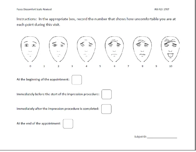
At the end of the dental appointment, the patient was asked to complete a discomfort questionnaire
(Fig. 4), which illustrates the FACES scale. The questionnaire posed the following question: “In
the appropriate box, record the number that represents how uncomfortable you are at each point
during this visit.” Beside each of 4 boxes were the following statements: “At the start of the
treatment appointment; immediately before the impression procedure was initiated; immediately
after the impression procedure was completed; at the completion of the treatment appointment.”
The patient had the ability to rate each statement on a scale of 0 to 10. The scores of the 4 questions
were analyzed.
The clinician completed the clinician impression form (Fig. 5), recording variables such as:
the research group (CL vs. CD); tooth number; whether tray adhesive was used; the tray material;
the type of tray used; the number of units requested; the number of impressions required to obtain
an acceptable impression; time required to make the study impression; patient age and gender; and
the department and year of resident provider making the impression.
The impressions were disinfected with spray disinfectant (CaviCide, Desident), sealed in a
biohazard bag, and sent to be evaluated. All evaluations were performed by 2 calibrated examiners
for each impression using an impression evaluation form (Fig. 6), and were completed
independently. In cases which had multiple prepared teeth, only the distal most prepared tooth was
evaluated. The evaluators recorded criteria for errors in the finish line, errors in the tray/material,
and errors with gingival displacement/hemostasis. The evaluation criteria were borrowed and
modified from the previous impression evaluation study by Rau et al., and are listed in Table 1. If
a conflict existed in evaluations between examiners, the examiners met to form a
consensus. Impressions were deemed acceptable or not acceptable. There were no attempts to
secondary outcomes were also analyzed. 1) The time required to perform the impression
procedure, and 2) Patient discomfort assessed using the FACES visual scale.
2.3 Statistical Analysis
The Fisher’s exact tests were used to compare the corded (CD) versus cordless (CL) groups
for all nominal variables as well as to compare the major and minor error variables between the
acceptable and unacceptable impressions. Wilcoxon rank sum tests were used to compare the
corded and cordless groups for all continuous variables except age, for which an unpaired t-test
was used. Level of significance was set at 0.05 for all analysis.
2.4 Results
With the randomization of all minor variables to the 2 groups (CD and CL) without any
statistical significance detected, we can say the minor variables did not have an effect on the
evaluation outcomes (acceptable or not acceptable). (Table 2.) Thus, the two groups (CD and CL)
are similar. We can say this with statistical confidence, but due to the small sample size, we cannot
definitively exclude the effect that these minor variables may have once the sample size meets a
more powerful number.
Of the 4 critical variables, voids and bubbles at finish line (VOID), lack of wash material at
finish line (WASH), tear at finish line (TEAR), and tissue over finish line (TISSUE), only TEAR
was significantly different between CD and CL groups. The cordless group had statistically
significant more finish line tears of the impression material. (Table 2.)
Of the 4 critical variables, only VOID was significantly different between acceptable and
not acceptable impression groups. The not acceptable group had statistically significant more voids
and bubbles at the finish line of the evaluated impressions. (Table 3.)
difference between CD and CL. The cordless group had statistically significant more unacceptable
impressions. (Table 2.)
Patient age (Table 4.) did not have statistical significance, nor did gender, tooth type (molar,
premolar, anterior) or location (maxilla, mandible). The clinical department and the provider year
in training did not have statistical significance, nor did the type of impression tray.
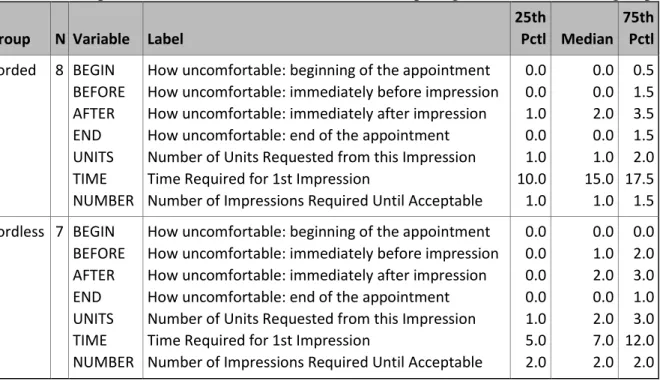
The amount of time recorded for corded impressions was median of 15 minutes and for
cordless a median of 7 minutes, however this was not detected statistically. (Table 5.)
2.5 Discussion
Background
Gingival displacement is defined as “the deflection of the marginal gingiva away from the
tooth,” according to The Glossary of Prosthodontic Terms.12 In 1984, Nemetz et al.13 described
the basic criteria for acceptable gingival displacement as: 1) the creation of sufficient lateral and
vertical space between the finish line and gingival tissues to allow the preparation margin to be
recorded in an impression medium, 2) provide absolute control of gingival fluid seepage and
hemorrhage, 3) no significant, irreversible soft or hard tissue damage resulting from the procedure,
and 4) not produce any potentially dangerous side effects. To accomplish proper gingiv al
displacement, techniques classified as mechanical, chemical, surgical, or a combination of these
methods are used.6,13,14
Gingival Retraction Cords and Medicaments
The most traditional method, and most frequently utilized,5,15 is the chemicomechanical
technique for gingival displacement described by Schillingburg.16 It is taught as the most
traditional method of gingival displacement in dental institutions. This technique utilizes 1 or 2