ISSN Online: 2160-6927 ISSN Print: 2160-6919
Structural and Optical Properties of Cu
2+
+ Ce
3+
Co-Doped ZnO by Solution Combustion Method
S. López-Romero, M. J. Quiroz Jiménez, M. García-Hipólito
Departamento de Materia Condensada y Criogenia, Instituto de Investigaciones en Materiales, Departamento de Materia Condensada y Criogenia, Universidad Nacional Autónoma de México, Coyoacán, México
Abstract
In this work, ZnO, Ce3+ doped ZnO (ZnO/Ce3+) and Cu2+ + Ce3+ co-doped ZnO
(ZnO/Cu2+ + Ce3+ ) solid solutions powders were synthesized by a solution
combus-tion method maintaining the Ce3+ ion concentration constant in 3%Wt while the
Cu2+ ion concentration was varied in 1, 2, 3, 10 and 20%Wt. After its synthesis, all
the samples were annealed at 900˚C by 24 h. The ZnO, ZnO/Ce3+ and ZnO/Cu2+ +
Ce3+ powders were structurally characterized using X-ray diffraction (XRD)
tech-nique, and the XRD patterns showed that for pure ZnO, Cu2+ undoped ZnO/Ce3+
and ZnO/Ce3+ doped with the Cu2+ ion, the three samples exhibited the hexagonal
wurtzite ZnO crystalline structure. However, the morphology and particle size of both samples were observed by means of a scanning electron microscopy (SEM); from SEM image, it is observed that the crystallites of both samples are agglomerated forming bigger amorphous particles with an approximate average size of 1 μm. In addition, the photoluminescence of the ZnO, Ce3+ doped ZnO and Cu2+ + Ce3+ doped
ZnO samples was measurement under an illumination of 209 nm wavelength (UV region): for the ZnO/Ce3+ sample, your emission spectrum is in the visible region
from blue color until red color; the UV band of the ZnO is suppressed. The multico-lor emission visible is attributed to the Ce3+ ion photoluminescence, while for the
ZnO/Cu2+ + Ce3+, its emission PL spectrum is quenching by the Cu2+ ion, present in
the ZnO crystalline.
Keywords
Zinc Oxide, Copper-Cerium, Co-Doped, Solution-Combustion
1. Introduction
Between the II-VI semiconductors compounds, the Zinc oxide (ZnO) by its
extraordi-How to cite this paper: López-Romero, S., Jiménez, M.J.Q. and García-Hipólito, M. (2016) Structural and Optical Properties of Cu2+ + Ce3+ Co-Doped ZnO by Solution
Combustion Method. World Journal of Con- densed Matter Physics, 6, 300-309.
http://dx.doi.org/10.4236/wjcmp.2016.64029
Received: September 6, 2016 Accepted: November 27, 2016 Published: November 30, 2016
Copyright © 2016 by authors and Scientific Research Publishing Inc. This work is licensed under the Creative Commons Attribution International License (CC BY 4.0).
http://creativecommons.org/licenses/by/4.0/
nary properties such as exhibiting an high energy band gap of 3.37 eV and an exciton binding energy of 60 meV [1] [2], has been converted in a strategic material for basic science investigation and technological applications [3] [4] [5], such as in the fabrica-tion of solar cells [6], electro-optical devices [7], gas sensors [8], catalyst [9], piezoelec-tricdevice [10], paramagneticmaterials [11] [12] [13],etc. In the scientific literature, there are various synthesis methods to obtain undoped and doped ZnO with various dopants types, for example: rare heart, metals, lanthanides, etc. [14], such synthesis methods include: electrodeposition [15], evaporation [16], vapor-liquid-solid (VLS) growth [17], metal organic catalyst assisted vapor-phase epitaxy [18], aqueous thermal decomposition [19], microwave activated chemical bath deposition (MW-CBD) [20], chemical bath deposition (CBD) [21], surfactant-assisted hydrothermal method [22], solution combustion [23] etc. Of the before methods, solution combustion presents some advantages because it is very fast, less expensive; it has an easier composition control, and coating can be deposited on large area etc. Lanthanide doped ZnO semi-conductor nanoparticles have important technological properties: they have been used to make efficient photoluminescent materials when lanthanide ions are intercalated in-to ZnO crystal lattice, by means of an energy transfer process from the ZnO lattice in-to the Ce3+ ions to produce internal transitions atomic in the lanthanide ions. Between the
lanthanide ions family members, the cerium ion (Ce) is the most emblematic according to the studies realized by various researchers [24] [25] [26] [27], from its results the authors show that the Ce3+ doped ZnO can present anomalous emission from the UV
until red color region; this multiple emission is studied in the present work. As a com-plement for the present work, it is necessary to mention that the doping of the ZnO matrix with Cu2+ ions leads to sum or produce changes that improve the ZnO
proper-ties such as: band gap tailored, magnetic, electrical and optical. It can also produce pas-sivation of defects and surfaces, as p-type dopant in the original n-type ZnO semicon-ductor. However, actually it is well known that due to the co-doping between the Cu2+
and Eu3+ ions in co-doped glasses matrix, the Cu2+ ion can quench the Eu3+
photolumi-nescence [28] [29] [30] [31], recently. S. Lopez-Romero et al. experimentally demon-strated that such quenching also can occur in solid ZnO semiconductor, this quenching effect can be used to tune between ultra-violet and visible PL emission in devises pho-toluminescent. In this study, ZnO/Ce3+ and co-doped ZnO/Cu2+ + Ce3+ solid solutions
powders have been synthesized by solution combustion method as a function of the Cu2+ ion concentration in %Wt. maintaining the Ce3+ ion concentration constant at a
value of 3%Wt. for both compounds Cu2+ undoped and doped. Finally the samples
were annealing at 900˚C by 24 h. In addition, the influence of the Cu2+ ion on the
mor-phology, structure and photoluminescence of the compounds is analyzed.
2. Experimental Details
metallic, water vapor, carbon dioxide and nitrogen molecular. We used zinc nitrate hexa- hydrate (Zn(NO3)2∙6H2O) as oxidizer agent and urea (NH2CONH2) as fuel, the REDOX
reaction between them is:
(
3)
2 2 2 2 2 2 23Zn NO ⋅6H O 5H NCONH+ →3ZnO 16H O 5CO+ + +8N +heat (1)
Complementary information about how the Equation (1) was obtained can be seen in reference [22]. Using the Equation (1) intrinsic ZnO, Ce3+ doped ZnO and Cu2+ +
Ce3+ co-doped ZnO were synthesized and after annealing at 900˚C by 24 h, the source
of the dopants Cu2+ and Ce3+ were CuCl
2 and Ce2O3. The Ce3+ ion concentration was
fixed at 3%Wt, while the Cu2+ ion concentration takes the values of 0, 1, 2, 3, 10 and
20%Wt. The ZnO/Ce3+ and ZnO/Cu2+ + Ce3+ samples thus obtained were structurally
characterized by x-ray diffraction (XRD) technique using a Philips PW 1800 diffracto-meter with CuKα radiation (1.5406 Å), the morphology of the samples was recorded using a scanning electron microscopy SEM JEOL JSM 840.), the properties photolumi-nescent (PL) of the ZnO/Ce3+ and ZnO/Cu2+ + Eu3+ samples was studied by means of a
spectrofluorometer FluoroMax-P that uses a xenon lampas excitation source, the wave-length excitation was of 209 nm.
3. Results and Discussion
3.1. Structural Characterization
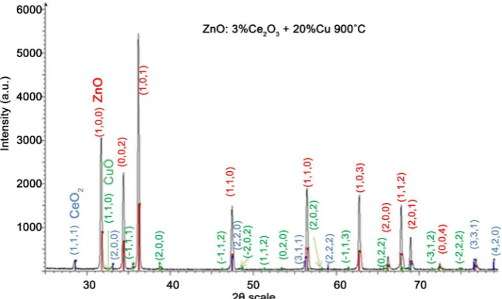
Figure 1 shows the XRD patterns of the as prepared samples of ZnO, ZnO/Ce3+ and
ZnO/Cu2+ + Ce3+ as a function of the Cu2+ ion concentration with the Ce3+ ion
concen-tration fixed in 3%wt. From the diffractograms can be see that all the diffraction peaks can be indexed to the hexagonal wurtzite ZnO structure (JCPDS CARD #89-(102)), no change of the peaks toward lesser angles was observed, however in Figure 2 can be ob-served the XRD pattern of the ZnO joint to two diffraction peaks at 28.4˚ and 32.5˚: the first is assigned to the Ce2O3 phase and the second to the CuO compound. Also is
ob-served that the diffraction peaks are very sharp indicating good crystallization of the products.
3.2. Morphology Study
Figures 3(a)-(d) are the SEM images of the ZnO/Cu2+ + Ce3+ samples with Cu2+ ion
concentration of 0, 3, 10 and 20%wt respectively, it is observed that for low Cu2+ ion
concentration the ZnO nanocrystals have nanoplate form and are agglutinated in amorphous geometry with bigger sizes of about 0.75 μm side and 1 μm large, while in the sample doped with 20%Wt the nanocrystals present ovoid form and are agglome-rated in particles measuring up to 2.5 μm side and 3 μm large.
3.3. Photoluminescence Study
3.3.1. ZnO and ZnO/Ce3+ Photoluminescence
Figure 4(a) and Figure 4(b) show the photoluminescence PL spectra of the pristine
radiation of 209 nm at room temperature. The PL spectra exhibited by the ZnO is typi-cal of the pure ZnO: present a strong UV band peaking about 395 nm and a little broad green band centered about 460 nm. The UV emission band is due to excitonic recom-bination corresponding to the near band edge emission of the ZnO. The green emission band is attributed to deep-level or trap-state emission. Effectively, actually various me-chanism have been proposed to understand the origin of the visible light of the ZnO which involve oxygen vacancies with various oxidation state: the single ionized oxygen vacancy, and the doubly ionized oxygen vacancy. Finally Vanheused et al. [31] found that only the singly ionized oxygen vacancies can emit green light in the ZnO. For the Cu2+ undoped ZnO/Ce3+ samples doped with 3%wt (see Figure 4(b)) and illuminated
Figure 1. XRD pattern of ZnO, Ce3+ doped ZnO and Cu2+ + Ce3+ co-doped ZnO as a function of
the Cu2+ ion concentration.
[image:4.595.194.555.472.687.2](a) ZnO/Ce (b) ZnO/Ce + 3% Cu2+
[image:5.595.43.552.66.511.2](c) ZnO/Ce + 10% Cu2+ (d) ZnO/Ce + 20% Cu2+
Figure 3. SEM images of the (a) Ce3+ doped ZnO, (b) Ce3+ co-doped ZnO with 3%Wt of Cu2+, (c) Cu2+ + Ce3+ co-doped ZnO with 10%
Cu2+, (d) Cu2+ + Ce3+ co-doped ZnO with 20% Cu2+.
with 209 nm wavelength. The emission spectra now present bands at 418 nm and 432 nm belong to the blue color, the peak centered about 452 nm correspond to purple col-or, the bands centered at 468 nm and 480 nm belong to the green region, the peak at 496 is green color, the band centered at 541 nm is yellow color, the peak at 648 nm is orange color, and the band at 687 nm is red color. This multicolor PL emission spectra is possible because when any matrix is doped with the Ce ion this attain two oxidation states namely the Ce3+ and Ce4+, consequently f-d transitions occur by effects field in
the ZnO matrix and 5 d state of the Ce3+ is splitting in two states: E
g and T2g attributed
to the new intern electronic ambient of the Ce3+ ion in the ZnO matrix [23]. It is
the pure ZnO sample. This quenching effect is caused by defects on the ZnO surface generated by organic residues of the fuel (more studies are necessary with this tem).
3.3.2. Ce3+ Photoluminescence Quenching by the Cu2+ Ion
Figure 5 represent the excitation spectrum applied to a ZnO/Cu2+ + Ce3+ sample doped
with 1%Wt of the Cu2+ ion, from the wavelength scale of the spectrum can be see clearly
that for the excitation wavelength of 209 nm no PL emission of the ZnO/Ce3+ + Cu2+
sample was observed, only until a excitation wavelength of 378nm can be see a new emission spectrum of the Ce43+ ion. The suppressing of the Ce3+ is due to that the Cu2+
ion quenching the Ce3+ photoluminescence [28] [29] [30]. This same quenching effect
also occur in glass matrix doped with the europium and copper ions [29]: the authors
(a)
[image:6.595.229.516.236.673.2](b)
Figure 4. (a) Photoluminescence spectra of pure ZnO; (b) Photoluminescence spectra of Ce3+
Figure 5. Excitation spectrum used to illuminate the ZnO/Cu2+ + Ce3+ sample doped with 1%Wt.
of the Cu 2+ ion.
explain the quenching effect by the occurrence of an overlap between Eu3+ emission
band and a Cu2+ absorption band. In our case, we suppose that this quenching effect is
due to an overlap between Ce3+ emission band and a Cu2+ absorption band. The Ce3+ PL
quenching occur for all Cu2+ ion concentration. (More studies are necessary in this
tem).
4. Conclusion
In this work, ZnO, Ce3+ doped ZnO and Ce3+ + Cu2+ co-doped ZnO were synthesized
by a solution combustion method maintaining the Ce3+ ion concentration constant and
changing the Cu2+ ion concentration. The ZnO resultant crystalline structure was the
hexagonal wurtzite structure for the three samples types. From PL studies, it is found that the UV band of 395 nm in the pure ZnO is suppressed in the Ce3+ doped ZnO and
the PL emission spectrum of the Ce3+ + Cu2+ co-doped ZnO samples also was
quench-ing by the Cu2+ ion.
Acknowledgements
The authors wish to thank to Dr. Ciro Falcony (IPN), Adriana Tejeda (IIM) for the XRD measurements, to Omar Novelo Peralta (IIM) for his SEM study, to M.A. Canseco Martinez and Lázaro Huerta (IIM) for their chemical analysis.
References
[1] Klingshirn, C. (2007) ZnO: From Basics towards Applications. Physica Status Solidi (b), 244, 3027-3073. http://dx.doi.org/10.1002/pssb.200743072
Synthesized ZnO Powders. Physica B: Condensed Matter, 405, 866-874. http://dx.doi.org/10.1016/j.physb.2009.10.005
[3] Sharma, S.K., Pitale, S.K., Manzar Malik, M., Dubey, R.N., Qureshi, M.S. and Lumin, J. (2009) Luminescence Studies on the Blue-Green Emitting Sr4Al14O25:Ce3+ Phosphor
Syn-thesized through Solution Combustion Route. Journal of Luminescence, 129, 140-147. http://dx.doi.org/10.1016/j.jlumin.2008.09.002
[4] Leea, J.-B., Leea, H.-J., Seob, S.-H. and Parka, J.-S. (2001) Characterization of Undoped and Cu-Doped ZnO Films for Surface Acoustic Wave Applications. Thin Solid Films, 398-399, 641-646. http://dx.doi.org/10.1016/S0040-6090(01)01332-3
[5] Qi, J., Gao, D.Q., Zhang, L. and Yang, Y.H. (2010) Room-Temperature Ferromagnetism of the Amorphous Cu-Doped ZnO Thin Films. Applied Surface Science, 256, 2507-2508. http://dx.doi.org/10.1016/j.apsusc.2009.10.096
[6] Wei, H.M., Gong, H.B., Wang, Y.Z., Hu, X.L., Chen, L., Xu, H.Y., Liu, P. and Cao, B.Q. (2011) Three Kinds of Cu2O/ZnO Heterostructure Solar Cells Fabricated with
Electro-chemical Deposition and Their Structure-Related Photovoltaic Properties. CrystEngComm, 13, 6065-6070. http://dx.doi.org/10.1039/c1ce05540b
[7] Kundu, T.K., Karak, N., Barik, P. and Saha, S. (2011) Optical Properties of ZnO Nanopar-ticles Prepared by Chemical Method Using Poly (VinylAlcohol ) (PVA) as Capping Agent
International Journal of Soft Computing and Engineering (IJSCE), 1, 2231-2307,
[8] Ge, C.Q. and Xie, C.S. (2007) Preparation and Gas-Sensing Properties of Ce-Doped ZnO Thin-Film Sensors by Dip-Coating. Materials Science and Engineering: B, 137, 53-58.
http://dx.doi.org/10.1016/j.mseb.2006.10.006
[9] Anandana, S. and Miyauchi, S. (2011) Ce-Doped ZnO (CexZn1−xO) Becomes an Efficient
Visible-Light-Sensitive Photocatalyst by Co-Catalyst (Cu2+) Grafting. Physical Chemistry
Chemical Physics, 13, 14937-14945. http://dx.doi.org/10.1039/c1cp21514k
[10] Ahn, C.-W., Nahm, S., Ryu, J., Uchino, K., Yoon, S.-J., Jung, S.-J. and Song, J.-S. (2004) Ef-fects of CuO and ZnO Additives on Sintering Temperature and Piezoelectric Properties of 0.41 Pb(Ni1/3Nb2/3)O3 – 0.36 PbTiO3 – 0.23 PbZrO3 Ceramics. Japanese Journal of Applied
Physics, 43, No. 1
[11] Morinaga, Y., Sakuragi, K., Fujimura, K. and Ito, T. (1997) Effect of Ce Doping on the Growth of ZnO Thin Films. Journal of Crystal Growth, 174, 691-695.
http://dx.doi.org/10.1016/S0022-0248(97)00045-6
[12] Sinhaa, N., Raya, G. and Bhandaria, S. (2014) Synthesis and Enhanced Properties of Cerium Doped ZnO Nanorods. Ceramics International, 40, 12337-12342.
http://dx.doi.org/10.1016/j.ceramint.2014.04.079
[13] Shukla, S., Agorku, E., Mittal, H. and Mishra, A. (2013) Synthesis, Characterization and Photoluminescence Properties of Ce3+ Doped ZnO-Nanophosphors. Chemical Papers, 68,
217-222. http://dx.doi.org/10.2478/s11696-013-0442-5
[14] Sofiania, Z., Derkowskab, B., Dalasińskib, P. and Wojdyłab, M. (2006) Optical Properties of ZnO and ZnO:Ce Layers Grown by Spray Pyrolysis. Optics Communications, 267, 433-439.
http://dx.doi.org/10.1016/j.optcom.2006.06.049
[15] Varughese, G., Jithin, P.W. and Usha, K.T. (2015) Determination of Optical Band Gap Energy of Wurtzite ZnO:Ce Nanocrystallites. Physical Science International Journal, 5, 146- 154. http://dx.doi.org/10.9734/PSIJ/2015/14151
[16] Koaoa, L.F., Dejenea, F.B. and Swartb, H.C. (2013) The Effect of Ce3+ on Structure,
http://dx.doi.org/10.1016/j.jlumin.2013.05.045
[17] Yousefia, M., Amirib, M. and Azimiradc, R. (2011) Enhanced Photoelectrochemical Activi-ty of Ce Doped ZnO Nanocomposite Thin Films under Visible Light. Journal of Electroa-nalytical Chemistry, 661, 106-112.http://dx.doi.org/10.1016/j.jelechem.2011.07.022
[18] Pandey, P., Kurchania, R. and Haque, F.Z. (2015) Structural, Diffused Reflectance and Photoluminescence Study of Cerium Doped ZnO Nanoparticles Synthesized through Sim-ple Sol-Gel Method. Optik-International Journal for Light and Electron Optics, 126, 3310- 3315. http://dx.doi.org/10.1016/j.ijleo.2015.06.026
[19] Karunakaran, C., Gomathisankar, P. and Manikandan, G. (2010) Preparation and Charac-terization of Antimicrobial Ce-Doped ZnO Nanoparticles for Photocatalytic Detoxification of Cyanide Materials. Chemistry and Physics, 123, 585-594.
http://dx.doi.org/10.1016/j.matchemphys.2010.05.019
[20] Samadia, M., Ziraka, M. and Naserib, A. (2016) Recent Progress on Doped ZnO Nano-structures for Visible-Light Photocatalysis. Thin Solid Films, 605, 2-19
http://dx.doi.org/10.1016/j.tsf.2015.12.064
[21] Koaoa, L.F., F.B. Dejenea, F.B. and Tsegaa, M. (2016) Annealed Ce3+-Doped ZnO Flower-
Like Morphology Synthesized by Chemical Bath Deposition Method. Physica B: Condensed Matter, 480, 53-57.http://dx.doi.org/10.1016/j.physb.2015.09.010
[22] Patil, K.C., Hegde, M.S., Tanu, R. and Aruna, S.T. (2008) Chemistry of Nanocrystalline Oxide Materials. Combustion Synthesis, Properties and Applications. World Scientific, 52- 58.
[23] George, A. and Sharma, S.K. (2011) Detailed of X-Ray Diffraction and Photoluminescence Studies of Ce Dopes ZnO Nanocrystals. Journal of Alloys and Compounds, 509, 5942-5946. http://dx.doi.org/10.1016/j.jallcom.2011.03.017
[24] Ge, C., Xie, C. and Cai, S. (2007) Preparation and Gas-Sensing Properties of Ce-Doped ZnO Thin-Film Sensors by Dip-Coating. Materials Science and Engineering: B, 137, 53-58. http://dx.doi.org/10.1016/j.mseb.2006.10.006
[25] Li, F., Yan, B., Zhang, J., Jiang, A.X., Shao, C.H., Kong, X.J. and Wang, X. (2007) Study on Desulfurization Efficiency and Products of Ce-Doped Nanosized ZnO Desulfurizer at Am-bient Temperature. Journal of Rare Earths, 25, 306-310.
http://dx.doi.org/10.1016/S1002-0721(07)60427-X
[26] Yang, J., Gao, M., Yang, L., Zhang, Y., Lang, J., Wang, D., Wang, Y., Liu, H. and Fan, H. (2008) Low-Temperature Growth and Optical Properties of Ce-Doped ZnO Nanorods. Ap-plied Surface Science, 255, 2646-2650. http://dx.doi.org/10.1016/j.apsusc.2008.08.001 [27] Iqbal, J., Liu, X., Zhu, H., Wu, Z.B., Zhang, Y., Yu, D., Yu, R. and Yu, R. (2009) Raman and
Highly Ultraviolet Red-Shifted near Band-Edge Properties of LaCe-Co-Doped ZnO Nano-particles. Acta Materialia, 57, 4790-4796. http://dx.doi.org/10.1016/j.actamat.2009.06.056 [28] López-Romero, S., Quiroz Jiménez, M.J. and García-Hipólito M. () Quenching
Photolumi-nescence of Eu(III) by Cu(II) in ZnO:Eu3+ + Cu2+ Compounds by Solution Combustion
Method. World Journal of Condensed Matter Physics, 6, 269-275. http://dx.doi.org/10.4236/wjcmp.2016.63025
[29] Jiménez, J.A. (2015) Photoluminescence of Eu³⁺-Doped Glasses with Cu²⁺ Impurities.
Spectrochimica Acta Part A: Molecular and Biomolecular Spectroscopy, 145, 482-486. http://dx.doi.org/10.1016/j.saa.2015.03.047
[31] Vanheusden, K., Seager, C.H., Warren, W.L., Tallent, D.R., Voigt, J.A. and Gnade, B.E. (1966) Mechanisms behind Green Photoluminescence in ZnO Phosphor Powders. Journal of Applied Physics, 79, 7983. http://dx.doi.org/10.1063/1.362349
Submit or recommend next manuscript to SCIRP and we will provide best service for you:
Accepting pre-submission inquiries through Email, Facebook, LinkedIn, Twitter, etc. A wide selection of journals (inclusive of 9 subjects, more than 200 journals)
Providing 24-hour high-quality service User-friendly online submission system Fair and swift peer-review system
Efficient typesetting and proofreading procedure
Display of the result of downloads and visits, as well as the number of cited articles Maximum dissemination of your research work
Submit your manuscript at: http://papersubmission.scirp.org/