N A N O E X P R E S S
Open Access
Biosynthesis, Antibacterial Activity and
Anticancer Effects Against Prostate Cancer
(PC-3) Cells of Silver Nanoparticles Using
Dimocarpus Longan Lour
. Peel Extract
Yan He
1, Zhiyun Du
1, Shijing Ma
1, Shupeng Cheng
1, Sen Jiang
1, Yue Liu
2, Dongli Li
1, Huarong Huang
1,
Kun Zhang
1*and Xi Zheng
1,2*Abstract
Metal nanoparticles, particularly silver nanoparticles (AgNPs), are developing more important roles as diagnostic and therapeutic agents for cancers with the improvement of eco-friendly synthesis methods. This study demonstrates the biosynthesis, antibacterial activity, and anticancer effects of silver nanoparticles usingDimocarpus Longan Lour. peel aqueous extract. The AgNPs were characterized by UV-vis absorption spectroscopy, X-ray diffraction (XRD), high-resolution transmission electron microscopy (HRTEM), scanning electron microscopy (SEM), and Fourier transform infrared spectroscope (FTIR). The bactericidal properties of the synthesized AgNPs were observed via the agar dilution method and the growth inhibition test. The cytotoxicity effect was explored on human prostate cancer PC-3 cells in vitro by trypan blue assay. The expressions of phosphorylated stat 3, bcl-2, survivin, and caspase-3 were examined by Western blot analysis. The longan peel extract acted as a strong reducing and stabilizing agent during the synthesis. Water-soluble AgNPs of size 9–32 nm was gathered with a face-centered cubic structure. The AgNPs had potent bactericidal activities against gram-positive and gram-negative bacteria with a dose-related effect. AgNPs also showed dose-dependent cytotoxicity against PC-3 cells through a decrease of stat 3, bcl-2, and survivin, as well as an increase in caspase-3. These findings confirm the bactericidal properties and explored a potential anticancer application of AgNPs for prostate cancer therapy. Further research should be focused on the comprehensive study of molecular mechanism and in vivo effects on the prostate cancer.
Keywords:Silver nanoparticles,Dimocarpus Longan Lour., Bactericidal, Anticancer, Prostate cancer
Background
Metal nanoparticles are most promising for their long use for medicinal purposes since ancient time and have attracted more attentions for potential applications in can-cers [1, 2]. Among silver nanoparticles (AgNPs) are one of the significant members due to their unique physical-chemical properties, biological compatibility, relative lower toxicity, and biological activities such as antimicro-bial, inflammatory, and anticancer activities [3–5].
AgNPs have been synthesized by various physical, chemical, electrochemical, photochemical, irradiative, and recently improved biological methods. Most of the ways are quite expensive or potentially dangerous to the environment [6]. While greener biological synthesis has better eco-friendly, cost-effective, and easily large-scaled properties and are motivating a research upsurge in ex-ploring the plants as reducing and protecting agents [7], such as Pulicaria glutinosa [8], P. glutinosa [9], and
Moringa oleifera [10]. Plant extracts are rich of func-tional molecules such as phenolic compounds, which have been regarded as potent natural reducers with high antioxidant activity [11, 12]. Longan (Dimocarpus Longan Lour.), an edible fruit, is cultivated widely in southern China and south-east Asia. With rich bioactive * Correspondence:[email protected];[email protected]
1Allan H. Conney Laboratory for Anticancer Research, School of Chemical
Engineering and Light Industry, Guangdong University of Technology, Guangzhou, China
Full list of author information is available at the end of the article
polysaccharides, phenolic acids, and flavonoids [13, 14], its flesh or seed is a good source of functional ingredi-ents for antioxidant, immunity-modulatory, insomnia re-lief, and memory-enhancing effects [15–19]. Whereas peels, regarded as a waste usually, have an untapped po-tential in the resource utilization and might be available for potential green synthesis purposes.
The improvement of biosynthesized AgNPs has
allowed better control of shape and size more suitable for the increasing biomedical applications, such as im-plantable biomaterials, tumor radiotherapy sensitization agent, molecular imaging agents, biological markers, and drug delivery [20, 21]. Silver is now extending its appli-cations in cancer treatment as antitumor molecules, and many attempts turned up meaningful and positive [22, 23]. It was reported that AgNPs had antitumor effects against the cervical carcinoma cells [10], embryo fibro-blast 3T3 cells [24], lung cancer H1299 cells [25], breast cancer MCF-7 cells [26], and glioblastoma multiforme U-87 cells [27]. AgNPs could be delivered into the cell by the Trojan effect and inhibit the RNA polymerase ac-tivity and the gene transcription via a direct reciprocal interaction [28]. The tumor cells were more sensitive to AgNPs damage than normal cells [29]. The particle size and surface features of AgNPs are very important for biomedical considerations. AgNPs with smaller particle size seemed to have a stronger penetration ability and greater toxicity for cancer cells [30]. The biochemical molecular mechanisms have not been fully revealed so far and more efforts should be tried.
Eco-friendly green synthesis with plant extracts to ob-tain ideal AgNPs for bactericidal and anticancer applica-tions attracted our attenapplica-tions. The present work investigated the synthesis of the AgNPs with a smaller size by peel extract of longan for the first time and fur-ther tested the antibacterial and in vitro anticancer ef-fects against prostate cancer cells.
Methods
Materials
Silver nitrate (AgNO3) and 3-(4,5-dimethylthiazol-2-yl)-2,5-diphenyltetrazolium bromide (MTT) were sourced from Sigma-Aldrich (St Louis, MO, USA). Dried longan was obtained from Chinese Herbal Medicine Market in Guangzhou.Escherichia coli(ATCC25922),Staphylococcus aureus (ATCC 6538), Bacillus subtilis (ATCC6633),
Pseudomonas aeruginosa (ATCC15442), and Candida albicans (ATCC10231) were offered by Guangdong Insti-tute of Microbiology (Guangzhou, China). Prostate cancer PC-3 cells were from the ATCC (Rockville, MD, USA). RPMI-1640 medium, fetal bovine serum (FBS), L-glutamine, and penicillin-streptomycin were purchased from Gibco (Grand Island, NY, USA). Trypan blue stain was bought from Cambrex (Walkersville, MD, USA).
Antibodies of β-actin, p-stat 3, survivin, and caspase-3 were purchased from Millipore Corporation (Billerica, MA, USA). Bcl-2 antibody and all secondary antibodies were provided by Santa Cruz Biotechnology (Dallas, TX, USA). Thermo assay kit with SuperSignal West Femto Luminol/Enhancer solution was from Thermo Scientific (Rockford, IL, USA).
Synthesis of AgNPs
Longan peel was selected as the plant reducer for the synthesis of AgNPs. The peels were immersed in dis-tilled water (10 g/250 mL) at 50 °C for 1 h. The extract filtrates were cooled and centrifuged at 3000 rpm for 5 min. Then, the filtrates were added drop by drop to AgNO3 (2 mM) aqueous solution at the ratio of 1:1. After the reaction at 80 °C for 5 h under stirring, the Ag colloid was then precipitated by the centrifugation (12,000 rpm, 20 min) and washed thrice with distilled water to make sure of the complete removal of extracts. The purified AgNPs were obtained after drying at 50 °C under vacuum oven for 24 h.
The effects of key factors such as (i) amount of longan peel extract, (ii) concentration of silver nitrate, and (iii) reaction temperature were explored to determine their effect on the synthesized nanoparticles. Different amounts of the aqueous extract (10, 20, 30, 40, and 50 mL) were added to a fixed concentration of AgNO3 (2.0 mM) at 80 °C. Different concentrations (0.5, 1.0, 2.0, 3.0, and 5.0 mM) of silver nitrate were also assessed when reacting with a 50 mL of the extract at 80 °C. Dif-ferent reaction temperatures (room temperature and 80 °C) were explored with 50 mL of extract and 2.0 mM of silver nitrate. The experiment was continuously ob-served via color change with naked eye as well as UV-vis spectrophotometer [31].
Characterization of AgNPs
4000–500 cm−1. Phase formation was analyzed XRD Analyzer (Ultima III, Rigaku, Tokyo, Japan) with Cukα radi-ation (λ= 1.5406 Å) in the 2θrange from 10° to 80° at the scanning rate of 4°/min. Mean crystallite diameter of the powder was determined by Debye-Scherrer equation from half width of diffraction peaks:
D¼ð Þkλ =ðβ cosθÞ ð1Þ
whereDis the mean crystallite diameter,kis a constant,
λ is the wavelength of Cukα, θ is the Bragg diffraction angle, andβis the full width at half-maximum.
Antibacterical Activity of AgNPs
The bactericidal activities of AgNPs were tested against the gram-positive bacteria (B. subtilis and S. aureus), gram-negative bacteria (E. coli and P. aeruginosa), and the fungus (C. albicans), via agar dilution method and growth inhibition test assay [11]. For the agar dilution method, the 6-mm sterile plates were loaded with 20μL of test samples of the biosynthesized AgNPs (2.0 mM), AgNO3 (2.0 mM), aqueous longan peel extract, or the penicillin solution (200 μg/mL). Each plate was inocu-lated with 1.0 × 10⁷ CFU of bacteria. Plates without AgNPs or with penicillin were used as a blank and posi-tive control separately. The bacteria were cultured in Muller-Hinton agar (Luogang, Guangzhou, China) at 37 °C for 24 h and the fungus in Sabouraud’s dextrose agar (Luogang, Guangzhou, China) at 28 °C for 36 h. After in-cubation, the bactericidal activities were measured by the inhibition zones. For bacteria growth inhibition test, 100 μL of bacteria or fungus suspension with 1.0 × 10⁸ CFU were inoculated into 45 mL fresh LB media and treated series concentrations of AgNPs at 37 °C. The op-tical densities of the cultures were determined at 600 nm (OD600) by a UV-vis spectrophotometer.
Cell Culture and Trypan Blue Exclusion Assay
PC-3 cells were cultured in RPMI-1640 medium with 10 % FBS, 100 units/mL streptomycin, 100 units/mL peni-cillin, and 300 μg/mL L-glutamine; 2 mL of cells were seeded at the density of 2 × 10⁴ cells/mL in the 35-mm dishes and cultured for 24 h at 37 °C in a humidified at-mosphere of 5 % CO2. Then, the cells were exposed to dif-ferent concentrations of AgNPs (2–30μg/mL). After 72 h of treatment, 80μL of the cell suspension was mixed with 20μL of 0.4 % trypan blue stain solution for 2 min at RT. The cells were determined using a hemocytometer under a light microscope (Nikon Optiphot, Japan). The cells that did not absorb dye were regarded as live cells, and blue cells were counted as dead cells.
Western Blot Analysis
The role of AgNPs for prostate cancer was further ex-amined on the level of phosphorylated stat 3, bcl-2, sur-vivin, and caspase-3 in PC-3 cells at the concentration of 10 μg/mL by Western blot according to our protocol [32, 33]. PC-3 cells were seeded at a density of 1 × 10⁵ cells/mL and incubated for 24 h. The cells were treated with 10μg/mL of AgNPs. After 24 h treatment, the cells were washed with ice-cold PBS and lysed with 200μL of lysis buffer (1 mM phenylmethylsulfonyl fluoride, 100 μM sodium orthovandate, 30 mM sodium pyro-phosphate, 50 mM sodium chloride, 50 mM sodium fluoride, 5 mM ZnCl2, 2 mM iodoacetic acid, 10 mM Tris–HCl, and 0.5 % Triton X-100). Cell homogenates were centrifuged at 12,000gfor 15 min at 4 °C. The pro-tein concentrations were determined by the Bio-Rad protein assay kit. Theβ-actin protein was used as a load-ing control. Equal amounts of protein (50 μg) were loaded on a Bio-Rad Precast Gel (10 %) and then trans-ferred to a PVDF membrane. The membranes were sub-sequently incubated with phosphor-stat 3, bcl-2, survivin, or caspase-3 primary antibodies (Millipore) at 4 °C overnight, respectively. At the end of incubation, all membranes were washed with 0.05 % Tween 20 Tris-buffered saline (TBST, 15 min) four times, then treated by the secondary antibody (Santa Cruz Biotechnology) for 1 h at RT. After washing the membranes again four times with TBST, immunoreactivity was detected by Thermo assay kit and analyzed by the Quantity One software (Bio-Rad).
Results and Discussion
Synthesis and Characterization of AgNPs
Eco-friendly green synthesis with plant extracts plays a very important role in nanotechnology without any harmful chemicals. Many natural plants extracts with a rich source of functional molecules has been verified as the capping and reducing agents for the synthesis of AgNPs [16]. We have before screened some edible plants, such as Chrysanthemum morifolium Ramat and
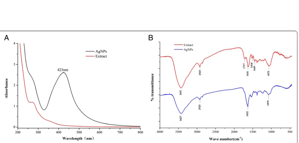
also be monitored by the UV-vis spectrum with the broad SFR at 425 nm (Figs. 2 and 3) [31].
We have obtained the well-synthesized AgNPs using longan extracts by exploring the effects of extract, silver nitrate, and reaction temperature. The reactions were observed under the UV-vis spectroscopy to examine the bioreduction of AgNPs from aqueous AgNO3 solution. Some small changes in synthetic factors lead to dramatic modifications in nanoparticle structure, size, morph-ology, stability, and self-assembly patterns [30], which could be indicated by the SPR band [34, 35]. The narrow peaks observed in the Fig. 2 give indirect evidence to the ability of the reductant to function as a stabilizer of the nanoparticles against aggregation. Figure 2a shows the effects of amount of plant extracts on the reaction process of AgNPs. No significant shift change of absorp-tion peak appeared indicating no difference in the morphology and size of the formed silver particles. More stable samples could be collected with amount of the ex-tract, while 50 mL above of the extract solutions could promote the agglomeration of nanosilver and stimulate the particle size larger. The spectra in Fig. 2b shows the influence of AgNO3on the reaction. There was a wave-length shift following the concentration changes with
absorption peak of 412, 427, 438, and 440 nm when reacted with 0.5, 1.0, 3, and 5.0 mM of AgNO3,
respect-ively. The SPR wavelength shifts towards
long-wavelength region which indicated larger particle size. The reaction with 20 mM and above of AgNO3showed an obvious increase of yield. There are no significant dif-ference between the peak absorption values between 20, 30, and 50 mM of AgNO3. To obtain the AgNPs with ideal size for the future investigation, 20 mM was finally decided for the synthesis. The UV-vis absorption spectrum of the obtained AgNPs was compared under room temperature (Fig. 2c) with 80 °C (Fig. 2d) at differ-ent time intervals. The process under 80 °C is more ad-vantageous as it results in the formation of nanoparticles at a faster rate, higher yield, and slightly smaller size when compared with room temperature. Finally, we have developed an optimum green synthesis method for AgNPs by treating 2.0 mM AgNO3with 50 mL of nat-ural longan peel extract at 80 °C for 3 h without any harmful reducing, capping, or dispersing agents. The optimum AgNPs were further characterized and deter-mined the antibacterial and anticancer effects.
[image:4.595.58.541.88.396.2]Fig. 2UV-vis absorption spectra of AgNPs during synthesis process, with various amount of extract at 80 °C for 3 h (a), with various concentrations of AgNO3at 80 °C for 3 h (b), at RT (c), and at 80 °C (d)
[image:5.595.59.539.90.452.2] [image:5.595.59.541.485.716.2]absorption peak was observed at 425 nm, which is a characteristic band for the Ag, arising from the excita-tion of longitudinal plasmon vibraexcita-tions of AgNPs in the solution. FTIR measurements were performed to identify the potential biomolecules in longan peel extract respon-sible for capping and stabilizing the silver nanoparticles. The FTIR spectrum of silver nanoparticles in Fig. 3b showed very strong peak at 3427 cm−1which is assigned as −OH stretching in alcohols and phenolic compounds [36]. The medium intense band 1632 cm−1is assigned to the C=C aromatic vibrations [37]. The band at 2925 cm−1 ascribes to the C–H stretching vibration of methyl, methy-lene, and methoxy groups. The peak 1079 cm−1indicates the presence of C–O stretching of alcohols, amide, ester, and ether groups. There represented some similar absorp-tion peaks at 3431, 2925, 1631, and 1073 cm−1in the ex-tract spectrum. The peak at 1717 cm−1 could be attributed to the C=O aromatic vibrations. The broad band at 1518 and 1449 cm−1 assigned for the presence stretching vibrations of C=C in aromatic ring. The con-sistent vibrational bands such as –O–H, –C–H, –C=C,
and–C=O are derived from the compounds such as flavo-noids and terpeflavo-noids in longan peel. These functional groups may play an important role in the synthesis as the capping and reducing agent. Fully understanding of the underlying mechanism demands further exploration.
The morphology of the well-designed AgNPs was characterized by XRD, HRTEM, and SEM micrographs (Fig. 4). Figure 4c depicted four main characteristic diffraction peaks for Ag observed at 2θ of 38.2, 44.3, 64.4, and 77.6, which correspond to the (111), (200), (220), and (311) based on the band for face-centered cubic structures of silver, respectively (JCPDS Card No. 04-0783). No peaks from any other phase were observed showing that single-phase Ag with cubic structure nano-particles has been obtained directly. The (111) plane was chosen to calculate crystalline size, and the average size of the synthesized AgNPs is 15 nm from the Debye-Scherrer equation. The HRTEM and SEM image pre-sented that AgNPs were spherical and single crystalline with a narrow size range 9–32 nm and the average size 20.35 nm, which is in good agreement with the shape of
[image:6.595.59.540.356.715.2]the SPR band in the UV-vis spectrum and the particle size calculated from XRD analysis. The well-controlled AgNPs, especially their sizes, is of critical importance to the activities and toxicity [38]. The AgNPs with smaller particle size have a stronger penetration ability and greater toxicity [39]. The cancer cells are different in pore size compared to other cells, and so a size-controlled targeting of AgNPs can prove effective in the case of cancer treatment [34]. Our synthesized AgNPs was spherical with such a size which is wildly recognized among a good range for anticancer drug delivery appli-cations and medical purposes [40].
Stability of AgNPs in Aqueous Solution
As to the stability of silver nanoparticles in aqueous so-lution, we evaluated the size distribution and plasmonic properties for 6 months at RT by TEM and UV-vis mea-surements (Fig. 5). It was found from TEM images that AgNPs are well-dispersed without any aggregation, as well as with no significant change in particle size and shape. There was also stable in plasmonic properties, in-dicating good stability of AgNPs for 6 months. This is corresponding to the previous study [41]. Good stability of nano-silver in many researches is discussed under dif-ferent standard cell incubation conditions [42, 43]. Foldbjerg et al. studied the cytotoxicity and genotoxicity of silver nanoparticles in the human lung cancer A549 cell, which indicated that the size and stability of the AgNPs remain the same in DMEM and RPMI also used as the exposure medium in our study [44].
Antimicrobial Activity of AgNPs
As indicated in Table 1, the inhibition zones of AgNPs were 15, 20, 16, 19, and 13 mm forS. aureus,B. subtilis,E. coli, P. aeruginosa, and C. albicans, respectively. The
extract tested had no bactericidal activity. The synthesized AgNPs shows a higher inhibitory effect compared with AgNO3 and could compete commercial antimicrobial agents penicillin used for the treatment of bacterial infec-tions. Figure 6 shows the good growth inhibition of AgNPs onE. coliandS. aureus.The growth ofE. coliwas completely inhibited in 35 h at 100, 24 h at 50, and 16 h at 20μg/mL. Meanwhile, the growth ofS. aureus was totally suppressed in 35 h at 100, 20 h at 50, and 16 h at 20μg/mL. AgNPs had better effects on bacteria than fungi, as well as greater impacts on negative bacteria than gram-positive bacteria in this study, which is corresponded with the previous researches [45]. The strong bactericidal activity of silver nanoparticles is verified because they can easily enter the pathogen with a unique small size and surface ef-fects [46]. The cell membrane of the bacteria might be destroyed due to the reaction of amino (−NH), −SH, −COOH, and other functional groups in the cell [47]. AgNPs could also depress the synthesis of bacterial metab-olism enzyme, destroy the genetic factor, and eventually lead to death [48]. The presence of peptidoglycan layer in the cell wall of gram-positive bacteria could attribute to less activity, and it can prevent the nanoparticles from penetrating into bacteria [49].
[image:7.595.307.540.99.197.2]Fig. 5Stability of AgNPs in aqueous solution for 6 months at RT. Photographs ofaUV-vis andbTEM images
Table 1Antibacterial activity of the biosynthesized AgNPs
Microorganisms Zone of inhibition(mm in diameter)
Penicillin AgNPs AgNO3 Extract
S. aureus 24 15 10 –
B. subtilis 23 20 12 –
E. coli 21 16 10 –
P. aeruginosa 25 19 9 –
[image:7.595.56.544.518.713.2]Anticancer Effects of AgNPs on PC-3 Cells
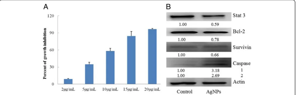
Cancers figure among the leading causes of morbidity and mortality worldwide, with approximately 14 million new cases and 8.2 million cancer-related deaths in 2012 [50]. Prostate cancer is the second most common cancer and the fifth leading cause of cancer death in men in 2012 [51]. AgNPs have been shown to have important bacteri-cidal and antiangiogenic activities, which are attractive for the research of their potent anticancer effects. The effect and the underlying mechanism of our AgNPs with a smaller size range on prostate cancer attracted our curios-ity. In this study, the cytotoxicity was evaluated on PC-3 cells in vitro by the trypan blue exclusion assay. As shown in Fig. 7a, the synthesized AgNPs of size 9–32 nm had dose-dependent cytotoxic effects on prostate cancer PC-3 cells in vitro.About 50 % of PC-3 cells died when treated with AgNPs at the concentrations between 5 and 10 μg/mL. The IC50 was less than 10 μg/mL, which is consistent with the previous findings [52]. The pro-tein expressions were explored by Western blot to clarify the mechanism by which AgNPs exert a cyto-toxic effect on PC-3 cells. The antitumor activities
were mediated by a decrease of stat 3, bcl-2, and sur-vivin, as well as an increase in caspase-3 shown in Fig. 7b.
The cytotoxic effect of AgNPs on cell viability has a major role in antitumor activity, thereby reducing dis-ease progression. Cellular internalization of silver might provide the basis for the cytotoxicity of AgNPs [39, 53]. Sriram et al. found that AgNPs could enter the cell through endocytosis, acted on the nucleus to interfere with chromosome stability, and interrupt the mitosis [29]. Active anticancer components are known to exhibit a consistent antitumor effect by blocking abnormal ex-pression alterations of multiple signaling proteins, such as VEGF, p53, JAK-STAT, and CDC14A [45, 54, 55]. The antitumor activity of AgNPs was also shown to be medi-ated by induction of proliferation and apoptosis by active caspase 9 and active caspase 3 in a GBM tumor system in vitro [56]. These biological responses eventually lead to the survival or apoptosis of the cancer cells and prompt a promising way of AgNPs for prostate cancer. The molecular mechanism of targeting has not fully understood for the inhibitory effects of AgNPs on the Fig. 6Growth curves ofE. coli(a),S. aureus(b) andC. albicans(c) in LB media with different concentrations of AgNPs
[image:8.595.59.540.88.226.2] [image:8.595.58.540.548.703.2]prostate cancer cells. Further research efforts are de-served to elucidate the latent mechanism and then lead to more effective measures for prostate cancer.
Conclusions
With better developments of the green synthesis methods and understanding of the molecular mechan-ism, the applications of AgNPs are likely to expand fur-ther in the cancer fur-therapy fields. In the present work, we have proposed first time a simple, high efficiency, eco-friendly green synthesis method for AgNPs using longan peel extract as reducing and stabilizing agent demon-strated. The biosynthezied AgNPs are spherical and sin-gle crystalline, with a narrow particle size range from 9 to 32 nm. The synthesized AgNPs showed good bacteri-cidal activities against gram-positive and gram-negative bacteria and fungus with a dose-dependent effect. AgNPs exhibited a strong inhibitory effect on the pros-tatic cancer PC-3 cells, which might be associated with a decrease of stat 3, bcl-2, and survivin, as well as an in-crease in caspase-3. AgNPs might be a potential active candidate for prostate cancer treatments. Further re-search should be focused on the comprehensive study of molecular mechanism and in vivo effects of AgNPs on prostate cancer.
Competing Interests
The authors declare that they have no competing interests.
Authors’Contributions
YH, ZD, and SM came up with the idea and participated in the design, preparation, characterization, and antibacterial activity of AgNPs and also writing of the manuscript. YH, YL, DL, and HH performed the cell assay and other biochemical assays. SC and SJ participated in the coordination of this study. KZ and XZ developed the conceptual framework and supervised the whole work. All authors read and approved the final manuscript.
Acknowledgements
This work was financially supported by grants from the Postdoctoral Science Foundation China (2013M540649), international collaboration Project from Guangzhou Science & Technology (2013J4500014), and Leadership Grant in Guangdong Province (Allan H. Conney). All authors dedicate this paper to Dr. Allan H. Conney, a respectable scientist who passed away on September 10, 2013.
Author details
1Allan H. Conney Laboratory for Anticancer Research, School of Chemical
Engineering and Light Industry, Guangdong University of Technology, Guangzhou, China.2Susan Lehman Cullman Laboratory for Cancer Research,
Department of Chemical Biology, Ernest Mario School of Pharmacy, Rutgers, The State University of New Jersey, 164 Frelinghusen Road, Piscataway, NJ 08854, USA.
Received: 18 April 2016 Accepted: 30 May 2016
References
1. Rai M, Ingle AP, Gupta I, Brandelli A (2015) Bioactivity of noble metal nanoparticles decorated with biopolymers and their application in drug delivery. Int J Pharm 496(2):159–172
2. Sharma H, Mishra PK, Talegaonkar S, Vaidya B (2015) Metal nanoparticles: a theranostic nanotool against cancer. Drug Discov Today 20(9):1143–1151
3. Austin LA, Mackey MA, Dreaden EC, El-Sayed MA (2014) The optical, photothermal, and facile surface chemical properties of gold and silver nanoparticles in biodiagnostics, therapy, and drug delivery. Arch Toxicol 88(7):1391–1417
4. Dos Santos CA, Seckler MM, Ingle AP, Gupta I, Galdiero S, Galdiero M, Gade A, Rai M (2014) Silver nanoparticles: therapeutical uses, toxicity, and safety issues. J Pharm Sci 103:1931–1944
5. Majdalawieh A, Kanan MC, El-Kadri O, Kanan SM (2014) Recent advances in gold and silver nanoparticles: synthesis and applications. J Nanosci Nanotechnol 14(7):4757–4780
6. Wei L, Lu J, Xu H, Patel A, Chen ZS, Chen G (2015) Silver nanoparticles: synthesis, properties, and therapeutic applications. Drug Discov Today 20(5):595–601
7. Chung IM, Park I, Seung-Hyun K, Thiruvengadam M, Rajakumar G (2016) Plant-mediated synthesis of silver nanoparticles: their characteristic properties and therapeutic applications. Nanoscale Res Lett 11(1):40
8. Khan M, Khan M, Adil SF, Tahir MN, Tremel W, Alkhathlan HZ, Al-Warthan A, Siddiqui MR (2013) Green synthesis of silver nanoparticles mediated by Pulicaria glutinosa extract. Int J Nanomedicine 8:1507–1516
9. Khan M, Khan ST, Khan M, Adil SF, Musarrat J, Al-Khedhairy AA, Al-Warthan A, Siddiqui MR, Alkhathlan HZ (2014) Antibacterial properties of silver nanoparticles synthesized using Pulicaria glutinosa plant extract as a green bioreductant. Int J Nanomedicine 9:3551–3565
10. Vasanth K, Ilango K, MohanKumar R, Agrawal A, Dubey GP (2014) Anticancer activity of Moringa oleifera mediated silver nanoparticles on human cervical carcinoma cells by apoptosis induction. Colloids Surf B: Biointerfaces 117:354–359
11. He Y, Du Z, Lv H, Jia Q, Tang Z, Zheng X, Zhang K, Zhao F (2013) Green synthesis of silver nanoparticles by Chrysanthemum morifolium Ramat. extract and their application in clinical ultrasound gel. Int J Nanomed 8:1809–1815
12. Harshiny M, Matheswaran M, Arthanareeswaran G, Kumaran S, Rajasree S (2015) Enhancement of antibacterial properties of silver nanoparticles-ceftriaxone conjugate through Mukia maderaspatana leaf extract mediated synthesis. Ecotoxicol Environ Saf 121:135–141
13. Chen J, Ge ZZ, Zhu W, Xu Z, Li CM (2014) Screening of key antioxidant compounds of longan (Dimocarpus Longan Lour.) seed extract by combining online fishing/knockout, activity evaluation, Fourier transform ion cyclotron resonance mass spectrometry, and high-performance liquid chromatography electrospray ionization mass spectrometry methods. J Agric Food Chem
62(40):9744–9750
14. Rangkadilok N, Worasuttayangkurn L, Bennett RN, Satayavivad J (2005) Identification and quantification of polyphenolic compounds in longan (Euphoria Longana Lam.) fruit. J Agric Food Chem 53:1387–1392 15. Jiang YM, Zhang ZQ, Joyce DC, Ketsa S (2002) Postharvest biology and
handling of longan fruit (Dimocarpus Longan Lour.). Postharvest Biol Technol 26(3):241–252
16. Prasad KN, Yang B, Shi J, Yu C, Zhao M, Xue S, Jiang Y (2010) Enhanced antioxidant and antityrosinase activities of longan fruit pericarp by ultra-high-pressure-assisted extraction. J Pharm Biomed Anal 51(2):471–477 17. Zhong K, Wang Q, He Y, He X (2010) Evaluation of radicals scavenging,
immunity-modulatory and antitumor activities of longan polysaccharides with ultrasonic extraction on in S180 tumor mice models. Int J Biol MacroMol 47(3):356–360
18. Park SJ, Park DH, Kim DH, Lee S, Yoon BH, Jung WY, Lee KT, Cheong JH, Ryu JH (2010) The memory enhancing effects of Euphoria longan fruit extract in mice. J Ethnopharmacol 128:160–165
19. Yang B, Jiang YM, Shi J, Chen F, Ashraf M (2011) Extraction and pharmacological properties of bioactive compounds from longan (Dimocarpus Longan Lour.) fruit—a review. Food Res Int 44(7):1837–1842 20. Chaloupka K, Malam Y, Seifalian AM (2010) Nanosilver as a new
generation of nanoproduct in biomedical applications. Trends Biotechnol 28(11):580–588
21. Stensberg MC, Wei Q, McLamore ES, Porterfield DM, Wei A, Sepúlveda MS (2011) Toxicological studies on silver nanoparticles: challenges and opportunities in assessment, monitoring and imaging.
Nanomedicine 6:879–898
23. Lima R, Seabra AB, Durán N (2012) Silver nanoparticles: a brief review of cytotoxicity and genotoxicity of chemically and biogenically synthesized nanoparticles. J Appl Toxicol 32:867–879
24. Arokiyaraj S, Arasu MV, Vincent S, Prakash NU, Choi SH, Oh YK, Choi KC, Kim KH (2014) Rapid green synthesis of silver nanoparticles from Chrysanthemum indicum L and its antibacterial and cytotoxic effects: an in vitro study. Int J Nanomedicine 9:379–388
25. Gengan RM, Anand K, Phulukdaree A, Chuturgoon A (2013) H1299 lung cell line activity of biosynthesized silver nanoparticles using Albizia adianthifolia leaf. Colloids Surf B: Biointerfaces 105:87–91
26. Farah MA, Ali MA, Chen SM, Li Y, Al-Hemaid FM, Abou-Tarboush FM, Al-Anazi KM, Lee J (2016) Silver nanoparticles synthesized from Adenium obesum leaf extract induced DNA damage, apoptosis and autophagy via generation of reactive oxygen species. Colloids Surf B: Biointerfaces 141:158–169
27. Locatelli E, Naddaka M, Uboldi C, Loudos G, Fragogeorgi E, Molinari V, Pucci A, Tsotakos T, Psimadas D, Ponti J, Franchini MC. Targeted delivery of silver nanoparticles and alisertib: in vitro and in vivo synergistic effect against glioblastoma. Nanomedicine (Lond). 2014;9(6):839-49.
28. Wang Z, Liu S, Ma J, Qu G, Wang X, Yu S, He J, Liu J, Xia T, Jiang GB (2013) Silver nanoparticles induced RNA polymerase-silver binding and RNA transcription inhibition in erythroid progenitor cells. ACS Nano 7(5):4171–4186
29. Sriram MI, Kanth SB, Kalishwaralal K, Gurunathan S (2010) Antitumor activity of silver nanoparticles in Dalton’s lymphoma ascites tumor model. Int J Nanomedicine 5:753–762
30. Li L, Sun J, Li X, Zhang Y, Wang Z, Wang C, Dai J, Wang Q (2012) Controllable synthesis of monodispersed silver nanoparticles as standards for quantitative assessment of their cytotoxicity. Biomaterials 33(6):1714–1721
31. Shivshankar S, Ahmad A, Rai A, Sastry M (2004) Rapid synthesis of Au, Ag and bimetallic Au core-Ag shell nanoparticles by using neem (Azadirachta indica) leaf broth. J Colloid Interface Sci 275(2):496–502
32. Huang H, He Y, Cui XX, Goodin S, Wang H, Du ZY, Li D, Zhang K, Tony Kong AN, DiPaola RS, Yang CS, Conney AH, Zheng X (2014) Potent inhibitory effect ofδ-tocopherol on prostate cancer cells cultured in vitro and grown as xenograft tumors in vivo. J Agric Food Chem 62(44):10752–10758
33. Dongli Li DZ, Li C, Liu Y, Goodin S, Huang H, He Y, Zhang Y, Wang H, Zheng X, Zhang K (2015) Potent inhibitory effect of terpenoids from Acanthopanax trifoliatus on growth of PC-3 prostate cancer cells in vitro and in vivo is associated with suppression of NF-kB and STAT3 signalling. J Funct Foods 15:274–283
34. Amendola V, Bakr OM, Stellacci F (2010) Study of the surface plasmon resonance of silver nanoparticles by the discrete dipole approximation method: effect of shape, size, structure, and assembly. Plasmonics 5(1):85–97 35. Smitha SL, Nissamuddin KM, Philip D, Gopchandran KG (2008) Studies on
surface plasmon resonance and photoluminescence of silver nanoparticles. Spectrochim Acta A Mol BioMol Spectrosc 71(1):186–190
36. Kasturi J, Veerapandian S, Rajendiran N (2009) Biological synthesis of silver nanoparticles using apiin as reducing agent. Colloid Surf B: Biointerfaces 68:55–60
37. Subarani S, Sabhanayakam S, Kamaraj C. Studies on the impact of biosynthesized silver nanoparticles (AgNPs) in relation to malaria and filariasis vector control against Anopheles stephensi Liston and Culex quinquefasciatus Say (Diptera: Culicidae). Parasitol Res. 2013;112(2):487-99. 38. Park MV, Neigh AM, Vermeulen JP, de la Fonteyne LJ, Verharen HW, Briedé JJ,
van Loveren H, de Jong WH (2011) The effect of particle size on the cytotoxicity, inflammation, developmental toxicity and genotoxicity of silver nanoparticles. Biomaterials 32(36):9810–9817
39. Carlson C, Hussain SM, Schrand AM, Braydich-Stolle LK, Hess KL, Jones RL, Schlager JJ (2008) Unique cellular interaction of silver nanoparticles: size-dependent generation of reactive oxygen species. J Phys Chem B 112(43):13608–13619
40. Albanese A, Tang PS, Chan WC (2012) The effect of nanoparticle size, shape, and surface chemistry on biological systems. Ann Rev Biomed Eng 14:1–16 41. Muniyappan N, Nagarajan NS (2014) Green synthesis of silver nanoparticles
withDalbergia spinosaleaves and their applications in biological and catalytic activities [J]. Process Biochem 49:1054–1061
42. Reidy B, Haase A, Luch A, Dawson KA, Lynch I (2013) Mechanisms of silver nanoparticle release, transformation and toxicity: a critical review of current
knowledge and recommendations for future studies and applications. Materials 6:2295–2350
43. Hansen U, Thünemann AF (2015) Characterization of silver nanoparticles in cell culture medium containing fetal bovine serum. Langmuir 31(24): 6842–6852
44. Foldbjerg R, Dang DA, Autrup H (2011) Cytotoxicity and genotoxicity of silver nanoparticles in the human lung cancer cell line, a549. Arch Toxicol 85:743–750
45. Fayaz AM, Balaji K, Girilal M, Yadav R, Kalaichelvan PT, Venketesan R (2010) Biogenic synthesis of silver nanoparticles and their synergistic effect with antibiotics: a study against gram-positive and gram-negative bacteria. Nanomedicine 6(1):103–109
46. Li WR, Xie XB, Shi QS, Zeng HY, Ou-Yang YS, Chen YB (2010) Antibacterial activity and mechanism of silver nanoparticles on Escherichia coli. Appl MicroBiol Biotechnol 85(4):1115–1122
47. Priyadarshini S, Gopinath V, Meera PN, MubarakAli D, Velusamy P (2013) Synthesis of anisotropic silver nanoparticles using novel strain, Bacillus flexus and its biomedical application. Colloids Surf B: Biointerfaces 102:232–237 48. Rai M, Yadav A, Gade A (2009) Silver nanoparticles as a new generation of
antimicrobials. Biotechnol Adv 27(1):76–83
49. Xu L, Li X, Takemura T, Hanagata N, Wu G, Chou LL (2012) Genotoxicity and molecular response of silver nanoparticle (NP)-based hydrogel. J Nanobiotechnology 10:16
50. Stewart BW, Wild CP (eds) (2014) World cancer report 2014. International Agency for Research on Cancer, Lyon
51. Ouvinha de Oliveira R, de Santa Maria LC, Barratt G (2014) Nanomedicine and its applications to the treatment of prostate cancer. Ann Pharm Fr 72(5):303–316
52. Firdhouse MJ, Lalitha P (2013) Biosynthesis of silver nanoparticles using the extract of Alternanthera sessilis—antiproliferative effect against prostate cancer cells. Cancer Nanotechnol 4(6):137–143
53. Söderstjerna E, Bauer P, Cedervall T, Abdshill H, Johansson F, Johansson UE (2014) Silver and gold nanoparticles exposure to in vitro cultured retina—studies on nanoparticle internalization, apoptosis, oxidative stress, glial- and microglial activity. PLoS One 9(8):e105359
54. Sheikpranbabu S, Kalishwaralal K, Venkataraman D, Eom SH, Park J, Gurunathan S (2009) Silver nanoparticles inhibit VEGF-and IL-1-induced vascular permeability via Src dependent pathway in porcine retinal endothelial cells. J Nanobiotechnology 7:8
55. Satapathy SR, Mohapatra P, Preet R, Das D, Sarkar B, Choudhuri T, Wyatt MD, Kundu CN (2013) Silver-based nanoparticles induce apoptosis in human colon cancer cells mediated through p53. Nanomedicine 8(8):1307–1322 56. Urbańska K, Pająk B, Orzechowski A, Sokołowska J, Grodzik M, Sawosz E,
Szmidt M, Sysa P (2015) The effect of silver nanoparticles (AgNPs) on proliferation and apoptosis of in ovo cultured glioblastoma multiforme (GBM) cells. Nanoscale Res Lett 10:98
Submit your manuscript to a
journal and benefi t from:
7 Convenient online submission 7 Rigorous peer review
7 Immediate publication on acceptance 7 Open access: articles freely available online 7 High visibility within the fi eld
7 Retaining the copyright to your article