A new serological assay, the recombinant flow cytometric immunofluorescence assay (r-FIFA), was developed
for the early detection of human immunodeficiency virus type 1 (HIV-1) antibodies by using recombinant
insoluble forms of HIV-1 Gag-p45, Gag-gp41 chimeric protein, gp160, and Pol97 polyprotein as antigens and
autologous carriers through flow cytometry. These recombinant proteins were expressed in insect cells by a
baculovirus expression system. Eight anti-HIV-1 seroconversion panels, a low-titer anti-HIV-1 panel from
Boston Biomedica Inc. (BBI), and three HIV-1 seroconversion specimens from the Provincial Health
Labora-tory of Ontario, Toronto, Ontario, Canada (PHL), were tested and analyzed by r-FIFA. In sensitivity
compar-isons between r-FIFA and tests licensed by the U.S. Food and Drug Administration, which were used to test all
of the HIV-1 panels from BBI, detection of HIV-1 antibody by r-FIFA was on average greater than 20 days
earlier than that by enzyme immunoassay. The sensitivity of r-FIFA has permitted the detection of
HIV-1-specific immunoglobulin G (IgG), IgM, and IgA antibodies during seroconversion. A kinetic analysis of HIV-1
antibody production by r-FIFA has shown that either IgG or IgM, or both, can be detected, depending on the
phase and type of the immune response in the HIV-1-infected individual. Both primary and secondary immune
responses were observed during this period. The r-FIFA results suggest that implementation of r-FIFA may
significantly reduce the ‘‘window’’ period from the time of infection to the time of seroconversion, with earlier
detection of antibodies after initial infection. This would also make it possible for us to understand the immune
response and the precise mechanisms of immunopathogenesis in the early period of HIV-1 infection.
Enzyme immunoassays (EIAs) and Western blot
(immuno-blot) assays (WB) have been routinely used to detect human
immunodeficiency virus type 1 (HIV-1) infection since the tests
were first licensed in 1985 (3). The implementation of these
tests has significantly reduced the risk of transfusion-related
HIV infection. However, some recent studies (14, 17) based on
the detection of HIV DNA and RNA by PCR and virus
iso-lation by viral culture techniques have demonstrated that some
HIV-infected individuals do not have anti-HIV antibodies that
are detectable by tests currently approved by the U.S. Food
and Drug Administration (FDA). There is a ‘‘window’’ period
estimated to span from a few weeks to several months between
the time of initial HIV infection and the time of
seroconver-sion. Reduction of the residual risk of HIV-1 infection from
blood transfusion is still one of the most important issues in
transfusion medicine. Another major concern is the reliability
of the WB as a confirmatory test. Since the improved EIA was
implemented in recent years, WB’s levels of sensitivity and
specificity for the detection of anti-HIV-1 antibodies during
seroconversion have been called into question (22). A task
force to identify acceptable alternatives to WB and
immuno-fluorescence assays (IFAs) was recently recommended (13).
We have developed the recombinant flow cytometric
indi-rect immunofluorescence assay (r-FIFA), which is based on the
flow cytometric indirect immunofluorescence assay (FIFA)
(24). The purified insoluble forms of the HIV-1 gag precursor,
Gag-gp41 chimeric proteins, the pol precursor polyprotein
Pol97, and cell-associated gp160 expressed in insect cells by
baculovirus vectors instead of HIV-1-infected human cells
(H9) were used directly as carriers and antigens for r-FIFA.
The baculovirus expression system has several advantages over
bacterial and mammalian systems, including superior yields of
recombinant proteins, safety (baculovirus is not infectious to
humans), and the fidelity of its products (9). HIV-1 gag, pol,
and env are initially translated as precursor proteins (16). The
antibodies to these precursors may be dominant and more
specific than the antibodies to a linear sequence of amino acid
residues of degraded viral gene products at the early stage of
HIV-1 infection (1, 15). The HIV-1 gag precursor has potential
for use as a carrier. Inserting defined relevant HIV-1 epitopes
into the HIV-1 gag precursor could allow for the presentation
of the selected epitopes by the particular antigen and carrier
(12, 26). The HIV-1 Pol97 polyprotein, containing reverse
transcriptase and integrase, is among the enzymatically active
forms of the HIV-1 pol gene products (6). Pol97 is an excellent
antigen for use in the detection of antibodies to HIV-1 and
HIV-2 (20). Furthermore, the use of insoluble forms of HIV-1
recombinant protein for r-FIFA would solve the problems of
antibody cross-reactivity to human cell antigens and the
bio-hazard concerns of the original FIFA (24). In this report, we
compare the sensitivity and specificity of r-FIFA with those of
the licensed screening and confirmatory tests using HIV-1
se-roconversion panels from Boston Biomedica Inc. (BBI) and
* Corresponding author. Mailing address: Molecular Virology,Na-tional Testing Laboratory, The Canadian Red Cross Society, 1800 Alta Vista Dr., Ottawa, Ontario K1G 4J5, Canada. Phone: (613) 739-2439. Fax: (613) 739-2426.
1412
on May 15, 2020 by guest
the HIV-1 seroconversion specimens provided by the
Provin-cial Public Health Laboratory of Ontario, Toronto, Ontario,
Canada (PHL). We show that r-FIFA detects antibodies to
HIV-1 significantly earlier than EIAs and confirmatory tests
do. To our knowledge, this is the first time that the
serocon-version profiles of anti-HIV-1 immunoglobulin G (IgG), IgM,
and IgA antibody production during the window period have
been determined by r-FIFA.
MATERIALS AND METHODS
Subjects.HIV-1 seroconversion panels obtained from BBI were used for the
evaluation of r-FIFA. All of the panels were tested with several FDA-licensed EIAs (second-generation EIAs). BBI provided comprehensive data including EIA, WB, radioimmunoprecipitation assay, and antigen (p24) results for all BBI panels for comparative analysis. Each set of BBI HIV-1 seroconversion panels included 5 to 10 aliquots assembled from a repository of frozen plasma units (4% sodium citrate was used as the anticoagulant). The samples were from closely spaced serial bleedings from paid donors from clinics who were later determined to have seroconverted (BBI data). The following were analyzed by r-FIFA: a low-titer anti-HIV-1 panel (PRB104) in which the specimens are aliquots from serum or plasma units collected from asymptomatic blood donors; anti-HIV-1 seroconversion panels D (PRB904), E (PRB905), H (PRB908), J (PRB910), K (PRB911), P (PRB916), Q (PRB917), R (PRB918); and three HIV-1 serocon-version panels, as well as a series of negative specimens provided by PHL. To confirm the specificity of r-FIFA, 295 plasma or serum samples from random blood donors, 105 WB-positive samples, and 138 EIA-reactive, WB-indetermi-nate samples from National Testing Laboratory, The Canadian Red Cross So-ciety, were also tested.
Construction of recombinant baculoviruses.A coding region of HIV-1
(in-cluding intact p17-, p24-, and part of p16-coding sequences) was isolated from plasmid pHxB-2D (19) and was inserted into the baculovirus genome under control of the polyhedrin gene promoter by the following procedure. A
ClaI-BglII fragment was modified and subcloned into plasmid pUC19 by using
syn-thetic oligonucleotide linkers. Linker 1 contained a BamHI site and the missing sequence including the translation initiation codon (ATG) at the N-terminal part of the gag gene. Linker 2 created a translation termination codon (TAA) fol-lowed by a BamHI site. The BamHI fragment was isolated from pUC19-Gag-p45 and was inserted into the BamHI site of the transfer vector pAcYM1 (25). The recombinant plasmid pAcYM1-Gag-p45 was used for cotransfection of
Spodopt-era frugiperda Sf9 cells with wild-type baculovirus AcNPV (Autographa californica
nuclear polyhedrosis virus) DNA, and then the recombinant baculovirus Gag-p45 was isolated to express the recombinant protein Gag-Gag-p45. The recombinant baculovirus expressing the Gag-gp41 chimeric protein was constructed; this is referred to as Gag-gp41-C. The gag coding sequence used in the construction of Gag-gp41-Cwas the same as that of Gag-p45. The gp41-coding sequence (nu-cleotides 7737 to 8264; 176 amino acids) was inserted at the BglII site at the end of the Gag-p45 gene. The chimeric DNA was isolated and was inserted into the
BamHI cloning site of the transfer vector plasmid pVL 1393, and cotransfection
was performed by the procedure provided with the BaculoGold system (Phar-Mingen). The recombinant virus expressing Pol97 and gp160 was constructed as described previously (4, 6).
Cell culture and HIV-1 recombinant protein production.Recombinant
bacu-loviruses were grown and assayed in Sf9 cells by using complete TNM-FH medium (Sigma) at 278C as described previously (12). The cells were harvested at the optimum time of 72 h postinfection. The cell processing and the recom-binant protein purification were carried out by using modifications of the pro-cedure described previously (12). Briefly, the cell pellet was washed in 30 ml of phosphate-buffered saline (PBS) and was then centrifuged at 250 3g (GP
centrifuge; Beckman) at room temperature (23658C). The pellet was resus-pended in 10 ml of PBS and was then sonicated on ice for 45 to 50 s (40% power setting) with a sonicating homogenizer (Cole-Parmer Ultrasonic Homogenizer 4710). The cell lysates were centrifuged in the GP centrifuge at 9003g for 10
min. The pellet (fraction I) was discarded. The supernatant was centrifuged at 12,1003g (Sorvall RC-5) and 48C for 30 min. The pellet (fraction II) was resuspended in 1 to 2 ml of PBS and was stored at 48C. The supernatant was centrifuged again at 120,0003g (Beckman ultracentrifuge) and 208C for 1.5 h. The resulting pellet (fraction III) was resuspended in 1 to 2 ml of PBS, and the supernatant was discarded. Fractions II and III were combined to form the intracellular protein stock. The protein stock was further purified by sucrose gradient (20 to 60%) ultracentrifugation at 120,0003g for 3 h at 208C.
Flow cytometry and r-FIFA assay for the detection of HIV-1 antibodies. (i)
Flow cytometry.Flow cytometric analysis was performed on a Becton Dickinson
FACSort instrument equipped with an argon ion laser tuned at 488 nm. Data acquisition was done with Lysis II software, version 1.1 (Becton Dickinson). Forward light scattering, orthogonal light scattering, and two fluorescence signals were determined on logarithmic settings for each of 20,000 events and were stored in data files. The detector settings had been optimized and were stored in data files for recall by the operator. Data analysis was also performed with the Lysis II software. A two-dimensional dot intensity plot of forward light scatter versus orthogonal light scatter was observed on ungated events. A region (R1) FIG. 1. Statistical analysis of histograms by the Kolmogorov-Smirnov test. (A) Histograms showing HIV-1 IgM antibody-positive signals (line peaks) of samples K1, K2, and K3 from BBI panel K and the negative background signal (solid black peaks) of the negative control are presented. (B) The Kolmogorov-Smirnov D statistics for comparison of the vertical displacement between distribution functions of the samples and control. The D statistics for samples K1, K2, and K3 are 0.40, 0.50, and 0.50, respectively, with a P value of!0.01 for all three samples, which were determined to be weakly positive (sample K1) and positive (samples K2 and K3) by r-FIFA.
VOL. 34, 1996 HIV-1 ANTIBODY DETECTION 1413
on May 15, 2020 by guest
http://jcm.asm.org/
FIG. 2. Primary (A) and secondary (B) antibody response patterns during the window period of HIV-1 infection. The results from kinetic analysis of IgM and IgG antibody production are expressed as the median fluorescence intensity ratio of the s/c value (left y axis). The broken line represents an antibody s/c value of 1.0. An
s/c value greater than or equal to 1.0 is considered positive. The right y axis represents the s/c value for the HIV-1 p24 antigen. EIA data, in parallel with results of
r-FIFA, are a summary of data from 13 commercial tests:1, positive;1/2, positive by some tests and negative by others;2, negative. A mixture of Gag-p45 and Gag-gp41-C chimeric proteins was used as antigen.E, IgM antibody to HIV-1;F, IgG antibody to HIV-1;ç, HIV-1 p24 antigen.
1414
was set on the dot plot, and single-parameter histograms of FL1 (green emission for fluorescein isothiocyanate [FITC] is 530 nm) and FL2 (red emission for R-phycoerythrin [R-PE] E is 585 nm) were examined. The median value of fluorescence intensity (channel number) was used to determine whether the test samples were positive (10, 11). This analysis procedure has also been automated and has been stored as a command file.
(ii) r-FIFA procedure.In 1.5-ml microcentrifuge tubes (Sarstedt), 4ml of
control or sample (plasma or serum) and a predetermined amount of recombi-nant protein were combined with PBS (containing NaN3) to a final volume of 100
ml. The tubes were gently vortexed and were then incubated at room temperature for 20 min on a rocker (model 1105; Adams Nutator). The mixture was washed three times in 1 ml of PBS per tube and was centrifuged at 12,7003g (IEC
Micro-MB centrifuge) for 5 min at room temperature. The supernatant was aspirated, and 10ml of FITC or R-PE-conjugated goat antibody to human IgG (gchain specific), IgM (mchain specific), or IgA (achain specific) (Southern
Biotechnology) was added. The mixture was incubated for 20 min at room temperature in the dark and was then washed two times with 1 ml of PBS per tube, as before. After the second aspiration, 500ml of PBS was added to each sample tube and the mixture was sonicated for 10 s with the sonicating homog-enizer. The contents were then transferred to polystyrene tubes (12 by 75 mm; specific for the flow cytometer; Becton Dickinson), stored for 2 h in the dark at room temperature, and then read on the flow cytometer.
[image:4.612.83.531.74.255.2]To determine the cutoff values of fluorescence intensity for r-FIFA for each of the antigens, 100 samples from healthy donors (50 serum and 50 plasma samples) were tested by using double staining with FITC-conjugated anti-human IgG and R-PE-conjugated anti-human IgM. Each sample was tested for median fluores-cence intensity (S) in duplicate, and the result was reported in relation to the mean value of the median fluorescence intensity of a known seronegative control (N) (S/N). The seronegative control was used for all of the tests in the study. The cutoff value (c) was calculated by taking the mean S/N value of the population (x#) FIG. 3. Fluorescence histograms of samples in BBI panel K represent the profiles of IgG, IgM, and IgA antibody production at a very early stage of HIV-1 infection. Ag, antigen.
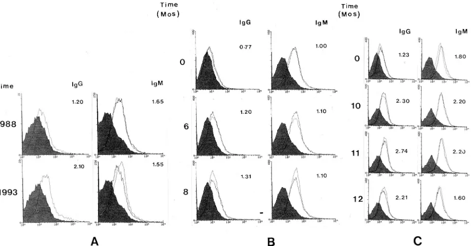
FIG. 4. Detection of early HIV-1 antibodies (IgG and IgM) by r-FIFA in the samples from three individuals (PHL-A, PHL-B, and PHL-C [panels A to C, respectively]) who were infected but seronegative by recently licensed screening tests with blood from the first bleeding (upper row). The numbers under “IgG” and “IgM” indicate the s/c values for the samples; a value of 1.0 or greater is considered positive. A mixture of Gag-p45 and Gag-gp41-C was used as the antigen for testing the samples.
VOL. 34, 1996 HIV-1 ANTIBODY DETECTION 1415
on May 15, 2020 by guest
http://jcm.asm.org/
[image:4.612.76.539.449.691.2]ments with the flow cytometer and Lysis II software.
Fifty-seven samples from eight BBI HIV-1 seroconversion panels
were compared with a control which was used for all tests in
the study. The IgG and IgM titers in each sample were
mea-sured. A total of 114 Kolmogorov-Smirnov D statistics were
computed, and the histograms for each pair were plotted on a
common graph. The results show that the histograms for the
samples and the control tend to be clearly differentiated, with
the samples shifted to the right of the control for all but eight
pairs for IgG (BBI panel members Q3, P1, P2, P3, P4, K1, K2,
and H2) and three pairs for IgM (BBI panel members Q3, H2,
and H5), for which the samples were negative by all anti-HIV-1
assays used in the study, including r-FIFA. The sample sizes
began at 12,907 for the control and 15,147 for the samples in
the r-FIFA tests. By using the smallest sample sizes in the
study, a P value of 0.05 would be achieved by a D value of 0.016
and a P value of 0.01 would be achieved by a D value of 0.019.
The smallest D statistic in the study is 0.03, with a P value of 7
3
10
210. Within the limits of machine accuracy, the P values in
the present study are all effectively 0. The statistical analysis of
the Kolmogorov-Smirnov test has resulted in strong proof of
the significance of the histograms for the samples and the
control (Fig. 1).
Analysis of serial samples from donors who were later
de-termined to have seroconverted has proven to be very useful
for the evaluation of serological assays and for documenting
their enhanced sensitivities. The r-FIFA serological profiles of
eight HIV-1-infected individuals during seroconversion are
shown in Fig. 2. The results indicated that r-FIFA detected
seroconversion on average more than 20 days earlier (range, 0
to 126 days) than the EIAs did. For example, the samples in
panel Q were collected over 72 days, but 53 days elapsed
between the first and second bleedings, making it appear
prob-able that sample 1 was collected prior to infection; however,
sample 2 was positive for the p24 antigen and a very faint p24
band appeared on WB of samples 1 through 5 (BBI data). The
r-FIFA result shows that sample 1 was positive for HIV-1 IgG
antibody and that sample 3, in which the p24 antigen
concen-tration was increased, then became negative, suggesting that an
antigen-antibody complex was formed at this time point. The
result was confirmed by a radioimmunoprecipitation assay
(RIPA) with
35S-labelled HIV-1 recombinant gp160 (data not
shown). Sample 1 in panel Q was positive and was therefore
collected postinfection, not preinfection.
The high degree of sensitivity of r-FIFA and its
multiple-detection feature have made it possible to study the profiles of
the antibody response in the early stage of HIV-1 infection. As
shown in Fig. 2, examples of the primary response were found
TABLE 1. Comparison of r-FIFA and FDA-licensed confirmatory tests on the basis of the sensitivity of detection of antibodies the three open reading frames of the HIV-1 genome in the low-titer anti-HIV-1 panel PRB104 PRB104 identification no. Result of FDA-licensed test a Bio-Rad WB Ortho/Cambridge WB Organon Teknika WB HIV-1 gag pol env Res. gag pol env Res. gag pol env Res. gag pol 01 1 2 1 1 11 1 1 1 2 1 1 12 02 1 2 1 1 12 1 1 1 2 1 1 12 03 1 2 1 1 12 1 1 1 1 1 1 11 04 1 2 1 1 12 1 1 1 2 1 1 12 05 1 2 1 1 11 1 1 1 1 1 1 11 06 1 1 1 1 12 1 1 1 1 1 1 11 07 12 2 IND 12 2 IND 12 2 IND 22 08 22 2 2 1 2 2 IND 12 2 IND 22 09 12 1 1 1 2 1 1 12 1 1 2 2 10 c 2 2 2 2 22 2 2 2 2 2 2 22 11 1 2 1 1 12 1 1 1 2 1 1 12 12 1 2 1 1 12 1 1 1 2 1 1 11 13 12 1 1 1 2 2 IND 12 f160 12 2 14 f24 22 IND 22 2 2 f24 22 IND 22 15 2 2 2 2 22 2 2 2 2 2 2 22 Total no. of positive results 12 1 10 10 12 2 9 9 13 3 10 10 8 4 aWB results were interpreted by using the criteria of the Centers for Disease Control and Prevention and the Association of State and Territorial Public Health IND, indeterminate; Res., result. By the Bio-Rad WB, Ortho/Cambridge WB, Organon Teknika WB, HIV-1 RIPA RL15, and HIV-1 r-FIFA assays, 71, 64, 71, respectively, and 7, 14, 21, 29, and 57% of the samples were found to be gag 1 pol 1 env 1, respectively. bThe chimeric Gag-gp41-C antigen was used to detect antibodies to env . Later, the results were confirmed by using insoluble gp160 polyprotein as the antigen cSample 10 is the negative control.
on May 15, 2020 by guest
[image:5.612.330.547.64.736.2]for panels J and K. Panels D and R showed similar patterns of
antibody production (Fig. 2A). However, for panels E, H, P,
and Q (Fig. 2B), the HIV-1 IgG antibody level remained varied
for a long period (longer than 1 to 3 months) prior to
sero-conversion, as detected by FDA-licensed tests. The IgM
anti-body became positive later (panel P). The pattern in panels E,
H, P, and Q (Fig. 2B) is apparently different from the pattern
in panels D, J, K, and R. Presumably, these individuals (panels
E, H, P, and Q) were not recently infected with HIV-1. For
some unknown reason, the virus antigen concentration rose,
boosting the antibody response in which the anti-HIV-1 IgG
antibodies increased to a higher level in a shorter time (as seen
for panels E, P, and Q), indicating a possible secondary
im-mune response. The fluorescence histograms of the samples
for panel K (Fig. 3) show the kinetics of specific IgG, IgM, and
IgA antibody production during the window period. The IgM
antibodies were detected before the p24 antigen became
de-tectable, and then IgA and IgG antibodies appeared as the p24
antigen level peaked. The IgM antibody response to HIV-1
overlapped with the HIV-1 antigen peak.
Three PHL HIV-1 seroconversion panels and a series of
negative samples (data not shown) were evaluated in a blind
test to confirm the sensitivity of r-FIFA. The first bleedings
from all three panels were positive for IgG and/or IgM by
r-FIFA but were negative by EIA (Fig. 4). The first bleedings
of patients PHL-B and PHL-C were positive for HIV-1 p24
antigen (data not shown).
Furthermore, to compare the sensitivity of r-FIFA with
those of FDA-licensed confirmatory tests, including WB and
RIPA, the BBI low-titer anti-HIV-1 panel was tested by
r-FIFA by using each of the antigens Gag-p45, Gag-gp41-C
chimeric protein, and Pol97 (Table 1). Since the chimeric
pro-tein Gag-gp41-C has specific epitopes from HIV-1 gag and
gp41 polyproteins, it is difficult to justify whether the samples
tested have specific antibodies to env. To confirm that
antibod-ies to env alone exist, all 15 of the samples were tested by
r-FIFA with an insoluble form of gp160 (cell-associated gp160
expressed in insect cells) as the antigen. The results correlated
100% with the results from the assay with the Gag-gp41
chi-meric protein as the antigen, as shown in Table 1. The
anti-bodies to all three antigens, Gag-p45, Gag-gp41-C (or gp160),
and Pol97, were detectable in 8 of the 14 samples (57%) by
r-FIFA. However, the antibodies to pol gene products (p66,
p51, and p31) were poorly detected in the FDA-licensed
con-firmatory tests. Only 1 to 4 of the 14 samples (7 to 29%) were
found to have antibodies to all three proteins by the WB and
RIPA. Statistical analysis as implemented by S-plus (Statistical
Science) for comparison of antibodies to gag, pol, and env
indicate that r-FIFA is more sensitive than the three licensed
WB (P
5
0.0052). However, the difference between r-FIFA
and RIPA is not significant (P
5
0.1573).
Reproducibility of r-FIFA.
To evaluate the reproducibility of
r-FIFA, two antigens, Gag-p45 and Gag-gp41-C, were used for
the analysis of panel D samples (Fig. 5). Similar patterns of
antibody production during the seroconversion were obtained
by using the two antigens in separate tests. The duplicates of
each assay were highly reproducible, as shown by the
overlap-ping of two-line peaks, which represent the fluorescence
[image:6.612.62.291.71.297.2]inten-FIG. 5. Early HIV-1 antibody detection in the samples of BBI anti-HIV-1 seroconversion panel D by r-FIFA with two different antigens. The flow cyto-metric histograms show the anti-HIV-1 antibody signals of the samples (line peaks) and the fluorescence signal (background) of the negative sample as a control (solid grey peak).
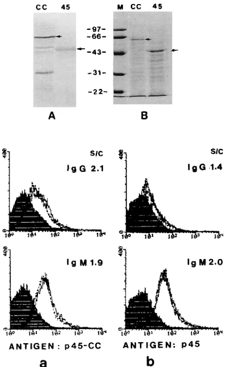
FIG. 6. Antibody detection by r-FIFA with antigen Gag-gp41-C (p45-CC; a) and Gag-p45 (p45; b) as antigens. The sample tested had weak antibodies to HIV-1 core antigen (A, lane 45) and strong antibodies to the Gag-gp41 antigen (A, lane CC) by WB analysis (anti-human IgG only). The Gag-p45 antigen (B, lane 45) and the Gag-gp41-C antigen (B, lane CC) were visualized by Coomassie blue staining of a 10% polyacrylamide gel.
VOL. 34, 1996 HIV-1 ANTIBODY DETECTION 1417
on May 15, 2020 by guest
http://jcm.asm.org/
[image:6.612.324.548.313.675.2]10% difference) or identical for most of the samples tested.
Specificity of r-FIFA.
A total of 295 random donor samples,
105 EIA- and WB-positive samples, and 128 samples reactive
by repeat EIA and indeterminate by WB were tested by
r-FIFA with the mixture of Gag-p45 and Gag-gp41-C as the
antigen. Only 1 of the 295 random donor samples was weakly
positive. The positive sample from the random donor was
negative by EIA. All 105 EIA- and WB-positive samples were
positive by r-FIFA. Only 30 of 128 samples (23%) repeatedly
reactive by EIA and indeterminate by WB were still positive
(most of those were expected to be false positive). These
re-sults suggest that r-FIFA’s specificity may be higher than those
of the EIAs and WB analysis; however, the very low percentage
of false-positive results that do occur is probably due to
anti-body cross-reactivity with the antigens. The results for samples
that test false positive will be confirmed further by r-FIFA with
Gag-p45, p97, and gp160 as antigens in separate tests, as for
Table 1.
Differences in antibody responses to different antigens,
Gag-p45, Gag-gp41-C, and Pol97, in r-FIFA.
Figure 6 shows that
chimeric protein Gag-gp41-C binds more IgG antibodies than
Gag-p45. Little difference in the detection of HIV-1 IgM
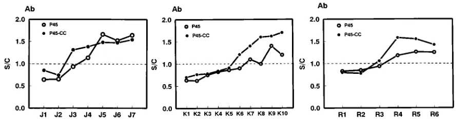
an-tibodies by the antigens was found. We found that IgG
anti-body was detected earlier by Gag-gp41-C than by Gag-p45
(Fig. 7). In some of the seroconversion panels HIV-1 IgG
antibodies to Pol97 were detected as early as those to
Gag-gp41-C. The HIV-1 IgM antibodies to Pol97 were not detected
as frequently as HIV-1 IgG antibodies during the window
period (data not shown).
DISCUSSION
We have created a unique way to detect both IgG and IgM
antibodies using insoluble forms of recombinant proteins as
autologous carriers through flow cytometry. IgM antibody is
one of the early markers of HIV-1 infection (5). This is
prob-ably one of the reasons why r-FIFA can detect HIV-1 antibody
during the early stages of HIV-1 infection, as do the
third-generation EIAs (28). However, in some HIV-1-infected
indi-viduals, only IgG antibodies were detectable by r-FIFA, such
as in BBI panels H and E. These results confirm the previous
findings of other investigators (18). These patients presumably
had a secondary immune response at a stage of HIV-1
infec-tion when the virus or the viral antigen was released at high
concentrations in blood or body fluids. This suggests that the
individual was not recently exposed to HIV-1 and had a longer
window period. This could result from factors such as host
genetics (7, 21), the route of viral transmission (2, 23, 27), the
that the antibodies to env and pol were, strikingly, restricted to
the IgG isotype (8).
The insoluble antigens used for r-FIFA are easily processed
and purified without denaturation, keeping intact their natural
molecular folding, which may optimize the presentation of
antigen epitopes (15). This may be one of the explanations that
can account for r-FIFA’s higher sensitivity for the detection of
early HIV-1 antibodies. Furthermore, an agglutination effect
involving the reaction between IgM antibodies and the
anti-gens in r-FIFA may enhance the sensitivity of the test for
detecting specific antibodies because all the insoluble
polypro-teins used are particulate antigens.
r-FIFA is a simple test. By using the same principle
de-scribed here, r-FIFA can be used to detect any antibody with
an insoluble form of recombinant protein, such as hepatitis B
virus core antigen or human T-cell leukemia type I or II gag
chimeric proteins, as carriers (data not shown). We have
con-structed all three recombinant HIV-1 precursors encoded in
the gag, pol, and env open reading frames of the HIV-1
ge-nome. These are the only natural precursor forms produced in
the viral life cycle and cover
.90% of the regions of the viral
structural proteins. Thus, r-FIFA may have the potential to be
a better confirmatory test because of its high degrees of
sen-sitivity and specificity and its ability to identify antibodies
di-rected to individual HIV-1 polyproteins, and for these same
reasons, it would also be a useful tool for basic research.
ACKNOWLEDGMENTS
This research was partially supported by a grant from the Miles (Bayer)/Canadian Red Cross Society Research and Development Fund.
We thank Catherine Dalzell, statistician, Fractionation Department, Canadian Red Cross, Ottawa, Ontario, Canada, for statistical evalua-tion of the data.
REFERENCES
1. Benjouad, A., J.-C. Gluckman, L. Montagnier, and E. Bahraoui. 1993. Spec-ificity of antibodies produced against native or desialylated human immuno-deficiency virus type 1 recombinant gp160. J. Virol. 67:1693–1697. 2. Borkowsky, W., K. Krasinski, H. Pollack, W. Hoover, A. Kaul, and T.
Ilmet-Moore.1992. Early diagnosis of human immunodeficiency virus infection in
children.6 months of age: comparison of polymerase chain reaction, cul-ture, and plasma antigen capture techniques. J. Infect. Dis. 166:616–619. 3. Busch, M. P. 1994. HIV and blood transfusions: focus on seroconversion.
23rd Congress of the International Society of Blood Transfusion. Vox Sang.
67(Suppl. 3):13–18.
4. Farmer, J. L., R. G. Hampton, and E. Boots. 1989. Flow cytometric assays for monitoring production of recombinant HIV-1 gp160 insect cells infected with a baculovirus expression vector. J. Virol. Methods 26:279–290. 5. Gallarda, J. L., D. R. Henrard, D. Liu, S. Harrington, S. L. Stramer, J. E.
on May 15, 2020 by guest
[image:7.612.82.538.71.188.2]Valinsky, and P. Wu.1992. Early detection of antibody to human immuno-deficiency virus type 1 by using an antigen conjugate immunoassay correlates with the presence of immunoglobulin M antibody. J. Clin. Microbiol. 30: 2379–2384.
6. Hu, Y.-W., and C. Y. Kang. 1991. Enzyme activities in four different forms of human immunodeficiency virus 1 pol gene products. Proc. Natl. Acad. Sci. USA 88:4596–4600.
7. Kashala, O., K. Kayembe, P. Kanki, P. Mukeba, M. Diese, M. Kalengayi,
K.-W. Izzia, and M. Essex.1993. Humoral aspects of anti-HIV immune
responses in Zairians with AIDS: lower antigenemia does not correlate with immune complex levels. AIDS Res. Hum. Retroviruses 9:251–258. 8. Khalife, J., B. Guy, M. Capron, M.-P. Kieny, J.-C. Ameisen, L. Montagnier,
J.-P. Lecocq, and A. Capron.1988. Isotypic restriction of the antibody
re-sponse to human immunodeficiency virus. AIDS Res. Hum. Retroviruses
4:3–9.
9. Kidd, I. M., and V. C. Emery. 1993. The use of baculoviruses as expression vectors. Appl. Biochem. Biotechnol. 42:137–159.
10. Laerum, O. D., and R. Bjerknes. 1992. Flow cytometry in hematology. Academic Press, London.
11. Longobardi Givan, A. 1992. Flow cytometry: first principles. Wiley-Liss Inc., New York.
12. Luo, L., L. Yan, P. M. Cannon, K. Sunyoung, and C. Y. Kang. 1992. Chimeric
gag-V3 like particles of human immunodeficiency virus induce
virus-neutralizing antibodies. Proc. Natl. Acad. Sci. USA 89:10527–10531. 13. Martin, R., J. R. George, M. Hanson, J. Pearson, R. Turnicky, and J.
Wethers.1993. Report of the Eighth Annual Conference on Human
Retro-virus Testing. Association of State and Territorial Public Health Laboratory Directors.
14. Mayo, D. J., A. M. Rose, S. E. Matchett, P. A. Hoppe, J. M. Solomon, and
K. K. McCurdy.1991. Screening potential blood donors at risk for human
immunodeficiency virus. Transfusion 31:466–474.
15. Nair, B. C., G. Ford, V. S. Kalyanaraman, M. Zafari, C. Fang, and M. G.
Sarngadharan.1994. Enzyme immunoassay using native envelope
glycopro-tein (gp160) for detection of human immunodeficiency virus type 1 antibod-ies. J. Clin. Microbiol. 32:1449–1456.
16. Park, J., and C. D. Morrow. 1992. The nonmyristylated Pr160gag-pol polypro-tein of human immunodeficiency virus type 1 interacts with Pr55gagand is incorporated into viruslike particles. J. Virol. 66:6304–6313.
17. Petersen, L. R., G. A. Satten, R. Dodd, M. Busch, S. Kleinman, A. Grindon,
B. Lenes, and the HIV Seroconversion Study Group.1994. Duration of time
from onset of human immunodeficiency virus type 1 infectiousness to devel-opment of detectable antibody. Transfusion 34:283–289.
18. Race, E. M., K. M. Ramsey, H. L. Lucia, and M. W. Cloyd. 1991. Human
immunodeficiency virus infection elicits early antibody not detected by stan-dard tests: implications for diagnostics and viral immunology. Virology 184: 716–722.
19. Ratner, L., W. Haseltine, R. Patarca, K. J. Livak, B. Starcich, S. F. Josephs,
E. R. Doran, J. A. Rafalski, E. A. Whitehorn, K. Baumeister, L. Ivanoff, S. R. Petteway, Jr., M. L. Pearson, J. A. Lautenberger, T. S. Papas, J. Ghrayeb,
N. T. Chang, R. C. Gallo, and F. Wong-Staal.1985. Complete nucleotide
sequence of the AIDS virus, HTLV-III. Nature (London) 313:277–284. 20. Reagan, K. J., C. C. Lile, Y. Devash, J. Turner, Y.-W. Hu, and C. Y. Kang.
1990. Use of HIV-1 pol gene precursor to detect HIV-1 and HIV-2. Lancet
335:236.
21. Simmonds, P., D. Beatson, R. J. G. Cuthbert, H. Watson, B. Reynolds, J. F.
Peutherer, J. V. Parry, C. A. Ludlam, and C. M. Steel.1991. Determinants
of HIV disease progression: six-year longitudinal study in the Edinburgh haemophilia/HIV cohort. Lancet 338:1159–1163.
22. Simon, F., J. M. Pe´pin, and F. Brun-Vezinet. 1992. Reliability of Western blotting for the confirmation of HIV-1 seroconversion. Lancet 340:1541– 1542.
23. Sinicco, A., R. Fora, M. Sciandra, A. Lucchini, P. Caramello, and P.
Gio-annin.1993. Risk of developing AIDS after primary acute HIV-1 infection.
J. Acquired Immune Defic. Syndr. 6:575–581.
24. Sligh, J. M., B. S. Stanford, T. Roodman, and C. C. Tsai. 1989. Flow cytometric indirect immunofluorescence assay with high sensitivity and spec-ificity for detection of antibodies to human immunodeficiency virus (HIV). Am. J. Clin. Pathol. 91:210–214.
25. Takehara, K., D. Ireland, and D. H. L. Bishop. 1988. Co-expression of the hepatitis B surface and core antigens using baculovirus multiple expression vectors. J. Gen. Virol. 69:2763–2777.
26. Wagner, R., H. Fliessbach, G. Wanner, M. Motz, M. Niedrig, G. Deby, A. von
Brunn, and H. Wolf.1992. Studies on processing particle formation and
immunogenicity of the HIV-1 gag gene product: a possible component of a HIV vaccine. Arch. Virol. 127:117–137.
27. Willerford, D. M., J. J. Bwayo, M. Hensel, W. Emonyi, F. A. Plummer, E. N.
Ngugi, N. Nagelkerke, W. M. Gallatin, and J. Kreiss.1993. Human
immu-nodeficiency virus infection among high-risk seronegative prostitutes in Nairobi. J. Infect. Dis. 167:1414–1417.
28. Zaaijer, H. L., P. V. Exel-Oehlers, T. Kraaijeveld, E. Altena, and P. N. Lelie. 1992. Early detection of antibodies to HIV-1 by third generation assays. Lancet 340:770–772.
29. Zhu, T., H. Mo, N. Wang, D. S. Nam, Y. Cao, R. A. Koup, and D. D. Ho. 1993. Genotypic and phenotypic characterization of HIV-1 in patients with pri-mary infection. Science 261:1179–1181.
VOL. 34, 1996 HIV-1 ANTIBODY DETECTION 1419