Mycobacterium tuberculosis
Clinical Isolates by Pyrosequencing
Anna Engström,a,bNora Morcillo,cBelen Imperiale,cSven E. Hoffner,a,band Pontus Juréena,b
Department of Microbiology, Tumor and Cell Biology, Karolinska Institutet, Stockholm, Swedena; Department of Preparedness, Swedish Institute for Communicable
Disease Control, Solna, Swedenb; and Reference Laboratory of Tuberculosis Control Program of Buenos Aires Province, Dr. Cetrángolo Hospital, Buenos Aires, Argentinac
Conventional phenotypic drug susceptibility testing (DST) methods forMycobacterium tuberculosisare laborious and very time-consuming. Early detection of drug-resistant tuberculosis (TB) is essential for prevention and control of TB transmission. We have developed a pyrosequencing method for simultaneous detection of mutations associated with resistance to rifampin, isoniazid, ethambutol, amikacin, kanamycin, capreomycin, and ofloxacin. Seven pyrosequencing assays were optimized for fol-lowing loci:rpoB,katG,embB,rrs,gyrA, and the promoter regions ofinhAandeis. The molecular method was evaluated on a panel of 290 clinical isolates ofM. tuberculosis. In comparison to phenotypic DST, the pyrosequencing method demonstrated high specificity (100%) and sensitivity (94.6%) for detection of multidrug-resistantM. tuberculosisas well as high specificity (99.3%) and sensitivity (86.9%) for detection of extensively drug-resistantM. tuberculosis. The short turnaround time combined with multilocus sequencing of several isolates in parallel makes pyrosequencing an attractive method for drug resistance screen-ing inM. tuberculosis.
T
uberculosis (TB), caused byMycobacterium tuberculosis, per-sists as a global public health problem. Drug-resistant bacteria, especially multidrug-resistant (MDR)M. tuberculosisand exten-sively drug-resistant (XDR)M. tuberculosis, pose challenges for the prevention and control of this deadly disease (64). MDRM. tuberculosisis defined as an isolate resistant to at least the two main first-line anti-TB drugs, rifampin (RIF) and isoniazid (INH), whereas XDRM. tuberculosisis defined as an MDR isolate that is also resistant to a fluoroquinolone (FQ) and at least one of the following second-line injectable agents: amikacin (AMK), kana-mycin (KAN), and capreokana-mycin (CAP) (8). TB control and pre-vention programs are based on early diagnosis followed by rapid identification of drug resistance (62). Generally, detection of drug-resistantM. tuberculosisis performed by culture and drug susceptibility testing (DST) in liquid or on solid media. However, these procedures are laborious and take several weeks to months to complete. The development of rapid molecular methods, which can be performed within 1 or 2 days, is important for the timely identification of appropriate TB treatment. In fact, the World Health Organization (WHO) endorses the use of genotypic meth-ods that target specific molecular mutations associated with resis-tance to individual drugs (63). Two examples of commercially available methods are Xpert MTB/RIF and GenoType MTBDR (16–18), which are predominately based on a lack of probe hy-bridization to wild-type loci, indirectly indicating the presence of mutations. The Xpert MTB/RIF system is also limited in that it detects only RIF resistance.Spontaneous chromosomal mutations are the genetic basis for drug resistance inM. tuberculosis(40,68), and a limited number of mutations account for the majority of phenotypic resistance to anti-TB drugs. RIF interacts with the ß-subunit (encoded byrpoB) of the RNA polymerase and inhibits the early steps of transcrip-tion (5). Resistance to RIF is almost entirely coupled to mutations within an 81-bp region of therpoBgene, called the RIF resistance-determining region (RRDR), comprising codons 507 to 533 (14,
40,46). In accordance with standard practice, these codon num-bers correspond to those found in theEscherichia coli rpoB
se-quence, as designated on the basis of alignment of the translated sequence, and do not represent the actual codon positions inM. tuberculosis rpoB(30,32,58). The mechanisms of resistance to isoniazid (INH), though extensively investigated, remain incom-pletely understood (60). INH is a prodrug that is activated by the catalase-peroxidase enzyme KatG, encoded by thekatGgene (22). The primary target of INH inM. tuberculosisis believed to be the enoyl-acyl carrier protein reductase InhA, which is involved in mycolic acid synthesis (3,39,43). Mutations inkatG, specifically at codon 315, lead to high-level INH resistance (40,67). Low-level resistance to INH is attributed to mutations in the promoter re-gion of themabA-inhA operon (here denotedinhA) leading to overexpression of InhA (26,60). Ethambutol (EMB) inhibits ara-binosyl transferases, encoded by theembCABoperon, thereby in-terfering with biosynthesis of the cell wall component arabinoga-lactan (55,59). Mutations associated with resistance to EMB are primarily found in theembBgene (40). Amikacin (AMK), kana-mycin (KAN), and capreokana-mycin (CAP) inhibit protein synthesis by binding to the ribosome (21,31,38). Resistance to all three drugs is associated with mutations in the 16S rRNA generrs, spe-cifically at nucleotide positions 1401, 1402, and 1484 (1,11,28,54,
57). A mutation at nucleotide 1401 or 1484 is associated with resistance to all these agents, whereas a mutation at nucleotide 1402 is associated with CAP resistance and low-level KAN resis-tance (11,28). Overexpression ofeis(encoding the aminoglyco-side acetyltransferase Eis), caused by mutations in the promoter region, confers low-level resistance to KAN (66).
Fluoroquinolo-Received17 December 2011Returned for modification17 January 2012
Accepted21 March 2012
Published ahead of print29 March 2012
Address correspondence to Anna Engström, [email protected].
Supplemental material for this article may be found athttp://jcm.asm.org/. Copyright © 2012, American Society for Microbiology. All Rights Reserved.
doi:10.1128/JCM.06664-11
on May 16, 2020 by guest
http://jcm.asm.org/
nes (FQs), e.g., ofloxacin (OFL), which bind to DNA gyrase, in-hibit proper regulation of supercoiling and cause chromosomal double-strand breaks (65). DNA gyrase is a heterotetramer con-sisting of two A and two B subunits, encoded by the genesgyrAand gyrB(34,61). FQ resistance-associated mutations are predomi-nantly found in a short region, codons 88 to 94, of thegyrAgene (53,56,65).
Pyrosequencing is a semiautomated sequencing method based on real-time monitoring of DNA synthesis, optimized to analyze short DNA sequences. It is based on the quantitative detection of released pyrophosphate during DNA synthesis. In a cascade of enzymatic reactions, light is generated at intensities proportional to the numbers of incorporated nucleotides (33, 42). Pyrose-quencing has previously been described as a method for detection of drug resistance inM. tuberculosis(2,4,23).
The objective of this study was to develop and evaluate a mo-lecular method for detection of drug resistance-associated muta-tions in clinical isolates ofM. tuberculosisby pyrosequencing tech-nology. Apart from further optimization of the currentrpoBassay (23), we have designed pyrosequencing assays forkatG,embB,rrs, gyrA, and the promoter regions ofinhAandeis. These assays fa-cilitate rapid molecular detection of resistance to RIF, INH, EMB, AMK, KAN, CAP, and OFL in clinical isolates. Phenotypic and genotypic DST results were compared for determination of the sensitivity and specificity of the molecular assays for each drug.
MATERIALS AND METHODS
Bacterial strains and phenotypic drug susceptibility testing.The pan-susceptible reference strainM. tuberculosisH37Rv (ATCC 25618) and 290 clinical isolates ofM. tuberculosisfrom Europe, Asia, Africa, and Latin America were selected from the culture collection at the WHO Suprana-tional TB Reference Laboratory at the Swedish Institute for Communica-ble Disease Control in Solna, Sweden, and included in this study. The classification of strains as susceptible or resistant was based on DST per-formed earlier using WHO-recommended standardized and quality as-sured methods forM. tuberculosisand, in some cases, complemented by MIC determinations (11).
Gene amplification.Genomic DNA was isolated as described else-where (23), or by the method of Sandegren and colleagues (45), but using only one chloroform extraction step. Parts of genesrpoB,katG,embB,rrs, andgyrAand of the promoter regions ofinhAandeiswere amplified using PCR. Primers and amplicon sizes are presented in Table S1 in the supple-mental material. Amplifications were performed in a final volume of 50l with 1⫻PCR buffer, 2 mM MgCl2for all loci (except forrpoB, for which
1 mM MgCl2was used), 100M each dNTP (Applied Biosystems,
War-rington, United Kingdom) (forrpoB,inhA,eisandgyrA) or 200M each dNTP (forembB,rrsandkatG), 200 nM each primer, 1 U of AmpliTaq Gold (Applied Biosystems, Branchburg, NJ), and 10 ng of DNA. Thermo-cycling conditions are specified in Table S1 in the supplemental material. Pyrosequencing.Seven pyrosequencing assays were optimized for the following loci:rpoB,katG,embB,rrs,gyrA, and the promoter regions of inhAandeis. A previously reportedrpoBassay was further optimized by using only two sequencing primers, F1 and F9 (see Table S1 in the sup-plemental material) and a directed dispensation order instead of cyclic dispensation (23), thus reducing the number of sequencing reactions needed per sample. The directed dispensation order forrpoBwas designed to detect all mutations in the RRDR reported by Ramaswamy and Musser (40). All four nucleotides are repeatedly dispensed in cyclic dispensation, whereas the nucleotide dispensation order is preprogrammed according to a known sequence in directed dispensation. Cyclic dispensation order was used forkatG,embB,gyrA, and the promoter region ofinhA. Apart from therpoBlocus, directed dispensation order was also used forrrsand the promoter region ofeis.
Preparation of templates and sequencing reactions were performed according to the instructions of the manufacturers (Biotage AB, Uppsala, Sweden, and Qiagen GmbH, Hilden, Germany). Briefly, each locus was amplified by PCR with a biotinylated reverse primer for immobilization onto streptavidin-coated Sepharose beads (GE Healthcare, Uppsala, Swe-den). The PCR product was converted into a single-stranded DNA tem-plate and purified using a vacuum preparation tool. A sequencing primer (0.4M) was subsequently annealed to the single-stranded DNA (see Table S1 in the supplemental material). Pyrosequencing was carried out in a PSQ 96MA instrument (Pyrosequencing AB, Uppsala, Sweden) using a PSQ Gold 96 SQA reagent kit (Biotage AB, Uppsala, Sweden, and Qiagen GmbH, Hilden, Germany) and a nucleotide dispensation order as speci-fied in Table S1 in the supplemental material. Pyrograms were interpreted and mutations were detected using both peak height and sequence output. Sequences were analyzed in BioEdit version 7.0.4.1 (13) and Geneious Pro version 4.8.4 (Biomatters Ltd., Auckland, New Zealand) by alignment to the M. tuberculosisH37Rv loci (GenBank accession no. NC_000962; NCBI bank) (7) and the sequence results ofM. tuberculosisH37Rv (ATCC 25618).
Sensitivity and specificity.Phenotypic DST was considered the gold standard for comparison with the results obtained by pyrosequencing. Sensitivity was calculated as the number of true-positive specimens (phe-notypically resistant and pyrosequencing resistant) divided by the num-ber of true-positive specimens plus the numnum-ber of false-negative speci-mens (phenotypically resistant and pyrosequencing susceptible). Specificity was calculated as the number of true-negative specimens (phe-notypically susceptible and pyrosequencing susceptible) divided by the number of true-negative specimens plus the number of false-positive specimens (phenotypically susceptible and pyrosequencing resistant).
RESULTS
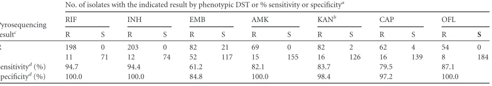
We have developed a pyrosequencing method for simultaneous detection of mutations associated with resistance to RIF, INH, EMB, AMK, KAN, CAP, and OFL inM. tuberculosisclinical iso-lates. In comparison to phenotypic DST, the molecular method demonstrated an overall specificity of 97.2% and a sensitivity of 83.2%. For detection of MDRM. tuberculosisisolates, a specificity of 100% and a sensitivity of 94.6% were noted. The corresponding figures for detection of XDRM. tuberculosisisolates were 99.3% and 86.9%, respectively.
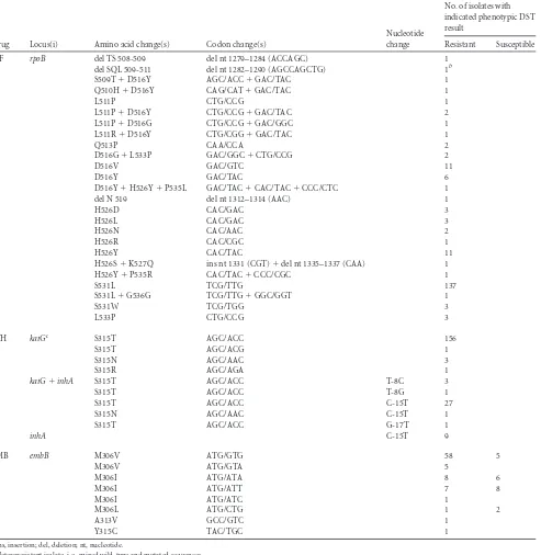
For RIF resistance detection, the sensitivity was 94.7% and the specificity was 100% (Table 1). Of 209 RIF-resistant isolates, 198 had a mutation in the RRDR, and the vast majority (189 of 198) had a mutation in codon 516, 526, or 531. TherpoBlocus of all but five isolates could be sequenced using directed dispensation order and sequencing primers F1 and F9. Four of these had insertions or deletions not designed for the directed dispensation order; in-stead, these were sequenced using four primers (F1, F7, F9, and F13) (23) and cyclic dispensation order. One isolate could not be sequenced with primer F1, and as a control, the RRDR was se-quenced by Sanger sequencing (24). This isolate harbored an L511P mutation but also a mutation in codon 505 (F¡V), which is where the F1 primer anneals. The vast majority of isolates had a missense mutation in the RRDR (Table 2). In one case, the silent mutation G536G occurred together with the S531L mutation. Eleven of the RIF-resistant clinical isolates had a wild-typerpoB locus.
The sensitivity and specificity for INH were 94.4% and 100%, respectively (Table 1). In total, 215 isolates were resistant to INH, and 203 of those had a mutation inkatG(the majority had an S315T mutation), in the promoter region ofinhA(the majority had a C-15T mutation), or in both loci (Table 2). Twelve clinical
on May 16, 2020 by guest
http://jcm.asm.org/
isolates were phenotypically determined to be resistant to INH but had wild-typekatGandinhAloci.
The sensitivity for EMB was 61.2%, and the specificity was 84.8% (Table 1). Only 82 of the 134 phenotypically resistant iso-lates harbored a mutation in theembBlocus. Also, among the EMB-susceptible isolates, 21 had anembB306 mutation. Muta-tions inembBcodons 313 and 315 were detected in EMB-resistant isolates (Table 2).
The sensitivity and specificity for AMK were 81.2% and 100%, respectively. For KAN, the sensitivity was 83.7% and the specific-ity was 98.4% (Table 1). The majority the AMK- and KAN-resis-tant isolates harbored a mutation in therrslocus (Table 3). All but one isolate with⫺14 or⫺37eismutations were KAN resistant, and the majority of⫺10 and⫺12 mutations were found among susceptible isolates. A C-15G mutation was detected in a KAN-susceptible isolate. The sensitivity and specificity for CAP were 79.5% and 97.2%, respectively (Table 1). Most CAP-resistant iso-lates had anrrsmutation at position 1401; however, this mutation was also detected in four susceptible isolates. Few possible muta-tions inrrsand a directed dispensation order made it possible to achieve a read length of 95 bp, which covered nucleotide positions 1401, 1402, and 1484 in one sequence run.
For OFL, the sensitivity and specificity were 87.1% and 100%, respectively (Table 1). Missense mutations ingyrA, specifically in codon 94, were detected in the vast majority of OFL-resistant iso-lates but were absent in all OFL-susceptible isoiso-lates.
By investigation of the pyrosequencing pyrograms, five hetero-resistant isolates, i.e., a mixed template consisting of both wild-type and mutated sequences, were identified (Tables 2and3). The total turnaround time from inactivation of bacteria to interpreted sequencing results was approximately 8 h.
DISCUSSION
Bacteriological methods are highly sensitive and specific for detec-tion of resistance to most anti-TB drugs; however, they are labor-intensive and very time-consuming. Consequently, molecular di-agnostics can be used for rapid detection of drug-resistantM. tuberculosis. We have developed seven pyrosequencing assays for detection of mutations associated with resistance to first- and sec-ond-line anti-TB drugs and evaluated the method on a panel of 290 clinical isolates ofM. tuberculosis.
Pyrosequencing is a robust technique that is easy to perform in a high-throughput manner. In the method developed here, 12 strains could simultaneously be sequenced in a 96-well plate. Some of the most frequent resistance-associated mutations inM.
tuberculosisare located in very short regions of genes or promoter regions, whereas others are more dispersed. By using directed pyrosequencing dispensation order forrpoB, rrs, and the pro-moter region ofeis, the entire resistance-associated locus could be covered in only one or two sequencing reactions. The successful sequencing of 95 bp of therrsgene shows not only that it is possi-ble to optimize longer reads for pyrosequencing but also that it is possible to do so using a GC-rich template (65%) (7). Cyclic dis-pensation was used forkatG, the promoter region ofinhA,embB, andgyrA, where mutations are confined to a short stretch of the locus. Cyclic dispensation increases tolerance for variations and eliminates the risk of failing to detect as-yet-unidentified muta-tions but typically renders shorter sequence reads. An advantage of sequencing compared to line-probe assays is the discriminatory power on the genotypic level. For instance, sequencing can distin-guish between missense and silent mutations and discriminate against mutations not associated with drug resistance. This is in contrast to the commercially available GenoType MTBDR assays, which partly define resistance by the absence of wild-type se-quence (17,18). Pyrosequencing also requires less hands-on time than GenoType MTBDR and is more flexible, as the number of assays and the choice of included loci easily can be adjusted. Fur-thermore, the pyrosequencing method offers sensitivity and spec-ificity comparable to those of Sanger sequencing (6) but is easier to handle and thus more suitable for investigation of a large number of samples. On the other hand, a drawback with pyrosequencing, as well as with other commercially available molecular methods (16–18), is that they cannot cover whole genes. Thus,pncAgene mutations, associated with resistance to the first-line drug pyrazi-namide (48), cannot be sufficiently covered by these methods. Pyrogram analysis revealed that five phenotypically resistant iso-lates were heteroresistant. Heteroresistance is due either to segre-gation of a single strain into resistant and susceptible organisms or to superinfection with two different strains (one susceptible and one resistant) (19). The former scenario describes the evolution of resistance that arises from suboptimal treatment.
[image:3.585.44.547.80.168.2]Introduction of directed dispensation order for therpoB locus reduced the number of sequencing reactions needed for the RRDR to only two without compromising the detection of missense mutations. However, four isolates had in-frame dele-tions and inserdele-tions; thus, cyclic dispensation and four se-quencing primers (F1, F7, F9, and F13) had to be employed in those cases. TherpoBassay is suboptimal in cases where mis-sense mutations, insertions, or deletions have occurred specif-ically at the site where the sequencing primer anneals. This is,
TABLE 1Performance of pyrosequencing as a molecular drug susceptibility test method
Pyrosequencing resultc
No. of isolates with the indicated result by phenotypic DST or % sensitivity or specificitya
RIF INH EMB AMK KANb CAP OFL
R S R S R S R S R S R S R S
R 198 0 203 0 82 21 69 0 82 2 62 4 54 0
S 11 71 12 74 52 117 15 155 16 126 16 139 8 184
Sensitivityd(%) 94.7 94.4 61.2 82.1 83.7 79.5 87.1
Specificityd(%) 100.0 100.0 84.8 100.0 98.4 97.2 100.0
aR, resistant; S, susceptible.
b
Isolates harboringeispromoter mutations at positions⫺10 and⫺12 were considered pyrosequencing susceptible.
cR, isolates with a mutation inrpoB(RIF),katGorinhA(INH),embB(EMB),rrs(AMK and CAP),rrsandeis(KAN), orgyrA(OFL); S, isolates with a wild-type locus. d
Data were determined on the basis of phenotypic drug susceptibility testing as the gold standard.
on May 16, 2020 by guest
http://jcm.asm.org/
however, not a concern for the downstream sequences of F1, as the read length is generally sufficient to cover two sequencing primers, and mutations at the F1 primer site are rarely found among clinical isolates. In fact, sequencing primer F1 anneals upstream of the RRDR. Still, repeatedly failed sequencing re-actions provide an indication that the sequencing primer can-not anneal to the specific annealing site, and we consider that the method failed to detect drug resistance in the case where an isolate
had mutations in codons 505 and 511. Eleven phenotypically RIF-resistant isolates in this study did not harbor a mutation in the RRDR and were thus misclassified as susceptible by the molecular method. This may reflect the inherent limitations of phenotypic DST but could also be due torpoBmutations outside the RRDR (14,49) or elsewhere in the genome.
[image:4.585.48.541.87.593.2]The INH assay (katGandinhApromoter region) was 100% specific and demonstrated high sensitivity. The link between INH
TABLE 2Mutations identified within loci associated with resistance to the first-line drugs rifampin, isoniazid, and ethambutol in clinical isolates of M. tuberculosisa
Drug Locus(i) Amino acid change(s) Codon change(s)
Nucleotide change
No. of isolates with indicated phenotypic DST result
Resistant Susceptible
RIF rpoB del TS 508-509 del nt 1279–1284 (ACCAGC) 1
del SQL 509-511 del nt 1282–1290 (AGCCAGCTG) 1b
S509T⫹D516Y AGC/ACC⫹GAC/TAC 1
Q510H⫹D516Y CAG/CAT⫹GAC/TAC 1
L511P CTG/CCG 1
L511P⫹D516Y CTG/CCG⫹GAC/TAC 2
L511P⫹D516G CTG/CCG⫹GAC/GGC 1
L511R⫹D516Y CTG/CGG⫹GAC/TAC 1
Q513P CAA/CCA 2
D516G⫹L533P GAC/GGC⫹CTG/CCG 2
D516V GAC/GTC 11
D516Y GAC/TAC 6
D516Y⫹H526Y⫹P535L GAC/TAC⫹CAC/TAC⫹CCC/CTC 1
del N 519 del nt 1312–1314 (AAC) 1
H526D CAC/GAC 3
H526L CAC/GAC 3
H526N CAC/AAC 2
H526R CAC/CGC 1
H526Y CAC/TAC 11
H526S⫹K527Q ins nt 1331 (CGT)⫹del nt 1335–1337 (CAA) 1
H526Y⫹P535R CAC/TAC⫹CCC/CGC 1
S531L TCG/TTG 137
S531L⫹G536G TCG/TTG⫹GGC/GGT 1
S531W TCG/TGG 3
L533P CTG/CCG 3
INH katGc S315T AGC/ACC 156
S315T AGC/ACG 1
S315N AGC/AAC 3
S315R AGC/AGA 1
katG⫹inhA S315T AGC/ACC T-8C 3
S315T AGC/ACC T-8G 1
S315T AGC/ACC C-15T 27
S315N AGC/AAC C-15T 1
S315T AGC/ACC G-17T 1
inhA C-15T 9
EMB embB M306V ATG/GTG 58 5
M306V ATG/GTA 5
M306I ATG/ATA 8 6
M306I ATG/ATT 7 8
M306I ATG/ATC 1
M306L ATG/CTG 1 2
A313V GCC/GTC 1
Y315C TAC/TGC 1
ains, insertion; del, deletion; nt, nucleotide.
b
Heteroresistant isolate, i.e., mixed wild-type and mutated sequences.
cThekatGlocus could not be amplified for one of the phenotypically resistant isolates.
on May 16, 2020 by guest
http://jcm.asm.org/
and catalase peroxidase (KatG) originally came from the observa-tion that some highly resistant clinical isolates ofM. tuberculosis, withkatGdeleted, were catalase negative (67). We suspect that one of the INH-resistant isolates in this study had this genotype, as we were consistently unable to amplify thekatGgene of this isolate, and this result was confirmed by the failure of another indepen-dentkatGPCR assay. As the pyrosequencing assay is designed to detect mutations, we consider that the method failed to detect drug resistance in this case. It is possible that the 12 phenotypically resistant isolates lacking a mutation in either locus have a muta-tion elsewhere in the genome. Mutamuta-tions in the open reading frame ofinhAconfer INH resistance (3); however, these muta-tions are rarely found in clinical isolates and usually occur to-gether with mutations inkatGand the promoter region ofinhA (60). Consequently, we chose not to include the opening reading frame ofinhAin the molecular method, as well as other genes, such asaphCandkasA, as there are limited data on their role in INH resistance (25,60).
TheembBpyrosequencing assay detected mutations in both phenotypically EMB-resistant and EMB-susceptible isolates ( Ta-ble 2), suggesting that these mutations are a poor indicator of EMB resistance. The sensitivity (61.2%) and specificity (84.8%) of detection of EMB resistance were indeed lower than were seen with the other drugs; however, this does not necessarily mean that the assay has a low predictive value for clinically relevant EMB resistance. Conventional phenotypic EMB DST forM. tuberculosis is notoriously problematic (12,27). Several studies, including
al-lelic exchange experiments, have demonstrated a strong associa-tion betweenembBmutations, specifically at codon 306, and EMB resistance (35–37,44,51,52). It has also been shown that there is a narrow range between the MICs of susceptible and EMB-resistant isolates ofM. tuberculosis(15,47). Some mutations, e.g., the M306I substitution, give rise to a modest increase in the MIC (37,44,52), which may explain why this particular mutation was found in so many EMB-susceptible isolates in this study. It is likely that the EMB MIC increases only moderately for theembBA313V substitution, as we detected it in a phenotypically resistant isolate, and others have found it in EMB-susceptible isolates (6). A possi-ble clinical implication is that allembB306 mutants should be treated as isolates with altered EMB susceptibility, even if they appear to be EMB susceptible by conventional DST. Mutations in codon 497 in theembBgene have been reported in EMB-resistant clinical isolates (20) and could possibly be included to increase the sensitivity of the method. Furthermore, mutations inembCand embAmay also be involved (41).
[image:5.585.42.546.89.375.2]Despite the relatively long distance between the mutations in rrs, the pyrosequencing assay for this locus could be optimized for only one sequencing reaction without compromising mutation detection. The specificities were high for AMK, KAN, and CAP (100%, 98.4%, and 97.2%, respectively), whereas the sensitivities were lower (82.1%, 83.7%, and 79.5%). The A1401G mutation in rrswas observed in isolates resistant to all the three agents. One isolate, which was found to be phenotypically resistant to AMK, KAN, and CAP, was identified as susceptible by the molecular
TABLE 3Mutations identified within loci associated with resistance to the second-line drugs amikacin, kanamycin, capreomycin, and ofloxacin in clinical isolates ofM. tuberculosis
Drug Locus(i)
Amino acid
change(s) Codon change(s) Nucleotide change(s)
No. of isolates with indicated phenotypic DST result
Resistant Susceptible
AMK rrs A1401G 69a
KAN rrs A1401G 65b
rrs⫹eis A1401G⫹G-6T 1
A1401G⫹C-12T 1
A1401G⫹G-37T 1c
eis G-10A 14 38
C-12T 1 5
C-14T 11 1
C-15G 1
G-37T 3
CAP rrs A1401G 62b 4b
OFL gyrA D89N GAC/AAC 1
A90V GCG/GTG 23
A90V⫹S91P GCG/GTC⫹TCG/CCG 1
A90V⫹D94N GCG/GTG⫹GAC/AAC 1
S91P TCG/CCG 4b
D94G GAC/GGC 13
D94G GAC/GGT 1
D94A GAC/GCC 6
D94N GAC/AAC 3
D94H GAC/CAC 1
D94Y GAC/TAC 1
a
Two isolates were heteroresistant, i.e., mixed wild-type and mutated sequences. bOne isolate was heteroresistant.
c
Heteroresistant isolate.
on May 16, 2020 by guest
http://jcm.asm.org/
method. It is possible that this isolate was heteroresistant but that the proportion of resistant organisms was not large enough to be detected by the pyrosequencing assay. Nevertheless, alternative drug resistance mechanisms cannot be ruled out. Four CAP-sus-ceptible isolates were identified as resistant by the pyrosequencing assay (rrs1401 mutation); however, the MIC (4 mg/liter) for these strains was close to the critical concentration for CAP (11). Over-expression ofeisconfers low-level KAN resistance (66). Mutations at certain positions, such as⫺14 and⫺37, give rise to a higher overexpression ofeisthan othereispromoter mutations (66) and can thus be considered more sensitive genetic markers for KAN resistance. All but one of the isolates with⫺14 or⫺37 mutations in this study were classified as phenotypically resistant. Mutations at position⫺10 and, in particular, at position⫺12 seem to give rise to a MIC close to the critical concentration, as isolates harbor-ing these mutations have been reported as both KAN susceptible and KAN resistant (6,11,66). Because of their poor predictive value, we chose not to take mutations at these positions into ac-count when calculating sensitivity and specificity for KAN. The previously unreported C-15Teispromoter mutation was detected in a KAN-susceptible isolate; however, further studies must be undertaken in order to clarify its role ineisexpression. It should also be noted that all but one of the AMK-resistant clinical isolates lacking anrrsmutation did have a mutation (G-10A, C-14T, or G-37T) in the promoter region ofeis. Overexpression of the Eis protein also leads to inactivation of AMK, but the inactivation is 3-fold less than that seen for KAN (66). As with EMB, this suggests that isolates with eispromoter mutations should be treated as isolates with altered KAN, and plausibly AMK, susceptibility, re-gardless of the phenotypic DST results. CAP resistance is also known to be conferred by mutations intlyA(29); however, we chose not to include this gene in the molecular method, sincetlyA mutations are rare (6,11) and some have also been found also in CAP-susceptible isolates (11). Furthermore, in order to include tlyAas a genetic marker for CAP, the entire open reading frame must be analyzed, and this is not practically feasible by pyrose-quencing.
The specificity of the OFL assay was 100%, although the sensi-tivity was lower. Eight of the 62 phenotypically OFL-resistant iso-lates lacked a mutation in thegyrAlocus. In these cases, it is not certain that a mutation accounting for this phenotype would be found elsewhere ingyrA, or ingyrB, as other drug resistance mech-anisms, such as efflux pumps, have been suggested to contribute to FQ resistance inM. tuberculosis(10). ThegyrBlocus was not included in this assay, as mutations in this gene occur at a much lower frequency and usually in association withgyrAmutations (9,68). There were equal distributions of thegyrAS95T mutation among both susceptible and resistant clinical isolates; in fact, it was detected in 88% of the cases in both groups. This mutation is considered to be a polymorphism not associated with resistance to FQs (50).
In general, the noted sensitivity rates for detection of resistance (Table 1) reflect the incomplete understanding of resistance mechanisms associated with all drugs investigated in this study. Phenotypically resistant isolates lacking mutations in known re-sistance-associated loci present a problem for all molecular tests designed to detect mutations associated with resistance. This em-phasizes the need for intensifying research that aims at identifying novel resistance mechanisms inM. tuberculosis.
We have developed a high-throughput molecular method for
simultaneous detection of resistance to RIF, INH, EMB, AMK, KAN, CAP, and OFL inM. tuberculosis. A rapid and specific diag-nosis is highly important not only for the individual patient but also from the general public perspective, since rapid modification to an effective drug therapy would reduce the spread of TB in the society. The short turnaround time in combination with a multi-locus sequencing of several isolates in parallel makes pyrose-quencing an attractive method for drug resistance screening inM. tuberculosis. As the method is PCR based, it has a great potential to be further developed for application directly to clinical samples, e.g., sputum samples.
ACKNOWLEDGMENTS
This work was supported by EC project FP7-HEALTH-2007-A-201690 (FAST-XDR-DETECT) and the National Council of Science and Tech-nology (CONICET), Argentina.
We gratefully acknowledge Juan-Carlos Palomino, Anandi Martin, and Fabienne Paasch for assistance with the study design and providing clinical isolates, Jim Werngren and Juan Carlos Toro for technical assis-tance, Ramona Groenheit, Solomon Ghebremichael, and Alexandra Pennhag for kindly providing DNA samples, and Diarmaid Hughes, Mar-gareta Krabbe, and David Herthnek for valuable comments on the man-uscript.
REFERENCES
1.Alangaden GJ, et al.1998. Mechanism of resistance to amikacin and kanamycin in Mycobacterium tuberculosis. Antimicrob. Agents Che-mother.42:1295–1297.
2.Arnold C, et al.2005. Single-nucleotide polymorphism-based differen-tiation and drug resistance detection in Mycobacterium tuberculosis from isolates or directly from sputum. Clin. Microbiol. Infect.11:122–130. 3.Banerjee A, et al.1994. inhA, a gene encoding a target for isoniazid and
ethionamide in Mycobacterium tuberculosis. Science263:227–230. 4.Bravo LT, et al.2009. Pyrosequencing for rapid detection of
Mycobacte-rium tuberculosis resistance to rifampin, isoniazid, and fluoroquinolones. J. Clin. Microbiol.47:3985–3990.
5.Campbell EA, et al.2001. Structural mechanism for rifampicin inhibition of bacterial RNA polymerase. Cell104:901–912.
6.Campbell PJ, et al.2011. Molecular detection of mutations associated with first- and second-line drug resistance compared with conventional drug susceptibility testing of Mycobacterium tuberculosis. Antimicrob. Agents Chemother.55:2032–2041.
7.Camus JC, Pryor MJ, Medigue C, Cole ST.2002. Re-annotation of the genome sequence of Mycobacterium tuberculosis H37Rv. Microbiology
148:2967–2973.
8.Centers for Disease Control.2006. Emergence of Mycobacterium tuber-culosis with extensive resistance to second-line drugs—worldwide, 2000 – 2004. MMWR Morb. Mortal. Wkly. Rep.55:301–305.
9.Chakravorty S, et al.2011. Rapid detection of fluoroquinolone-resistant and heteroresistant Mycobacterium tuberculosis by use of sloppy molec-ular beacons and dual melting-temperature codes in a real-time PCR as-say. J. Clin. Microbiol.49:932–940.
10. da Silva PE, Von Groll A, Martin A, Palomino JC.2011. Efflux as a mechanism for drug resistance in Mycobacterium tuberculosis. FEMS Immunol. Med. Microbiol.63:1–9.
11. Engström A, Perskvist N, Werngren J, Hoffner SE, Jureen P. 2011. Comparison of clinical isolates and in vitro selected mutants reveals that tlyA is not a sensitive genetic marker for capreomycin resistance in Myco-bacterium tuberculosis. J. Antimicrob. Chemother.66:1247–1254. 12. Griffith ME, Bodily HL.1992. Stability of antimycobacterial drugs in
susceptibility testing. Antimicrob. Agents Chemother.36:2398 –2402. 13. Hall TA.1999. BioEdit: a user-friendly biological sequence alignment
editor and analysis program for Windows 95/98/NT. Nucleic Acids Symp. Ser.41:95–98.
14. Heep M, et al.2001. Frequency of rpoB mutations inside and outside the cluster I region in rifampin-resistant clinical Mycobacterium tuberculosis isolates. J. Clin. Microbiol.39:107–110.
15. Heifets LB, Iseman MD, Lindholm-Levy PJ.1986. Ethambutol MICs and
on May 16, 2020 by guest
http://jcm.asm.org/
MBCs for Mycobacterium avium complex and Mycobacterium tubercu-losis. Antimicrob. Agents Chemother.30:927–932.
16. Helb D, et al.2010. Rapid detection of Mycobacterium tuberculosis and rifampin resistance by use of on-demand, near-patient technology. J. Clin. Microbiol.48:229 –237.
17. Hillemann D, Rusch-Gerdes S, Richter E.2007. Evaluation of the Geno-Type MTBDRplus assay for rifampin and isoniazid susceptibility testing of Mycobacterium tuberculosis strains and clinical specimens. J. Clin. Mi-crobiol.45:2635–2640.
18. Hillemann D, Rusch-Gerdes S, Richter E.2009. Feasibility of the Geno-Type MTBDRsl assay for fluoroquinolone, amikacin-capreomycin, and ethambutol resistance testing of Mycobacterium tuberculosis strains and clinical specimens. J. Clin. Microbiol.47:1767–1772.
19. Hofmann-Thiel S, et al.2009. Mechanisms of heteroresistance to isoni-azid and rifampin of Mycobacterium tuberculosis in Tashkent, Uzbeki-stan. Eur. Respir. J.33:368 –374.
20. Huang WL, Chi TL, Wu MH, Jou R.2011. Performance assessment of the GenoType MTBDRsl test and DNA sequencing for detection of sec-ond-line and ethambutol drug resistance among patients infected with multidrug-resistant Mycobacterium tuberculosis. J. Clin. Microbiol.49: 2502–2508.
21. Johansen SK, Maus CE, Plikaytis BB, Douthwaite S.2006. Capreomycin binds across the ribosomal subunit interface using tlyA-encoded 2= -O-methylations in 16S and 23S rRNAs. Mol. Cell23:173–182.
22. Johnsson K, Schultz PG.1994. Mechanistic studies of the oxidation of isoniazid by the catalase peroxidase from Mycobacterium tuberculosis. J. Am. Chem. Soc.116:7425–7426.
23. Jureen P, et al.2006. Rapid detection of rifampin resistance in Mycobac-terium tuberculosis by pyrosequencing technology. J. Clin. Microbiol.44: 1925–1929.
24. Jureen P, Werngren J, Hoffner SE.2004. Evaluation of the line probe assay (LiPA) for rapid detection of rifampicin resistance in Mycobacte-rium tuberculosis. Tuberculosis (Edinb.)84:311–316.
25. Kiepiela P, Bishop KS, Smith AN, Roux L, York DF.2000. Genomic mutations in the katG, inhA and aphC genes are useful for the prediction of isoniazid resistance in Mycobacterium tuberculosis isolates from Kwa-zulu Natal, South Africa. Tuber. Lung Dis.80:47–56.
26. Larsen MH, et al.2002. Overexpression of inhA, but not kasA, confers resistance to isoniazid and ethionamide in Mycobacterium smegmatis, M. bovis BCG and M. tuberculosis. Mol. Microbiol.46:453– 466.
27. Madison B, et al.2002. Multicenter evaluation of ethambutol suscepti-bility testing of mycobacterium tuberculosis by agar proportion and ra-diometric methods. J. Clin. Microbiol.40:3976 –3979.
28. Maus CE, Plikaytis BB, Shinnick TM.2005. Molecular analysis of cross-resistance to capreomycin, kanamycin, amikacin, and viomycin in Myco-bacterium tuberculosis. Antimicrob. Agents Chemother.49:3192–3197. 29. Maus CE, Plikaytis BB, Shinnick TM.2005. Mutation of tlyA confers
capreomycin resistance in Mycobacterium tuberculosis. Antimicrob. Agents Chemother.49:571–577.
30. Miller LP, Crawford JT, Shinnick TM.1994. The rpoB gene of Myco-bacterium tuberculosis. Antimicrob. Agents Chemother.38:805– 811. 31. Moazed D, Noller HF.1987. Interaction of antibiotics with functional
sites in 16S ribosomal RNA. Nature327:389 –394.
32. Musser JM.1995. Antimicrobial agent resistance in mycobacteria: molec-ular genetic insights. Clin. Microbiol. Rev.8:496 –514.
33. Nyrén P.2007. The history of pyrosequencing. Methods Mol. Biol.373: 1–14.
34. Piton J, et al.2010. Structural insights into the quinolone resistance mechanism of Mycobacterium tuberculosis DNA gyrase. PLoS One
5:e12245.
35. Plinke C, et al.2009. Tuberculosis ethambutol resistance: concordance between phenotypic and genotypic test results. Tuberculosis (Edinb.)89: 448 – 452.
36. Plinke C, Rusch-Gerdes S, Niemann S.2006. Significance of mutations in embB codon 306 for prediction of ethambutol resistance in clinical My-cobacterium tuberculosis isolates. Antimicrob. Agents Chemother.50: 1900 –1902.
37. Plinke C, Walter K, Aly S, Ehlers S, Niemann S.2011. Mycobacterium tuberculosis embB codon 306 mutations confer moderately increased re-sistance to ethambutol in vitro and in vivo. Antimicrob. Agents Che-mother.55:2891–2896.
38. Prammananan T, et al.1998. A single 16S ribosomal RNA substitution is responsible for resistance to amikacin and other 2-deoxystreptamine
ami-noglycosides in Mycobacterium abscessus and Mycobacterium chelonae. J. Infect. Dis.177:1573–1581.
39. Quémard A, et al.1995. Enzymatic characterization of the target for isoniazid in Mycobacterium tuberculosis. Biochemistry34:8235– 8241. 40. Ramaswamy S, Musser JM.1998. Molecular genetic basis of
antimicro-bial agent resistance in Mycobacterium tuberculosis: 1998 update. Tuber. Lung Dis.79:3–29.
41. Ramaswamy SV, et al.2000. Molecular genetic analysis of nucleotide polymorphisms associated with ethambutol resistance in human isolates of Mycobacterium tuberculosis. Antimicrob. Agents Chemother.44:326 – 336.
42. Ronaghi M, Uhlen M, Nyren P.1998. A sequencing method based on real-time pyrophosphate. Science281:363, 365.
43. Rozwarski DA, Grant GA, Barton DH, Jacobs WR, Jr, Sacchettini JC.
1998. Modification of the NADH of the isoniazid target (InhA) from My-cobacterium tuberculosis. Science279:98 –102.
44. Safi H, Sayers B, Hazbon MH, Alland D.2008. Transfer of embB codon 306 mutations into clinical Mycobacterium tuberculosis strains alters sus-ceptibility to ethambutol, isoniazid, and rifampin. Antimicrob. Agents Chemother.52:2027–2034.
45. Sandegren L, et al.2011. Genomic stability over 9 years of an isoniazid resistant Mycobacterium tuberculosis outbreak strain in Sweden. PLoS One6:e16647.
46. Sandgren A, et al.2009. Tuberculosis drug resistance mutation database. PLoS Med.6:e2.
47. Schön T, et al.2009. Evaluation of wild-type MIC distributions as a tool for determination of clinical breakpoints for Mycobacterium tuberculosis. J. Antimicrob. Chemother.64:786 –793.
48. Scorpio A, Zhang Y.1996. Mutations in pncA, a gene encoding pyrazi-namidase/nicotinamidase, cause resistance to the antituberculous drug pyrazinamide in tubercle bacillus. Nat. Med.2:662– 667.
49. Siu GK, et al. 2011. Mutations outside the rifampicin resistance-determining region associated with rifampicin resistance in Mycobacte-rium tuberculosis. J. Antimicrob. Chemother.66:730 –733.
50. Sreevatsan S, et al.1997. Restricted structural gene polymorphism in the Mycobacterium tuberculosis complex indicates evolutionarily recent global dissemination. Proc. Natl. Acad. Sci. U. S. A.94:9869 –9874. 51. Sreevatsan S, et al.1997. Ethambutol resistance in Mycobacterium
tu-berculosis: critical role of embB mutations. Antimicrob. Agents Che-mother.41:1677–1681.
52. Starks AM, Gumusboga A, Plikaytis BB, Shinnick TM, Posey JE.2009. Mutations at embB codon 306 are an important molecular indicator of ethambutol resistance in Mycobacterium tuberculosis. Antimicrob. Agents Chemother.53:1061–1066.
53. Sun Z, et al.2008. Comparison of gyrA gene mutations between labora-tory-selected ofloxacin-resistant Mycobacterium tuberculosis strains and clinical isolates. Int. J. Antimicrob. Agents31:115–121.
54. Suzuki Y, et al.1998. Detection of kanamycin-resistant Mycobacterium tuberculosis by identifying mutations in the 16S rRNA gene. J. Clin. Mi-crobiol.36:1220 –1225.
55. Takayama K, Kilburn JO.1989. Inhibition of synthesis of arabinogalac-tan by ethambutol in Mycobacterium smegmatis. Antimicrob. Agents Chemother.33:1493–1499.
56. Takiff HE, et al.1994. Cloning and nucleotide sequence of Mycobacte-rium tuberculosis gyrA and gyrB genes and detection of quinolone resis-tance mutations. Antimicrob. Agents Chemother.38:773–780. 57. Taniguchi H, et al.1997. Molecular analysis of kanamycin and viomycin
resistance in Mycobacterium smegmatis by use of the conjugation system. J. Bacteriol.179:4795– 4801.
58. Telenti A, et al.1993. Detection of rifampicin-resistance mutations in Mycobacterium tuberculosis. Lancet341:647– 650.
59. Telenti A, et al.1997. The emb operon, a gene cluster of Mycobacterium tuberculosis involved in resistance to ethambutol. Nat. Med.3:567–570. 60. Vilchèze C, and Jacobs WR, Jr.2007. The mechanism of isoniazid killing:
clarity through the scope of genetics. Annu. Rev. Microbiol.61:35–50. 61. Wang JC.1985. DNA topoisomerases. Annu. Rev. Biochem.54:665– 697. 62. World Health Organization.2009. Guidelines for surveillance of drug resistance in tuberculosis. WHO/HTM/TB/2009.422. World Health Or-ganization, Geneva, Switzerland.
63. World Health Organization and Stop Partnership TB.2010. The global plan to stop TB 2011-2015: transforming the fight towards elimination of tuberculosis. World Health Organization, Geneva, Switzerland.
on May 16, 2020 by guest
http://jcm.asm.org/
64. World Health Organization.2010. Multidrug and extensively drug-resistant TB (M/XDR-TB): 2010 global report on surveillance and response, WHO/ HTM/TB/2010, 3rd ed. World Health Organization, Geneva, Switzerland. 65. Xu C, Kreiswirth BN, Sreevatsan S, Musser JM, Drlica K.1996.
Fluo-roquinolone resistance associated with specific gyrase mutations in clini-cal isolates of multidrug-resistant Mycobacterium tuberculosis. J. Infect. Dis.174:1127–1130.
66. Zaunbrecher MA, Sikes RD, Jr, Metchock B, Shinnick TM, Posey JE.
2009. Overexpression of the chromosomally encoded aminoglycoside acetyltransferase eis confers kanamycin resistance in Mycobacterium tu-berculosis. Proc. Natl. Acad. Sci. U. S. A.106:20004 –20009.
67. Zhang Y, Heym B, Allen B, Young D, Cole S. 1992. The catalase-peroxidase gene and isoniazid resistance of Mycobacterium tuberculosis. Nature358:591–593.
68. Zhang Y, Yew WW.2009. Mechanisms of drug resistance in Mycobacte-rium tuberculosis. Int. J. Tuberc. Lung Dis.13:1320 –1330.