Microarray analysis reveal differential expression of
microRNAs in triple negative breast cancer and normal
breast cells
Sherette S. Godfrey, Kashenya M. Gurley, Malcolm M. Moses, Brianna L. Arrington, and Checo J. Rorie, PhD
Department of Biology, North Carolina Agricultural and Technical State University, Greensboro, North Carolina, USA
Abstract- The Triple-Negative Breast Cancer (TNBC) subtype accounts for 15 percent of all breast cancers and is characterized by the lack of estrogen, progesterone, and HER2/Neu receptors. The objective of this study is to compare and contrast microRNA expression level profiles from triple-negative breast cancer versus normal breast cell lines. MicroRNA was extracted from normal (AG11132) and TNBC (HCC1806, HCC70, and MB157) cell lines and subjected to microarray analysis. Microarray analysis revealed that a number of microRNAs (miR-21, 34a, and let-7 family miRNAs) were shown to be differentially expressed in TNBC versus the normal breast cell lines. It was shown that TNBC cells have a differential microRNA expression profile when compared to normal cells. Microarray analysis revealed that microRNA expression profiles cluster the TNBC cells separately from the normal cells. The findings suggest that microRNAs could potentially be used as biomarkers to diagnose TNBC, and could potentially be used as drug-targeted therapies in the future.
Index Terms- triple negative breast cancer, breast cancer, microRNA, microarray
I. INTRODUCTION
reast cancer is the uncontrolled growth of abnormal tissues occurring in the breast. Some tumors in the breast can grow slowly, potentially up to 10 years by the time a lump can be felt, but some tumors are aggressive and grow much faster [1]. Breast cancer have four known subtypes: normal-like, luminal-like, HER-2-positive, and basal-like [2]. Triple-negative breast cancer or TNBC is a subtype of breast cancer that usually falls into the basal-like category, and is described as a heterogeneous disease compromising of multiple tumors that are associated with histological patterns and different biological and clinical behaviors and have no known biomarkers [3]. Triple-negative breast cancer accounts for about 15 percent of all breast cancers. It is characterized by cancer cells that lack estrogen, progesterone and HER2 receptors. For this reason, these tumors do not respond to hormone therapies or HER2-targeted treatments [4].
MicroRNAs are short single stranded molecules that are involved in regulation of gene expression and play a role in influencing the pathways that are known to be responsible for the progression of many diseases such as cardiovascular disease, fibrosis, and cancer [5, 6]. While microRNAs are transcribed from genes, they do not encode for proteins. The function of microRNAs is to prevent the translation of messenger RNAs/mRNAs into proteins by triggering the degradation of these mRNAs and can be classified as oncogenic or tumorigenic [6].
According to a study published in 2014 by Gasparini et al, microRNAs can be used to classify Triple-Negative Breast Cancer malignancy into six different subtypes, which could lead to new screening methods, prognostic markers, and perhaps new targeted treatments for this aggressive form of breast cancer. The six different TNBC subtypes were identified through gene expression profiling as: basal-like 1(BL1), basal-like 2(BL2), immunomodulatory, mesenchymal, mesenchymal stem-like, and luminal androgen receptor-expressing. The most common of these subtypes is basal-like as it is often clinically related to TNBC. All of these subtypes have different rates of incidence, risk factors, prognosis and response to treatment. Sub-classification is necessary to better identify molecular-based therapeutic targets, select biomarkers, discover new drugs, and design clinical trials that will enable alignment of TNBC patients to appropriate targeted therapies [7]. Therefore, this study seeks to report on baseline expression patterns of microRNAs in triple-negative breast cancer cell lines as compared to normal breast cells using microarray analysis. These baseline microRNA expression profiles may provide an insight into potential TNBC miRNA biomarkers that may be targeted for cancer therapies in the future.
II. MATERIALS AND METHODS
Cell Lines: Triple-negative breast cancer cell lines HCC1806, HCC70, and MDA-MB-157 were purchased from American Type Culture Collection (Manassas, VA). Normal breast cell line A11132 was purchased from Coriell Institute for Medical Research (Camden, NJ). Cell lines HCC1806 and HCC70 were both grown in RPMI-1640 (Thermo Scientific; Rockford, IL) containing 10% fetal bovine serum (FBS) and 1% penicillin and streptomycin (P/S) (Hyclone Laboratory; Logan Utah). AG11132 was grown in MEGM (Lonza; Walkersville, MD). MDA-MB-157 was grown in L-15 (Thermo Scientific; Rockford, IL). All cell lines were maintained in 5% CO2 at 37°C. All of the cells were grown to 95% confluence, counted, pelleted, and then stored at -80°C.
Microarray Hybridization and Analysis: A total of eight cell pellets, two of each cell line (AG11132, HCC1806, HCC70, and MB157) ran as duplicates, designated as 1&2and were sent frozen on dry ice to Beckman Coulter Genomics (Morrisville, NC) in accordance with Beckman Coulter Genomics’ specifications for microRNA extraction and processing. One hundred nanograms of total RNA were labeled with fluorescent dye Cy3 using the miRNAs Complete Labeling Kit (Agilent Technologies, Palo Alto, CA) following the manufacturer’s protocol. The Cy3 labeled RNA was hybridized to an Agilent Human miRNAs Microarray 8X15K Release 14.0 array and the generated data was analyzed using Partek Genomics Suite at North Carolina A&T State University. The microarray text data was imported into Partek Genomics Suite software for comparison analysis. The controls used in this experiment were the normal breast tissue cell line AG11132, and the TNBC cancer cell lines were the MDA-MB-157, HCC1806, and the HCC70 cell lines. The mean & median microRNA signals and gene name are the categories that were filtered out of the original cell line data, and then the data from the duplicate cell lines were merged. Determination of under and over expression of the microRNA values were done initially through literature reviews to assist in narrowing down microRNAs to analyze using quantitative or real time-PCR. Heat maps and hierarchical clustering of the microRNA signal expression profiles were performed using Partek Genomics Suite.
Partek Genomics Suite Analysis: Partek Genomic Suite incorporates data and allows for analysis of this information in a variety of different methods. Importation of data into Partek can be done in several different ways; data can come from different array chips (i.e., Agilent or Affymetrix) and/or text files. This study utilized the text file importation method which was simple set of steps where specific data that correlated to this research was included in analysis in Partek Genomic Suite. The process for using chips such as Affymetrix or Agilent has several more dialogue boxes that must be completed prior to importation of files. Controls used in this experiment were designated as the normal breast tissue cell line AG11132 (1&2) and TNBC cancer cell lines were identified as MDA-MB-157 (1&2), HCC1806 (1&2), and HCC70 (1&2). The raw hybridization data received from Beckman Coulter was filtered to include the mean & median signals for microarray analysis. From this data the individual cell lines were then filtered out individually and merged based on the cell line type. There were two sets of data for each cell line as it was run on the microarray chip in duplicates. Finally, all the cancer cell lines were combined onto one spreadsheet. Determination of under- and overexpression of these values were in part determined through literature reviews.
III. RESULTS
Microarray Analysis Reveal the Differential Expression of MicroRNAs
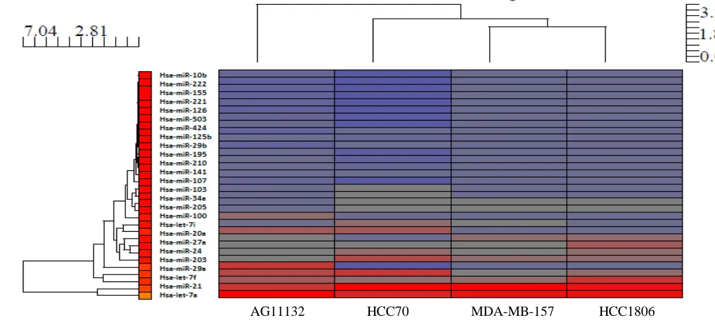
Figure1: Hierarchical clustering of g-mean average signals of microRNAs in breast cell lines ran as duplicates on Agilent Human miRNAs Microarray 8X15K Release 14.0 microarray
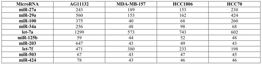
[image:3.612.27.585.469.606.2]The hierarchical clustering in Figure 1 contained signals from all of the 12,000 miRNA transcripts from the array chip. An extensive literature review was conducted to identify from the array chip miRNAs that were associated with some type of cancer. Table 1 list the miRNAs associated with cancer that were downregulated or suppressed in the TNBC cells when compared to the normal breast cell line. The signals represent the average of the g-mean signal and my not necessarily be significantly different from the control AG11132 cell line. The suppressed miRNAs that were revealed in the table were consistent with miRNAs that are believed to be tumor suppressive and therefore be downregulated in cancer cells.
Table 1: MicroRNAs downregulated or suppressed in TNBC Cell Lines (g-mean hybridization signal)
MicroRNA AG11132 MDA-MB-157 HCC1806 HCC70
miR-27a 243 189 153 230
miR-29a 560 153 162 424
miR-100 375 40 68 260
miR-34a 256 48 98 68
let-7a 1299 573 743 602
miR-125b 59 44 52 48
miR-203 647 43 49 43
let-7f 471 380 233 198
miR-503 67 43 47 45
miR-424 78 43 46 46
Table 2 list the miRNAs associated with cancer that were upregulated or overexpressed in the TNBC cells when compared to the normal breast cell line. The upregulated miRNAs from Table 2 are consistent with miRNAs that have been reported as having oncogenic properties.
Table 2: MicroRNAs upregulated or overexpressed in TNBC Cell Lines (g-mean hybridization signal)
MicroRNA AG11132 MDA-MB-157 HCC1806 HCC70
miR-222 43 45 53 44
miR-16-1 39 42 46 42
miR-195 41 80 47 43
miR-155 40 43 51 51
miR-421 39 41 45 43
miR-126 39 45 50 59
miR-335 38 42 43 41
miR-145 40 43 50 42
miR-34b 38 43 54 44
miR-10b 41 43 50 42
miR-103 103 121 119 147
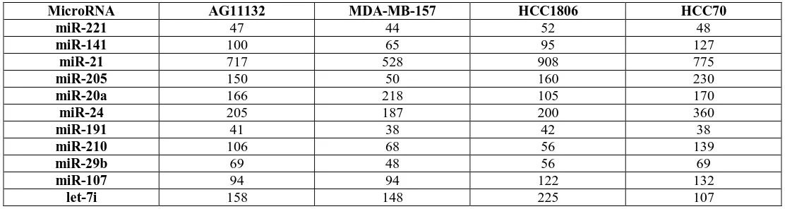
[image:4.612.28.589.304.452.2]Table 3 list the remaining miRNAs that were associated with breast cancer, but reveals that they were differentially expressed within the TNBC breast cancer subtype.
Table 3: MicroRNAs differentially expressed in TNBC Cell Lines (g-mean hybridization signal)
MicroRNA AG11132 MDA-MB-157 HCC1806 HCC70
miR-221 47 44 52 48
miR-141 100 65 95 127
miR-21 717 528 908 775
miR-205 150 50 160 230
miR-20a 166 218 105 170
miR-24 205 187 200 360
miR-191 41 38 42 38
miR-210 106 68 56 139
miR-29b 69 48 56 69
miR-107 94 94 122 132
Figure 2 is a hierarchical cluster of the 32 miRNAs listed in Tables 1, 2, and3 and have been reported to play a role in breast cancer. The cluster shows that the TNBC cells clustered together and separately from the normal breast cell line, AG11132.
Figure 2: Hierarchical clustering of g-mean average signals of 32 microRNAs associated with cancer in breast cell lines ran as duplicates on Agilent Human miRNAs Microarray 8X15K Release 14.0 microarray
IV. DISCUSSION
This study compared the baseline g-mean signal microRNA expression profiles from triple-negative breast cancer and normal breast tissue cell lines using microarray analysis. MicroRNA expression profiles revealed a differential expression pattern in triple-negative breast cancer cells when compared to normal breast tissue cells. Previous research reports suggest that the over or under expression of microRNAs have a significant impact on the expression of protein-coding genes. While some of the differences in the baseline expression levels of microRNAs are not significant or in some cases negligible, it has not been established what fold change in the expression level has an impact on the microRNAs role in the cell. MicroRNAs have the ability to play a role in regulation of gene expression or in tumorigenesis, by acting as oncogenes or tumor suppressors [8]. Other reports have described altered expression of microRNAs in cancer tissues compared to normal tissues, suggesting that these microRNAs could potentially represent novel clinical and prognostic markers [2]. This research is novel because of its usage of cell lines that could serve as model systems to investigate deeper molecular level potential TNBC biomarkers and drug therapeutic targets. These findings may help to provide individualized therapeutic options based on the microRNA expression profile of a TNBC patient. More studies need to be conducted to determine the physiological effects that these microRNAs at these basal expression levels have on the cell and whether they pose an increase risk to cancer.
ACKNOWLEDGMENT
The research study was funded in part from the North Carolina Agricultural and Technical State University College of Arts and Sciences Innovation Fund, and the RIMI/NIH-NIGMS Grant #5P20MD000546-07.
REFERENCES
1. Komen, S.G. What is Breast Cancer? 2015; Available from: http://ww5.komen.org/BreastCancer/WhatisBreastCancer.html.
2. Carey, L.A., et al., Race, breast cancer subtypes, and survival in the Carolina Breast Cancer Study. JAMA, 2006. 295(21): p. 2492-502.
3. Anders, C. and L.A. Carey, Understanding and treating triple-negative breast cancer. Oncology (Williston Park), 2008. 22(11): p. 1233-9; discussion 1239-40, 1243.
4. Anders, C.K. and L.A. Carey, Biology, metastatic patterns, and treatment of patients with triple-negative breast cancer. Clin Breast Cancer, 2009. 9 Suppl 2: p. S73-81.
5. Zampetaki, A. and M. Mayr, MicroRNAs in vascular and metabolic disease. Circ Res. 110(3): p. 508-22.
6. Ardekani, A.M. and M.M. Naeini, The Role of MicroRNAs in Human Diseases. Avicenna Journal of Medical Biotechnology, 2010. 2(4): p. 161-179. 7. Gasparini, P., et al., microRNA expression profiling identifies a four microRNA signature as a novel diagnostic and prognostic biomarker in triple negative
breast cancers. Oncotarget. 5(5): p. 1174-84.
8. Buffa, F.M., et al., microRNA-associated progression pathways and potential therapeutic targets identified by integrated mRNA and microRNA expression profiling in breast cancer. Cancer Res, 2011. 71(17): p. 5635-45.
AUTHORS
First Author – Sherette S. Godfrey, Masters of Science, North Carolina Agricultural and Technical State University, [email protected].
Second Author – Kashenya M. Gurley, Masters of Science, North Carolina Agricultural and Technical State University, [email protected].
Third Author – Malcolm M. Moses, Undergraduate, North Carolina Agricultural and Technical State University, [email protected].
Fourth Author – Brianna L. Arrington, Bachelor of Science, North Carolina Agricultural and Technical State University, [email protected].
Fifth Author – Checo J. Rorie, Doctorate of Philosophy, Assistant Professor, North Carolina Agricultural and Technical State University, [email protected].