major mRNA decay pathway.
HOLMES, L E A, CAMPBELL, Susan, DE LONG, S K, SACHS, A B and
ASHE, M P
Available from Sheffield Hallam University Research Archive (SHURA) at:
http://shura.shu.ac.uk/8291/
This document is the author deposited version. You are advised to consult the
publisher's version if you wish to cite from it.
Published version
HOLMES, L E A, CAMPBELL, Susan, DE LONG, S K, SACHS, A B and ASHE, M P
(2004). Loss of translational control in yeast compromised for the major mRNA
decay pathway. Molecular and cellular biology, 24 (7), 2998-3010.
Copyright and re-use policy
See
http://shura.shu.ac.uk/information.html
Copyright © 2004, American Society for Microbiology. All Rights Reserved.
Loss of Translational Control in Yeast Compromised for the Major
mRNA Decay Pathway
L. E. A. Holmes,
1S. G. Campbell,
1S. K. De Long,
2A. B. Sachs,
2and M. P. Ashe
1*
Department of Biomolecular Sciences, University of Manchester Institute of Science and Technology, Manchester M60 1QD, United Kingdom,1and Department of Molecular and Cell Biology, University of California at
Berkeley, Berkeley, California 947202
Received 8 October 2003/Returned for modification 12 November 2003/Accepted 15 January 2004
The cytoplasmic fate of mRNAs is dictated by the relative activities of the intimately connected mRNA decay and translation initiation pathways. In this study, we have found that yeast strains compromised for stages downstream of deadenylation in the major mRNA decay pathway are incapable of inhibiting global translation initiation in response to stress. In the past, the paradigm of the eIF2␣kinase-dependent amino acid starvation pathway in yeast has been used to evaluate this highly conserved stress response in all eukaryotic cells. Using a similar approach we have found that even though the mRNA decay mutants maintain high levels of general translation, they exhibit many of the hallmarks of amino acid starvation, including increased eIF2␣ phos-phorylation and activatedGCN4mRNA translation. Therefore, these mutants appear translationally oblivious to decreased ternary complex abundance, and we propose that this is due to higher rates of mRNA recruitment to the 40S ribosomal subunit.
Translation and mRNA decay are intimately linked to reg-ulate the gene expression profile in response to a variety of nutritional and environmental stresses in Saccharomyces cer-evisiae. Previously, a number of these yeast stresses have been found to inhibit translation at the initiation step, leading to a rapid loss of polysomes and an accumulation of inactive singlet ribosomes or monosomes. These stresses include glucose star-vation (3, 23), salt stresses (28, 42), rapamycin addition (5), fusel alcohol addition (4), and severe amino acid starvation (41). In general, the inhibition of translation initiation is ex-tremely rapid, and we rationalize these responses as follows. The inhibition of protein synthesis could allow the degradation of existing mRNAs and proteins, thus facilitating a switch in the genetic program that might be required to adapt to the particular stress. In addition, the inhibition of global transla-tion initiatransla-tion may allow specific specialized mRNAs to be translated and this activation could also support adaptation to the stress.
A precedent for this type of regulation exists in the general control pathway of yeast (17). Here global protein synthesis is rapidly inhibited upon severe amino acid starvation (41). It is thought that a specific protein kinase (Gcn2p) senses the levels of amino acids indirectly via the level of uncharged tRNA in the cell. An accumulation of uncharged tRNA stimulates the phosphorylation of the eIF2␣subunit at serine 51 by Gcn2p, leading to the sequestration of a guanine nucleotide exchange factor, eIF2B. The eIF2B complex normally catalyzes the re-cycling of GDP to the translationally active form, eIF2-GTP. eIF2-GTP is capable of interacting with the methionyl initiator tRNA (Met-tRNAiMet) to form the ternary complex
(eIF2-GTP Met-tRNAiMet). Therefore, inhibition of eIF2B
(fol-lowing severe starvation for amino acids and the subsequent phosphorylation of eIF2␣) ultimately leads to decreased levels of the ternary complex (17). Recently it has been suggested that the ternary complex might interact with other translation initiation factors (eIFs 1, 5, and 3) to form a multifactor com-plex (MFC) prior to interaction with the 40S ribosomal subunit (43).
As well as inhibiting global protein synthesis, starvation for amino acids induces the expression of a transcription factor, Gcn4p (17). The activation of this transcription factor at the translational level serves to overcome the amino acid stress by inducing the transcription of amino acid biosynthetic genes (18). The GCN4 mRNA contains four short upstream open reading frames (uORFs) that are involved in the translational activation via a complex mechanism involving translational reinitiation at downstream ORFs. The translation of this mRNA is exquisitely sensitive to the levels of ternary complex in the cell. Therefore, following translation of the first uORF (in the presence of high levels of ternary complex), reinitiation at downstream uORFs and subsequent translation termination is favored. In contrast, when ternary complex levels are low (for instance, following amino acid starvation), there is a greater chance that ternary complex will be acquired after ribosomes pass the uORFs, thus allowingGCN4ORF trans-lation (17).
A variety of other stresses have also been shown to activate GCN4translation initiation. These include purine starvation, glucose starvation, fusel alcohol addition, salt stress, and rapa-mycin addition (4, 8, 15, 22, 30, 44, 48). Although the recently described effects of rapamycin onGCN4do rely on increases in eIF2␣phosphorylation (8, 22, 44), at least some of these stress responses do not appear to require an induction of eIF2␣ phosphorylation. Therefore, it seems plausible that these re-sponses might decrease levels of ternary complex via alterna-tive mechanisms. For example, we have proposed that fusel
* Corresponding author. Mailing address: Department of Biomolec-ular Sciences, UMIST, Sackville St., P.O. Box 88, Manchester M60 1QD, United Kingdom. Phone: 44 161 200 4164. Fax: 44 161 200 0409. E-mail: [email protected].
alcohols directly regulate the activity of eIF2B and thus lead to decreased ternary complex levels (4).
Starvation for glucose has a complex effect on translation initiation. Immediately following glucose removal (i.e., within minutes) there is a rapid inhibition of translation initiation (3, 23, 42). This inhibition still occurs in mutants that are incapa-ble of phosphorylating eIF2␣. In wild-type cells, however, ac-tivation of GCN4 translation at later time points following glucose starvation has been described and this activation is Gcn2p dependent (48). The precise mechanisms for these ef-fects on global andGCN4translation initiation remain to be elucidated.
Two general pathways of mRNA decay have been described for yeast. In both of these pathways, mRNAs are first dead-enylated in a process thought to involve the Ccr4p/Pop2p deadenylase (40). In the major or 5⬘to 3⬘pathway, the 5⬘cap structure is then removed by the Dcp1p/Dcp2p decapping complex and this facilitates the degradation of the body of the mRNA via Xrn1p, the 5⬘to 3⬘exoribonuclease (36, 39). In the exosome-mediated pathway, the deadenylated mRNA is de-graded by the cytoplasmic exosome complex (which includes proteins with 3⬘ to 5⬘ exoribonuclease activity and putative RNA helicase activity) (26). As well as these general pathways for mRNA decay, pathways exist to degrade mRNAs contain-ing premature stop codons. These nonsense-mediated decay pathways utilize many of the same components described for the general pathways of mRNA decay; however, these mRNAs are targeted for decay via a specific complex containing the Upf proteins (14, 27).
In this study we have analyzed a catalogue of mutants in these mRNA decay processes and assessed translational con-trol in response to stress. As a result, we have found that major pathway mRNA decay mutants acting at stages downstream of deadenylation are globally resistant to stress-induced transla-tional inhibition. At least in the case of severe amino acid starvation, these strains are translationally resistant to the de-crease in ternary complex levels that occurs as a result of this stress. Thus, this study provides an example of the intimate relationship that exists between protein synthesis and mRNA turnover in the cytoplasm of cells and the interdependence of mechanisms involved in their regulation.
MATERIALS AND METHODS
Strains and growth conditions.Table 1 lists the strains used in this study. In general the yMK116 (yRP840) strain background has been used throughout except where this proved impractical. In these cases isogenic strains were used across individual experiments. Gene disruptions were carried out as previously described (47). General yeast molecular genetic techniques and growth of cul-tures on standard yeast extract-peptone-dextrose (YPD) or synthetic complete dextrose (SCD) medium were performed as detailed previously (16). Glucose starvation was brought about by the removal of glucose for 5 or 10 min. Severe amino acid starvation was brought about by the removal of every amino acid for 10 min. Butanol was added at 1 or 0.75% (vol/vol) in liquid culture for 10 min. Rapamycin (Sigma-Aldrich, Poole, Dorset, United Kingdom) in 90% ethanol– 10% Tween 20 was added to achieve a final concentration of 0.2g/ml for 1 h (an equal volume of drug vehicle was used as a control). Lithium acetate was added to galactose-grown cultures to achieve a final concentration of 30 mM for 15 min. Dilution series plate assays started with 3l of exponentially growing cultures at an optical density at 600 nm (OD600) of 0.3 and continued in fivefold dilutions. Analysis of ribosome distribution on sucrose gradients.Yeast cultures were grown to an OD600of 0.7 and treated by the removal of glucose or amino acids or by the addition of butanol, rapamycin, or lithium as described above. Extracts were prepared in 100g of cycloheximide/ml, and these were layered onto 15 to
50% sucrose gradients. The gradients were sedimented via centrifugation at 40,000 rpm in a Beckman ultracentrifuge for 2.5 h, and theA254was measured continuously to give the traces shown (3).
[35S]methionine incorporation assay.Yeast strains were grown to an OD600of
0.4 in SCD medium lacking methionine, and two 7.5-ml aliquots were pelleted and then resuspended in 20 ml of SCD-Met or SC-Met. Methionine was added to achieve a final concentration of 60 ng/ml, of which 0.5 ng/ml was [35 S]methi-onine (New England Nuclear, Boston, Mass.) (cell-labeling grade; 1,175 Ci/ mmol). Samples (1 ml) were taken at the indicated times and processed as described previously (3).
Assays ofGCN4-lacZreporter expression.Standard methods for measuring the-galactosidase activity for strains bearingGCN4-lacZfusions were used (24).-Galactosidase levels are expressed as nanomoles ofo-nitrophenol--D -galactopyranoside (ONPG) hydrolyzed per minute per microgram of total pro-tein.
Measurement of capped mRNA with the Xrn1p assay.Yeast strains were grown to an OD600of 0.7 in YPD and divided into three aliquots, and cells were pelleted. One of the cell pellets was immediately frozen in liquid nitrogen. The other two were resuspended in YPD or YP, incubated, pelleted, and frozen in liquid nitrogen such that the total incubation time was 10 min. RNA was ex-tracted from the frozen pellets and used for Northern blot analysis (7). The Xrn1p assay was performed as described previously (7). Total RNA (5g) was incubated with 400 ng of purified Xrn1p (a gift from A. Johnson, University of Texas, Austin) in a final volume of 5l in 33 mM Tris-HCl (pH 8.0)–50 mM NaCl–2.5 mM MgCl2–0.2 mM dithiothreitol in the presence or absence of 5 mM EDTA. Reactions were incubated at 37°C for 30 min and stopped using RNA loading buffer.
Immunoblotting of eIF2␣and phosphoserine 51 eIF2␣.Yeast strains were grown to an OD600of 0.7 in SCD-His. Three 15-ml aliquots were taken per strain, the cells were pelleted, and SCD-His medium, SCD minus all amino acids, or SCD-His plus 50 mM 3-aminotriazole (3-AT) was added to each of the three aliquots, respectively. The cells were incubated, pelleted at 30°C, and rapidly frozen in liquid N2such that the total incubation time was 15 min. The cells were lysed and protein samples were prepared, electrophoretically separated, and subjected to immunoblot analysis as described previously (3). The phosphospe-cific eIF2␣(Biosource International) and eIF2␣(a gift from G. Pavitt, University of Manchester Institute of Science and Technology, Manchester, United King-dom) antibodies were used for the detection.
RESULTS
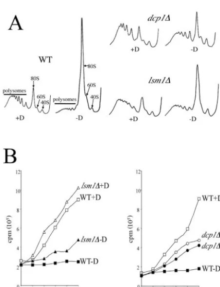
dcp1⌬ and lsm1⌬mutants maintain translation following glucose removal.Previously we have found that the removal of glucose from yeast growth medium results in a rapid inhibition of translation initiation (3). This inhibition is characterized by a loss of polysomes and a concomitant increase in 80S mono-somes (Fig. 1A). A candidate gene strategy was adopted to test a whole range of strains (either mutated in or overexpressing translation initiation factor and mRNA decay genes) (data not shown). Intriguingly, strains lacking either Dcp1p, a key com-ponent of the mRNA decapping enzyme, or Lsm1p, a member of the mRNA decay Lsm complex, maintain polysomal levels following glucose removal (Fig. 1A).
That polysomes were maintained in these mutants after glu-cose removal could be explained either by a continued synthe-sis of proteins or by decreased rates of downstream transla-tional steps such as elongation and termination. To distinguish between these possibilities, the overall rate of protein synthesis was measured by [35S]methionine incorporation assays in
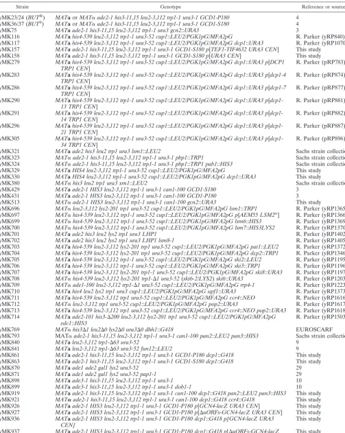
TABLE 1. Yeast strains used in this study
Strain Genotype Reference or source
yMK23/24 (BUTR) MATaorMAT␣ade2-1 his3-11,15 leu2-3,112 trp1-1 ura3-1 GCD1-P180 4
yMK36/37 (BUTS) MATaorMAT␣ade2-1 his3-11,15 leu2-3,112 trp1-1 ura3-1 GCD1-S180 4
yMK75 MATaade2-1 his3-11,15 leu2-3,112 trp1-1 ura3 gcn2::URA3 3
yMK116 MATahis4-539 leu2-3,112 trp1-1 ura3-52 cup1::LEU2/PGK1pG/MFA2pG R. Parker (yRP840) yMK117 MATahis4-539 leu2-3,112 trp1-1 ura3-52 cup1::LEU2/PGK1pG/MFA2pG dcp1::URA3 R. Parker (yRP1070) yMK157 MATaade2-1 his3-11,15 leu2-3,112 trp1-1 ura3-1 GCD1-S180 p[TEF1-TIF4632 URA3 CEN] This study
yMK158 MATaade2-1 his3-11,15 leu2-3,112 trp1-1 ura3-1 GCD1-S180 p[URA3 CEN] This study yMK279 MATahis4-539 leu2-3,112 trp1-1 ura3-52 cup1::LEU2/PGK1pG/MFA2pG dcp1::URA3 p[DCP1
TRP1 CEN] R. Parker (pRP783)
yMK283 MATahis4-539 leu2-3,112 trp1-1 ura3-52 cup1::LEU2/PGK1pG/MFA2pG dcp1::URA3 p[dcp1-4
TRP1 CEN] R. Parker (pRP874)
yMK286 MATahis4-539 leu2-3,112 trp1-1 ura3-52 cup1::LEU2/PGK1pG/MFA2pG dcp1::URA3 p[dcp1-7
TRP1 CEN] R. Parker (pRP877)
yMK290 MATahis4-539 leu2-3,112 trp1-1 ura3-52 cup1::LEU2/PGK1pG/MFA2pG dcp1::URA3 p[
dcp1-13 TRP1 CEN] R. Parker (pRP881)
yMK291 MATahis4-539 leu2-3,112 trp1-1 ura3-52 cup1::LEU2/PGK1pG/MFA2pG dcp1::URA3 p[
dcp1-14 TRP1 CEN] R. Parker (pRP882)
yMK296 MATahis4-539 leu2-3,112 trp1-1 ura3-52 cup1::LEU2/PGK1pG/MFA2pG dcp1::URA3 p[
dcp1-21 TRP1 CEN] R. Parker (pRP887)
yMK305 MATahis4-539 leu2-3,112 trp1-1 ura3-52 cup1::LEU2/PGK1pG/MFA2pG dcp1::URA3 p[
dcp1-34 TRP1 CEN] R. Parker (pRP896)
yMK321 MATaade2 his3 leu2 trp1 ura3 lsm1::LEU2 Sachs strain collection
yMK323 MAT␣ade2-1 his3-11,15 leu2-3,112 trp1-1 ura3-1 pbp1::TRP1 Sachs strain collection yMK324 MAT␣ade2-1 his3-11,15 leu2-3,112 trp1-1 ura3-1 pbp1::TRP1 pab1::HIS3 Sachs strain collection yMK329 MATaHIS4 leu2-3,112 trp1-1 ura3-52 cup1::LEU2/PGK1pG/MFA2pG This study
yMK330 MATaHIS4 leu2-3,112 trp1-1 ura3-52 cup1::LEU2/PGK1pG/MFA2pG dcp1::URA3 This study
yMK380 MAT␣his3 leu2 trp1 ura3 xrn1::LEU2 Sachs strain collection
yMK429 MATaade2-1 HIS3 leu2-3,112 trp1-1 ura3-1 can1-100 GCD1-S180 3 yMK431 MATaade2-1 HIS3 leu2-3,112 trp1-1 ura3-1 can1-100 GCD1-P180 3 yMK513 MAT␣ade2-1 HIS3 leu2-3,112 trp1-1 ura3-1 can1-100 gcn2::URA3 This study
yMK696 MAT␣leu2-3,112 lys2-201 trp1 ura3-52 cup1::LEU2/PGK1pG/MFA2pG lsm1::TRP1 R. Parker (yRP1365) yMK697 MAT␣his4-539 leu2-3,112 trp1-1 ura3-52 cup1::LEU2/PGK1pG/MFA2pG p[AEM55 LSM2ts] R. Parker (yRP1366)
yMK699 MAT␣his4-539 leu2-3,112 trp1-1 ura3-52 cup1::LEU2/PGK1pG/MFA2pG lsm6::HIS3 R. Parker (yRP1369) yMK700 MAT␣his4-539 leu2-3,112 trp1-1 ura3-52 cup1::LEU2/PGK1pG/MFA2pG lsm7::HIS3LYS2 R. Parker (yRP1370)
yMK701 MATaade2 his3 leu2 lys2 trp1 ura3 LHP1 R. Parker (yRP1402)
yMK702 MATaade2 his3 leu2 lys2 trp1 ura3 LHP1 lsm8-1 R. Parker (yRP1405)
yMK703 MATahis4-539 leu2-3,112 lys2-201 trp1 ura3-52 cup1::LEU2/PGK1pG/MFA2pG pat1::LEU2 R. Parker (yRP1372) yMK704 MATahis4-539 leu2-3,112 lys2-201 trp1 ura3-52 cup1::LEU2/PGK1pG/MFA2pG dcp2::TRP1 R. Parker (yRP1346) yMK705 MATahis4-539 leu2-3,112 trp1-1 ura3-52 cup1::LEU2/PGK1pG/MFA2pG ski2::LEU2 R. Parker (yRP1195) yMK706 MATahis4-539 leu2-3,112 trp1-1 ura3-52 cup1::LEU2/PGK1pG/MFA2pG ski3::TRP1 R. Parker (yRP1196) yMK707 MATahis4-539 leu2-3,112 lys2-201 trp1-1 ura3-52 cup1::LEU2/PGK1pG/MFA2pG ski8::URA3 R. Parker (yRP1197) yMK708 MAT␣his4-539 leu2-3,112 lys2-201 trp1-⌬1 ura3-52(ski6-2/LYS2)ski6::URA3 R. Parker (yRP1203) yMK709 MAT␣ade1-100 leu2-3,112 trp1-⌬1 ura3-52 cup1::LEU2/PGK1pG/MFA2pG rrp4-1 R. Parker (yRP1223) yMK710 MATahis4 leu2 lys2 trp1 ura3 cup1::LEU2/PGK1pG/MFA2pG upf1::URA3 R. Parker (yRP1373) yMK711 MATahis4-539 leu2-3,112 trp1 ura3-52 cup1::LEU2/PGK1pG/MFA2pG ccr4::NEO R. Parker (yRP1616) yMK712 MAT␣leu2-3,112 trp1 ura3-52 cup1::LEU2/PGK1pG/MFA2pG pop2::URA3 R. Parker (yRP1617) yMK713 MATahis4-539 leu2-3,112 trp1 ura3-52 cup1::LEU2/PGK1pG/MFA2pG ccr4::NEO pop2::URA3 R. Parker (yRP1618) yMK714 MATaade2-101 his3-⌬200 leu2-3,112 lys2-201 trp1 ura3-52 cup1::LEU2/PGK1pG/MFA2pG
edc1::HIS3 R. Parker (yRP1503)
yMK769 MAT␣his3⌬1 leu2⌬0 lys2⌬0 ura3⌬0 dhh1::G418 EUROSCARF
yMK793 MAT␣ade2-1 his3-11,15 leu2-3,112 trp1-1 ura3-1 can1-100 pan2::LEU2 pan3::HIS3 Sachs strain collection
yMK840 MATaleu2-3,112 trp1-⌬63 ura3-52 9
yMK841 MATaleu2-3,112 trp1-⌬63 ura3-52 fun12::LEU2 9
yMK861 MATaade2-1 his3-11,15 leu2-3,112 trp1-1 ura3-1 GCD1-P180 dcp1::G418 This study yMK863 MATaade2-1 his3-11,15 leu2-3,112 trp1-1 ura3-1 GCD1-S180 dcp1::G418 This study
yMK870 MATaade1 ade2 gal1 lys2 ura3-52 29
yMK871 MATaade1 ade2 gal1 lys2 ura3-52 pap1-1 29
yMK898 MATaade3-1 his3-11,15 leu2-3,112 trp1-1 ura3-1 10
yMK899 MATaade3-1 his3-11,15 leu2-3,112 trp1-1 ura3-1 dob1-1 10
yMK919 MATaade2-1 his3-11,15 leu2-3,112 trp1-1 ura3-1 can1-100 dcp1::G418 pan2::LEU2 pan3::HIS3 This study yMK921 MAT␣ade2-1 his3-11,15 leu2-3,112 trp1-1 ura3-1 can1-100 dcp1::G418 ccr4::G418 This study yMK926 MATaade2-1 HIS3 leu2-3,112 trp1-1 ura3-1 GCD1-P180 p[GCN4-lacZ URA3 CEN] This study yMK927 MATaade2-1 HIS3 leu2-3,112 trp1-1 ura3-1 GCD1-P180 p[⌬uORFs-GCN4-lacZ URA3 CEN] This study yMK936 MATaade2-1 HIS3 leu2-3,112 trp1-1 ura3-1 GCD1-P180 dcp1::G418 p[GCN4-lacZ URA3
CEN] This study
yMK937 MATaade2-1 HIS3 leu2-3,112 trp1-1 ura3-1 GCD1-P180 dcp1::G418 p[⌬uORFs-GCN4-lacZ
URA3 CEN] This study
mutant is lower than that seen with the wild type. For both the lsm1⌬anddcp1⌬strains, however, it is clear that (following a switch to medium with no glucose) the level of [35S]methionine
incorporation is significantly higher than that seen with the appropriate wild-type strains. Indeed thedcp1⌬mutant strain continues to translate at a rate typical of that seen with glu-cose-containing medium. This demonstrates that the observed maintenance of polysomes following glucose removal from
these mutants is due to sustained protein synthesis and does not occur due to slowed rates of elongation or termination in these strains.
Loss of decapping activity in DCP1 mutants allows the maintenance of translation initiation following glucose re-moval.Strains lacking the decapping component Dcp1p have been characterized by their lack of decapping function and a resulting inability to decay mRNAs in a 5⬘to 3⬘direction (6). In addition, the original loss-of-function mutation in LSM1 (SPB8) was shown to be partially inhibited for mRNA decap-ping (7). This is consistent with the partial maintenance of protein synthesis observed following glucose removal in an lsm1⌬mutant (Fig. 1). To investigate whether a decrease in decapping function was responsible for continued translation following glucose removal, a well-characterized series ofDCP1 mutants was assessed (37). Strains with mutations inDCP1that either have little effect (dcp1-13,dcp1-14, anddcp1-21) or are partial-loss-of-function mutations (dcp1-4) in terms of mRNA decapping (37) show a redistribution of polysomes upon glu-cose withdrawal typical of that observed for wild-type strains (Fig. 2A). However, dcp1-7and dcp1-34mutants, which are known to have a strong loss of mRNA decapping function (37), maintain protein synthesis following glucose withdrawal (Fig. 2A). Hence, where decapping activity is low in theDCP1 mu-tants, translation is maintained following glucose removal.
An activation of decapping does not explain the inhibition of translation initiation following glucose removal.Previously we showed that for wild-type strains, there is no major change in the abundance of several mRNA species after 10 min in the absence of glucose (3). As decapping mutants are translation-ally resistant to glucose removal, it is formtranslation-ally possible that in wild-type strains rapid global mRNA decapping might follow glucose removal and lead to the observed inhibition of protein synthesis. To explore whether there is a global decapping of mRNAs following glucose removal, an assay was used to assess the level of capped mRNA. Here RNA was prepared from cells without incubation (control) or incubated for short peri-ods in either rich medium or medium lacking glucose. The sensitivity of these RNA samples to in vitro incubation in the presence of active Xrn1p was assessed by Northern blotting (Fig. 2B). Xrn1p degrades noncapped RNAs in a 5⬘ to 3⬘ direction; hence, the rRNA (which is not modified by a 5⬘cap structure) is completely degraded during this treatment (Fig. 2B; cf. lanes 4 to 6 versus lanes 1 to 3). The presence of a cap structure protects the body of mRNAs from this in vitro Xrn1p exonucleolytic decay treatment (7). The levels of either the PGK1orPAB1mRNAs do not significantly differ between the samples prepared from yeast incubated in the presence or absence of glucose (Fig. 2B; cf. lanes 2 and 3). This suggests that the 5⬘ cap is present on these transcripts and inhibits degradation by the exogenously added Xrn1p regardless of the starvation for glucose. Hence, it appears that there is no global
FIG. 1. mRNA decapping mutants maintain protein synthesis fol-lowing glucose starvation. (A) Polysome traces from yMK116 (WT), yMK117 (dcp1⌬), and yMK321 (lsm1⌬). Yeast were grown in SCD medium, harvested, and then resuspended for 10 min in medium containing (⫹D) or lacking (⫺D) glucose. Polysomes were analyzed as described in Materials and Methods. The 40S (small ribosomal sub-unit), 60S (large ribosomal subsub-unit), 80S (monosome), and polysome peaks are labeled. Even though the mutant strains were derived from different backgrounds, only a single wild type is shown for simplicity, as the inhibition of translation is identical in the different wild-type strains (data not shown). (B) [35S]methionine incorporation into proteins over
time in the presence (⫹D) or absence (⫺D) of glucose for strains yMK23 (WT) and yMK321 (lsm1⌬) (left panel) and strains yMK116 (WT) and yMK117 (dcp1⌬) (right panel). Yeast strains were grown in SCD medium without methionine, harvested, and resuspended in a labeling mixture in the presence or absence of glucose. Aliquots were taken at the indicated times, and the levels of [35S]methionine
[image:5.603.48.274.265.559.2]incor-poration into proteins were determined.
TABLE 1—Continued
Strain Genotype Reference or source
decapping of mRNA following glucose removal from wild-type strains.
As the inhibition mechanism following glucose removal is as yet unknown, polysome profiles of adcp1⌬mutant were ex-amined following amino acid removal. Severe amino acid star-vation results in polysomal runoff indicative of an inhibition of translation initiation (41). The translational inhibition follow-ing amino acid starvation occurs via a well-described pathway that has not been shown to involve mRNA decay (17). There-fore, polysomal runoff in response to severe amino acid star-vation involves eIF2␣phosphorylation via Gcn2p kinase; thus, thegcn2⌬mutant is translationally resistant to this stress (Fig. 2C). Surprisingly, polysomal profiling of adcp1⌬mutant shows that in contrast to the results seen with the wild-type strain, active translation is maintained following amino acid removal as well as glucose removal (Fig. 2C). Therefore, rather than a global decapping of mRNA upon either amino acid starvation or glucose removal accounting for the polysomal runoff and
hence the translational resistance of thedcp1⌬strain, it seems more likely that the dcp1⌬ mutant is generally resistant to translational control.
An analysis of mRNA decay mutants following either glu-cose or amino acid starvation.To further investigate the con-nection between mRNA decay and the regulation of transla-tion initiatransla-tion in response to nutritransla-tional stress, we analyzed the translational phenotypes of a series of mRNA decay mutants. The mutants used harbored deficiencies in the nonsense-me-diated, exosome-menonsense-me-diated, or major 5⬘ to 3⬘ pathway of mRNA decay. Mutants were resuspended in rich medium and medium lacking either glucose or amino acids and were ana-lyzed by polysomal profiling (Fig. 3A). Figure 3B depicts a graph summarizing these data in which polysome maintenance is a measure of the level of polysomes remaining after each stress relative to the level of polysomes present in unstressed cells.
A comparison of the effects of amino acid and glucose removal
FIG. 2. Decapping activity is required for translational inhibition, but this is not via a global decapping activation in response to stress. (A) Polysome traces from thedcp1⌬strain (yMK117) containing various plasmid-borne (⫹p) alleles ofDCP1. Wild-typeDCP1(yMK279) or mutant alleles ofDCP1(yMK286 [dcp1-7], yMK305 [dcp1-34], yMK283 [dcp1-4], yMK290 [dcp1-13], yMK291 [dcp1-14], and yMK296 [dcp1-21]) were grown in SCD-TRP, harvested, and then resuspended in the same medium in the presence (⫹D) or absence (⫺D) of glucose for 10 min. (B) Northern blot stained with methylene blue to visualize rRNA levels (upper panel). The same blot was probed for eitherPAB1mRNA orPGK1
across this set of strains shows that each individual strain responds similarly to both nutritional stresses (Fig. 3B). Where inhibition of translation is observed, it is often more severe after glucose re-moval than after amino acid rere-moval.
For mutants specifically deficient in the exosome (e.g.,ski2⌬, ski3⌬, and ski6-2) or nonsense-mediated mRNA decay
path-ways (e.g., upf1⌬), translation was inhibited following either glucose or amino acid withdrawal in a manner similar to that seen with wild-type strains (Fig. 3). However, one exception to this was noted: that of therrp4-1mutant. This strain has mul-tiple phenotypes (26) and is analyzed further below.
Thelsm1⌬anddcp1⌬mutants thus far identified are
cient in the decapping step of the major 5⬘to 3⬘ pathway of mRNA decay. As deadenylation is a prerequisite to decapping in this decay pathway, deadenylation mutants were also ana-lyzed. For mutants such as the poly(A) nucleasepan2⌬pan3⌬ mutant or ccr4⌬, the deadenylation mutant, translation was largely inhibited following either glucose or amino acid re-moval (Fig. 3). Pop2p has also been found as part of the deadenylase complex. In this mutant, partial resistance to the nutritional stresses was observed. This agrees with previously published observations and probably relates more to another function of Pop2p in glucose repression (42). Indeed, previ-ously we have found that mutants which are constitutively derepressed for glucose repression genes are resistant to these stress-induced translational controls (3). Other mRNA decay mutants, affecting steps downstream of deadenylation (such as lsmmutants,pat1⌬,edc1⌬, anddhh1⌬), are at least partially resistant to the translational effects of glucose or amino acid removal. Mutants which directly affect either decapping (e.g., dcp1⌬and dcp2⌬) or 5⬘to 3⬘exonucleolytic digestion of the RNA (e.g., xrn1⌬) are almost completely resistant to these translational stresses. However, mutation of the nucleus-spe-cific LSM8gene (lsm8-1) showed wild-type levels of transla-tional inhibition. The majority of the mutants that have been identified as resistant or partially resistant to translational stress influence the decapping step of the major mRNA decay pathway. As Xrn1p acts downstream of this step by degrading the body of the mRNA, it is intriguing that anxrn1⌬mutant is resistant to stress-induced translational regulation. Recently, it has been shown that Xrn1p is found associated with both the Lsm complex and the decapping complex (13). This correlates with recent evidence suggesting that many of the mRNA decay factors colocalize to large complexes called cytoplasmic pro-cessing bodies (35). Therefore, mutation ofXRN1is likely to have more complex effects than previously anticipated and may influence upstream stages of the mRNA decay process.
All of the mRNA decay mutants described above that show resistance to translational stress actively deadenylate mRNAs. Therefore, these mutants accumulate mRNAs with reduced lengths of 3⬘ poly(A) tail (19, 38). The poly(A) tail impacts upon translation initiation via Pab1p in the closed loop model of translation initiation (31), possibly by facilitating the recruit-ment of the 60S large ribosomal subunit (32, 34). Therefore, it is plausible that the deadenylated mRNAs that accumulate in mRNA decay mutants might influence the control of transla-tion initiatransla-tion. We tested this model in a number of ways. We made use of the poly(A) polymerase mutantpap1-1, which at the nonpermissive temperature should accumulate mRNAs, without poly(A) tails, that get incorporated into polysomes in the cytoplasm (29). However, the translation rate for this mu-tant following a switch to nonpermissive temperature was al-ready too low to allow assessment of whether this mutant is capable of responding to stress in terms of translation (data not shown). Another approach was to attempt to rescue the con-trol of translation in the mRNA decay mutants by inactivating the deadenylation process. Therefore, we generated deadeny-lation mutants in thedcp1⌬background. Both thedcp1⌬ccr4⌬ mutant strains and the dcp1⌬ pan2⌬ pan3⌬ mutant strains were as resistant to the stress-induced translational inhibition as thedcp1⌬strain (Fig. 3B). However, it is unclear whether
disruption of these nucleases is sufficient to reverse the poly(A) tail deficiency in adcp1⌬mutant.
A further argument against the poly(A) deficiency model comes from studies of Pab1p. It seems likely that if there is a function for the poly(A) tail in maintaining the capacity for translational control, then this function would be mediated by Pab1p. Deletion ofPAB1is lethal forS. cerevisiae, but viability can be maintained by a number of suppressor mutations: for example, deletion ofpbp1(spb9) (25). Translation in both the pbp1⌬strains andpbp1⌬pab1⌬strains is regulated normally following glucose and amino acid removal (Fig. 3B). This shows that Pab1p is not required for translational regulation and suggests that the resistance of mRNA decay mutants does not result from an accumulation of deadenylated mRNA tran-scripts.
Therefore, this comprehensive analysis of many mRNA de-cay mutants shows that mutants affecting the major pathway of mRNA decay at stages downstream of deadenylation are re-sistant to certain translational stresses.
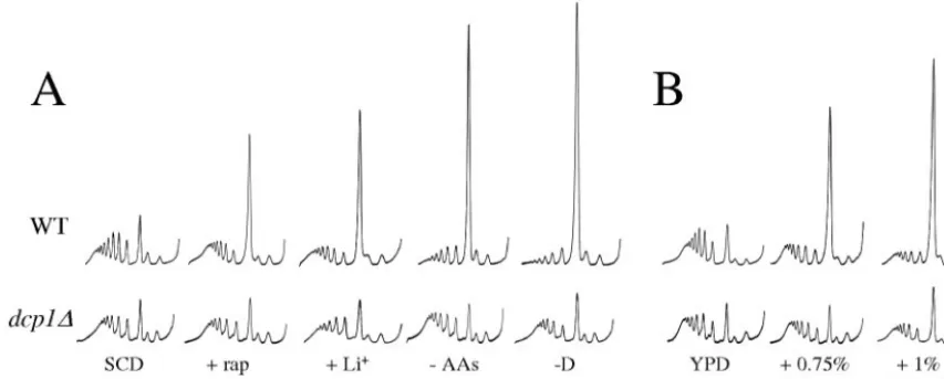
Mutation ofDCP1generates resistance to a host of trans-lational controls. A number of stress conditions have been shown to elicit effects on translational initiation. To further examine how compromised mRNA decay pathways might in-fluence these processes, thedcp1⌬mutant was subjected to a host of these translational inhibitory stresses. Figure 4A shows that following the addition of rapamycin or lithium (in galac-tose medium) or the removal of amino acids or glucose, a redistribution of polysomes into the monosome peak is ob-served for the wild-type strain. In contrast, when the dcp1⌬ mutant was subjected to these stresses no change in the poly-some profile was observed.
Recently we have found that the addition of fusel alcohols such as 1-butanol to yeast can inhibit translation initiation, leading to a redistribution of polysomes (4). This regulation relies on specific allelic variation in the GCD1 gene, which encodes the␥subunit of eIF2B. Therefore, we disrupted the DCP1gene in a butanol-sensitive (BUTS) strain background.
Addition of 1-butanol to the parental strain for as little as 10 min leads to a dramatic inhibition of translation initiation, as shown by the change in polysome profile (Fig. 4B). However, in the dcp1⌬mutant the same stress had little effect on the polysome profile, suggesting that translation is maintained in this mutant (Fig. 4B). Indeed, we have now shown thatDCP1 deletion in three different strain backgrounds leads to resis-tance to translational inhibition. In terms of growth this dis-ruption has background-dependent effects, resulting in quite different growth rates for individual dcp1⌬ strains. However, the translational regulation characteristics and shapes of the polysome profiles across these different backgrounds were in-distinguishable (data not shown). Therefore, mutation of the DCP1gene of the major mRNA decay pathway causes resis-tance to many different forms of translational stress.
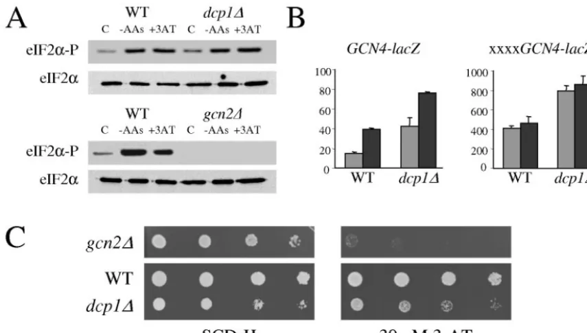
gcn2⌬mutant cannot phosphorylate eIF2␣and maintains poly-somes following severe amino acid starvation (12) (Fig. 2C). Surprisingly, thedcp1⌬mutant was capable of phosphorylating eIF2␣to an extent similar to that seen with the wild-type strain in response to either severe amino acid starvation or addition of the competitive inhibitor of histidine biosynthesis 3-AT for 15 min (Fig. 5A). The rapid phosphorylation of eIF2␣ by Gcn2p following amino acid starvation inhibits eIF2B, leading to lower levels of eIF2 GTP Met-tRNAiMet, the ternary
com-plex. Translation of theGCN4mRNA is exquisitely sensitive to changes in ternary complex abundance. Over a period of 6 h following amino acid starvation, there is a gradual increase in Gcn4p protein levels which relies upon this decrease in ternary complex abundance (1). As the level of ternary complex has never been directly assayed, theGCN4-lacZreporter (contain-ing the GCN4 5⬘ leader that includes the four uORFs) has therefore become an accepted indirect in vivo predictor for ternary complex levels (17). Adcp1⌬mutant maintained poly-somes and protein synthesis for more than 1 h following amino acid starvation (data not shown); therefore, we used a 1-h time point to evaluateGCN4-lacZlevels. In keeping with the results seen with previous analyses, it is clear that amino acid starva-tion inducesGCN4-lacZapproximately two- to threefold in the wild-type strain over this time frame (Fig. 5B). This induction was not observed when a gcn2⌬ mutant in the same strain background was used (data not shown). However, even though in terms of general translation thedcp1⌬mutant behaved like agcnmutant,GCN4-lacZwas still induced upon amino acid starvation (Fig. 5B). This induction relies upon the four up-stream ORFs, as no induction was observed in a mutant lack-ing these (Fig. 5B).
Interestingly, the basal level of activity from both theGCN4 -lacZ reporter and a reporter bearing mutant uORFs was higher in thedcp1⌬strain than in the wild type (Fig. 5B). The
results of Northern analysis of GCN4 mRNA levels suggest that this difference is due to higher levels of theGCN4mRNA in this mutant, probably resulting from deficient mRNA decay (data not shown).
The induction of GCN4 reporter activity following amino acid starvation in the dcp1⌬ mutant correlates with the fact that unlike thegcn2⌬mutant, thedcp1⌬strain is not sensitive to 3-AT, the histidine starvation inducer, in terms of growth (Fig. 5C). Under conditions of severe starvation for amino acids, therefore, adcp1⌬mutant strain responds by increasing the phosphorylation of eIF2␣ and the subsequent levels of Gcn4p. As eIF2␣ phosphorylation is known to decrease ter-nary complex levels (leading to an induction ofGCN4 trans-lation), these results suggest that the ternary complex levels decrease in adcp1⌬mutant strain following amino acid star-vation and yet that global protein synthesis is sustained.
Both 60S ribosomal subunit biogenesis mutant and eIF4G overexpression strains are translationally resistant to nutri-tional stress.Given the apparent normal response to amino acid starvation in the dcp1⌬ strain (as measured using the Gcn4p regulatory system), the obvious question is why do global rates of translation initiation not decrease following translational stress? It seems that this mutant strain back-ground is somehow capable of maintaining translation even though the levels of ternary complex decrease. This further indicates that in this mutant strain background, the rate of translation initiation is less dependent upon the rate of ternary complex recruitment to the 40S ribosomal subunit.
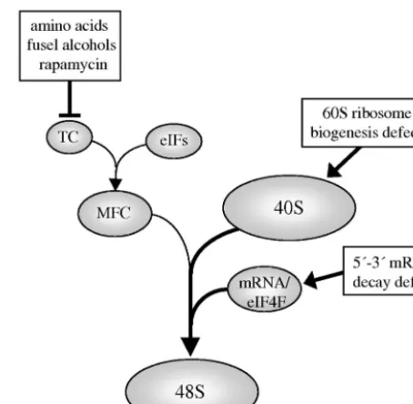
[image:9.603.79.506.73.244.2]One possible explanation is that increased levels of other components in the translation initiation process (such as the 40S ribosomal subunit or the eIF4F-bound mRNA) might overcome the decreased ternary complex and thus the trans-lational inhibition by a mass action mechanism. To test this idea we have taken two broad approaches.
FIG. 4. dcp1⌬is resistant to many forms of translational stress. (A) Polysome analyses for the wild-type (WT) strain (yMK329) and thedcp1⌬
strain (yMK330). Strains were grown in SCD medium and then subjected to a number of stresses. Rapamycin (⫹rap) was added to achieve a final concentration of 200 ng/ml for 1 h. A drug vehicle control gave a polysome trace identical to that of unstressed cells (data not shown). Lithium (⫹Li⫹) was added to achieve a final concentration of 30 mM for 15 min to yeast grown with galactose as the sole carbon source (the nontreated
galactose grown control is not shown for simplicity, as the polysome profiles are indistinguishable from those of glucose-grown strains). The removal of amino acids for 10 min (⫺AAs) or glucose for 5 min (⫺D) was performed as described in previous figures. (B) Polysome analyses for theBUTSstrain background, yMK36 (WT), and yMK863 (dcp1⌬). Strains were grown in rich medium (YPD), and 1-butanol was added to achieve
First, we have used mutants in the 60S ribosomal biogenesis pathway, reasoning that the excess of 40S ribosomal subunits in these strains could facilitate a level of translational resistance similar to that observed in the mRNA decay mutants. More specifically, Dob1p is a putative ATP-dependent RNA helicase that is involved in biogenesis of the 5.8S rRNA. Mutation of this gene leads to a deficiency in 60S ribosomal subunits and an increase in both the free 40S ribosomal subunits and “half-mers” (10). Half-mers are characteristic peaks which sediment just after each ribosomal peak and represent mRNAs with a 40S subunit that has not yet complexed with the 60S subunit. In contrast to the results seen with a wild-type strain, polysomes are maintained or partially maintained in the dob1-1mutant following amino acid starvation or glucose starvation, respec-tively (Fig. 6A). Interestingly, Dob1p interacts with compo-nents of the nuclear exosome (including Rrp4p) and is thought to facilitate 5.8S rRNA biogenesis without affecting cytoplas-mic 3⬘ to 5⬘ mRNA decay (2, 10). This suggests that the ex-planation for the translational resistance of therrp4-1mutant (Fig. 3B and 6A) lies more in the 60S ribosomal subunit bio-genesis defect for this strain rather than in a 3⬘to 5⬘mRNA decay defect.
This translational resistance observed for the 60S biogenesis mutants might be due to a rate-limiting step at the 60S joining step of translation initiation rather than to an excess of free 40S subunits. However, a fun12⌬ mutant (which has a 60S subunit joining deficiency) was completely inhibited for trans-lation initiation following glucose removal (data not shown).
Therefore it seems more likely that an accumulation of 40S ribosomal subunits can overcome translational inhibition.
It is also possible that general alterations to the relative levels of ribosomal subunits could allow resistance to transla-tional stress. To investigate this possibility we analyzed a 40S subunit biogenesis mutant,fal1-1, which accumulates free 60S subunits (21). In contrast to the results seen with 60S biogen-esis mutants, fal1-1 was largely inhibited following either amino acid or glucose withdrawal (Fig. 6B). Therefore, the accumulation of 60S subunits (in 40S biogenesis mutants) did not give the same resistance to translational stress as the ac-cumulation of 40S subunits (in 60S biogenesis mutants).
[image:10.603.80.508.71.313.2]In a second approach designed to test whether excess down-stream components can increase the level of preinitiation com-plex formation (especially under conditions of limiting ternary complex), we overexpressedTIF4632. This encodes eIF4G, the least abundant translation initiation factor in the eIF4F cap binding complex (46). We reasoned that accumulation of the eIF4F complex in a wild-type strain might alter the existing cap competition between Dcp1p and eIF4F, thereby allowing ulti-mately greater concentrations of mRNA/eIF4F complexes. Therefore, this scenario may mimic the results seen with a dcp1⌬ mutant. As shown in Fig. 6C and in contrast to the results seen with a strain containing plasmid vector alone, overexpression of eIF4G from a plasmid vector allows contin-ued translation following both amino acid and glucose starva-tion. Clearly this effect is less substantial than that seen with thedcp1⌬mutant, but it does suggest a plausible explanation
for the continued synthesis of proteins in mRNA decay mu-tants under stress conditions. That is, an excess of downstream components of the translation pathway can counter the stress-dependent inhibition of translation initiation. Ultimately, these results suggest that there is a delicate stoichiometrical balance of translational, ribosomal, and mRNA decay complexes which gives cells the capacity to rapidly control translation initiation.
DISCUSSION
Many studies over the years have highlighted the connection between translation and mRNA decay (33). The most obvious example is nonsense-mediated mRNA decay, in which a pre-requisite to this decay pathway is the translational recognition of a termination codon (20). Studies on the connection be-tween the general pathways of mRNA decay and translation have usually assessed the effects of alterations in translation on mRNA stability. For instance, the inhibition (using cyclohexi-mide or via conditional mutations) of translation elongation results in mRNA stabilization. In addition, it has been shown that in a variety of translation initiation mutants, the rate of decay for specific mRNAs increases (reviewed in reference 33). This has led to the hypothesis that mRNA decay and transla-tion initiatransla-tion in some way compete for the capped end of a mRNA species (33).
In this paper, we describe studies that approach the problem of translation and mRNA decay coordination from a different angle. Our goal was to determine how the various pathways of
mRNA decay might impact upon the control of translation initiation. We show that specific mutants in the major 5⬘to 3⬘ pathway of mRNA decay (most notably adcp1⌬mutant strain) are almost completely resistant to stress-induced translational inhibitory pathways. To further evaluate the mechanism in-volved in this connection between translational control and mRNA decay, we studied the well-characterized amino acid starvation pathway of global translational inhibition and Gcn4p induction. We show that even though thedcp1⌬strain is translationally resistant to severe amino acid starvation, this mutant does not bear other hallmarks of a gcn mutant. For instance, this mutant is capable of phosphorylating eIF2␣and activating GCN4 translation. As eIF2␣ phosphorylation is known to decrease ternary complex levels and consequently activateGCN4translation, these results suggest that (in terms of global translation initiation)dcp1⌬mutants are resistant to decreases in ternary complex abundance.
The fact that we have identified mutants such asdcp1⌬that are resistant to almost all forms of translational stress in yeast might mean that these translational regulatory responses have a common mechanism. For amino acid starvation, the pathway of translational regulation is well characterized and ultimately impacts upon eIF2B activity and ternary complex levels (17). It seems likely that fusel alcohols affect translation initiation via a similar mechanism except that this regulation does not rely on eIF2␣ kinases but instead probably affects the activity of eIF2B more directly (4). Rapamycin has recently been shown to inhibit translation initiation by activating the yeast eIF2␣
kinase, Gcn2p, and therefore also inhibits eIF2B activity, lead-ing to lower ternary complex levels (8). All three of these translational stresses lead to increased translation of GCN4, which demonstrates the reduction in ternary complex levels (4, 8, 17, 22, 44). Glucose starvation also leads to increased trans-lation ofGCN4 (48). However, the kinetics of this increased GCN4translation are relatively slow compared with the results seen with other stresses such as amino acid starvation and fusel alcohol addition (data not shown). This suggests that the pri-mary cause of the translational inhibition following glucose removal may not be the decreased ternary complex levels. The other stress that we have used in this study was the addition of lithium when yeast was grown in galactose medium. For this stress it has been found that overexpression of the gene for eIF4A suppresses the translational inhibitory effects (28). In addition, we have measuredGCN4-lacZreporter activity; ac-cording to the results seen with this indirect assay, lithium addition does not decrease ternary complex levels (data not shown). Overall it seems that even though mRNA decay mu-tants are resistant to many forms of translational stress, it is likely that these stresses affect translation via distinct mecha-nisms.
Translational control via eIF2␣kinases is an evolutionarily conserved response to stress. For example, four eIF2␣kinases have been identified in mammalian cells: HRI (which responds to the depletion of heme), PKR (which is activated during viral infection), PEK/PERK (which responds to misfolded proteins within the lumen of the endoplasmic reticulum), and mGCN2 (which responds to nutrient availability). All of these kinases target the guanine nucleotide exchange function of eIF2B, highlighting the role of eIF2B and the subsequent regulation of ternary complex levels as a major control point of protein synthesis (reviewed in reference 11). Furthermore, inherited mutations in eIF2B have recently been shown to cause the fatal human brain disease childhood ataxia with central nervous system hypomyelination (45). Therefore, the ternary complex level within eukaryotic cells is considered a key rate-limiting determinant for translation initiation. In this study we have identified specific yeast mutants where cells can maintain pro-tein synthesis even though ternary complex levels are de-creased. In addition, these mutants are resistant to transla-tional stresses that have no obvious impact upon ternary complex levels (as described above). One possible explanation for these observations is that a specific mRNA(s) might be upregulated in mRNA decay mutants and have direct effects upon translational regulation. However, it seems unlikely that changes in expression of specific mRNAs can generate resis-tance to so many divergent translational stresses that are thought to act via distinct mechanisms. Therefore, we favor a model in which the mRNA decay mutants have global effects on the gene expression profile of cells. Indeed, these mutants have globally relevant characteristics which may affect transla-tion initiatransla-tion and prevent translatransla-tion regulatransla-tion. These in-clude an increased abundance of mRNA and accumulation of mRNA decay intermediates (19).
A model that has been proposed to explain the connection between translation initiation and mRNA decay is the cap competition model. In this scenario, the lack of decapping in a dcp1⌬mutant would allow an accumulation of translationally competent or eIF4F-bound mRNA (33). Here we have shown
that stabilization of the eIF4F complex in a wild-type strain reduces the extent of translational regulation following stress. It seems that decreasing either the activity of Dcp1p (Fig. 2) or its access to the 5⬘cap (Fig. 6C) allows resistance to reduced levels of ternary complex and to translational stresses consid-ered to act via alternate mechanisms. We favor a model whereby an accumulation of eIF4F bound mRNA might glo-bally increase the recruitment rate of 43S complexes by mass action, therefore negating the effect of changes in ternary com-plex level (Fig. 7).
The hypothesis that other components of the translational initiation process can act by mass action to overcome changes in ternary complex level is further supported by an analysis of mutants in the biogenesis of the large ribosomal subunit. These mutants accumulate 40S ribosomal subunits and (in similarity to mRNA decay mutants) are resistant to translational stress (Fig. 6A). The excess 40S subunits may drive the various equi-libria within the translation initiation process towards the preinitiation complex, thus overcoming effects on ternary com-plex levels (Fig. 7).
[image:12.603.306.533.70.292.2]The results presented in this study graphically demonstrate the intimate relationship that exists between protein synthesis, mRNA decay, and ribosomal biogenesis. Although these con-nections have been implicated previously in a number of stud-ies, this study represents the first analysis in which the global control of translation in response to stress has been added to the equation. Surprisingly, mRNA decay mutants can maintain translation under stress even though ternary complex levels appear to decrease normally. As regulation of ternary complex
FIG. 7. A model depicting how 5⬘to 3⬘mRNA decay or 60S bio-genesis defects might overcome decreases in ternary complex. Mutants that accumulate either 40S subunits or mRNA/eIF4F complexes drive the assembly of the preinitiation machinery by mass action. TC and MFC represent the ternary complex (eIF2-GTP Met-tRNAiMet) and the
is a highly conserved mechanism of translational control, this may have wide ranging implications. Our interpretation of these results is that there is a delicate balance in the stoichi-ometry of different translation initiation components (such as MFC, 40S ribosomes, and mRNA) and that the maintenance of this equilibrium is critical in preserving the cell’s capacity to globally control protein synthesis.
ACKNOWLEDGMENTS
This work was supported largely by Wellcome Trust project grant 061867/Z/00/Z to M.P.A. L.E.A.H. is supported by an MRC student-ship. S.G.C. is supported by Wellcome Trust project grant 067328/Z/ 02/Z to M.P.A. In addition, the work was supported by NIH grant GM50308 to A.B.S. and by an EMBO long-term fellowship to M.P.A. while at University of California at Berkeley.
We thank G. Pavitt, C. Grant, A. Johnson, C. Wilutz, S. Peltz, S. Butler, and P. Linder for reagents and advice. We especially thank R. Parker for his generous donation of yeast strains and plasmids, without which this work would not have been possible.
REFERENCES
1. Albrecht, G., H. U. Mosch, B. Hoffmann, U. Reusser, and G. H. Braus.1998. Monitoring the Gcn4 protein-mediated response in the yeastSaccharomyces cerevisiae. J. Biol. Chem.273:12696–12702.
2. Anderson, J. S. J., and R. P. Parker.1998. The 3⬘to 5⬘degradation of yeast mRNAs is a general mechanism for mRNA turnover that requires the SKI2 DEVH box protein and 3⬘ to 5⬘ exonucleases of the exosome complex. EMBO J.17:1497–1506.
3. Ashe, M. P., S. K. De Long, and A. B. Sachs.2000. Glucose depletion rapidly inhibits translation initiation in yeast. Mol. Biol. Cell11:833–845. 4. Ashe, M. P., J. W. Slaven, S. K. De Long, S. Ibrahimo, and A. B. Sachs.2001.
A novel eIF2B-dependent mechanism of translational control in yeast as a response to fusel alcohols. EMBO J.20:6464–6474.
5. Barbet, N. C., U. Schneider, S. B. Helliwell, I. Stansfield, M. F. Tuite, and M. N. Hall.1996. TOR controls translation initiation and early G1 progres-sion in yeast. Mol. Biol. Cell7:25–42.
6. Beelman, C. A., A. Stevens, G. Caponigro, T. E. LaGrandeur, L. Hatfield, D. M. Fortner, and R. Parker.1996. An essential component of the decap-ping enzyme required for normal rates of mRNA turnover. Nature382:642– 646.
7. Boeck, R., B. Lapeyre, C. E. Brown, and A. B. Sachs.1998. Capped mRNA degradation intermediates accumulate in the yeastspb8–2mutant. Mol. Cell. Biol.18:5062–5072.
8. Cherkasova, V. A., and A. G. Hinnebusch.2003. Translational control by TOR and TAP42 through dephosphorylation of eIF2␣kinase GCN2. Genes Dev.17:859–872.
9. Choi, S. K., J. H. Lee, W. L. Zoll, W. C. Merrick, and T. E. Dever.1998. Promotion of met-tRNAiMet binding to ribosomes by yIF2, a bacterial IF2 homolog in yeast. Science280:1757–1760.
10. de la Cruz, J., D. Kressler, D. Tollervey, and P. Linder.1998. Dob1p (Mtr4p) is a putative ATP-dependent RNA helicase required for the 3⬘end forma-tion of 5.8S rRNA inSaccharomyces cerevisiae.EMBO J.17:1128–1140. 11. Dever, T. E.1999. Translation initiation: adept at adapting. Trends Biochem.
Sci.24:398–403.
12. Dever, T. E., L. Feng, R. C. Wek, A. M. Cigan, T. F. Donahue, and A. G. Hinnebusch.1992. Phosphorylation of initiation factor 2 alpha by protein kinase GCN2 mediates gene-specific translational control ofGCN4in yeast. Cell68:585–596.
13. Fromont-Racine, M., A. E. Mayes, A. Brunet-Simon, J. C. Rain, A. Colley, I. Dix, L. Decourty, N. Joly, F. Ricard, J. D. Beggs, and P. Legrain.2000. Genome-wide protein interaction screens reveal functional networks involv-ing Sm-like proteins. Yeast17:95–110.
14. Gonza´lez, C. I., A. Bhattacharya, W. Wang, and S. W. Peltz.2001. Nonsense-mediated mRNA decay inSaccharomyces cerevisiae.Gene274:15–25. 15. Goossens, A., T. E. Dever, A. Pascual-Ahuir, and R. Serrano.2001. The
protein kinase Gcn2p mediates sodium toxicity in yeast. J. Biol. Chem. 276:30753–30760.
16. Guthrie, C., and G. R. Fink (ed.).1991. Guide to yeast genetics and molec-ular biology. Academic Press, San Diego, Calif.
17. Hinnebusch, A. G.2000. Mechanism and regulation of initiator methionyl-tRNA binding to ribosomes, p. 185–244.InN. Sonenberg, J. W. B. Hershey, and M. B. Mathews (ed.), Translational control of gene expression. Cold Spring Harbor Laboratory Press, Cold Spring Harbor, N.Y.
18. Hinnebusch, A. G., and K. Natarajan.2002. Gcn4p, a master regulator of gene expression, is controlled at multiple levels by diverse signals of starva-tion and stress. Eukaryot. Cell1:22–32.
19. Hsu, C. L., and A. Stevens.1993. Yeast cells lacking 5⬘33⬘exoribonuclease 1 contain mRNA species that are poly(A) deficient and partially lack the 5⬘
cap structure. Mol. Cell. Biol.13:4826–4835.
20. Jacobson, A., and S. W. Peltz.2000. Destabilization of nonsense-containing transcripts inSaccharomyces cerevisiae, p. 827–847.InN. Sonenberg, J. W. B. Hershey, and M. B. Mathews (ed.), Translational control of gene expression. Cold Spring Harbor Laboratory Press, Cold Spring Harbor, N.Y. 21. Kressler, D., J. de la Cruz, M. Rojo, and P. Linder.1997. Fal1p is an
essential DEAD-box protein involved in 40S-ribosomal-subunit biogenesis in
Saccharomyces cerevisiae. Mol. Cell. Biol.17:7283–7294.
22. Kubota, H., T. Obata, K. Ota, T. Sasaki, and T. Ito.2003. Rapamycin-induced translational derepression ofGCN4mRNA involves a novel mech-anism for activation of the eIF2␣kinase GCN2. J. Biol. Chem.278:20457– 20460.
23. Kuhn, K. M., J. L. DeRisi, P. O. Brown, and P. Sarnow.2001. Global and specific translational regulation in the genomic response ofSaccharomyces cerevisiaeto a rapid transfer from a fermentable to a nonfermentable carbon source. Mol. Cell. Biol.21:916–927.
24. Lucchini, G., A. G. Hinnebusch, C. Chen, and G. R. Fink.1984. Positive regulatory interactions of theHIS4gene ofSaccharomyces cerevisiae.Mol. Cell. Biol.4:1326–1333.
25. Mangus, D. A., N. Amrani, and A. Jacobson.1998. Pbp1p, a factor interact-ing withSaccharomyces cerevisiaepoly(A)-binding protein, regulates polyad-enylation. Mol. Cell. Biol.18:7383–7396.
26. Mitchell, P., E. Petfalski, A. Shevchenko, M. Mann, and D. Tollervey.1997. The exosome: a conserved eukaryotic RNA processing complex containing multiple 3⬘35⬘exoribonucleases. Cell91:457–466.
27. Mitchell, P., and D. Tollervey.2003. An NMD pathway in yeast involving accelerated deadenylation and exosome-mediated 3⬘35⬘degradation. Mol. Cell11:1405–1413.
28. Montero-Lomeli, M., B. L. Morais, D. L. Figueiredo, D. C. Neto, J. R. Martins, and C. A. Masuda.2002. The initiation factor eIF4A is involved in the response to lithium stress inSaccharomyces cerevisiae.J. Biol. Chem. 277:21542–21548.
29. Proweller, A., and S. Butler.1994. Efficient translation of poly(A)-deficient mRNAs inSaccharomyces cerevisiae.Genes Dev.8:2629–2640.
30. Rolfes, R. J., and A. G. Hinnebusch.1993. Translation of the yeast transcrip-tional activator GCN4 is stimulated by purine limitation: implications for activation of the protein kinase GCN2. Mol. Cell. Biol.13:5099–5111. 31. Sachs, A. B.2000. Physical and functional interactions between the mRNA
cap structure and the poly(A) tail, p. 447–465.InN. Sonenberg, J. W. B. Hershey, and M. B. Mathews (ed.), Translational control of gene expression. Cold Spring Harbor Laboratory Press, Cold Spring Harbor, N.Y. 32. Sachs, A. B., and R. W. Davis.1989. The poly(A) binding protein is required
for poly(A) shortening and 60S ribosomal subunit-dependent translation initiation. Cell58:857–867.
33. Schwartz, D. C., and R. Parker.2000. Interaction of mRNA translation and mRNA decay inSaccharomyces cerevisiae, p. 807–825.InN. Sonenberg, J. W. B. Hershey, and M. B. Mathews (ed.), Translational control of gene expression. Cold Spring Harbor Laboratory Press, Cold Spring Harbor, N.Y. 34. Searfoss, A., T. E. Dever, and R. Wickner.2001. Linking the 3⬘poly(A) tail to the subunit joining step of translation initiation: relations of Pab1p, eu-karyotic translation initiation factor 5b (Fun12p), and Ski2p-Slh1p. Mol. Cell. Biol.21:4900–4908.
35. Sheth, U., and R. Parker.2003. Decapping and decay of messenger RNA occur in cytoplasmic processing bodies. Science300:805–808.
36. Steiger, M., A. Carr-Schmid, D. C. Schwartz, M. Kiledjian, and R. Parker. 2003. Analysis of recombinant yeast decapping enzyme. RNA9:231–238. 37. Tharun, S., and R. Parker.1999. Analysis of mutations in the yeast mRNA
decapping enzyme. Genetics151:1273–1285.
38. Tharun, S., W. He, A. E. Mayes, P. Lennertz, J. D. Beggs, and R. Parker. 2000. Yeast Sm-like proteins function in mRNA decapping and decay. Na-ture404:515–518.
39. Tucker, M., and R. Parker.2000. Mechanisms and control of mRNA decap-ping inSaccharomyces cerevisiae.Annu. Rev. Biochem.69:571–595. 40. Tucker, M., R. R. Staples, M. A. Valencia-Sanchez, D. Muhlrad, and R.
Parker.2002. Ccr4p is the catalytic subunit of a Ccr4p/Pop2p/Notp mRNA deadenylase complex inSaccharomyces cerevisiae.EMBO J.21:1427–1436. 41. Tzamarias, D., I. Roussou, and G. Thireos.1989. Coupling ofGCN4mRNA
translational activation with decreased rates of polypeptide chain initiation. Cell57:947–954.
42. Uesono, Y., and A. Toh-E.2002. Transient inhibition of translation initiation by osmotic stress. J. Biol. Chem.277:13848–13855.
43. Valasek, L., A. A. Mathew, B. S. Shin, K. H. Nielsen, B. Szamecz, and A. G. Hinnebusch.2003. The yeast eIF3 subunits TIF32/a, NIP1/c, and eIF5 make critical connections with the 40S ribosome in vivo. Genes Dev.17:786–799. 44. Valenzuela, L., C. Aranda, and A. Gonzalez.2001. TOR modulatesGCN4 -dependent expression of genes turned on by nitrogen limitation. J. Bacteriol. 183:2331–2334.
Oudejans, R. B. Schutgens, and J. C. Pronk.2002. Mutations in each of the five subunits of translation initiation factor eIF2B can cause leukoencepha-lopathy with vanishing white matter. Ann. Neurol.51:264–270.
46. von der Haar, T., and J. E. McCarthy.2002. Intracellular translation initi-ation factor levels inSaccharomyces cerevisiaeand their role in cap-complex function. Mol. Microbiol.46:531–544.
47. Wach, A., A. Brachat, R. Pohlmann, and P. Philippsen.1994. New heterol-ogous modules for classical or PCR-based gene disruptions inSaccharomyces cerevisiae.Yeast10:1793–1808.
48. Yang, R., S. A. Wek, and R. C. Wek.2000. Glucose limitation inducesGCN4