In Vitro
Biocompatibility of Ni-Free Ti-Based Shape Memory Alloys
for Biomedical Applications
Hiroyasu Kanetaka
1;2, Hideki Hosoda
3, Yoshinaka Shimizu
4, Tada-aki Kudo
5,
Ye Zhang
1, Mitsuhiro Kano
4, Yuya Sano
2;*and Shuichi Miyazaki
61Department of Physical Medicine and Rehabilitation, Graduate School of Biomedical Engineering, Tohoku University, Sendai 980-8575, Japan
2Division of Advanced Prosthetic Dentistry, Graduate School of Dentistry, Tohoku University, Sendai 980-8575, Japan 3Precision and Intelligence Laboratory, Tokyo Institute of Technology, Yokohama 226-8503, Japan
4Division of Oral and Craniofacial Anatomy, Graduate School of Dentistry, Tohoku University, Sendai 980-8575, Japan 5Division of Oral Physiology, Graduate School of Dentistry, Tohoku University, Sendai 980-8575, Japan
6Institute of Materials Science, University of Tsukuba, Tsukuba 305-8573, Japan
This study was undertaken to evaluate thein vitrobiocompatibility of newly developed Ni-free Ti-based shape memory alloys (SMAs) in comparison to that of commercial pure titanium (cpTi). This study compared Ti-24 mol%Nb-3 mol%Al (Ti-Nb-Al) and Ti-7 mol%Cr-3 mol%Sn (Ti-Cr-Sn) to cpTi from a cell-compatibility perspective. In all, 63 samples (21 samples for each group) were prepared, which were machined into 10-mm-diameter, 0.15-mm-thick, mirror-polished disks. Their surface morphology was evaluated using scanning electron microscopy (SEM). The chemical composition of the sample surface was determined using an energy dispersive X-ray analyzer (EDX). Sample surface roughness was measured using a non-contact 3D profiler. After sample surface observations, the cell proliferation and viability of African green monkey kidney fibroblast cell line COS7 in direct contact with these new alloys were evaluated by DNA quantification, by live cell imaging using CelLuminate Red fluorescent cell stain as a new method, and by cytoskeletal observations by immunofluorescent actin labelling. Cell proliferation was examined after 1, 3, and 5 days of culture. Results were the following. (i) Each sample showed high purity and a very smooth surface, showing no morphological differences among groups. (ii) The COS7 cells took in sufficient CelLuminate Red to visualize the cells using epifluorescent microscopy, and (iii) cell proliferation with Ti-Cr-Sn was lower than with either cpTi or Ti-Nb-Al. These results suggest that Ti-Nb-Al alloy showed biocompatibility as high as that of cpTi and that it is more suitable for biomedical applications.
[doi:10.2320/matertrans.M2010038]
(Received February 1, 2010; Accepted July 21, 2010; Published September 8, 2010)
Keywords: nickel-free titanium-based shape memory alloy, in vitro, biocompatibility, cell proliferation, deoxypentose nucleic acid (DNA) quantification, live cell imaging
1. Introduction
Shape memory alloys (SMAs) belong to a group of
materials known assmart functional materials. These SMAs,
as typified by nickel–titanium (Ni–Ti) alloy, exhibit unique properties of shape memory effect and superelasticity. The shape memory effect relates to the fact that the alloy reverts to its original shape after heating above a specific temper-ature; superelasticity is a phenomenon by which the stress value remains nearly constant up to a certain point of wire
deformation.1,2)These properties can be put to excellent use
in various biomedical applications,3–7)such as catheter guide
wires, stents, orthodontic wires, and orthopedic implants for osteosynthesis.
Ni–Ti alloy is the only practical SMA in biomedical use because of its excellent mechanical stability and function-ability. However, the biocompatibility of Ni–Ti is
question-able because it contains a large amount of nickel.8,9)
Therefore, Ni–Ti might induce nickel hypersensitivity;10,11)
we recommend avoiding the use of this alloy in nickel-sensitive patients. In contrast, some studies have shown high biocompatibility of Ni–Ti because of the passive oxide layer on the alloy surface and the strong interatomic bonding between nickel and titanium ions. However, recent studies have revealed that mechanical stress or some chemical environments cause corrosion and that substantial quantities
of nickel ions can be released.12–14)Therefore, the long-term
biomedical use of Ni–Ti in a living body might not be desirable. Moreover, results of recent numerous animal studies evaluating the carcinogenicity of nickel have
sup-ported the view that nickel is a potent carcinogen.15–17)
From the perspective of allergenicity and toxicity for humans, reduction of Ni content or development of Ni-free SMAs is necessary to reduce the risks of nickel hyper-sensitivity and carcinogenicity. Therefore, we have been systematically investigating new Ni-free Ti-based SMAs composed of non-toxic elements, demonstrating that
Ti-24 mol%Nb-3 mol%Al (Ti-Nb-Al)18–20) and Ti-7
mol%Cr-3 mol%Sn (Ti-Cr-Sn)21,22)exhibit good mechanical
perform-ance among Ni-free shape memory and superelastic alloys. For in vitrobiocompatible evaluation of alloys, cells on the metallic samples were conventionally observed using epifluorescent microscopy, because metallic samples were not transmissive to light. However, some fluorescent stain methods need lipofection, which is toxic to cells to some degree, and even some fluorescent pigments themselves have some kinds of toxicity. Therefore a new simple and safe method for observation of the cells on the metallic sample has been expected, changing to the conventional methods.
The purpose of this study was to evaluate relativein vitro
biocompatibility of newly developed Ni-free Ti-based SMAs compared to commercial pure titanium (abbreviated as cpTi). In this study, the cell proliferation and viability of African green monkey kidney fibroblast cell line COS7 in direct
*Graduate Student, Graduate School of Dentistry, Tohoku University
contact with these new alloys were evaluated using DNA quantification, new live cell imaging on metallic samples using epifluorescent microscopy by CelLuminate Red fluorescent cell stain concurrently with phase microscopy, and cytoskeletal observations by immunofluorescent actin labeling.
2. Materials and Methods
2.1 Samples
For this study, 24 mol%Nb-3 mol%Al (Nb-Al), Ti-7 mol%Cr-3 mol%Sn (Ti-Cr-Sn) and commercial pure tita-nium (cpTi, JIS H 4600 (JIS Grade 1); Nippon Steel Corp., Tokyo, Japan) were used. The Ti-Nb-Al and Ti-Cr-Sn alloys used in this study were fabricated by nonconsumable W-electrode type arc melting method in Ar-1%H2 reduction atmosphere using a Cu heath and high purity elemental materials with 99.99% (4N). In all, 63 samples (21 samples for each group) were prepared. The samples were machined into 10-mm-diameter, 0.15-mm-thick disks. To create a mirror-like surface, Ti-Nb-Al and Ti-Cr-Sn were
mechan-ically polished with 3-mm and 1-mm diamond paste; cpTi
was mechanically polished with 3-mm diamond paste and
0.03-mmcolloidal silica. After polishing, the samples were
cleaned ultrasonically for 5 min twice in acetone, and for 20 min twice in distilled water. Subsequently, samples were
sterilized using an autoclave at 121C for 20 min and then
oven-dried.
2.2 Surface observation
The surface morphology of the samples before and after polishing was evaluated using scanning electron microscopy (SEM; JSM-6390LA; JEOL, Tokyo, Japan) operated at the
accelerating voltage of 10 kV and filament current of 67mA
at 500 magnification. The chemical composition of the
samples after polishing was determined using an energy dispersive X-ray analyzer (EDX; JED-2300; JEOL, Tokyo, Japan) operated at the accelerating voltage of 15 kV and
filament current of 60mA. Several points on the sample
surfaces were selected randomly. They were examined using EDX point analysis. Roughness of the 10 sample surfaces after polishing of each group was measured using a non-contact 3D Profiler (Talysurf CCI 3000; Taylor Hobson Ltd., Leicester, UK).
2.3 Cell culture
To evaluate in vitro biocompatibility of the Ti-Nb-Al,
Ti-Cr-Sn, and cpTi, African green monkey kidney fibroblast cell line COS7 was cultured on each sample in a Dulbecco’s Modified Eagle Medium (Wako Pure Chemical Industries Ltd., Osaka, Japan) containing 10% horse serum (Invitrogen Corp., Carlsbad, CA, USA), 100 U/ml penicillin
(Meiji-Seika Kaisha Ltd., Tokyo, Japan) and 100mg/ml
streptomy-cin (Meiji-Seika Kaisha Ltd., Tokyo, Japan). The cells were
maintained in a humid incubator at 37C with 5% CO
2.
The sterilized samples (10 mm diameter) were set onto the bottom of a 24-well non-tissue culture treated plate (Becton, Dickinson and Co., Franklin Lakes, NJ, USA). A cell suspen-sion consisting of 20 000 cells was seeded onto the surfaces of the Ti-Nb-Al, Ti-Cr-Sn, and cpTi. Cells were grown in
300ml of medium. The cell proliferation was examined after
1, 3, and 5 days of culture. In this study, the cell proliferation and viability with these alloys were evaluated using DNA quantification, new live cell imaging, and cytoskeletal observation by immunofluorescence. Seven samples of each group were used per day; five samples for DNA quantifica-tion, one sample for live cell imaging, one sample for cytoskeletal observation staining by immunofluorescence. A total of 21 samples of each group were used for this study.
2.4 Quantification of DNA from the cells
To evaluate the cell proliferation, the isolation of DNA from the incubated cells on each sample material was performed using the AllPrep DNA/RNA/Protein Mini Kit (Qiagen Inc., Valencia, CA, USA) and QIA shredder (Qiagen Inc.) according to the manufacturer’s protocol. For extraction from cells, harvesting was conducted using a cell scraper (Sumitomo Bakelite Co. Ltd., Tokyo, Japan) instead of trypsinization. Following extraction, the DNA was quantified by absorbance at 260 nm using a spectrophotometer (GeneQuant pro, GE Healthcare, Buckinghamshire, UK).
2.5 CelLuminate Red fluorescent cell stain
For live cell imaging on the sample material, CelLuminate Red (Biocompatibles UK Ltd., Farnham, UK), a fluorescent stain for live cells made of non-toxic nanoparticles including the fluorophore rhodamine, was added to experimental wells at a concentration of 10% (v/v) in culture medium and was incubated for 24 h. After incubation, each sample attached by cells onto the upper surface was removed from the bottom of the experimental well and was set inversely on another well with fresh culture medium for removal of residual CelLuminate Red and for visualization using epifluorescent microscopy. Live cells that took in CelLuminate Red were imaged using an epifluorescent microscope (CKX41; Olympus Corp., Tokyo, Japan) with a 480–550 nm excitation filter. The cells were also visualized using phase-contrast microscopy. The images were captured with a digital camera (DP72; Olympus Corp., Tokyo, Japan) and were processed with software (DP2-BSW; Olympus Corp., Tokyo, Japan).
2.6 Immunofluorescence stain for cytoskeletal observa-tion
In order to label actin filaments in COS7 cells, samples of each group attached by COS7 cells at 3 days of culture were washed in phosphate-buffered saline (PBS) and fixed with 4% paraformaldehyde and permeabilized using 0.5% Triton X-100 (Sigma, St. Louis, USA) in PBS for 10 min. After washing with Tris-buffered saline (TBS, pH 7.6), samples
were incubated in 100ml of blocking buffer, then incubated
with conjugate phalloidin (5 IU, Alexa Fluor 488; Cambrex Corp., Walkersville, MD, USA) for 20 min at room tem-perature. After washing, samples were mounted on PLA-coated glass plates with mounting medium for fluorescence (Vectashield; Vector Laboratories Inc., Burlingame, CA, USA). The cytoskeleton was observed with an epifluorescent microscope.
2.7 Statistical analysis
Data were compared between groups using analysis of
variance (ANOVA) followed by a Tukey test; P<0:05
indicated a statistically significant result.
3. Results
3.1 Surface observation
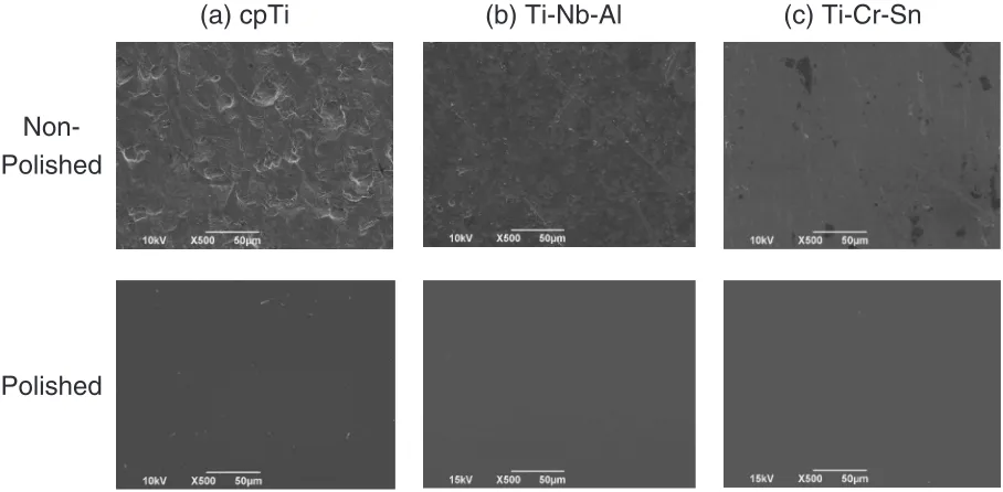
The surface morphologies of the samples before and after polishing are shown in Fig. 1, as evaluated using scanning electron microscopy. The surfaces of non-polished samples showed many small pits, wrinkles, and scratches. In contrast, the polished samples showed smooth surfaces. Results of EDX analysis of the sample surfaces after polishing are shown in Fig. 2. The EDX results show that the surface of cpTi showed only the element Ti, Ti-Nb-Al showed only Ti, Nb, Al, and Ti-Cr-Sn showed only Ti, Cr, and Sn. The arithmetic mean roughness (Ra) and maximum height of roughness (Rz) of the 10 sample surfaces after polishing of each group are shown in Table 1. No significant difference in either Ra or Rz was found among the three groups.
3.2 Cell proliferation
To evaluate the cell proliferation, DNA quantification and live cell imaging with CelLuminate Red were performed in
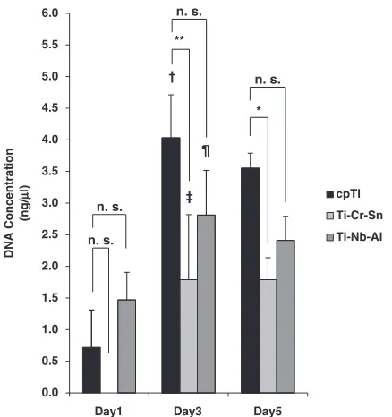
this study. Figure 3 shows the time course of cell prolifer-ation with DNA quantificprolifer-ation. A significant increase was found in the amount of DNA from cells between day 1 and day 3 in the culture with Ti; no significant difference was found between day 3 and day 5. In the culture with both Ti-Nb-Al and Ti-Cr-Sn, the same progress was observed with Ti. However, in a comparison among sample materials, we detected that the amount of DNA from the cells on Ti-Cr-Sn was significantly less in comparison with cpTi at both day 3 and day 5. Nevertheless, no significant difference was found between cpTi and Ti-Nb-Al at day 1, day 3, or day 5. The value of Ti-Cr-Sn on day 1 was below the spectrophotometer detection limit. Next, live COS7 cells on each material were observed at day 1, day 3, and day 5 with epifluorescent microscopy to monitor cell proliferation with treatment of CelLuminate Red. The cells on each material were also
(a) cpTi
(b) Ti-Nb-Al
(c) Ti-Cr-Sn
Non-Polished
Polished
Fig. 1 SEM images (Original magnification500, Scale bar 50mm) of non-polished and polished surfaces of samples: (a) cp-Ti, (b) Ti-Nb-Al, (c) Ti-Cr-Sn.
(a) cpTi (b) Ti-Nb-Al (c) Ti-Cr-Sn
[image:3.595.71.525.74.297.2]Fig. 2 EDX spectrometries of the sample surfaces: (a) cpTi, (b) Ti-Nb-Al, (c) Ti-Cr-Sn.
Table 1 Roughness of sample surfaces after polishing of each group (n¼10).
Roughness cpTi Ti-Nb-Al Ti-Cr-Sn
Ra (nm) 1:030:37 0:870:25 1:190:27
Rz (nm) 7:834:11 5:741:47 7:631:88
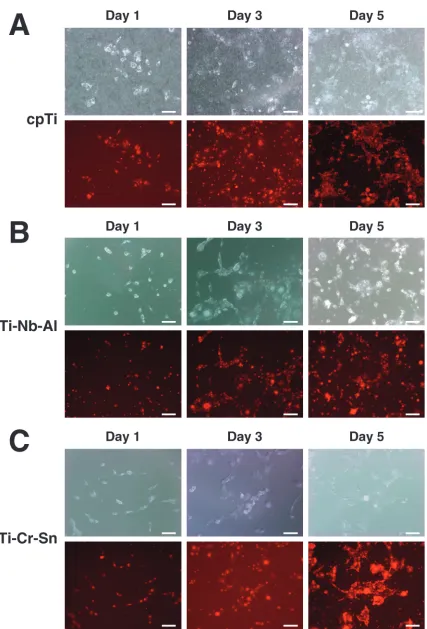
[image:3.595.80.523.341.466.2] [image:3.595.305.551.527.569.2]observed using phase contrast method in parallel. Results show that (i) COS7 cells took in sufficient CelLuminate Red to enable visualization of the cells with epifluorescent microscopy, at least in the experimental conditions of this study. Furthermore, (ii) cell proliferation with Ti-Cr-Sn was apparently lower than with either cpTi or Ti-Nb-Al (Fig. 4). Both live cell imaging with phase microscopy and with epifluorescent microscopy using CelLuminate Red revealed the similar tendency (Fig. 4), but the live cell imaging using CelLuminate Red yielded a clearer image and was more sensitive to active cells (Fig. 5).
3.3 Cell viability
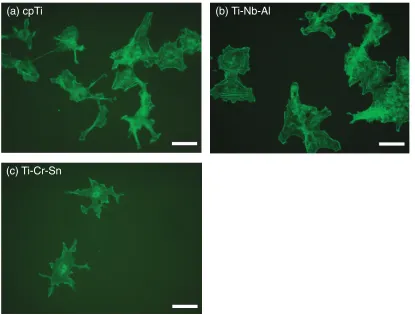
Observation of CelLuminate Red pinocytosed by cells on each sample surface revealed that the rate of cell growth in the culture with Ti-Cr-Sn was less than with either cp-Ti or Ti-Nb-Al (Fig. 4). Figure 6 depicts cytoskeleton morpholo-gies of the cells at day 3 on the cpTi, Ti-Nb-Al, and Ti-Cr-Sn observed using fluorescent microscopy. The COS7 cells on the cpTi and Ti-Nb-Al showed clear cytoskeleton, grew better, and spread more widely. Therefore, COS7 cells on the cpTi and Ti-Nb-Al were apparently larger and showed higher cell viability than those on Ti-Cr-Sn.
4. Discussion
4.1 Newly developed Ni-free Ti-based SMAs
Our group has systematically examined Ni-free Ti-based SMAs composed of non-toxic element and the Ti-Nb-Al ternary system as a promising candidate for practical applications. With decreasing Nb content, the transformation strain of Ti-Nb alloys increases but, simultaneously, the
martensitic transformation start temperature (abbreviated as
Ms) increases.23) Therefore, for the appearance of
super-elasticity of Ti-Nb alloys with large superelastic strain, decreasing the Nb content should be achieved simultaneously with compensation of Ms by addition of ternary elements. In fact, Al is an effective ternary element to reduce Ms while preventing degradation of the transformation strain
when substituting to Nb.24) Furthermore, Al addition is
effective to suppress !embrittlement.25)A Ti-Nb-Al alloy,
Ti-24 mol%Nb-3 mol%Al, shows good superelastic
perform-ance.18–20)
To fabricate homogenized ingots, it is beneficial to use
other stabilizing elements having lower melting points.
Based on the background presented above, Ti-Cr-Sn is a
promising alloy system. Actually, Cr is a stabilizing
element having a comparable melting point to Ti (1943 K) of
2136 K; Sn is also known to suppress ! embrittlement.
Moreover, Ti-Nb-Sn is a Ni-free biomedical superelastic
alloy.26)In actuality, Ti-7 mass%Cr-4 mass%Sn exhibits 60%
shape recovery at 573 K.27) Then, with decreasing Ms
through controlled alloy composition, Ti-Cr-Sn alloys are promising candidate as biomedical shape memory alloys; good mechanical properties and superelastic behavior have
been reported.22,28)Therefore, Ti-Nb-Al and Ti-Cr-Sn alloys
were used for this study.
Our group demonstrated the clinical applicability of the
Ti-Nb-Al alloy as orthodontic superelastic wire.29,30)The
Ti-Nb-Al and Ti-Cr-Sn alloys are expected to be composed of a
single-Ti phase at usual temperatures of the human body.
A single-phase alloy usually has better resistance against galvanic corrosion than a multi-phase alloy because it does
not cause any difference in potential.31,32)Even under stress,
the stress-induced martensite still contains the same chemical composition as that of the parent phase. In this case, galvanic corrosion between parent and martensite phases must be slight because of their chemical similarity. Moreover, each element composing the Ti-Nb-Al and Ti-Cr-Sn alloy shows high-to-medium biocompatibility. Previous studies of cyto-toxicity of metal salts for human and mouse origin cells
revealed that the 50% inhibitory concentration (IC50) of Ti,
Nb, Al, and Sn showed a large value and that IC50 of Cr
showed a medium value.33,34)
4.2 Samples
Cell proliferation can be affected by the sample surface morphology: surfaces of the non-polished samples showed many small pits, wrinkles, and scratches. For that reason, all samples used for this study were mirror-polished to eliminate the influence of surface morphology. The composing elements of each sample were also evaluated by EDX analysis and showed no impurities. The Ti-Nb-Al and Ti-Cr-Sn alloys used in this study were fabricated by nonconsum-able W-electrode type arc melting method in Ar-1%H2 reduction atmosphere using a Cu heath and high purity elemental materials with 99.99% (4N). Therefore, the amount of impurities from the starting materials and contaminations during alloying were lowered as possible as we could. Then, although we did not the chemical analysis for the impurities, the effects of the impurities on cell culture were minimized as possible as we could. These results 5.0 5.5 6.0 ** n. s. n. s. † 3.5 4.0 4.5 cpTi * ‡ ¶ 1.5 2.0 2.5 3.0 DNA Concentration (ng/ µµ l) Ti-Cr-Sn Ti-Nb-Al n. s. n. s. 0.0 0.5 1.0
Day1 Day3 Day5
[image:4.595.61.280.72.305.2]suggest that each sample showed high purity, and that the surface of each sample was very smooth, presenting no morphological differences among groups.
4.3 CelLuminate red fluorescent cell stain
In this study, CelLuminate Red was applied to observe live cells cultured on metallic materials with epifluorescent Fig. 4 Evaluation of cell proliferation with live cell imaging. COS7 cells on each sample were observed at day 1, day 3, and day 5 using
[image:5.595.86.513.63.692.2]microscopy. It was confirmed that COS7 cells were visible
with epifluorescent microscopy at 24 h after adding
CelLuminate Red to the culture (Fig. 4). This new reagent
consists of biomimetic nanoparticles35)that include a
rhod-amine, a fluorescent reagent; it is designed to be pinocytosed easily by cells. Use of this reagent enables live fluorescence staining of cells while obviating methods such as lipofection for transient gene transfection of cDNA encoding green fluorescent protein (GFP), a protein that exhibits green fluorescence when exposed to blue excitation light, into
the cells.36,37) Because gene transfer such as lipofection is
toxic to cells to some degree, non-cytotoxic CelLuminate Red is expected to be more useful for investigating cell
compatibility of metallic materials than the reagents for gene
transfer.38) Moreover, CelLumiante is designed to perform
live cell imaging; it is another feature by which we can perform non-toxic time-lapse assay for real-time monitoring of the cells on metallic materials. The metallic disks used for this study were 0.15-mm-thick. Because all probed materials used in this study were transmissive to light from microscopy to some degree, we visualized live cells using phase microscopy concurrently with epifluorescent microscopy. Though both live cell imaging with phase microscopy and with epifluorescent microscopy using CelLuminate Red revealed the similar tendency, the live cell imaging using CelLuminate Red was thought to be more sensitive to active cells, because more active cells took in more CelLuminate Red following to clear visualization of the cells with epifluorescent microscopy (Fig. 5).
4.4 In vitrobiocompatibility
The cell proliferation and viability of COS7 cells in direct contact with the mirror-polished materials were evaluated using DNA quantification, live cell imaging, and cytoskeletal observations by immunofluorescent actin labelling. Results indicate that cell proliferation was greater on sample surfaces of cpTi and Ti-Nb-Al than on Ti-Cr-Sn; cell viability was apparently higher on cpTi and Ti-Nb-Al than on Ti-Cr-Sn. Observation of CelLuminate Red pinocytosed by cells on each sample surface revealed that the rate of cell growth in the culture with Ti-Cr-Sn was less than with either cp-Ti or Ti-Nb-Al. The COS7 cells on the cpTi and Ti-Nb-Al Fig. 5 Comparison of the both live cell imaging with phase microscopy
and with epifluorescent microscopy using CelLuminate Red. The images of active cells (thin arrows) and inactive cell (thick arrow) are similar using phase microscopy (a). On the other hand, the images of active cells (thin arrows) show clearer visualization than inactive cell (thick arrow) using epifluorescent microscopy with CelLuminate Red (b). (Original magnification100, Scale bar 25mm).
(a) cpTi
(b) Ti-Nb-Al
(c) Ti-Cr-Sn
[image:6.595.55.285.73.165.2] [image:6.595.92.507.443.758.2]showed clear cytoskeleton, grew better, and spread more widely by observation using fluorescent microscopy. These results suggest that Ti-Nb-Al shows biocompatibility as high as that of cpTi, and that Ti-Cr-Sn showed lower biocompat-ibility than that of cpTi. Both Ti-Nb-Al and Ti-Cr-Sn alloys
were expected to be composed of a single phase. Their
surface morphologies, which can affect the cell proliferation and viability, of both materials were almost identical. The difference in biocompatibility between the Nb-Al and Ti-Cr-Sn might be attributable to differences of the elements composing each material. For example, Ti, which accounts for 73 mol% of Ti-Nb-Al and 90% of Ti-Cr-Sn, exhibits good mechanical properties, high corrosion resistance, and excellent biocompatibility. It is used for many dental and
medical applications and instruments.39) We selected
addi-tional elements for improving the mechanical properties from elements that have been used in biomedical applications and thereby developed Ti-Nb-Al and Ti-Cr-Sn alloys as new Ni-free Ti-based SMAs. However, the cytotoxicities of metal salts of the respective additional elements were different.
The IC50 of Nb5þ, Al3þ, and Sn4þ showed large values,
indicating higher cell compatibility. On the other hand, IC50
of Cr3þ and Sn2þ showed a medium value, which means
medium cell compatibility.34,35)It is recommended that more
cell-compatible elements should be selected as new alloys for biomedical applications.
5. Conclusions
This study evaluated the cell proliferation and viability of COS7 cells in direct contact with newly developed Ni-free SMAs using DNA quantification, new live cell imaging using CelLuminate Red, and cytoskeletal observations by immunofluorescent actin labelling. The results suggest that Ti-Nb-Al alloy has biocompatibility as high as that of cpTi and could be more suitable for biomedical applications.
Acknowledgments
This work was supported by Grants-in-Aid for Scientific Research (B) (No. 21390522, 2009-2011) from the Japan Society for the Promotion of Science.
REFERENCES
1) K. Otsuka and C. M. Wayman (eds.): Shape Memory Materials, (Cambridge University Press, NY, 1998) pp. 49–96.
2) P. D. Wilkinson, P. S. Dysart, J. A. A. Hood and G. P. Herbison: Am. J. Orthod. Dentofacial Orthop.115(2002) 483–495.
3) C. J. Yoon, J. W. Chung, H. B. Kim, J. W. Lee and J. H. Park: Cardiovasc Intervent Radiol.25(2002) 200–204.
4) P. T. Esposito and C. J. Cunningham: J. Endod.21(1995) 173–176. 5) J. J. Malal, G. Hegde and R. D. Ferdinand: J. Foot Ankle Surg.45
(2006) 113–117.
6) C. J. Burstone, B. Qin and J. Y. Morton: Am. J. Orthod.87(1985) 445– 452.
7) F. Miura, M. Mogi, Y. Ohoura and H. Hamanaka: Am. J. Orthod. Dentofac Orthop.90(1986) 1–10.
8) H. Kim and J. W. Johnson: Angle Orthod.69(1999) 39–44. 9) M. Assad, S. Lombardi, S. Berne´che, E. A. Desrosiers, L. H. Yahia and
C. H. Rivard: Ann. Chir.48(1994) 731–736.
10) A. L. Counts, M. A. Miller, M. L. Khakhria and S. Strange: J. Orofac. Orthop/Fortschr Kieferorthop.63(2002) 509–515.
11) G. Rahilly and N. Price: J. Orthod.30(2003) 171–174.
12) D. Bogdanski, M. Ko¨ller, D. Mu¨ller, G. Muhr, M. Bram, H. P. Buchkremer, D. Sto¨ver, J. Choi and M. Epple: Biomater.23(2002) 4549–4555.
13) O. Mockers, D. Deroze and J. Camps: Dent. Mater.18(2002) 311– 317.
14) L. Ponsonnet, D. Tre´heux, M. Lissac, N. Jaffrezic and B. Grosgogeat: Int. J. Appl. Electromagn. Mech.23(2006) 147–151.
15) C. Z. Deng, M. P. Fons, J. Rosenblatt, R. A. El-Zein, S. Z. Abel-Rahman and T. Albrecht: Environ. Mol. Mutagen.43(2006) 150–161. 16) M. P. Waalkes, J. Liu, K. S. Kasprzak and B. A. Diwan: Int. J. Toxicol.
24(2005) 215–220.
17) T. K. Grimsrud, S. R. Berge, T. Haldorsen and A. Andersen: Epidemiology16(2005) 146–154.
18) H. Hosoda, Y. Kinoshita, Y. Fukui, T. Inamura, K. Wakashima, H. Y. Kim and S. Miyazaki: Mater. Sci. Eng. A438–440(2006) 870–874. 19) T. Inamura, Y. Fukui, H. Hosoda, K. Wakashima and S. Miyazaki:
Mater. Trans.45(2004) 1083–1089.
20) T. Inamura, Y. Fukui, H. Hosoda, K. Wakashima and S. Miyazaki: Mater. Trans.45(2004) 1077–1082.
21) Y. Kusano, T. Inamura, H. Hosoda, K. Wakashima and S. Miyazaki: Adv. Mater. Res.89–91(2010) 307–312.
22) Y. Kusano, K. Kasuya, T. Inamura, H. Hosoda and S. Miyazaki: New Research and Development of Shape Memory Alloy for Advanced Functional and Bio-Materials, Association of Shape Memory Alloys, (2009) pp. 22–25.
23) H. Y. Kim, Y. Ikehara, J. I. Kim, H. Hosoda and S. Miyazaki: Acta Mater.54(2006) 2419–2429.
24) T. Inamura, Y. Ono, H. Hosoda, K. Wakashima, S. Miyazaki and T. Higuchi: IEEE Trans. Sensors Micromach.126(2006) 164–165. 25) T. Nishimura, M. Nishigaki and S. Ohtani: J. Jpn. Inst. Met.40(1976)
219–226.
26) E. Takahashi, T. Sakurai, S. Watanabe, N. Masahashi and S. Hanada: Mater. Trans.43(2002) 2978–2983.
27) M. Ikeda, S. Masuda and M. Ogawa: Ti-2007 Science and Technology, eds. N. Miinomi, S. Akiyama, M. Ikeda, M. Hagiwara and K. Maruyama, The Japan Institute of Metals, Vol. II (2007) pp. 1433– 1436.
28) K. Kasuya, T. Inamura, K. Wakashima, H. Hosoda and S. Miyazaki: Proc. Int. Conf. on Shape Memory and Superelastic Technologies, edited by S. Miyazaki, (ASM International, Materials Park, OH, 2008) pp. 553–558.
29) H. Kanetaka, Y. Shimizu, H. Hosoda, R. Tomizuka, A. Suzuki, S. Urayama, T. Inamura, S. Miyazaki and T. Takano-Yamamoto: Mater. Trans.48(2007) 367–372.
30) A. Suzuki, H. Kanetaka, Y. Shimizu, R. Tomizuka, H. Hosoda, S. Miyazaki, O. Okuno, K. Igarashi and H. Mitani: Angle Orthod.76 (2006) 1041–1046.
31) R. Ambat, N. N. Aung and W. Zhou: Corrosion Sci.42(2000) 1433– 1455.
32) A. Gebert, U. Wolff, A. John and J. Eckert: Scr. Mater.43(2000) 279– 283.
33) A. Yamamoto, R. Honma and M. Sumita: J. Biomed. Mater. Res.39 (1998) 331–340.
34) A. Yamamoto, R. Honma, A. Tanaka and M. Sumita: J. Biomed. Mater. Res.40(1999) 396–403.
35) H. Lomas, I. Canton, S. MacNeil, J. Du, S. P. Armes, A. J. Ryan, A. L. Lewis and G. Battaglia: Adv. Mater.19(2007) 4238–4243. 36) P. L. Felgner, T. R. Gadek, M. Holm, R. Roman, H. W. Chan, M.
Wenz, J. P. Northrop, G. M. Ringold and M. Danielsen: Proc. Natl. Acad. Sci. USA84(1987) 7413–7417.
37) C. Douglas: Trends Genet.11(1995) 320–323.
38) L. T. Nguyen, K. Atobe, J. M. Barichello, T. Ishida and H. Kiwada: Biol. Pharm. Bull.30(2007) 751–757.