REVIEW ARTICLE
Recognizing Autoimmune-Mediated Encephalitis in the
Differential Diagnosis of Limbic Disorders
X A.J. da Rocha,XR.H. Nunes, A.C.M. Maia Jr, and L.L.F. do Amaral
ABSTRACT
SUMMARY: Limbic encephalitis is far more common than previously thought. It is not always associated with cancer, and it is potentially treatable. Autoantibodies against various neuronal cell antigens may arise independently or in association with cancer and cause autoim-mune damage to the limbic system. Neuroimaging plays a key role in the management of patients with suspected limbic encephalitis by supporting diagnosis and excluding differential possibilities. This article describes the main types of autoimmune limbic encephalitis and its mimic disorders, and emphasizes their major imaging features.
ABBREVIATIONS:AME⫽autoimmune-mediated encephalopathy; AMPAR⫽␣-amino-3-hydroxy-5-methyl-4-isoxazolepropionic acid; CASPR2, contactin-asso-ciated protein-like 2; GAD65⫽65-kD isoform of glutamic acid decarboxylase; GABA⫽gamma-aminobutyric acid; HSE⫽herpes virus encephalitis; LE⫽limbic encephalitis; LGI1⫽leucine-rich glioma inactivated 1; PLE⫽paraneoplastic limbic encephalitis; TL⫽temporal lobe; VGKC⫽voltage-gated potassium channel
L
imbic encephalitis (LE) was initially described in 3 patients with malignancies (and in the absence of a better explanation) as a subacute encephalitis of later adult life that mainly affected the limbic areas.1More than half a century later, most forms of LE have been recognized as a potentially treatable nonparaneoplastic autoimmune encephalopathy with a broad spectrum of recogniz-able symptoms that include psychiatric or behavioral features, seizures, hallucinations, and cognitive abnormalities.2,3Current knowledge has improved our recognition of the neu-rologic presentation and outcomes of patients with LE. Early di-agnosis is always desirable because a satisfactory response to im-munotherapy can be achieved.3On electroencephalography or MR imaging, most patients with LE present inflammatory fea-tures in the CSF associated with temporal lobe (TL) abnormalities and detectable antineuronal antibodies.3,4However, LE is not the first diagnosis in clinical practice because clinical and paraclinical markers are often unavailable. In addition, symptoms can precede the diagnosis of cancer, and T2/FLAIR hyperintensity in the me-dial aspect of the TL may mimic several other disorders.4-12
MR imaging plays a key role in the management of patients with suspected LE and is used as part of the LE diagnostic criteria to rule out differential diagnoses. Certain imaging and clinical peculiarities may narrow the list of possible diagnoses; however, a complete list of differential diagnoses remains beyond the scope of this article. Our current aim was to describe the most com-monly reported MR features of LE and its mimic disorders.
Autoimmune Encephalopathies
Both paraneoplastic LE (PLE) and nonparaneoplastic LE present a similar clinical picture that includes CSF and MR imaging abnormal-ities. It is estimated that 60% to 70% of cases are PLE; however, a neurologic disorder can precede neoplasia by months or even years.2,3
Autoimmune-mediated encephalopathy (AME) can be distin-guished by its association with autoantibodies3,13and by certain rec-ognizable features on MR imaging, which (besides LE) include cere-bellar degeneration, striatal encephalitis, brain stem encephalitis, and leukoencephalopathy.14-16A comprehensive search for an underly-ing malignancy is always considered when AME is suspected.3The position of the causal antigens is correlated with the disease mecha-nism and with concurrent cancer.2,3,13In general, antibodies against intracellular antigens are associated with cytotoxic T-cell mecha-nisms; in these cases, neuronal damage seems to be irreversible, as-sociations are found with underlying malignancies and poor prog-nosis, and structural abnormalities are not restricted to the limbic structures.10Conversely, in restricted LE, neuronal cell-surface anti-gens are targeted, an associated malignancy is unusual, and its ex-pected response to immunotherapy is superior.3
From the Division of Neuroradiology (A.J.d.R., R.H.N., A.C.M.M., L.L.F.d.A.), Santa Casa de Sa˜o Paulo School of Medical Sciences, Sa˜o Paulo, Brazil; Division of Neuro-radiology (A.J.d.R., R.H.N., A.C.M.M.), Fleury Medicina e Sau´de, Sa˜o Paulo, Brazil; Research Fellow, University of North Carolina (R.H.N.), Chapel Hill, North Carolina; and Division of Neuroradiology (L.L.F.d.A.), Med Imagem, Hospital da Beneficeˆncia Portuguesa de Sa˜o Paulo, Sa˜o Paulo, Brazil.
Please address correspondence to Antonio Jose da Rocha, MD, PhD, Santa Casa de Miserico´rdia de Sa˜o Paulo – Servic¸o de Diagno´stico por Imagem, Rua Dr Cesa´rio Motta Junior 112, Vila Buarque, Sa˜o Paulo – SP, Brazil 01221-020; e-mail: [email protected]
Indicates open access to non-subscribers at www.ajnr.org
Paraneoplastic LE
The classic mechanism reported in PLE is a systemic neoplasia that expresses coincident antigens within the CNS, which results in the production of antibodies that target neoplastic tissue (on-coneural antigens) as well as intracellular antigens.2,13,14The cor-rect diagnosis of PLE is relevant because earlier recognition often allows the discovery and treatment of the underlying malignancy. Cancer control is a crucial step in the management of PLE, which is usually followed by the remission of the paraneoplastic syndrome.17
PLE Associated with Autoantibodies against Intracellular Antigens
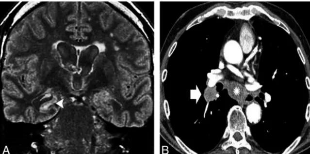
Hu Antibodies. The Hu antineuronal nuclear antibody is a type IIa antineuronal nuclear antibody type I, which can appear in any part of the nervous system. Approximately 75% of the patients have small cell lung carcinoma and often develop symptoms re-lated to inflammation across widespread areas of the CNS or the peripheral nervous system.18MR imaging reveals variable abnor-malities according to clinical features, including T2/FLAIR hyper-intensity in the mesial TL (Fig 1), cerebellar edema or atrophy, and brain stem abnormalities.4 Rarely, patients have epilepsia partialis continua, which results from restricted lesions in non-limbic cortical areas.19First-line immunotherapies often fail, and the prognosis of this condition is usually poor despite immunotherapy.20
Ma2 Antibodies. Patients with Ma2 antineuronal nuclear anti-body–related encephalitis often have accompanying symptoms of diencephalic inflammation (sleep disturbances, dysthermia, and endocrine abnormalities) and upper brain stem inflammation (eye movement abnormalities and hypokinetic syndrome). Ap-proximately 75% of patients have abnormal MRI, usually with classic LE findings.21The remaining patients have signal abnor-malities that are either isolated or associated with the hypothala-mus and thalahypothala-mus or with the brain stem.22Nodular parenchy-mal enhancement in the affected regions has been reported, which may mimic a brain tumor or an infection.21,22This AME
occurs mostly in association with testic-ular germinal cell tumors in younger male individuals; but, in older individu-als, there may be an underlying non– small cell lung carcinoma or breast cancer.23Improvement with immuno-therapy is more likely than in other forms of LE that involve antibodies against intracellular antigens.22
CV2/Collapsing Response Mediator Protein-5 Antibodies. Bilateral striatal encephalitis with T2/FLAIR hyperinten-sity is a typical finding, which causes choreiform movement disorders and is highly suggestive of CV2/collapsing re-sponse mediator protein-5 antineuronal nuclear antibody–related encephalitis associated with underlying small cell lung carcinoma or malignant thymoma, among others disorders.24,25However, patients may also present with a range of imaging patterns that rarely include LE and typically do not include striatal restriction on DWI, which may help to distinguish this AME from prion diseases.25
PLE Associated with Autoantibodies against Extracellular Antigens
N-Methyl-D-Aspartate Receptor Antibodies. A specific immu-noglobulin G antibody against the GluN1 subunit of the anti–N -methyl-D-aspartate receptor results in a highly characteristic and recognizable LE that is far more common than previously be-lieved26and mostly affects young women and children.27Two major well-characterized stages are noticeable.28A viral-like pro-drome followed by severe psychiatric features characterizes the earliest involvement of the cortical regions. In addition, patients may develop amnesia and seizures.29After a few days to a few weeks, subcortical areas are affected and a movement disorder appears (often dyskinesia of the mouth and face) followed by a decreased level of consciousness and dysautonomia, which re-quires intensive care support. A lymphocytic pleocytosis is ob-served in the CSF, and, less commonly, increased protein and/or oligoclonal bands are present.27
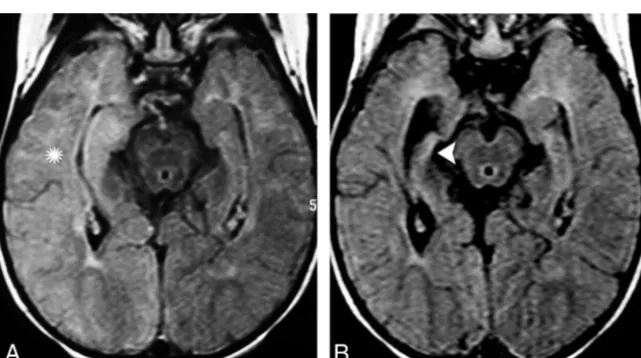
The most common MR imaging abnormality is unilateral or bilateral LE30,31; however, approximately 66% of patients have an unremarkable MR imaging. Cerebellitis, striatal abnormalities, and brain stem encephalitis have also been described.30,31 Gado-linium enhancement is uncommon, and imaging follow-up could reveal complete recovery or focal atrophy (Fig 2).27,30,31
The concurrence of tumors is reportedly age dependent. Whereas approximately 45% of adult woman had ovarian tera-toma, only 9% of younger girls had this type of tumor. Identifi-cation and removal of the tumor were crucial because patients without tumor removal recovered less frequently and had an in-creased risk of relapse.27In patients older than 45 years, the out-come was reportedly favorable, whereas 23% of patients had un-derlying carcinomas instead of teratomas.32Despite this ominous FIG 1. A 62-year-old man with subacute cognitive impairment and seizures.A, An enlarged and
[image:2.594.56.376.47.206.2]clinical presentation, approximately 50% of patients respond to first-line immunotherapies, often with full remission, whereas patients who do not respond to treatment or who experience re-lapse should be reassessed for the presence of an underlying con-tralateral or recurrent teratoma.33
Gamma-Aminobutyric Acid Receptor Antibodies. Anti– gam-ma-aminobutyric acid (GABA) B-receptor antibody-related en-cephalitis usually presents as LE. Most patients have early and frequent seizures associated with unilateral or bilateral T2/FLAIR hyperintensity in the mesial TL that are potentially reversible after treatment.34
As is most commonly reported in older patients, approxi-mately 50% of patients with GABA B-receptor AMEs have under-lying small cell lung carcinoma or lung neuroendocrine tumors.35 This AME usually precedes a cancer diagnosis but represents the second most common cause of LE related to small cell lung carcinoma.36
An AME associated with anti–GABA A-receptor antibodies was recently described in children and adults who developed a
rapidly progressive encephalopathy with refractory seizures, status epilepti-cus, and/or epilepsia partialis continua that was preceded by or associated with behavioral changes.37 Unlike patients with other LEs in whom MR imaging is either normal or shows predominant in-volvement of the limbic system, these patients have multifocal and extensive T2/FLAIR brain abnormalities. In addi-tion, they respond well to immunother-apy and rarely have an underlying tu-mor. When a tumor is present, it is usually a thymoma. Patients are often misdiagnosed with the 65-kD isoform of glutamic acid decarboxylase (GAD65) antibody-associated encephalitis or Hashimoto encephalitis due to the fre-quent co-occurrence of GAD65 and an-tithyroid antibodies.16,37
Other PLEs with Autoantibodies against Extracellular Antigens. In anti–␣ -amino-3-hydroxy-5-methyl-4-isoxazolepropionic acid receptor (AMPAR) encephalitis, pa-tients develop antibodies against the GluR1 and GluR2 subunits of the AMPAR, and present with symptoms and MR imaging features of unilateral or bilat-eral LE that rarely involve extrahippocam-pal limbic structures. In some cases, the manifestations are purely psychiatric. Most of these patients are women who are harboring a tumor in the lung, breast, or thymus.38
Hodgkin lymphoma is the third most common cause of LE after small cell lung carcinoma and testicular germ cell tumors.4This association has been called Ophelia syndrome, and it is characterized by generalized or partial complex seizures in 50% of the patients. It is also more commonly associated with short-term memory loss or amnesia, psychiatric changes, and even frank psychosis with visual or au-ditory hallucinations or paranoid ideation.17,39
Intriguingly, AME is not typically associated with non-Hodg-kin lymphoma.17Although Hodgkin lymphoma rarely infiltrates the CNS, the onset of an LE in this setting should be attributable to either a concurrent infection or an AME (Fig 3). Successful treat-ment of the tumor results in complete neurologic recovery, prob-ably due to an association with an antibody against the metabo-tropic glutamate receptor 5, which is highly expressed in the hippocampus and presumably promotes reversible neuronal dys-function rather than neuronal death.17,40
Nonparaneoplastic LE
It is assumed that nonparaneoplastic LE is more common than classic PLE and affects a wider age range of patients, though predominantly young patients. Nonparaneoplastic LE is a re-FIG 2. A previously healthy 44-year-old woman presented with subacute psychiatric disturbance
[image:3.594.55.376.45.404.2]sult of antibodies against neuronal cell surface or synaptic receptors.2
Nonparaneoplastic LE Associated with Autoantibodies against Intracellular Antigens
GAD65 Antibodies. Some patients with nonparaneoplastic LE have antibodies against the intracellular antigen GAD65. How-ever, unlike other intracellular antibodies, anti-GAD65 is not typ-ically related to underlying malignancies. Patients typtyp-ically pres-ent with stiff man syndrome or cerebellar ataxia, but they also may
present with severe TL epilepsy, with less pronounced cognitive-behavioral fea-tures and a poorer response to first-line epilepsy drugs.41
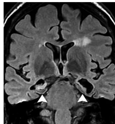
MR imaging frequently shows signal abnormalities and swelling predomi-nantly in the amygdala and hippocam-pus, which may resolve or progress to mesial temporal sclerosis on follow-up imaging (Fig 4).41
Nonparaneoplastic LE Associated with Autoantibodies against Extracellular Antigens
Voltage-Gated Potassium Channel-Complex Antibodies. Anti–leucine-rich glioma inactivated 1 (LGI1) and anti– contactin-associated protein-like 2 (CASPR2) antibodies have been de-scribed as voltage-gated potassium channel (VGKC) antibodies and the most common cause of nonparaneo-plastic LE.16 Results of recent studies highlight the relevance of discriminating both LGI1 and CASPR2 from VGKC-complex antibodies. Although LGI1 and contactin-associated protein-like 2 anti-bodies are specifically associated with limited subsets of syndromes, VGKC-complex antibodies lack specificity and may be found in nonautoimmune diseases, including Creutzfeldt-Jakob disease.42,43
LGI1 antibodies occur most often in young male patients (2:1) who develop a classic LE with peculiar features, such as hyponatremia (60%), rapid eye move-ment–sleep behavior disorders, and normal CSF. In a few patients, a charac-teristic clinical manifestation described as faciobrachial dystonic or tonic sei-zures is observed. Fewer than 10% of pa-tients with LGI1 antibodies have an un-derlying neoplasm, which is usually a thymoma.16,44
Approximately 78.6% of patients present with typical LE MR imaging findings (Fig 5). Restricted DWI is observed in approximately 50% of these patients, whereas up to 25% have associated mild, ill-defined contrast enhancement and extrahippocampal involve-ment, including striatal encephalitis.11,45
Antibodies against the VGKC-complex have been identified in a subgroup of patients with epilepsy that appears on imaging as mesial temporal sclerosis, which indicates that some patients with epilepsy who are poorly responsive to conventional antiepileptic drugs may have an immune-mediated etiology.11,46Recognition and appropriate treatment with immunotherapy are recom-FIG 3. A 22-year-old man with Hodgkin lymphoma presented with acute onset of short-term
memory loss and mental confusion.A, An evident hyperintensity and subtle enlargement of the right hippocampus and amygdala were noticed on an axial FLAIR image (arrow).B, No parenchy-mal enhancement was observed (arrowhead). In addition to the fact that CNS involvement is not expected in Hodgkin lymphoma, a lack of enhancement is not the expected imaging pattern. After the patient did not respond to antiviral treatment, PLE was considered. The findings fulfilled the criteria for Ophelia syndrome, which consists in an interval of⬍4 years between the onset of neuropsychiatric disturbance and the diagnosis of the Hodgkin lymphoma, exclusion of other cancer-related complications, and evidence of hippocampal abnormalities on MR imaging.
[image:4.594.55.376.47.226.2] [image:4.594.56.375.325.516.2]mended to prevent structural damage due to severe encephalitis as well as cognitive dysfunction.16,47
LGI1 antibodies are almost exclusively expressed in the CNS. They often result in LE or epilepsy but primarily result in non-paraneoplastic LE (Fig 5). Conversely, CASPR2 antibodies ex-pressed in the peripheral nervous system are involved in Morvan disease or peripheral nerve hyperexcitability–neuromyotonia spectrum disorders and are typically associated with thymomas. Myasthenia gravis and LE can also be found in some patients.48
Limbic Disorders That Mimic AME
Abnormal MR signal intensity that involves the TL has a broad dif-ferential diagnosis that includes a range of unrelated disorders that are rarely reported, for example, Whipple disease,49
4-aminopyri-dine toxicity,50and hypoglycemia.51Neuroradiologists must recog-nize these disorders and their imaging features more often.
Infectious LE
Herpes Virus Encephalitis. Herpes virus encephalitis (HSE) causes at least 20% of acute LE cases.52Although human herpes virus 6 is associated with posttransplantation acute LE,10the most common agent is herpes virus type 1, which has high mortality and morbidity rates.52
The clinical and imaging findings of LE caused by either AME or HSE may overlap. Although almost 50% of patients with AME present with or develop fever during their disease course and have prodromal symptoms with abnormal CSF, these findings favor HSE. The absence of psychiatric symp-toms and the sudden and rapid progres-sion also support the early administra-tion of antiviral therapy based on a presumed diagnosis of HSE.10In addi-tion, even though both HSE and LE in-volve the TL, basal ganglia inin-volvement on MR imaging favors nonherpetic etiologies.12
It has been demonstrated that some types of viral encephalitis can trigger au-toimmune LE,53,54particularly anti-N -methyl-D-aspartate receptor encephali-tis.55 This phenomenon occurs when prolonged or atypical neurologic symp-toms recur after successful control of the viral infection. Some patients with neg-ative viral results develop a syndrome described as relapsing post-HSE or cho-reoathetosis post-HSE. A few weeks af-ter recovery from HSE, children present with abnormal movement and adults FIG 5. A healthy 46-year-old woman presented with an acute onset of psychiatric disturbance
and hyponatremia.A, Bilateral hyperintensity and mild enlargement were noticed on an axial FLAIR image in both hippocampi and amygdalae.B, No abnormal restricted diffusion was ob-served on DWI. The final diagnosis was anti-VGKC encephalitis. Restricted diffusion may occur in approximately 50% of patients at this phase and is usually restricted to the limbic system. The presence of faciobrachial dystonic or tonic seizures, hyponatremia, and unremarkable CSF in the setting of LE should raise concern that anti-LGI1 encephalitis is present.
[image:5.594.55.375.219.402.2] [image:5.594.55.530.485.661.2]present with behavioral changes that are not associated with ad-ditional brain lesions on MR imaging or response to antiviral therapy.
Neurosyphilis. The incidence of neurosyphilis, caused by a spi-rochete (Treponema pallidum), has once again begun to increase in the era of acquired immunodeficiency syndrome.56MR imag-ing shows a variety of usually nonspecific findimag-ings, includimag-ing se-lective involvement of the TL that mimics HSE and LE.10,56In older subjects with a long latency period of infection or in patients who are immunocompromised, T2/FLAIR hyperintensities in the mesial TL areas that may or may not be associated with either atrophic or gadolinium-enhanced areas increase the likelihood that neurosyphilis is present rather than other etiologies (Fig 6).
Neoplastic Limbic Disorders
Diffuse gliomas and gliomatosis cerebri may mimic the imaging features of LE. The hallmark feature on MR imaging is an infiltrative pattern with poorly de-marcated boundaries that is usually not restricted to the limbic system.8,14,57 Gliomatosis cerebri, as well as low-grade diffuse gliomas, may progress slowly, with seizures or even focal deficits. Moreover, high-grade tumors might present with atypical imaging features that rarely mimic LE but then progress invariably to a recognizable MR imaging pattern of necrotic lesions (Fig 7). In this setting, MR-perfusion and MR-spec-troscopy techniques are useful to detect brain tumors and enable surgical planning.58
Vascular Limbic Disorders
Differentiation between primary vascu-litis and LE may represent a real challenge under certain condi-tions of subacute presentation. Abnormal vessels on angiography and cytotoxic edema on DWI that usually extends throughout the compromised vascular territory and is not restricted to the limits of the limbic system are helpful to confirm imaging suspicions.9 Transient global amnesia also affects the hippocampal formation, but its clinical and imaging presentation is rather typical.59
Seizure-Related Limbic Disorders
Hippocampal sclerosis associated with TL abnormalities is the multifactorial hallmark of mesial temporal sclerosis. This condi-tion could be a consequence of prolonged unilateral febrile sei-zures or status epilepticus, which occurs mainly in children when FIG 7. A previously healthy 67-year-old man presented with a transient isolated episode of partial complex seizures and dysphasia.A, A cortical abnormality that involved the lateral aspect of the left TL (asterisk) and a subtle hyperintensity on coronal T2 were noticed in the ipsilateral hippocampus.B, Restricted diffusion on DWI was visible in the same areas (arrowhead), and a diagnosis of postictal edema was considered.C, After 2 months and a worsening of the clinical manifestations, a necrotic mass in the left TL (arrow) was observed on a T1 postcontrast image. A diagnosis of glioblastoma was confirmed after surgery. High-grade gliomas can manifest early as ill-defined lesions that usually have restricted diffusion and involve the cortex with a lack of a mass effect. Follow-up imaging and advanced imaging techniques are crucial for making the diagnosis.
[image:6.594.56.532.48.239.2] [image:6.594.56.377.320.499.2]the hippocampus is more vulnerable to convulsion-induced exci-totoxic damage and involves the sectors of the hippocampus rich in kainate orN-methyl-D-aspartate receptors and, therefore, that lack protection against calcium overload.60
Prolonged seizures or status epilepticus may appear as TL ab-normalities on MR imaging, including cortical hyperintensities on DWI that mimic LE and are attributable to hippocampal pos-tictal edema.9,61Imaging follow-up with typical clinical and elec-troencephalographic features may aid diagnosis. This condition is potentially reversible or can result in atrophy with mesial tempo-ral sclerosis (Fig 8).62
Hippocampal sclerosis may also be related to a rare neurode-generative condition called pure hippocampal sclerosis dementia. Despite its similarity to mesial temporal sclerosis on imaging, de-mentia is always observed in the absence of epilepsy and usually occurs in the elderly.63
Febrile infection–related epilepsy syndrome, or acute enceph-alitis with refractory repetitive partial seizures, is considered a severe epileptic encephalopathy with multifocal refractory status epilepticus, which occurs mostly in young children but also in adult patients.64 The initial phase is characterized by a simple febrile infection, followed by an acute phase with recurrent focal seizures that evolve rapidly into refractory status epilepticus, gen-erally without fever and additional neurologic features. The diag-nosis is made after an exhaustive negative search for an active CNS infection and autoimmune or metabolic disorders. Early MR im-aging may be normal in approximately half of the cases; however, T2 abnormalities are detected in some patients, predominantly in the temporal regions but also in the insula and basal ganglia, which mimics LE.64,65In the chronic phase, MR imaging shows mesial temporal sclerosis in half of the patients, and bilateral hy-pometabolism of orbitofrontal and temporoparietal regions is of-ten demonstrated on PET.64The etiology and mechanisms that underlie it are still unknown, and, even though an autoimmune mechanism could be considered and autoantibodies have previ-ously been described in epilepsy, up to now there is no evidence to support that autoantibodies are the etiology of febrile infection-related epilepsy syndrome.64,66
Other Autoimmune Disorders
Autoimmune systemic disorders are associated with LE.2Sjo¨gren syndrome, lupus erythematosus, Bec¸het disease, primary angiitis of the CNS, and antiphospholipid syndrome can occasionally cause clinical and/or radiologic abnormalities in the limbic sys-tem that are not antibody mediated but that are accompanied by histopathologic evidence of cellular inflammation.3
Hashimoto encephalopathy or steroid-responsive encepha-lopathy associated with autoimmune thyroiditis67manifests as a diffuse progressive AME characterized by dementia, psychiatric disturbances, and seizures; there also is a vasculitic type charac-terized by multiple strokelike episodes, seizures, and fluctuating consciousness.68This disorder is more common in women and is associated with autoimmune antithyroid antibodies. There is in-creasing evidence that these antibodies are not pathogenic but rather are markers of autoimmunity for other associated but cur-rently unclassified antineuronal antibodies.69MR imaging may mimic patterns of LE (Fig 9),70but leukoencephalopathy with
bilateral patchy or confluent supratentorial subcortical and periventricular white matter T2/FLAIR hyperintensities is the most common abnormality and is usually reversible after corticotherapy.67
A rare cause of LE is relapsing polychondritis, in which clini-coradiologic involvement of the limbic system might be more common than was previously thought.71,72This condition is a disorder of unknown etiology that manifests as episodic and pro-gressive inflammation of the cartilaginous structures of the body, as is suggested by the detection of autoantibodies against type II collagen restricted to the cartilage in the sera of 30%–50% of affected patients.73MR imaging findings are coincident with LE; however, peculiar cartilage involvement may help identify this entity.
Recommended Diagnostic Approach to Limbic Disorders
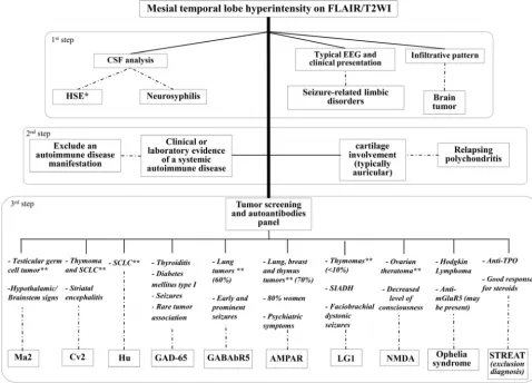
An algorithm that describes an approach to the diagnosis of lim-bic disorders by using clinical and neuroimaging features is pre-sented inFig 10.
CONCLUSIONS
[image:7.594.319.513.45.254.2]recognizing limbic disorders and is useful for differentiating among them and for improving their investigation.
REFERENCES
1. Brierley JB, Corsellis JA, Hierons R, et al.Subacute encephalitis of later adult life mainly affecting the limbic areas.Brain1960;83: 357– 68CrossRef
2. Vernino S, Geschwind M, Boeve B.Autoimmune encephalopathies.
Neurologist2007;13:140 – 47CrossRef Medline
3. Tu¨zun E, Dalmau J.Limbic encephalitis and variants: classification, diagnosis and treatment. Neurologist 2007;13:261–71 CrossRef Medline
4. Gultekin SH, Rosenfeld MR, Voltz R, et al.Paraneoplastic limbic encephalitis: neurological symptoms, immunological findings and tumour association in 50 patients.Brain2000;123(pt 7):1481–94
CrossRef Medline
5. Vieira Santos A, Matias S, Saraiva P, et al.Differential diagnosis of mesiotemporal lesions: case report of neurosyphilis. Neuroradiol-ogy2005;47:664 – 67CrossRef Medline
6. Kararizou E, Markou I, Zalonis I, et al.Paraneoplastic limbic en-cephalitis presenting as acute viral enen-cephalitis.J Neurooncol2005; 75:229 –32CrossRef Medline
7. Tyler KL.Emerging viral infections of the central nervous system: part 1.Arch Neurol2009;66:939 – 48CrossRef Medline
8. Nagata R, Ikeda K, Nakamura Y, et al.A case of gliomatosis cerebri mimicking limbic encephalitis: malignant transformation to glio-blastoma.Int Med2010;49:1307–10CrossRef Medline
9. Fo¨rster A, Griebe M, Gass A, et al.Diffusion-weighted imaging for the differential diagnosis of disorders affecting the hippocampus.
Cerebrovasc Dis2012;33:104 –15CrossRef Medline
10. Armangue T, Leypoldt F, Dalmau J.Autoimmune encephalitis as differential diagnosis of infectious encephalitis.Curr Opin Neurol
2014;27:361– 68CrossRef Medline
11. Kotsenas AL, Watson RE, Pittock SJ, et al.MRI findings in autoim-mune voltage-gated potassium channel complex encephalitis with seizures: one potential etiology for mesial temporal sclerosis.AJNR Am J Neuroradiol2014;35:84 – 89CrossRef Medline
12. Oyanguren B, Sa´nchez V, Gonza´lez FJ, et al.Limbic encephalitis: a clinical-radiological comparison between herpetic and autoim-mune etiologies.Eur J Neurol2013;20:1566 –70CrossRef Medline
13. Dalmau J, Bataller L.Clinical and immunological diversity of limbic encephalitis: a model for paraneoplastic neurologic disorders. He-matol Oncol Clin North Am2006;20:1319 –35CrossRef Medline
14. Demaerel P, Van Dessel W, Van Paesschen W, et al. Autoimmune-mediated encephalitis. Neuroradiology 2011;53:837–51 CrossRef Medline
15. Darnell RB, Posner JB.Paraneoplastic syndromes involving the ner-vous system.N Engl J Med2003;349:1543–54CrossRef Medline
16. Dalmau J, Rosenfeld MR.Autoimmune encephalitis update.Neuro Oncol2014;16:771–78CrossRef Medline
17. Graus F, Arin˜o H, Dalmau J. Paraneoplastic neurological syn-dromes in Hodgkin and non-Hodgkin lymphomas.Blood2014;123: 3230 –38CrossRef Medline
[image:8.594.54.533.49.393.2]neoplastic encephalomyelitis: analysis of 200 patients.Brain2001; 124(pt 6):1138 – 48CrossRef Medline
19. Shavit YB, Graus F, Probst A, et al.Epilepsia partialis continua: a new manifestation of anti-Hu-associated paraneoplastic encephalomy-elitis.Ann Neurol1999;45:255–58CrossRef Medline
20. Keime-Guibert F, Graus F, Fleury A, et al.Treatment of paraneoplas-tic neurological syndromes with antineuronal antibodies (Anti-Hu, anti-Yo) with a combination of immunoglobulins, cyclophos-phamide, and methylprednisolone.J Neurol Neurosurg Psychiatry
2000;68:479 – 82CrossRef Medline
21. Rosenfeld MR, Eichen JG, Wade DF, et al.Molecular and clinical diversity in paraneoplastic immunity to Ma proteins.Ann Neurol
2001;50:339 – 48CrossRef Medline
22. Dalmau J, Graus F, Villarejo A, et al.Clinical analysis of anti-Ma2-associated encephalitis. Brain 2004;127(pt 8):1831– 44 CrossRef Medline
23. Dalmau J, Gultekin SH, Voltz R, et al.Ma1, a novel neuron- and testis-specific protein, is recognized by the serum of patients with paraneoplastic neurological disorders.Brain1999;122(pt 1):27–39
CrossRef Medline
24. Honnorat J, Cartalat-Carel S, Ricard D, et al.Onco-neural antibodies and tumour type determine survival and neurological symptoms in paraneoplastic neurological syndromes with Hu or CV2/CRMP5 antibodies.J Neurol Neurosurg Psychiatry2009;80:412–16CrossRef Medline
25. Vernino S, Tuite P, Adler CH, et al.Paraneoplastic chorea associated with CRMP-5 neuronal antibody and lung carcinoma.Ann Neurol
2002;51:625–30CrossRef Medline
26. Gable MS, Sheriff H, Dalmau J, et al.The frequency of autoimmune N-methyl-D-aspartate receptor encephalitis surpasses that of indi-vidual viral etiologies in young indiindi-viduals enrolled in the Califor-nia Encephalitis Project.Clin Infect Dis2012;54:899 –904CrossRef Medline
27. Titulaer MJ, McCracken L, Gabilondo I, et al.Treatment and prog-nostic factors for long-term outcome in patients with anti-NMDA receptor encephalitis: an observational cohort study.Lancet Neurol
2013;12:157– 65CrossRef Medline
28. Irani SR, Bera K, Waters P, et al.N-methyl-D-aspartate antibody encephalitis: temporal progression of clinical and paraclinical ob-servations in a predominantly non-paraneoplastic disorder of both sexes.Brain2010;133(pt 6):1655– 67CrossRef Medline
29. Kayser MS, Titulaer MJ, Gresa-Arribas N, et al.Frequency and char-acteristics of isolated psychiatric episodes in anti–N-methyl-d-as-partate receptor encephalitis. JAMA Neurol 2013;70:1133–39
CrossRef Medline
30. Dalmau J, Gleichman AJ, Hughes EG, et al.Anti-NMDA-receptor encephalitis: case series and analysis of the effects of antibodies.
Lancet Neurol2008;7:1091–98CrossRef Medline
31. Dalmau J, Tu¨zu¨n E, Wu HY, et al.Paraneoplastic anti-N-methyl-D-aspartate receptor encephalitis associated with ovarian teratoma.
Ann Neurol2007;61:25–36CrossRef Medline
32. Titulaer MJ, McCracken L, Gabilondo I, et al.Late-onset anti-NMDA receptor encephalitis. Neurology 2013;81:1058 – 63 CrossRef Medline
33. Johnson N, Henry C, Fessler AJ, et al.Anti-NMDA receptor enceph-alitis causing prolonged nonconvulsive status epilepticus. Neurol-ogy2010;75:1480 – 82CrossRef Medline
34. Lancaster E, Lai M, Peng X, et al.Antibodies to the GABA(B) recep-tor in limbic encephalitis with seizures: case series and characteri-sation of the antigen.Lancet Neurol2010;9:67–76CrossRef Medline
35. Ho¨ftberger R, Titulaer MJ, Sabater L, et al.Encephalitis and GABAB receptor antibodies: novel findings in a new case series of 20 pa-tients.Neurology2013;81:1500 – 06CrossRef Medline
36. Boronat A, Sabater L, Saiz A, et al.GABA(B) receptor antibodies in limbic encephalitis and anti-GAD-associated neurologic disorders.
Neurology2011;76:795– 800CrossRef Medline
37. Petit-Pedrol M, Armangue T, Peng X, et al.Encephalitis with refrac-tory seizures, status epilepticus, and antibodies to the GABAA
receptor: a case series, characterisation of the antigen, and analysis of the effects of antibodies.Lancet Neurol2014;13:276 – 86CrossRef Medline
38. Graus F, Boronat A, Xifro´ X, et al. The expanding clinical profile of anti-AMPA receptor encephalitis. Neurology 2010;74:857–59
CrossRef Medline
39. Carr I.The Ophelia syndrome: memory loss in Hodgkin’s disease.
Lancet1982;1:844 – 45Medline
40. Mat A, Adler H, Merwick A, et al.Ophelia syndrome with metabo-tropic glutamate receptor 5 antibodies in CSF.Neurology2013;80: 1349 –50CrossRef Medline
41. Malter MP, Helmstaedter C, Urbach H, et al.Antibodies to glutamic acid decarboxylase define a form of limbic encephalitis.Ann Neurol
2010;67:470 –78CrossRef Medline
42. Grau-Rivera O, Sa´nchez-Valle R, Saiz A, et al.Determination of neu-ronal antibodies in suspected and definite Creutzfeldt-Jakob dis-ease.JAMA Neurol2014;71:74 –78CrossRef Medline
43. Paterson RW, Zandi MS, Armstrong R, et al.Clinical relevance of positive voltage-gated potassium channel (VGKC)-complex antibodies: experience from a tertiary referral centre.J Neurol Neu-rosurg Psychiatry2014;85:625–30CrossRef Medline
44. Irani SR, Michell AW, Lang B, et al.Faciobrachial dystonic seizures precede Lgi1 antibody limbic encephalitis.Ann Neurol2011;69:892– 900CrossRef Medline
45. Hiraga A, Kuwabara S, Hayakawa S, et al.Voltage-gated potassium channel antibody-associated encephalitis with basal ganglia le-sions.Neurology2006;66:1780 – 81CrossRef Medline
46. Majoie HJ, de Baets M, Renier W, et al.Antibodies to voltage-gated potassium and calcium channels in epilepsy.Epilepsy Res2006;71: 135– 41CrossRef Medline
47. Vincent A, Buckley C, Schott JM, et al.Potassium channel antibody-asso-ciated encephalopathy: a potentially immunotherapy-responsive form of limbic encephalitis.Brain2004;127(pt 3):701–12CrossRef Medline
48. Lancaster E, Huijbers MG, Bar V, et al.Investigations of caspr2, an autoantigen of encephalitis and neuromyotonia.Ann Neurol2011; 69:303–11CrossRef Medline
49. Blanc F, Ben Abdelghani K, Schramm F, et al.Whipple limbic en-cephalitis.Arch Neurol2011;68:1471–73CrossRef Medline
50. Badruddin A, Menon RS, Reder AT.4-Aminopyridine toxicity mim-ics autoimmune-mediated limbic encephalitis.Neurology2009;72: 1100 – 01CrossRef Medline
51. Boeve BF, Bell DG, Noseworthy JH.Bilateral temporal lobe MRI changes in uncomplicated hypoglycemic coma.Can J Neurol Sci
1995;22:56 – 8Medline
52. Baringer JR.Herpes simplex infections of the nervous system. Neu-rol Clin2008;26:657–74, viiiCrossRef Medline
53. Armangue T, Leypoldt F, Ma´laga I, et al.Herpes simplex virus en-cephalitis is a trigger of brain autoimmunity.Ann Neurol2014;75: 317–23CrossRef Medline
54. Scha¨bitz WR, Rogalewski A, Hagemeister C, et al.VZV brainstem encephalitis triggers NMDA receptor immunoreaction.Neurology
2014;83:2309 –11CrossRef Medline
55. Ho¨ftberger R, Armangue T, Leypoldt F, et al.Clinical neuropathol-ogy practice guide 4 –2013: post-herpes simplex encephalitis: N-methyl-Daspartate receptor antibodies are part of the problem.
Clin Neuropathol2013;32:251–54CrossRef Medline
56. Karsan N, Barker R, O’Dwyer JP.Clinical reasoning: the “great im-itator.”Neurology2014;83:e188 –96CrossRef Medline
57. Vates GE, Chang S, Lamborn KR, et al.Gliomatosis cerebri: a review of 22 cases.Neurosurgery2003;53:261–71, discussion 271CrossRef Medline
58. Maia AC Jr, Malheiros SM, da Rocha AJ, et al.Stereotactic biopsy guidance in adults with supratentorial nonenhancing gliomas: role of perfusion-weighted magnetic resonance imaging.J Neurosurg
2004;101:970 –76CrossRef Medline
59. Sedlaczek O, Hirsch JG, Grips E, et al.Detection of delayed focal MR changes in the lateral hippocampus in transient global amnesia.
60. Cendes F.Febrile seizures and mesial temporal sclerosis.Curr Opin Neurol2004;17:161– 64CrossRef Medline
61. Kim JA, Chung JI, Yoon PH, et al.Transient MR signal changes in patients with generalized tonicoclonic seizure or status epilepticus: periictal diffusion-weighted imaging.AJNR Am J Neuroradiol2001; 22:1149 – 60Medline
62. Cox JE, Mathews VP, Santos CC, et al.Seizure-induced transient hippocampal abnormalities on MR: correlation with positron emission tomography and electroencephalography. AJNR Am J Neuroradiol1995;16:1736 –38Medline
63. Hatanpaa KJ, Blass DM, Pletnikova O, et al.Most cases of dementia with hippocampal sclerosis may represent frontotemporal demen-tia.Neurology2004;63:538 – 42CrossRef Medline
64. Caraballo RH, Reyes G, Avaria MF, et al.Febrile infection-related epilepsy syndrome: a study of 12 patients.Seizure2013;22:553–59
CrossRef Medline
65. van Baalen A, Ha¨usler M, Boor R, et al.Febrile infection-related epilepsy syndrome (FIRES): a nonencephalitic encephalopathy in childhood.Epilepsia2010;51:1323–28CrossRef Medline
66. Venkatesan A, Benavides DR.Autoimmune encephalitis and its re-lation to infection.Curr Neurol Neurosci Rep2015;15:3CrossRef
67. Castillo P, Woodruff B, Caselli R, et al.Steroid-responsive
encepha-lopathy associated with autoimmune thyroiditis.Arch Neurol2006; 63:197–202CrossRef Medline
68. Hollowell JG, Staehling NW, Flanders WD, et al.Serum TSH, T(4), and thyroid antibodies in the United States population (1988 to 1994): National Health and Nutrition Examination Survey (NHANES III).J Clin Endocrinol Metab2002;87:489 –99CrossRef Medline
69. Ferracci F, Carnevale A.The neurological disorder associated with thyroid autoimmunity.J Neurol2006;253:975– 84CrossRef Medline
70. Song YM, Seo DW, Chang GY.MR findings in Hashimoto enceph-alopathy.AJNR Am J Neuroradiol2004;25:807– 08Medline
71. Fujiki F, Tsuboi Y, Hashimoto K, et al.Non-herpetic limbic enceph-alitis associated with relapsing polychondritis.J Neurol Neurosurg Psychiatry2004;75:1646 – 47CrossRef Medline
72. Kumar N, Leep Hunderfund AN, Kutzbach BR, et al.A limbic en-cephalitis MR imaging in a patient with Behcet disease and relaps-ing polychondritis.AJNR Am J Neuroradiol2009;30:E96CrossRef Medline