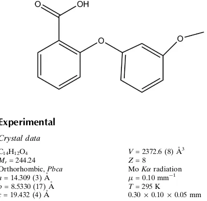
2-(3-Methoxyphenoxy)benzoic acid
Zhi-Fang Zhang
School of Chemistry and Chemical Engineering, Yulin University, Yulin 719000, People’s Republic of China
Correspondence e-mail: [email protected]
Received 19 March 2011; accepted 24 March 2011
Key indicators: single-crystal X-ray study;T= 295 K; mean(C–C) = 0.005 A˚; Rfactor = 0.062;wRfactor = 0.133; data-to-parameter ratio = 13.3.
In the crystal structure of the title compound, C14H12O4, the molecules form classical O—H O hydrogen-bonded carb-oxylic acid dimers. These dimers are linked by C—H pi; interactions into a three-dimensional network. The benzene rings are oriented at a dihedral angle of 69.6 (3).
Related literature
For applications of the title compound, see: Jackson et al. (1993); Gapinskiet al.(1990). For related structures, see: Shiet al.(2011); Raghunathanet al.(1982). For the synthesis of the title compound, see: Pello´net al.(1995). For bond-length data, see: Allenet al.(1987).
Experimental
Crystal data
C14H12O4 Mr= 244.24 Orthorhombic,Pbca a= 14.309 (3) A˚ b= 8.5330 (17) A˚ c= 19.432 (4) A˚
V= 2372.6 (8) A˚3 Z= 8
MoKradiation
= 0.10 mm1 T= 295 K
0.300.100.05 mm
Data collection
Enraf–Nonius CAD-4 diffractometer
Absorption correction: scan (Northet al., 1968) Tmin= 0.970,Tmax= 0.995 4273 measured reflections
2175 independent reflections 1056 reflections withI> 2(I) Rint= 0.093
3 standard reflections every 200 reflections
intensity decay: 1%
Refinement
R[F2> 2(F2)] = 0.062 wR(F2) = 0.133 S= 1.00 2175 reflections
163 parameters
H-atom parameters constrained
max= 0.16 e A˚
3
min=0.16 e A˚
[image:1.610.47.261.455.662.2]3
Table 1
Hydrogen-bond geometry (A˚ ,).
Cg1 is the centroid of the C2–C7 ring.
D—H A D—H H A D A D—H A
O4—H4A O3i
0.82 1.82 2.633 (3) 173
C1—H1B Cg1ii
0.96 2.89 3.784 (4) 155
Symmetry codes: (i)xþ2;y;zþ2; (ii)xþ3 2;yþ
1 2;z.
Data collection: CAD-4 Software (Enraf–Nonius, 1985); cell refinement: CAD-4 Software; data reduction: XCAD4 (Harms & Wocadlo, 1995); program(s) used to solve structure: SHELXS97
(Sheldrick, 2008); program(s) used to refine structure:SHELXL97
(Sheldrick, 2008); molecular graphics:SHELXTL(Sheldrick, 2008); software used to prepare material for publication:SHELXTL.
The author gratefully acknowledges financial support from the Natural Science Foundation of the Education Department of Shaanxi Provincial Government (09JK844) and is grateful for support provided by the key industry problem plan of Yulin (gygg200807) and the special research projects of Yulin University (08YK17).
Supplementary data and figures for this paper are available from the IUCr electronic archives (Reference: HG5013).
References
Allen, F. H., Kennard, O., Watson, D. G., Brammer, L., Orpen, A. G. & Taylor, R. (1987).J. Chem. Soc. Perkin Trans.2, pp. S1–19.
Enraf–Nonius (1985).CAD-4 Software. Enraf–Nonius, Delft, The Nether-lands.
Gapinski, D. M., Mallett, B. E., Froelich, L. L. & Jackson, W. T. (1990).J. Med. Chem.33, 2798-2807.
Harms, K. & Wocadlo, S. (1995).XCAD4. University of Marburg, Germany. Jackson, W. T., Boyd, R. J., Froelich, L. L., Gapinski, D. M., Mallett, B. E. &
Sawyer, J. S. (1993).J. Med. Chem.36, 1726–1734.
North, A. C. T., Phillips, D. C. & Mathews, F. S. (1968).Acta Cryst.A24, 351– 359.
Pello´n, R. F., Carrasco, R., Milia´n, V.& Rodes, L. (1995).Synth. Commun.25, 1077–1083.
Raghunathan, S., Chandrasekhar, K. & Pattabhi, V. (1982).Acta Cryst.B38, 2536–2538.
Sheldrick, G. M. (2008).Acta Cryst.A64, 112–122.
Shi, L., Zhang, Q., Xiao, Q., Wu, T. & Zhu, H.-J. (2011).Acta Cryst.E67, o748.
Acta Crystallographica Section E
Structure Reports Online
supporting information
Acta Cryst. (2011). E67, o1078 [doi:10.1107/S1600536811011019]
2-(3-Methoxyphenoxy)benzoic acid
Zhi-Fang Zhang
S1. Comment
The title compound, 2-(3-methoxyphenoxy)benzoic acid is an important intermediate of xanthone dicarboxylic acids
(Jackson et al., 1972). Xanthone dicarboxylic acid, such as LY223982, which inhibited the binding of LTB4 to receptors
on intact human neutrophils nearly as well as nonradioactive LTB4 (Gapinski et al., 1990). Knowledge of the crystal
structure of such benzoicacid derivatives gives us not only information about nuclearity of the complex molecule, but is
important in understanding the behaviour of these compounds with respect to the mechanisms of pharmacological
activities and physiological activities. Therefore, we have synthesized the title compound, (I), and report its crystal
structure here.
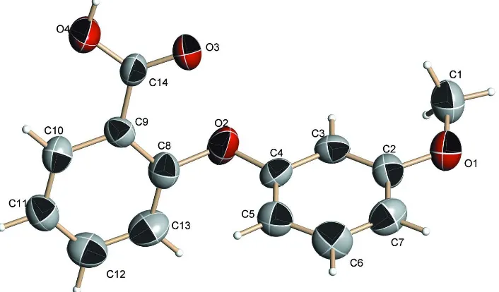
The molecular structure of (I) is shown in Fig. 1, and the intermolecular O—H···O hydrogen bond (Table 1) results in
the formation of carboxylic acid dimers (Fig. 2.). The bond lengths are within normal ranges (Allen et al., 1987). Similar
crystal structure of some compounds have been reported (Shi et al., 2011; Raghunathan et al., 1982).
In the molecule of (I), the dihedral angle of the rings(C3—C6) and (C8—C13) is 69.6 (3)°, the molecules were
connected together via O—H···O intermolecular hydrogen bonds to form dimers. These dimers are linked by C—H···π
and weak C—H···O interactions to give a three-dimensional network, which seems to be very effective in the stabilization
of the crystal structure.
S2. Experimental
The title compound (I) was prepared by the method of Ullmann condensation reaction reported in literature (Pellón et al.,
1995). A mixture of 2-chlorobenzoic acid (6.26 g, 0.04 mol), 3-methoxyphenol (9.93 g, 0.08 mol), anhydrous K2CO3
(11.04 g′ 0.08 mol), pyridine (1.58 g′ 0.02 mol), Cu powder (0.2 g) and cuprous iodide (0.2 g) in 25 ml water was kept at
reflux for two hours. The mixture was then basified with Na2CO3 solution and extracted with diethyl ether. The aqueous
solution was acidified with HCl, the precipitated solid was filtered off and disolved in NaOH; the basic solution was
filtered (charcoal) and acidified with acetic acid. The 2-(3-methoxyphenoxy)benzoic acid was crystalized from the
mixture.
S3. Refinement
H atoms were positioned geometrically and refined as riding groups, with O—H = 0.82 and C—H = 0.93 Å for aromatic
H, and constrained to ride on their parent atoms, with Uiso(H) = xUeq(C), where x = 1.2 for aromatic H, and x = 1.5 for
Figure 1
The molecular structure of (I) (thermal ellipsoids are shown at 30% probability levels).
Figure 2
[image:3.610.131.485.324.626.2]2-(3-Methoxyphenoxy)benzoic acid
Crystal data
C14H12O4 Mr = 244.24 Orthorhombic, Pbca Hall symbol: -P 2ac 2ab a = 14.309 (3) Å b = 8.5330 (17) Å c = 19.432 (4) Å V = 2372.6 (8) Å3 Z = 8
F(000) = 1024 Dx = 1.367 Mg m−3
Mo Kα radiation, λ = 0.71073 Å Cell parameters from 25 reflections θ = 9–12°
µ = 0.10 mm−1 T = 295 K Block, colourless 0.30 × 0.10 × 0.05 mm
Data collection
Enraf–Nonius CAD-4 diffractometer
Radiation source: fine-focus sealed tube Graphite monochromator
ω/2θ scans
Absorption correction: ψ scan (North et al., 1968)
Tmin = 0.970, Tmax = 0.995 4273 measured reflections
2175 independent reflections 1056 reflections with I > 2σ(I) Rint = 0.093
θmax = 25.4°, θmin = 2.1° h = 0→17
k = 0→10 l = −23→23
3 standard reflections every 200 reflections intensity decay: 1%
Refinement
Refinement on F2 Least-squares matrix: full R[F2 > 2σ(F2)] = 0.062 wR(F2) = 0.133 S = 1.00 2175 reflections 163 parameters 0 restraints
Primary atom site location: structure-invariant direct methods
Secondary atom site location: difference Fourier map
Hydrogen site location: inferred from neighbouring sites
H-atom parameters constrained w = 1/[σ2(F
o2) + (0.039P)2] where P = (Fo2 + 2Fc2)/3 (Δ/σ)max < 0.001
Δρmax = 0.16 e Å−3 Δρmin = −0.16 e Å−3
Special details
Geometry. All e.s.d.'s (except the e.s.d. in the dihedral angle between two l.s. planes) are estimated using the full covariance matrix. The cell e.s.d.'s are taken into account individually in the estimation of e.s.d.'s in distances, angles and torsion angles; correlations between e.s.d.'s in cell parameters are only used when they are defined by crystal symmetry. An approximate (isotropic) treatment of cell e.s.d.'s is used for estimating e.s.d.'s involving l.s. planes.
Refinement. Refinement of F2 against ALL reflections. The weighted R-factor wR and goodness of fit S are based on F2, conventional R-factors R are based on F, with F set to zero for negative F2. The threshold expression of F2 > σ(F2) is used only for calculating R-factors(gt) etc. and is not relevant to the choice of reflections for refinement. R-factors based on F2 are statistically about twice as large as those based on F, and R- factors based on ALL data will be even larger.
Fractional atomic coordinates and isotropic or equivalent isotropic displacement parameters (Å2)
x y z Uiso*/Ueq
O1 0.60412 (16) 0.2617 (3) 0.77635 (10) 0.0641 (7)
C1 0.6361 (3) 0.4135 (4) 0.79576 (18) 0.0738 (11)
H1A 0.6239 0.4863 0.7592 0.111*
H1C 0.6040 0.4468 0.8366 0.111*
O2 0.70190 (16) 0.0652 (2) 0.99264 (12) 0.0668 (7)
C2 0.6162 (2) 0.1419 (4) 0.82240 (16) 0.0510 (8)
O3 0.88741 (15) 0.0003 (3) 0.97959 (10) 0.0654 (7)
C3 0.6528 (2) 0.1601 (4) 0.88657 (16) 0.0495 (8)
H3A 0.6720 0.2584 0.9016 0.059*
O4 0.95145 (16) −0.0699 (3) 1.07827 (11) 0.0783 (8)
H4A 0.9992 −0.0465 1.0573 0.117*
C4 0.6614 (2) 0.0306 (4) 0.92945 (16) 0.0472 (8)
C5 0.6323 (2) −0.1143 (4) 0.90941 (18) 0.0570 (9)
H5A 0.6371 −0.2000 0.9388 0.068*
C6 0.5953 (3) −0.1298 (4) 0.84420 (18) 0.0660 (10)
H6A 0.5751 −0.2279 0.8296 0.079*
C7 0.5878 (2) −0.0056 (5) 0.80078 (18) 0.0638 (10)
H7A 0.5636 −0.0193 0.7568 0.077*
C8 0.7034 (2) −0.0465 (4) 1.04428 (17) 0.0533 (8)
C9 0.7889 (2) −0.0922 (3) 1.06981 (15) 0.0454 (7)
C10 0.7896 (2) −0.1884 (4) 1.12863 (16) 0.0535 (9)
H10A 0.8463 −0.2195 1.1477 0.064*
C11 0.7077 (3) −0.2363 (4) 1.15792 (17) 0.0598 (9)
H11A 0.7090 −0.3012 1.1964 0.072*
C12 0.6238 (3) −0.1898 (5) 1.1312 (2) 0.0666 (10)
H12A 0.5683 −0.2236 1.1512 0.080*
C13 0.6218 (2) −0.0919 (5) 1.0743 (2) 0.0699 (10)
H13A 0.5650 −0.0575 1.0567 0.084*
C14 0.8796 (2) −0.0495 (4) 1.03863 (15) 0.0488 (8)
Atomic displacement parameters (Å2)
U11 U22 U33 U12 U13 U23
O1 0.0741 (17) 0.0676 (16) 0.0506 (13) 0.0054 (14) −0.0066 (12) 0.0027 (13)
C1 0.091 (3) 0.067 (3) 0.064 (2) 0.008 (2) −0.008 (2) 0.010 (2)
O2 0.0799 (17) 0.0490 (13) 0.0716 (15) −0.0051 (12) −0.0318 (14) 0.0080 (12)
C2 0.049 (2) 0.055 (2) 0.0488 (19) 0.0094 (18) 0.0042 (15) 0.0037 (18)
O3 0.0670 (15) 0.0839 (17) 0.0452 (12) −0.0097 (13) −0.0051 (11) 0.0177 (14)
C3 0.046 (2) 0.0420 (19) 0.061 (2) −0.0060 (16) −0.0011 (16) −0.0044 (16)
O4 0.0547 (15) 0.115 (2) 0.0654 (16) −0.0057 (16) −0.0086 (13) 0.0296 (15)
C4 0.0391 (18) 0.046 (2) 0.0565 (19) 0.0019 (14) −0.0081 (16) −0.0041 (18)
C5 0.062 (2) 0.045 (2) 0.064 (2) 0.0033 (19) 0.0009 (19) −0.0012 (18)
C6 0.081 (3) 0.043 (2) 0.074 (2) −0.0001 (19) 0.002 (2) −0.014 (2)
C7 0.064 (2) 0.070 (2) 0.057 (2) 0.001 (2) −0.0037 (17) −0.020 (2)
C8 0.062 (2) 0.0448 (19) 0.0530 (19) 0.0070 (18) −0.0061 (17) 0.0012 (16)
C9 0.0518 (18) 0.0384 (17) 0.0461 (16) −0.0025 (15) 0.0024 (16) −0.0004 (14)
C10 0.058 (2) 0.059 (2) 0.0439 (19) 0.0086 (18) −0.0046 (18) −0.0023 (15)
C11 0.070 (2) 0.061 (2) 0.0485 (19) −0.002 (2) 0.0065 (19) 0.0034 (17)
C12 0.053 (2) 0.075 (3) 0.072 (3) 0.003 (2) 0.015 (2) −0.002 (2)
C13 0.048 (2) 0.079 (3) 0.083 (3) 0.011 (2) 0.001 (2) −0.006 (2)
Geometric parameters (Å, º)
O1—C2 1.369 (3) C5—H5A 0.9300
O1—C1 1.425 (4) C6—C7 1.360 (5)
C1—H1A 0.9600 C6—H6A 0.9300
C1—H1B 0.9600 C7—H7A 0.9300
C1—H1C 0.9600 C8—C13 1.362 (4)
O2—C8 1.384 (3) C8—C9 1.377 (4)
O2—C4 1.389 (3) C9—C10 1.407 (4)
C2—C3 1.362 (4) C9—C14 1.477 (4)
C2—C7 1.388 (4) C10—C11 1.366 (4)
O3—C14 1.229 (3) C10—H10A 0.9300
C3—C4 1.389 (4) C11—C12 1.366 (4)
C3—H3A 0.9300 C11—H11A 0.9300
O4—C14 1.296 (3) C12—C13 1.386 (5)
O4—H4A 0.8200 C12—H12A 0.9300
C4—C5 1.362 (4) C13—H13A 0.9300
C5—C6 1.379 (4)
C2—O1—C1 117.7 (2) C6—C7—C2 119.7 (3)
O1—C1—H1A 109.5 C6—C7—H7A 120.1
O1—C1—H1B 109.5 C2—C7—H7A 120.1
H1A—C1—H1B 109.5 C13—C8—C9 121.8 (3)
O1—C1—H1C 109.5 C13—C8—O2 119.6 (3)
H1A—C1—H1C 109.5 C9—C8—O2 118.1 (3)
H1B—C1—H1C 109.5 C8—C9—C10 117.7 (3)
C8—O2—C4 120.0 (2) C8—C9—C14 124.2 (3)
O1—C2—C3 124.2 (3) C10—C9—C14 118.1 (3)
O1—C2—C7 116.2 (3) C11—C10—C9 120.5 (3)
C3—C2—C7 119.6 (3) C11—C10—H10A 119.8
C2—C3—C4 119.5 (3) C9—C10—H10A 119.8
C2—C3—H3A 120.2 C10—C11—C12 120.6 (3)
C4—C3—H3A 120.2 C10—C11—H11A 119.7
C14—O4—H4A 109.5 C12—C11—H11A 119.7
C5—C4—O2 125.0 (3) C11—C12—C13 119.8 (3)
C5—C4—C3 121.5 (3) C11—C12—H12A 120.1
O2—C4—C3 113.4 (3) C13—C12—H12A 120.1
C4—C5—C6 117.9 (3) C8—C13—C12 119.7 (4)
C4—C5—H5A 121.1 C8—C13—H13A 120.2
C6—C5—H5A 121.1 C12—C13—H13A 120.2
C7—C6—C5 121.7 (3) O3—C14—O4 122.0 (3)
C7—C6—H6A 119.1 O3—C14—C9 123.2 (3)
C5—C6—H6A 119.1 O4—C14—C9 114.8 (3)
C1—O1—C2—C3 3.1 (5) C13—C8—C9—C10 −0.1 (5)
C1—O1—C2—C7 −177.4 (3) O2—C8—C9—C10 171.1 (3)
O1—C2—C3—C4 179.6 (3) C13—C8—C9—C14 178.6 (3)
C8—O2—C4—C5 −9.0 (5) C8—C9—C10—C11 1.3 (4)
C8—O2—C4—C3 171.1 (3) C14—C9—C10—C11 −177.5 (3)
C2—C3—C4—C5 −1.3 (5) C9—C10—C11—C12 −1.0 (5)
C2—C3—C4—O2 178.6 (3) C10—C11—C12—C13 −0.6 (6)
O2—C4—C5—C6 −178.6 (3) C9—C8—C13—C12 −1.5 (5)
C3—C4—C5—C6 1.3 (5) O2—C8—C13—C12 −172.5 (3)
C4—C5—C6—C7 −0.1 (5) C11—C12—C13—C8 1.8 (6)
C5—C6—C7—C2 −1.1 (6) C8—C9—C14—O3 −16.7 (5)
O1—C2—C7—C6 −178.4 (3) C10—C9—C14—O3 162.0 (3)
C3—C2—C7—C6 1.1 (5) C8—C9—C14—O4 163.9 (3)
C4—O2—C8—C13 −67.7 (4) C10—C9—C14—O4 −17.4 (4)
C4—O2—C8—C9 121.0 (3)
Hydrogen-bond geometry (Å, º)
Cg1 is the centroid of the C2–C7 ring.
D—H···A D—H H···A D···A D—H···A
O4—H4A···O3i 0.82 1.82 2.633 (3) 173
C1—H1B···Cg1ii 0.96 2.89 3.784 (4) 155