R E S E A R C H
Open Access
Evaluation of steroidogenic capacity after follicle
stimulating hormone stimulation in bovine
granulosa cells of Revalor 200® implanted heifers
Andrea D Stapp
1, Craig A Gifford
1, Dennis M Hallford
2and Jennifer A Hernandez Gifford
1,3*Abstract
Background:Heifers not used as breeding stock are often implanted with steroids to increase growth efficiency thereby altering hormone profiles and potentially changing the environment in which ovarian follicles develop. Because bovine granulosa cell culture is a commonly used technique and often bovine ovaries are collected from abattoirs with no record of implant status, the objective of this study was to determine if the presence of an implant during bovine granulosa cell development impacts follicle stimulating hormone-regulated steroidogenic enzyme expression. Paired ovaries were collected from 16 feedlot heifers subjected to 1 of 3 treatments: non-implanted (n = 5), Revalor 200 for 28 d (n = 5), or Revalor 200 for 84 d (n = 6). Small follicle (1 to 5 mm) granulosa cells were isolated from each pair and incubated with phosphate buffered saline (n = 16) or 100 ng/mL follicle stimulating hormone (n = 16) for 24 h.
Results:Granulosa cells of implanted heifers treated with follicle stimulating hormone produced medium concentrations of progesterone similar (P= 0.22) to non-implanted heifers, while medium estradiol concentrations were increased (P< 0.10) at 28 and 84 d compared to non-implanted heifers indicating efficacy of treatment. Additionally, real-time PCR analysis in response to follicle stimulating hormone treatment demonstrated a decrease in steroidogenic acute regulatory protein (P= 0.05) mRNA expression in heifers implanted for 84 d and an increase in P450 side chain cleavage mRNA in granulosa cells of heifers implanted for 28 (P< 0.10) or 84 d (P< 0.05)
compared to non-implanted females. However, no difference in expression of 3-beta-hydroxysteroid dehydrogenase (P= 0.57) and aromatase (P= 0.23) were demonstrated in implanted or non-implanted heifers.
Conclusions:These results indicate follicles which develop in the presence of high concentrations of androgenic and estrogenic steroids via an implant tend to demonstrate an altered capacity to respond to follicle stimulating hormone stimulation. Thus, efforts should be made to avoid the use of implanted heifers to study steroidogenesis in small follicle granulosa cell culture systems.
Keywords:Bovine, Follicle stimulating hormone, Granulosa cells, Implant, Steroidogenesis
Background
Combination trenbolone acetate (TBA) and estradiol-17β (E2) implants are commonly used in feedlot cattle to in-crease feed efficiency and muscle mass [1]. However, expos-ure to exogenous hormones also influences other physiological functions. SJ Jones, RD Johnson, CR Calkins and ME Dikeman [2] demonstrated that implanted bulls had reduced cortisol and testosterone serum concentrations
and smaller testicular size compared to non-implanted bulls. These data indicate that combination implants alter adrenal and gonadal steroid production and normal go-nadal development.
In females, elevated concentrations of hormones, includ-ing estradiol, can alter ovarian function and steroid hor-mone synthesis. Anabolic agents used to enhance growth inhibit pituitary release of gonadotropins [3] as a result of the androgenic activity as exhibited by TBA [4] or the es-trogenic activity [5]. Consequently, implanting developing heifers has marked impacts on reproductive function. Heifers receiving a TBA and E2implant at 84 d of age had
* Correspondence:[email protected]
1
Department of Animal Science, Oklahoma State University, Stillwater, OK 74078, USA
Full list of author information is available at the end of the article
delayed puberty and retardation in reproductive tract devel-opment [6]. Ewes prenatally treated with testosterone dur-ing mid-gestation did not display a delay in onset of puberty but demonstrated absent or disrupted progesto-genic cycles, and had larger follicles with prolonged pres-ence [7]. Heifers receiving a TBA implant at estrus or d 13 of the estrous cycle were anestrus for a period of time dur-ing growth promotant release, thereafter d 13 implanted heifers remained anestrus due to follicle or luteal cysts [8].
Though steroid implants are not intended for use in breeding females, bovine ovaries are often harvested from abattoirs for GC culture to investigate mechanisms regulating follicle maturation and differentiation. To our knowledge there is no study demonstrating the impact of elevated levels of androgens and estrogens on the de-veloping follicle. Therefore, the objective of this study was to determine if the presence of anabolic and estro-genic steroids impacts follicle stimulating hormone (FSH)-regulated steroidogenic enzyme expression.
Methods Animals
All procedures involving animals were approved by the Oklahoma State University Institutional Animal Care and Use Committee (AG-12-4). Sixteen predominantly Angus heifers (361 kg) were randomly assigned to one of three im-plant groups: non-imim-planted (n = 5), imim-planted for 28 d with a combination implant (200 mg TBA + 20 mg E2; Revalor 200®; Intervet, Inc., Millsboro, DE, USA; 28 d; n = 5), and 84 d with Revalor 200® (84 d; n = 6). Assigned heifers were implanted on d 0 (group implanted for 84 d) or d 56 (group implanted for 28 d) and were not re-implanted. Heifers were harvested on d 84 and 85 and paired ovaries were harvested (Robert M. Kerr Food and Agriculture Products Center, Oklahoma State University, Stillwater, OK) from each heifer for GC collection and culture.
Granulosa cell culture
Small follicle (1 to 5 mm) GC were isolated from ovaries of each animal and each animal’s GC were cultured separately using methods previously described [9]. Follicle size selec-tion was based on the intent of investigating FSH signaling cascades in implanted and non-implanted heifers. Previous observations indicate that 1) recruitment of bovine follicles able to respond to FSH occurs at a diameter of 1 to 3 mm [10]; 2) GC acquire FSH receptors prior to follicular re-cruitment [11,12]; and 3) GC of recruited follicles express steroidogenic enzyme mRNAs before LH receptor mRNA is detected [13]. Briefly, GC were resupended and washed twice in short-term media (1:1 mixture of Dulbecco’s Modi-fied Eagle Medium (DMEM) and Ham’s F12 containing 0.12 mmol/L gentamycin and 38.5 mmol/L sodium bicar-bonate) obtained from Sigma-Aldrich (St. Louis, MO,
USA). After the final wash, cells were re-suspended in 0.5 to 2 mL of resuspension medium (serum-free medium with 2.5 mg/mL collagenase and 1 mg/mL DNase) (Sigma-Aldrich) to prevent cell clumping prior to plating. Cell number and viability were determined via hemocytometer using trypan blue dye exclusion. Granulosa cells from each animal were seeded in two-60-mm culture dishes at a dens-ity of 5.2 × 10⁵ cells in DMEM complete medium (1:1 DMEM and Ham’s F-12 containing 10% fetal bovine serum, 0.12 mmol/L gentamycin, 2.0 mmol/L glutamine, and 38.5 mmol/L sodium bicarbonate). Incubation of cells oc-curred at 38.5˚C and 5% CO2 and medium was changed every 24 h until cell confluency reached 70-75%. Once con-fluency was reached, medium and unattached cells were re-moved. To test how each animal’s GC responded to FSH treatment, one culture dish of GC from each animal were incubated with phosphate buffered saline (PBS; Con; n = 16) in serum free media supplemented with 10-7mol/L testosterone propionate (Sigma-Aldrich) for 24 h, allowing each animal to serve as its own control. The second culture dish was treated with 100 ng/mL purified human FSH (S1AFP-B-3; National Hormone and Peptide Program, Na-tional Institute of Diabetes and Digestive and Kidney Diseases, National Institutes of Health, Bethesda, MD, USA; n = 16) in serum free media supplemented with 10-7 mol/L testosterone propionate (Sigma-Aldrich) for 24 h. Treatment medium was collected and frozen at −80°C until analysis. Treatments were terminated by re-moving medium and rinsing cells once with ice cold PBS. Cells were scraped into 1 mL TRIzol (Invitrogen, Grand Island, NY, USA) reagent and stored at −80°C until isola-tion of RNA.
RNA extraction and quantitative real-time PCR
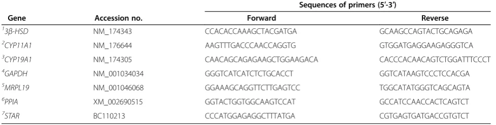
sequences are listed in Table 1. Primers were vali-dated at a concentration of 300 nmol/L using a 7-log dilution curve as previously reported [15].
A working solution of cDNA was prepared by dilut-ing 1:10 with DEPC-treated water. Five microliters of cDNA working solution was added to 20 μL master mix containing 13 μL SYBR green and fluorescein mix (Bioline, Taunton, MA, USA) and 0.75 μL of each
forward primer (10 μmol/L) and reverse primer
(10 μmol/L). Real-time PCR analysis for each sample was carried out in duplicate using a CFX real-time
PCR detection system (Bio-Rad Laboratories,
Hercules, CA, USA). Standard thermocycler condi-tions were as follows: 95°C for 10 min, followed by 40 cycles of 95°C for 15 s, 60°C for 30 s, and 72°C for 30 s. Relative fold change in target mRNAs was
quantified using the ΔΔCq method where the FSH
ΔΔCq for each animal was determined by subtracting each animals Con ΔCq from their FSH ΔCq [16]. All reverse-transcribed cDNA samples were assayed in duplicate for each gene, and melt curve analyses were performed to ensure specificity of amplification. Melt curve analysis was carried out for 81 cycles with 0.5°C temperature increase from 55°C to 95°C.
To determine the appropriate reference gene to normalize cDNA variability between samples, a panel of three reference genes was analyzed including, glyceraldehyde-3-phosphate dehydrogenase (GAPDH), cyclophilin A (PPIA), and mitochondrial ribosomal protein L19 (MRPL19). The raw Cq values were ob-tained for each gene in all samples and analyzed using GeNorm (Biogazell qbasePLUS2, Zwijnaarde, Belgium) to determine the most stable normalization factor. The most stable housekeeping gene for target
gene normalization was determined to be GAPDH
and was used as the reference gene [17].
Radioimmunoassay
Granulosa cell culture medium was analyzed for E2 and progesterone (P4) by solid-phase radioimmunoassay using components of Siemens Medical Diagnostics Corp (Los Angeles, CA, USA) commercial kits as previously described [9]. The E2concentration in samples of cell culture medium was determined in 200μL of medium and the specific bind-ing was 62.5%. Detection limit (95% of maximum bindbind-ing) of the assay was 2 pg/mL. Intra-assay CV for E2was 6.5% for cell culture medium. The P4concentration in samples of GC medium was assayed at 10μL. The specific binding was 58.8%. Detection limit (95% of maximum binding) of the assay was 0.1 ng/mL. Intra-assay CV for P4was 4.1% for cell culture medium.
Statistical analysis
Experiments were analyzed by analysis of variance for a completely randomized design in which three treatments were included; non-implanted (n = 5), Revalor 200® for 28 d (n = 5), and Revalor 200® for 84 d (n = 6). Relative fold changes in gene expression for steroidogenic acute regulatory protein (STAR), 3β-hydroxysteroid dehydro-genase (3β-HSD), P450 side chain cleavage (CYP11A1), and aromatase (CYP19A1) mRNA, and medium hormo-nal concentration of P4and E2are presented as the least square means ± standard error of the mean. For all cul-ture experiments, GC from each animal were kept separ-ate and each animal’s GC were subjected to either control treatment or FSH treatment. Thus, fold change values are each animal’s FSH response relative to that animal’s non-treated controls. A value in CYP11A1 mRNA expression of a non-implanted heifer at least three standard deviations from the mean and a missing fold change for CYP19A1 in the 84 d treatment group were excluded from statistical analysis. Quantitative real-time PCR data and hormone concentrations were
Table 1 Primer sequences used in real-time PCR
Sequences of primers (5′-3′)
Gene Accession no. Forward Reverse
13
β-HSD NM_174343 CCACACCAAAGCTACGATGA GCAAGCCAGTACTGCAGAGA
2CYP11A1 NM_176644 AAGTTTGACCCAACCAGGTG GTGGATGAGGAAGAGGGTCA
3CYP19A1 NM_174305 CAACAGCAGAGAAGCTGGAAGACA CACCCACAACAGTCTGGATTTCCCT
4GAPDH NM_001034034 GGGTCATCATCTCTGCACCT GGTCATAAGTCCCTCCACGA
5MRPL19 NM_001046068 GGAAAGCAGGTTCTTGAGTCC TGGCATATGGGTCAGCAGTA
6PPIA XM_002690515 GGTACTGGTGGCAAGTCCAT GCCATCCAACCACTCAGTCT
7STAR BC110213 CCCATGGAGAGGCTTTATGA CGTGAGTGATGACCGTGTCT
1
3β-HSD= 3β-hydroxysteroid dehydrogenase. 2
CYP11A1= P450 side chain cleavage. 3
CYP19A1= aromatase. 4
GAPDH= glyceraldehyde-3-phosphate dehydrogenase. 5
MRPL19= mitochondrial ribosomal protein L19. 6
PPIA= cyclophilin A. 7
analyzed using the GLM procedures of SAS (SAS Insti-tute, Cary, NC, USA). Data were tested for homogeneity of variance using Hartley’s F max test andSTAR and E2 were corrected by log transformation (log + 3 and log + 1, respectively). When a significant treatment effect was observed, means were separated using the least signifi-cant test computed by the predicted difference option of SAS. Statistical significance was set atP< 0.10.
Results and discussion
The anabolic effects of implants are likely a consequence of altering the endogenous hormonal milieu. This con-cept is supported by the demonstrated increase in plasma GH concentrations in response to E2 [18] or TBA and E2 [19] implants. High levels of anabolic hor-mones can also modulate reproduction as demonstrated by TBA induced anestrus in cows [8,20] and delayed pu-berty and decreased fertility in TBA plus E2 implanted heifers compared to non-implanted controls [6,21].
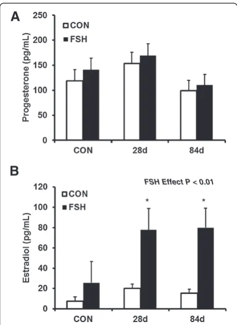
To determine the steroidogenic capacity of small fol-licle GC from an environment of elevated anabolic ste-roids, we first evaluated steroid accumulation in cell culture medium. Hormonal output was analyzed via radioimmunoassay. The ability of cultured GC to synthesize P4 was not affected by heifer implant status (P= 0.22; Figure 1A). These results are similar to previ-ous studies in which bovine GC were cultured with 200 ng/mL FSH and also did not show an effect on pro-duction of P4in cell culture medium compared to con-trols [22]. As expected, FSH treated GC had increased (P< 0.01) concentrations of E2compared to PBS-treated controls (Figure 1B) demonstrating successful induction of the FSH signaling pathway. Additionally, GC from heifers implanted for 28 d or 84 d produced greater con-centrations of E2 (78 and 80 ± 21 pg/mL, respectively) compared to non-implanted heifers (26 ± 21 pg/mL; P< 0.10; Figure 2B) in response to FSH.
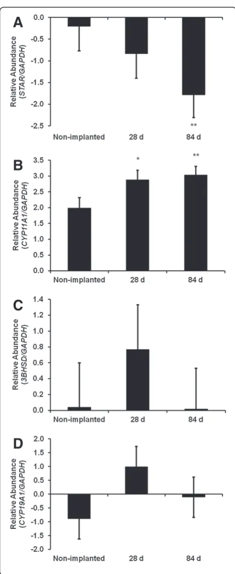
Based on the apparent change in estrogen production as a result of implant status and that estrogen produc-tion by the follicle is determined by FSH regulaproduc-tion of genes encoding key steroidogenic enzymes, we next eval-uated gene expression of the steroidogenic enzymes of non-implanted and implanted heifers in response to FSH. Analysis of steroidogenic enzyme mRNAs of pu-bertal heifers implanted with TBA and E2 in the pres-ence or abspres-ence of FSH demonstrated differpres-ences in expression as compared to non-implanted heifers. The first rate limiting step in steroid synthesis is the delivery of cholesterol to the inner mitochondrial membrane which is mediated by steroid acute regulatory protein (STAR). Expression of STAR was reduced (P< 0.05) in response to FSH in cells from heifers exposed to TBA and E2 for 84 d when compared to non-implanted heifers and heifers implanted for 28 d (Figure 2A).STAR
is fundamental to the biosynthesis of steroid hormones as it provides cholesterol to the cytochrome P450 side-chain cleavage enzyme (CYP11A1). Mitochondrial CYP11A1 catalyzes the cleavage of the cholesterol side chain to form pregnenolone and this reaction represents the first committed step in steroidogenesis. Follicle stimulating hormone increased mRNA expression of CYP11A1, in GC from 28 d (P< 0.10) and 84 d (P< 0.05)
implanted heifers as compared to non-implanted
(Figure 2B). Studies indicate that both delivery of choles-terol to the enzyme system and the expression of CYP11A1 are important factors controlling the rate of steroid hormone synthesis [23] and may contribute to
A
B
* *
FSH Effect P < 0.01
Figure 1Anabolic androgens and estrogens did not affect progesterone (P4) production but increased estradiol (E2)
concentrations.Small follicle granulosa cells from non-implanted heifers (n = 5) or heifers implanted for 28 d (n = 5) or 84 d (n = 6) were treated for 24 h with FSH (100 ng/mL, 24 h) or PBS, subse-quently cell culture medium were analyzed by RIA.(A)Combination implants of TBA and E2did not affect the ability of granulosa cells to
produce P4.(B)Medium E2concentrations were greater in cells
ex-posed to FSH (P< 0.01) compared to non-treated controls indicating successful FSH treatment. Additionally, medium E2concentrations
the increase in medium estrogen detected on d 28 and 84. In the female, ovarian androgens and estrogens regu-late release of LH and FSH by feedback mechanisms on the hypothalamus and pituitary and it is not unexpected that exposure to anabolic steroids may disrupt this deli-cate balance. Elevated concentrations of anabolic and es-trogenic steroids from the implants did not have a
marked effect on mRNA expression of 3β-HSD
(P= 0.57; Figure 2C) the enzyme responsible for convert-ing pregnenolone to progesterone. Next, we evaluated CYP19A1, the enzyme in granulosa cells responsible for converting androgens to estrogen. However, no change in gene expression was detected forCYP19A1in control versus heifers implanted for 28 or 84 d (P= 0.22; Figure 1D). This may be explained in part by the rela-tively short 3 h half-life ofCYP19A1 in FSH-stimulated bovine granulosa cells compared to the more stable 14 h half-life demonstrated forCYP11A1[24].
Additionally, although implant status did not result in significant changes in CYP19A1 mRNA expression, E2concentrations however, were elevated in both FSH treated implanted groups. This is consistent with pre-vious work in cattle showing that minimal regulation in CYP19A1 gene expression can contribute to meas-urable differences in E2 synthesis [9,25]. Normal follicular development relies on increasing concentra-tions of E2corresponding with follicle maturation and resultant proliferation and differentiation of GC [26]. Additionally, elevated levels of E2 in culture medium improves growth of oocytes from early antral follicles [27] supporting an important role of E2 in folliculo-genesis. However, elevated exogenous concentration of E2can increase the chance of developing cystic fol-licles and decreases fertility in heifers [6].
In conclusion, these results indicate that follicles which develop in the presence of high concentrations of androgenic and estrogenic steroids via an implant have an altered ability to respond to FSH stimulation as demonstrated by varied steroidogenic enzyme ex-pression and elevated estradiol production. Thus, ef-forts should be made to avoid the use of implanted heifers to study steroidogenesis in small follicle GC culture systems.
A
B
C
D
Figure 2Expression of key steroidogenic enzymes is altered in FSH stimulated granulosa cells from implanted heifers.Bovine small follicle granulosa cells were treated as described in Figure 1. Gene expression data are represented as fold change for each group compared to its respective control to demonstrate individual group response to FSH treatment. Fold change of(A)STAR,(B)CYP11A1,
Competing interests
The authors declare they have no competing interest.
Authors’contribution
ADS carried out granulosa cell isolation and cell culture including treatment and collection of cells and media. ADS conducted real-time PCR gene expression analysis. DMH carried out the radioimmunoassays. CAG and JAHG were involved in experimental design, interpretation of data and writing of this manuscript. All authors read and approved the final manuscript.
Acknowledgements
Research supported by the Oklahoma Agric. Exp. Sta., Stillwater (OKL02789). The authors would like to thank the NIDDK’s National Hormone & Peptide Program and A.F. Parlow for supplying the FSH reagent. Additionally, authors appreciate the Willard Sparks Beef Research Center personnel for overseeing care of experimental animals and lab members for assistance in granulosa cell collection.
Author details
1
Department of Animal Science, Oklahoma State University, Stillwater, OK 74078, USA.2Department of Animal and Range Science, New Mexico State University, Las Cruces, NM 88003, USA.3Department of Animal Science, 114B Animal Science Building, Oklahoma State University, Stillwater, OK 74078, USA.
Received: 17 September 2013 Accepted: 3 January 2014 Published: 7 January 2014
References
1. Johnson BJ, Anderson PT, Meiske JC, Dayton WR:Effect of a combined trenbolone acetate and estradiol implant on feedlot performance, carcass characteristics, and carcass composition of feedlot steers.J Anim Sci1996,74(2):363–371.
2. Jones SJ, Johnson RD, Calkins CR, Dikeman ME:Effects of trenbolone acetate on carcass characteristics and serum testosterone and cortisol concentrations in bulls and steers on different management and implant Schemes.J Anim Sci1991,69(4):1363–1369.
3. Cooper RA:Some aspects of the use of the growth promoter zeranol in ewe lambs retained for breeding: III. effect on plasma LH levels.Br Vet J
1985,141(4):424–426.
4. Neumann F:Pharmacological and endocrinological studies on anabolic agents.Environ Qual Saf Suppl1976,5:253–264.
5. Katzenellenbogen BS, Katzenellenbogen JA, Mordecai D:Zearalenones: characterization of the estrogenic potencies and receptor interactions of a series of fungal beta-resorcylic acid lactones.Endocrinology1979,
105(1):33–40.
6. Moran C, Prendiville DJ, Quirke JF, Roche JF:Effects of oestradiol, zeranol or trenbolone acetate implants on puberty, reproduction and fertility in heifers.J Reprod Fertil1990,89(2):527–536.
7. Manikkam M, Steckler TL, Welch KB, Inskeep EK, Padmanabhan V:Fetal programming: prenatal testosterone treatment leads to follicular persistence/luteal defects; partial restoration of ovarian function by cyclic progesterone treatment.Endocrinology2006,147(4):1997–2007. 8. Reynolds IP, Harrison LP, Mallinson CB, Harwood DJ, Heitzman RJ:The
effect of trenbolone acetate on the bovine estrous-cycle.Anim Reprod Sci
1981,4(2):107–116.
9. Castañon BI, Stapp AD, Gifford CA, Spicer LJ, Hallford DM, Gifford JAH:
Follicle-stimulating hormone regulation of estradiol production: possible involvement of WNT2 andβ-catenin in bovine granulosa cells.J Anim Sci
2012,90:3789–3797.
10. Jaiswal RS, Singh J, Adams GP:Developmental pattern of small antral follicles in the bovine ovary.Biol Reprod2004,71(4):1244–1251. 11. Xu Z, Garverick HA, Smith GW, Smith MF, Hamilton SA, Youngquist RS:
Expression of follicle-stimulating hormone and luteinizing hormone receptor messenger ribonucleic acids in bovine follicles during the first follicular wave.Biol Reprod1995,53(4):951–957.
12. Evans AC, Fortune JE:Selection of the dominant follicle in cattle occurs in the absence of differences in the expression of messenger ribonucleic acid for gonadotropin receptors.Endocrinology1997,138(7):2963–2971. 13. Bao B, Garverick HA, Smith GW, Smith MF, Salfen BE, Youngquist RS:
Changes in messenger ribonucleic acid encoding luteinizing hormone
receptor, cytochrome P450-side chain cleavage, and aromatase are associated with recruitment and selection of bovine ovarian follicles.
Biol Reprod1997,56(5):1158–1168.
14. Rozen S, Skaletsky H:Primer3 on the WWW for general users and for biologist programmers.Methods Mol Biol2000,132:365–386. 15. Gifford CA, Racicot K, Clark DS, Austin KJ, Hansen TR, Lucy MC, Davies CJ,
Ott TL:Regulation of interferon-stimulated genes in peripheral blood leukocytes in pregnant and bred, nonpregnant dairy cows.J Dairy Sci
2007,90(1):274–280.
16. Kubista M, Andrade JM, Bengtsson M, Forootan A, Jonák J, Lind K, Sindelka R, Sjöback R, Sjögreen B, Strömbom L,et al:The real-time polymerase chain reaction.Mol Aspects Med2006,27(2–3):95–125.
17. Vandesompele J, De Preter K, Pattyn F, Poppe B, Van Roy N, De Paepe A, Speleman F:Accurate normalization of real-time quantitative RT-PCR data by geometric averaging of multiple internal control genes.Genome Biol
2002,3(7). research0034.0031 - research0034.0011.
18. Rumsey TS, Elsasser TH, Kahl S:Roasted soybeans and an estrogenic growth promoter affect growth hormone status and performance of beef steers.J Nutr1996,126(11):2880–2887.
19. Hongerholt DD, Crooker BA, Wheaton JE, Carlson KM, Jorgenson DM:
Effects of a growth hormone-releasing factor analogue and an estradiol-trenbolone acetate implant on somatotropin, insulin-like growth factor I, and metabolite profiles in growing Hereford steers.J Anim Sci1992,
70(5):1439–1448.
20. Heitzman RJ, Harwood DJ:Residue levels of trenbolone and oestradiol-17beta in plasma and tissues of steers implanted with anabolic steroid preparations.Br Vet J1977,133(6):564–571.
21. Heitzman RJ, Harwood DJ, Kay RM, Little W, Mallinson CB, Reynolds IP:
Effects of implanting prepuberal dairy heifers with anabolic steroids on hormonal status, puberty and parturition.J Anim Sci1979,48(4):859–866. 22. Langhout DJ, Spicer LJ, Geisert RD:Development of a culture system for
bovine granulosa cells: effects of growth hormone, estradiol, and gonadotropins on cell proliferation, steroidogenesis, and protein synthesis.J Anim Sci1991,69(8):3321–3334.
23. Miller WL:Molecular biology of steroid hormone synthesis.Endocr Rev
1988,9(3):295–318.
24. Sahmi M, Nicola ES, Price CA:Hormonal regulation of cytochrome P450 aromatase mRNA stability in non-luteinizing bovine granulosa cells in vitro.J Endocrinol2006,190(1):107–115.
25. Luo W, Gumen A, Haughian JM, Wiltbank MC:The role of luteinizing hormone in regulating gene expression during selection of a dominant follicle in cattle.Biol Reprod2011,84(2):369–378.
26. Richards JS, Midgley AR Jr:Protein hormone action: a key to
understanding ovarian follicular and luteal cell development.Biol Reprod
1976,14(1):82–94.
27. Endo M, Kimura K, Kuwayama T, Monji Y, Iwata H:Effect of estradiol during culture of bovine oocyte–granulosa cell complexes on the mitochondrial DNA copies of oocytes and telomere length of granulosa cells.
Zygote2012:1–9.
doi:10.1186/2049-1891-5-2