STUDY OF COMPLEMENT C3 AND C4 LEVELS IN
CHRONIC OBSTRUCTIVE PULMONARY DISEASE
R. Prabhakar RaoA, S. Rama DeviB, B. Amrita SaiC
A- Professor and Head, Department of Medicine, Santhiram Medical College,Nandyal, Kurnool, Andhra Pradesh, India
B -Associate professor, Department of Pathology, Santhiram Medical College,Nandyal, Kurnool, Andhra Pradesh, India
C- Post graduate student, Department of Medicine, Santhiram Medical College, Nandyal, Kurnool, Andhra Pradesh, India
Abstract:
Chronic obstructive pulmonary disease (COPD) is a very common disease encountered in our hospitals. There are several conflicting studies which looked at the association of serum complement levels and various radiological and functional indices reflecting the severity of emphysema in patients with COPD. A few studies showed lower serum levels of complement components C3 and C4 compared to healthy subjects and this may indicate sustained compliment activation as a result of recurrent infection of respiratory tract.Some studies showed no correlation at all. We studied the same association in our hospital to find out for ourselves.
Objective: Evaluation of patients of COPD with reference to levels of complement C3 and C4 and correlate with PFT and radiological emphysema score.
Key Words: COPD, C3, C4, FEV1, FVC, PFT
Medicine
Corresponding Author
Dr. Amrita Sai,
Santhiram Medical College, Nandyal, Kurnool, Andhra Pradesh, India. Phone: +91-9390578889. E-mail: pearlamritha@gmail. com
Introduction
Chronic obstructive pulmonary disease (COPD) is defined as a disease state characterized by airflow limitation that is not fully reversible. COPD includes emphysema, an anatomically defined condition characterized by destruction and enlargement of the lung alveoli; chronic bronchitis, a clinically defined condition with chronic cough and phlegm; and small airways disease, a condition in which small bronchioles are narrowed. COPD is also a disease of increasing public health importance around the world. Estimates suggest that COPD will rise from the sixth to the third most common cause of death worldwide by 2020.1
Although a great deal is known about etio-pathogenesis of COPD, there are still significant lacunae in understanding of
the role of immunity in the part played by recurrent infections in COPD. There are several conflicting results in studies which looked at the association between serum complement levels and various radiological and functional indices reflecting severity of emphysema in patients with COPD.
Patients with COPD have been shown to exhibit lower serum levels of complement components C3 and C4 than healthy subjects, and this may indicate sustained complement activation as a result of recurrent respiratory tract infections.2
destruction. There have been studies which demonstrate that COPD patients with lower levels of C4 are those who experience respiratory infections and tend to have more radiological signs of Emphysema and have a predominant small airway resistance.
However, a small study from Turkey and a few other studies elsewhere found no correlation between the level of complement and severity of COPD indicating lack of clear cut knowledge about the complement role in COPD.3 The dichotomy in the results of various studies indicates the present gap in the knowledge about the role of complement in COPD. In view of very few studies carried out in our population regarding this matter hence we took up this study.
Methods
This was hospital-based, prospective, observational, and comparative study of COPD patients with emphysema during last 2year from October 2012 to September 2014.
Thirty five patients were included in study between age group of 40 and 70 years. Exclusion criteria are patients with immunological disorders that might interfere with compliment activation, patient’s unable to perform pulmonary function tests PFT, pregnancy, and bronchial asthma. PFT was done by Spiroback-G machine. Global Initiative for Chronic Obstructive Lung Disease’s criteria was used to stage the severity of COPD. Blood samples were collected in ethylenediaminetetraacetic impregnated Vacutainer and stored at 2o
C until they could be transported and analyzed for C3 and C4 by immunoturbidity method in a lab accredited with NABL.
Cases were divided into three groups based on PFT-Mild (forced expiratory volume in 1 s [FEV1]/forced vital capacity FVC<0.70 with FEV180% normal), moderate (FEV1/FVC <0.70 with FEV1 50-79% normal), severe (FEV1/FVC <0.70with FEV130-49% normal). Based on emphysema score0-6, 7-10, and >10. Controls were divided into different equal number of groups to match with subject
significant if P< 0.05. Statistical analysis was performed by SPSS version 18.0 for Microsoft Windows. Pearson’s correlation co-efficient was used for bivariate correlations.
Observations
This was a prospective study for a period of 2-year (October 2012- September 2014) comprising 35 patients with COPD admitted at Santhiram Medical College and General Hospital.
Out of the total 70 subjects, 35 were cases (50%) and 35 were controls (50%)[Table1 and Graph1].
Gender distribution of cases
Out of the total 35 patients with COPD, 20 patients (57%) were males and 15 patients (43%) were females [Table 2 and Graph2].
Cases were predominantly seen in age groups 61-65 (28%) and 66-70 (22%) years [Table 3 and Graph 3].
Total numbers of controls were 35, in which males were 18 (51%) and females were 17 (49%) [Table 4 and Graph 4].
Comparison of cases with different degrees of ob-struction based on PFTs with controls
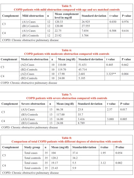
There was no statistically significant difference (P= 0.97) observed in complement C3 levels and complement C4 levels (P=0.61) between COPD patients with mild PFT obstruction compared to age and sex matched controls [Table 5 and Graph 5].
with mild PFT obstruction (P=0.97) and moderate PFT obstruction (P=0.662) compared to controls and statistical significance (P=0.017) observed among patients with severe PFT obstruction compared to age and sex matched controls for complement C3 [Graph 8].
There was no statistical significance observed among cases with mild PFT obstruction (P=0.616) and statistical significance observed among patients with moderate PFT obstruction (P=0.004) and severe PFT obstruction (P=0.005) compared to age and sex matched controls for complement C4 [Graph 9].
When total COPD patients with different degree of obstruction were compared with controls though there was no statistically significant difference (P=0.054) in serum C3 levels, but there was significant difference in mean C3 values observed and there was statistically significant difference (P=0.002) observed in serum C4 levels [Table 8].
Comparison of cases with controls based on emphy-sema scores
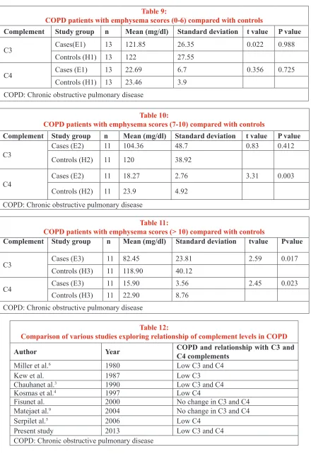
When COPD patients with emphysema score (0-6) were compared with age and sex matched controls, there was no significant difference between serum C3 (P=0.988), C4 levels (P=0.725) observed [Table 9 and Graph10].
When COPD patients with emphysema score (7-10) were compared with age and sex matched controls; there was no statistically significant difference in C3 levels (P=0.412), but there was significant difference in mean vales observed; and there was statistically significant difference (P=0.003) present in serum C4 levels [Table 10 and Graph 11].
When COPD patients with emphysema score (>10) were compared with age and sex matched controls, there was statistically significant difference in both C3 (P=0.017), C4 levels (P=0.023) observed [Table 11 and Graph 12].
There was no statistical significance (P=0.988) observed among cases with emphysema score (0-6) and emphysema score 7-10 (P=0.412) compared to controls, and statistical significance observed among patients with emphysema score (>10) compared (P=0.017) to controls for complement C3 [Graph 13].
There was no statistical significance (P=0.725) observed among cases with emphysema score (0-6) and statistical significance observed among patients with emphysema score 7-10 (P=0.003) and for patients with emphysema score (>10) (P=0.023) compared to controls for complement C4 [Graph 14].
Table 1:
Demographic characteristics of the study population
Total number of subjects Number
Cases 35
Controls 35
Table 2:
Gender distribution of cases Gender distribution of cases N (%)
Males 20 (57)
Females 15 (43)
Table 3:
Age distribution of cases (n=35) Age distribution of
cases (in years) patients (n) PercentageNumber of
40-45 2 5
46-50 3 8
51-55 5 14
56-60 7 20
61-65 10 28
66-70 8 22
Table 4:
Gender distribution of controls (n=35) Gender distribution
of controls subjects (n) PercentageNumber of
Males 18 51
Table 5:
COPD patients with mild obstruction compared with age and sex matched controls
Complement Mild obstruction n Mean complement level in mg/dl Standard deviation t value P value
C3 (A1) Cases(B1) Controls 1212 120.33120.00 26.92527.555 0.030 0.976
C4 (A1) Cases 12 22.75 7.034 0.508 0.616
(B1) Controls 12 23.92 3.704
COPD: Chronic obstructive pulmonary disease
Table 6:
COPD patients with moderate obstruction compared with controls
Complement Moderate obstruction n Mean (mg/dl) Standard deviation t value P value
C3 (A2) Cases 10 110.00 51.631 0.445 0.662
(B2) Controls 10 119.70 45.714
C4 (A2) Cases(B2) Controls 10 17.9010 24.00 2.6015.185 3.325** 0.004
COPD: Chronic obstructive pulmonary disease
Table 7:
COPD patients with severe obstruction compared with controls
Complement Severe obstruction n Mean (mg/dl) Standard deviation t value P value
C3 (A3) Cases 13 86.38 23.8 2.57 0.017
(B3) Controls 13 117.00 35.7
C4 (A3) Cases (B3) Controls 1313 16.0024.08 3.4168.789 3.088 0.005 COPD: Chronic obstructive pulmonary disease
Table 8:
Comparison of total COPD patients with different degrees of obstruction with controls
Complement Study group n Mean (mg/dl) Standarddeviation tvalue Pvalue
C3 Total cases 35 104 37.4 1.95 0.054
Total controls 35 120.2 34.2
C4 Total cases 35 19.17 5.5 3.12 0.002
Table 9:
COPD patients with emphysema scores (0-6) compared with controls
Complement Study group n Mean (mg/dl) Standard deviation t value P value
C3 Cases(E1) 13 121.85 26.35 0.022 0.988
Controls (H1) 13 122 27.55
C4 Cases (E1) 13 22.69 6.7 0.356 0.725
Controls (H1) 13 23.46 3.9 COPD: Chronic obstructive pulmonary disease
Table 10:
COPD patients with emphysema scores (7-10) compared with controls
Complement Study group n Mean (mg/dl) Standard deviation t value P value
C3 Cases (E2)Controls (H2) 1111 104.36120 48.738.92 0.83 0.412
C4 Cases (E2) 11 18.27 2.76 3.31 0.003
Controls (H2) 11 23.9 4.92
COPD: Chronic obstructive pulmonary disease
Table 11:
COPD patients with emphysema scores (> 10) compared with controls
Complement Study group n Mean (mg/dl) Standard deviation tvalue Pvalue
C3 Cases (E3) 11 82.45 23.81 2.59 0.017
Controls (H3) 11 118.90 40.12
C4 Cases (E3) 11 15.90 3.56 2.45 0.023
Controls (H3) 11 22.90 8.76 COPD: Chronic obstructive pulmonary disease
Table 12:
Comparison of various studies exploring relationship of complement levels in COPD
Author Year COPD and relationship with C3 and C4 complements
Miller et al.6 1980 Low C3 and C4
Kew et al. 1987 Low C3
Chauhanet al.3 1990 Low C3 and C4
Kosmas et al.4 1997 Low C4
Fisunet al. 2000 No change in C3 and C4
Matejaet al.9 2004 No change in C3 and C4
Serpilet al.5 2006 Low C4
Present study 2013 Low C3 and C4
Graph Legends
Graph 1: Study subjects (n=7)
Graph 2: Gender distribution of case (n = 35)
Graph 3: Age distribution of cases
Graph 4: Gender distribution of controls (n = 35)
Graph 6:Complementlevels C3 and C4 among cases with moderate pulmonary function tests obstruction
compared to age and sex matched controls
Graph 7: Complement levels C3 and C4 among cases with severe pulmonary function tests obstruction
compared to age and sex matched controls
Graph 8: Complement C3 levels among cases compared to age and sex matched controls
Graph 10: Complement levels C3 and C4 among cases with emphysema score (0-6) compared to age and sex
matched controls
Graph 11: Complement levels C3 and C4 among cases with emphysema score (7-10) compared to age and sex
matched controls
Graph 12: Complement levels C3 and C4 among cases with emphysema score (>10) compared to age and sex
matched controls
Graph 13: Complement levels of C3 among cases compared to age and sex matched controls
Graph 14: Complement levels
Discussion
In the present study, out of 35 cases 57% (n=20) were males and 43% (n=15) were females. The male to female sex ratio was 1.3:1. This was in accordance with study done by Jindal et al.1
In the present study, the maximum number of cases of COPD and exacerbations (60%) (n=21) were admitted during rainy and winter seasons (July-December). A similar trend was seen in a study done by Jenkins et al.4 Factors potentially contributing to this include increased exposure to viral infections, increased host susceptibility; greater time spent indoors, reduced physical activity, and temperature-related reduction in lung function.
In the present study, subjects with mild, moderate and severe PFT obstruction were compared with the age and sex matched controls. In patients with mild PFT obstruction, mean values of complements C3 and C4 were calculated for both cases and controls. There was no significant difference observed in complement C3, C4 levels between COPD patients with mild PFT obstruction and healthy age and sex matched controls.
controls, and we also observedstatistical significance for both C3 (P=0.017) and C4 (P=0.005). Similar observations were made by Chauhan et al.5 where they observed both serum C3 (IU) and C4 (IU) were lower in COPD patients (C3 = 95.9 ± 33.11, C4 = 113.6 ± 62.4) than in control (C3 = 167.3 ± 25.42, C4 = 205 ± 76.5; P<0.05).
In other studies done by Mateja et al.6 and Fishnet al.,7 there
was no significant difference observed in C3 and C4 levels between patients with COPD and healthy control subjects which was in contrast to our study.
When total COPD patients with different degree of obstruction were compared with controls though there was no statistically significant difference in serum C3 levels (P=0.054), there was significant difference in mean C3 values were observed, and there was statistically significant difference observed for serum C4 levels (P=0.002).
Similar observations were noticed by Kosmas et al.8 and Serpiletal.9
In this study, COPD patients with emphysema score 0-6 (E1) were compared with age and sex matched controls for complement levels C3 and C4. When COPD patients with emphysema score (0-6) were compared with healthy sex and age matched controls, there was no significant difference between serum C3, C4 levels observed.
When COPD patients with emphysema score (7-10) were compared with controls, although significant difference observed in mean C3 values, these values were statistically not significant (P=0.412). However, statistical significant difference (P=0.003) observed in C4 values.
When COPD patients with emphysema score (>10) were compared with controls, statistical significance observed in both C3 (P=0.017), C4 (P=0.023) levels, similar observations were made by Burki and Krumpelman10and
Chugh et al.11 in their studies.
Findings arising from this study showed that for patients with COPD, particularly moderate to severe degree COPD
Quantitatively, C3 and C4 comprise approximately two thirds of the complement system. The classic complement pathway is activated by either antibody-coated targets such as microorganisms or antigen-antibody complexes, while the alternative complement pathway is activated directly by bacterial polysaccharides.
Probable reason for lower serum levels of complement components C3 and C4 in COPD patients compared to healthy controls could be because of the following, sustained complement activation secondary to repeated respiratory infections leading to influx of inflammatory cells into the lung parenchyma with subsequent release of elastases and oxidants that cause damage to elastic lung tissue. This leads to the logical inference that there might be a quantitative relationship between complement consumption and degree of elastic tissue destruction.
Limitations of the study
There are certain limitations in our study, which we would like to put on record.
1. Although more number of patients were enrolled in this study compared to other similar studies, even more cases could have been taken
2. We could have used CT scan to measure the emphysema score instead of chest X-ray, although chest X-ray was also a well-known modality to know the lung destruction.
Strengths of the study
However, it is hereby restated that this study has several strengths, mentioned as follows:
Summary
The 35 patients with COPD were studied during the period October 2011-September 2013.
The majority of COPD patients were males comprising of 57% (n=20) and females 43% (n=15) and majority of patients (n=18) were above 60 years (50%), 44% (n=15) in between 45 and 60 years age group and only 5% of patients (n=2) in <45 years age group. The maximum number of cases (n=21) of COPD and exacerbations (60%) were seen during rainy and winter seasons (July-December).
The 35 patients with COPD and 35 controls were studied to know the relation between complement components C3, C4 levels and degree of obstruction and lung damage for this we classified cases in to three groups based on degree of airway obstruction and lung destruction with the help of PFTs and emphysema score.
Mean complement C3, C4 levels of the cases were compared with age and sex matched controls.
The observations made were
1. Mean complement component C3, C4 levels were significantly low in patients with moderate to severe PFT obstruction and moderate (7-10) to higher emphysema scores (>10) compared to controls(Table 12).
2. In cases with mild obstruction and low emphysema score (0-6) there was no significant difference observed.
3. It was observed that there was significant decrease in complement C3,C4 levels with increase in severity of obstruction and lung destruction.
Probable explanation for the lower serum levels of complement components C3 and C4 in COPD patients compared to healthy controls could be as follows: Sustained complement activation secondary to repeated respiratory infection leading to influx of inflammatory cells into the lung parenchyma with subsequent release of elastases and oxidants that cause damage to elastic lung tissue, this leads to the inference that there might be a quantitative relationship between complement consumption and degree
of elastic tissue destruction.
Hence, it is postulated that the severity of lung damage and morbidity are related to lower complement C3, C4 levels. These complement levels can be used as marker of severity of COPD. Monitoring of complement C3, C4 levels may help in assessment of severity and progression of COPD along with PFTs and emphysema score.
Conclusions
The analysis of results of the present study leads to the following conclusions:
There was a direct correlation between the severity of COPD as estimated by radiological emphysema score and PFTs with serum complement levels. That is lower levels of complements being seen in progressive severity of COPD. This is in agreement with postulates of other studies, that serum complements levels may well serve as marker for COPD severity, however further large scale studies needed in this regard.
References
1. John. J. Reilly et al, Chronic Obstructive Pulmonary Disease, Harrison’s principles of Internal Medicine, 19th Ed, Chapter 314, page-1700.
2. Miller RD, Kueppers F, Offord KP. Serum concentrations of C3 and C4 of the complement system in patients with chronic obstructive pulmonary disease. J Lab Clin Med 1980;95:266-71.
3. Marc MM, Korosec P, Kosnik M, Kern I, Flezar M, Suskovic S, et al. Complement factors c3a, c4a, and c5a in chronic obstructive pulmonary disease and asthma. Am J Respir Cell MolBiol 2004;31:216-9.
4. Jindal SK, Aggarwal AN, Gupta D, Agarwal R, Kumar R, Kaur T, et al. Indian study on epidemiology of asthma, respiratory symptoms and chronic bronchitis in adults (INSEARCH). Int J Tuberc Lung Dis 2012;16:1270-7.
levels in chronic obstructive pulmonary disease. Indian J Med Res 1990;92:241-5.
6. Karadad F, Polatli M, Cyldad O, AydinN. Serumcomplement and immunoglobulin levels in chronic obstructive pulmonary disease. Adnan Menderes UnivSchMed2000;1:13-6.
7. Hoidal JR, McCusker KT, Marshall BC, Rao NV. Oxidative damage and COPD. In: Cherniack NS, editor. Chronic Obstructive Pulmonary Disease. Philadelphia: WB Saunders; 1991. p. 44-9.
8. Kosmas EN, Zorpidou D, Vassilareas V, Roussou T, Michaelides S. Decreased C4 complement component serum levels correlate with the degree of emphysema in patients with chronic bronchitis. Chest 1997;112:341-7.
9. Serpil P, GulruP,Demir A, Büyükşirin M, Kömürcüoğlu B, Demirci F, et al. Evaluation of the results of lung function tests and COPD risk factors in COPD Tent. Turk Thorac J 2006;7:23-8.
10. Burki NK, Krumpelman JL. Correlation of pulmonary function with the chest roentgenogram in chronic airway obstruction. Am Rev Respir Dis 1980;121:217-23.