Research Article
Identification of species-specific
molecular markers in different farm
animals by PCR-RFLP analysis
Waqas Ahmad Khan
1*, Hamid Mustafa
2, Umara Amir-u-Din
1,
Maimoona Yousaf
1, Adeela Ajmal
2, Khalid Mehmood
1and Muhammad
Imran
31. Department of Biotechnology, Faculty of Science, University of Sargodha, Sargodha-Pakistan
2. Department of Livestock Production, University of Veterinary and Animal Sciences, Ravi Campus, Pattoki-Pakistan
3. Institute of Biochemistry and Biotechnology, University of Veterinary and Animal Sciences, Lahore-Pakistan *Corresponding author’s email: [email protected]
Citation
Waqas Ahmad Khan, Hamid Mustafa, Umara Amir-u-Din, Maimoona Yousaf, Adeela Ajmal, Khalid Mehmood and Muhammad Imran. Identification of species-specific molecular markers in different farm animals by PCR-RFLP analysis. Pure and Applied Biology. Vol. 7, Issue 1, pp338-342. http://dx.doi.org/10.19045/bspab.2018.70041 Received: 03/01/2018 Revised: 18/02/2018 Accepted: 20/02/2018 Online First: 21/02/2018
Abstract
Meat verification is relevant for public health and economic concerns. The present study identified species origin of raw meat samples of buffalo, cow, sheep, goat and chicken using Polymerase Chain Reaction-Restriction Fragment Length Polymorphism (PCR-RFLP) of a mitochondrial cytochrome b gene (359 bp). The amplified PCR products were digested using restriction enzymes
Tas1 and Hinf1. DNA fragments of different lengths cleaved by two different enzymes were obtained. Each animal species had a distinctive combination of restriction fragments. PCR-RFLP analysis was performed by resolving digested products through agarose gel electrophoresis. Lengths of most of DNA fragments obtained for different farm animals were same as expected with few exceptions. PCR fragment obtained for buffalo remained uncut by enzyme HinfI. In short, PCR-RFLP method was successfully applied to detect the origin of meat species. This method is simple, quick and reliable for differentiation of meat species. The restriction patterns of cytochrome b are helpful in the discrimination of meat identification in a blind fashion.
Keywords: DNA; Enzymes; Meat; PCR-RFLP
Introduction
Meat adulteration is a common practice during preparation of meat products in many countries and causes serious health, economic and religious concerns [1]. The identification of meat adulteration in meat products is very important for the implementation of labeling legislation and unfair competition prevention [2]. Different
high cost and inadequacy to differentiate closely related species [4]. Different molecular approaches have been developed to identify different meat species. These methods can reduce the shortcomings of conventional methods [5]. These molecular approaches include PCR, RAPD, AFLP, DNA hybridization and RFLP [6, 7, 8, 9]. Polymerase chain reaction based methods are very fast and reliable, and now they have become popular for meat identification in meat industry [2]. Hebert et al. [10] proposed PCR based method for the analysis of sequence diversity of cytochrome c oxidase subunits 1 (CO1). This study presents the identification of species origin of raw meat using PCR-RFLP.
Materials and methods
Sample collection and DNA extraction Samples of both meat and blood were collected from different commercial sources. Species selected for meat detection were Bos indicus (Cattle), Bubalus bubalis (Buffalo),
Ovis aries (Sheep), Capra hircus (Goat), and
Gallus gallus domesticus (Chicken). About 200 μL blood taken from each species was poured into separate vials having 0.5 M EDTA with pH 8.0 and stored at -20 °C for DNA extraction. Meat samples of the concerned species were stored in cryo vials at -80 °C for DNA extraction. DNA was extracted from blood samples by applying Phenol-Chloroform organic method as described by Sambrook et al. [11]. We followed the procedure of Koh et al. [12] to purify DNA from meat and meat mixtures. Gene quant spectrophotometer UK was used to determine the accuracy and purity of the extracted DNA. Absorbance values of 260/280 OD are indicators of purity of DNA. A ratio between 1.7 and 1.9 indicates a very high purity of DNA.
Primers design and synthesis
The common primers were used CYTb1 (5’-CCATCCAACATCTCAGCATGATGAAA -3’) and CYTb2
(5’-GCCCCTC-AGAATGATATTTGTCCTCA-3’) as presented earlier by Carr and Marshall [13]. These primers amplified a 359-bp DNA fragment. These primers were designed from the previously developed data of Mitochondrial Cytochrome b genes of Cow, Buffalo, Sheep, Goat and Chicken [14].
PCR optimization
A precise region of the DNA fiber (target DNA) is amplified by PCR. Amplification of the target DNA (Mitochondrial Cytochrome B gene) was carried out for each species with the help of forward and reverse primers. Each tube contained 2 µL of target DNA, 2.5 µL of 10× PCR buffer, 2.5 µL of MgCl2, 2 µL of deoxynucleotide triphosphate (dNTPs), 0.25 µL of Taq DNA polymerase and 0.5 µL of each primer (forward and reverse primer). Initial denaturation was carried out at 95 °C for 5 minutes followed by 35 cycles of denaturation 94 °C for 30 seconds, annealing of primers at 45 °C for 30 seconds and extension at 72 °C 45 seconds. A final extension step was given at 72 °C for 5 minutes followed by hold at 4 °C for infinity.
RFLP analysis
Products of amplification obtained from PCR were digested with the help of restriction enzymes. Restriction enzymes used were
Tas1 and Hinf1; 30 µL of reaction mixture was prepared by adding 10 ul amplified PCR product, 1 ul of each restriction enzyme, 2 µL buffer and adjustable amount of sterile distilled water. The mixture was allowed to incubate at 37°C for 5 hours. After incubation the digested products were analyzed on agarose gel electrophoresis.
Results
Analysis of PCR products
Different bands of DNA were seen under the UV light. DNA bands for five different species digested by two different enzymes
RFLP analysis
Cytochrome b gene amplicons were digested by two restriction enzymes TasI and HinfI.
Both enzymes cut the gene at different points.
Thus, DNA fragments of various sizes were produced by the two enzymes. The detail of RFLP of five species is given below and can also be seen in (Table 1 & Figure 1).
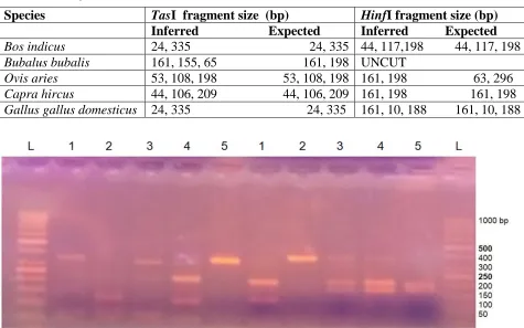
Table 1. Expected and inferred Cytochrome b gene fragment sizes produced with two restriction enzymes
Figure 1. Restriction profiles of Mitochondrial Cytochrome b gene amplicon with TasI and
HinfI restriction enzymes in the five Species. L: GeneRuler 50 bp DNA Ladder, 1: Cow, 2:
Buffalo, 3: Sheep, 4: Goat, 5: Chicken
Bos indicus (Cow)
TasI: Major fragment of 334 bp and a minor fragment of 24 bp were produced. HinfI: Three fragments were produced; major ones with 198 bp and 117 bp and a minor one with 44 bp.
Bubalus bubalis (Buffalo)
TasI: Three fragments of 161, 155 and 65 bp were generated. HinfI: DNA remained uncut.
Ovis aries (Sheep)
TasI: Major fragment produced was 198 bp while other two fragments were of 108 and 53 bp. HinfI: Two pieces of 198 bp and 161
bp were obtained.
Capra hircus (Goat)
TasI: The larger fragment produced was 209 bp while second one was of 106 bp and the third one was of 44 bp. HinfI: Two pieces of DNA having 198 bp and 161 bp were produced.
Gallus gallus domesticus (Chicken)
TasI: The larger fragment produced was of 335 bp while small one was only of 24 bp.
HinfI: Two fragments of moderate length 188 bp and 161 bp were generated and a very small one having only 10 bp was obtained.
Species TasI fragment size (bp) HinfI fragment size (bp)
Inferred Expected Inferred Expected
Bos indicus 24, 335 24, 335 44, 117,198 44, 117, 198
Bubalus bubalis 161, 155, 65 161, 198 UNCUT
Ovis aries 53, 108, 198 53, 108, 198 161, 198 63, 296
Capra hircus 44, 106, 209 44, 106, 209 161, 198 161, 198
Discussion
Detection of meat and meat products is crucial due to health, religious, ethical and economic reasons. A number of methods have been employed to distinguish meat of one specie from the other, which include immunodiffusion, immunoelectrophoresis, isoelectric focusing and DNA hybridization
[15]. In meat processing plants it is more likely that meat of one species is mixed with other species during processing by grinders, cutters, knives, choppers or by other ways. In such cases samples should be homogenized and multiple samples should be taken. PCR-RLFP technique is so sensitive that it can detect traces of meat mixtures [9]. As such violations occur unintended and at minor level, a number of precautions should be considered when interpreting the data of PCR products. Therefore in the present study PCR-RFLP method was applied in detection of origin of meat species. These results might be useful to detect adulterated meat products and wrong labeling. This method is also equally applied to find meat species in sea foods. Mitochondrial cytochrome b gene is an excellent molecular marker for identification of species as it is highly conserved and provides successful PCR amplification results. Moreover, Mitochondrial DNA is in abundance in cell nucleic acids. DNA was extracted from both blood and tissues of five species. 359 bp of Cytochrome b gene was successfully amplified with the help of two universal primers CYTb1 and CYTb2 [13]. These universal primers have been successfully used to amplify Cytochrome b gene of a range of vertebrate species which also helps to find evolutionary tendencies among vertebrates. Universal primers also work as an internal control and estimate the DNA amplification. Due to high copy number of Cytochrome b gene (1000 copies approximately in each cell) its success rate in
PCR amplification is much higher than the low copy number of nuclear DNA.
Conclusion
The present study reveals the authenticity of species identification by the amplification of mitochondrial Cytochrome b gene followed by restriction fragment length polymorphism. 359 bp Cytochrome b gene region of each species was amplified by PCR. Then it was cut with two restriction enzymes and agarose gel electrophoresis was performed. Each animal species had a distinctive combination of restriction fragments. Inferred and expected values for fragment lengths obtained by the digestion with TasI restriction enzyme were same for all of the five species under investigation except for one species Bubalus bubalis (Buffalo) where expected fragments were of 161 and 198 bp while three fragments of 161, 155 and 65 bp were obtained. It was noticeable that the buffalo gene remained un-cleaved by the restriction enzyme HinfI. Another controversy in the inferred and expected values of fragment lengths was observed in case of Ovis aries (Sheep) DNA fragment treated by the restriction enzyme HinfI. Expected fragment lengths were 63, 296 bp whereas two fragments of 161 and 198 bp were obtained. The results show that PCR-RLFP is a simple, quick and reliable method for verification of species. This method is equally applicable for animal breeding and protection of biodiversity.
Authors’ contributions
Conceived and designed the experiments: H Mustafa, WA Khan & M Imran, Performed the experiments: K Mehmood, WA Khan & M Imran, Analyzed the data: H Mustafa, WA Khan & M Imran, Contributed materials/ analysis/ tools: M Yousaf & UA Udeen, Wrote the paper: H Mustafa, M Imran & A Ajmal.
References
`Identification of 12 animal species meat by T-RFLP on the 12S rRNA gene. Meat Sci 85: 265–269.
2. Kesmen Z, Yetim H & Şahin, F (2010). Identification of different meat species used in sucuk production by PCR assay.
GIDA 35: 81–87.
3. Haider N, Imad N & Bassam AS (2012). Identification of meat species by PCR-RFLP of the mitochondrial COI gene.
Meat Sci 90: 490-493.
4. Abdel-Rahman SM, El-Saadani MA, Ashry KM & Haggag, AS (2009). Detection of Adulteration and Identification of Cat's, Dog's, Donkey's and Horse's Meat Using Species-Specific PCR and PCR-RFLP Techniques. Australian J Basic App Sci
3: 1716–1719.
5. Girish PS, Anjaneyulu ASR, Viswas KN, Shivakumar BM, Anand M & Patel M (2005). Meat species identification by polymerase chain reaction-restriction fragment length polymorphism (PCR-RFLP) of mitochondrial 12S rRNA gene. Meat Sci 70: 107–112.
6. Rodriguez MA, Gracia T, Gonalez I, Asensio L, Fernandiz A & Lobo E (1991). Identification of goose (Anser) and mule duck (Anas platyrhynchos x Carina moschata) foegras by multiplex polymerase chain reaction amplification of the 5S rDNA gene. J Agri Food Chem
49: 2717–2721.
7. Arslan A, Ilhak I, Calicioglu M & Karahan M (2005). Identification of meats using random amplified polymorphic DNA (RAPD) technique. J Musc Foods 16: 37–45.
8. Alves E, Castellanos C, Ovilo C, Silio L & Rodriguez C (2002). Differentiation of the raw material of the Iberian pig meat industry based on the use of
amplified fragment length polymorphism. Meat Sci 61: 157–162. 9. Chikuni K, Ozutsume K, Hoishi Kawa T
& Kato S (1990). Species identification of cooked meats by DNA hybridization assay. Meat Sci 27:119–128.
10. Hebert PDN, Cywinska A, Ball SL deWaard JR (2003). Biological identifications through DNA barcodes.
Pro. Royal Soc. London – Ser. B: Bio Sci
270:313–322.
11. Sambrook J & Russell DW (2001). Molecular cloning: A laboratory manual 3rd edition Cold Spring Harbour Laboratory Press, Cold Spring Harbour, New York.
12. Koh MC, Lim CH, Chua SB, Chew ST & Phang STW (1998). Random amplified polymorphic DNA (RAPD) fingerprints for identification of red meat animal species. Meat Sci 48: 275-28 13. Carr SM & Marshall HD (1991).
Detection of interspecies DNA sequence variation in the mitochondrial cytochrome b gene of Atlantic cod (Gadus morhua) by the polymerase chain reaction. Canadian J Fish Aqu Sci
48: 48-52.
14. Anderson AT, Bankier BG, Barrell MHL, DeBruijn A, Coulson J, Drouin IC, Eperon DP, Nierlich BA, Roe F, Sanger PH, Schreier AJH, Smith R & Staden IG (1981). Sequence and organization of the human mitochondrial genome. Young Nat 290: 457-465. 15. Winters AK, Thomsen PD & Davies W
(1990). A comparison of DNA-hybridization immunodiffusion, countercurrent immunoelectrophoresis and isoelectric focusing for detecting the admixture of pork to beef.