_____________________________________________________________________________________________________
www.sciencedomain.org
Effect of HbF Level among Different Severity of
Sickle Cell Disease
Ream Elzain Abdelgadir
1, Mawada Abdelsalam
1and Abdel Rahim Mahmoud Muddathir
2,3*1
Department of Hematology, Faculty of Medicine and Health Sciences, Kordofan University, Sudan.
2
Department of Clinical Laboratory Sciences, Faculty of Applied Medical Sciences, Taibah University, Medina, KSA.
3
Department of Hematology and Blood Transfusion, Faculty of Medical Laboratory Sciences, Alzaeim Alazhari University, Khartoum, Sudan.
Authors’ contributions
This work was carried out in collaboration between all authors. Authors REA and ARMM designed the study, performed the statistical analysis, wrote the protocol and wrote the first draft of the manuscript. Author MA managed the analyses of the study. Author REA managed the literature searches. All authors read and approved the final manuscript.
Article Information
DOI: 10.9734/IBRR/2017/34020 Editor(s): (1) Dr. Dharmesh Chandra Sharma, Incharge Blood Component & Aphaeresis Unit,
G. R. Medical College, Gwalior, India. Reviewers: (1) Kallol Kumar Bhattacharyya, Imambara Sadar Hospital, India. (2)Omesh Kumar Bharti, State Institute of Health and Family Welfare, India. Complete Peer review History:http://www.sciencedomain.org/review-history/19486
Received 9th May 2017 Accepted 9th June 2017 Published 12th June 2017
ABSTRACT
Background: Fetal hemoglobin (HbF) can inhibit the deoxygenation induced polymerization of sickle hemoglobin (HbS) that drives the Pathophysiology of sickle cell disease. The aim of this study was to determine fetal Hb level in Sudanese sickle cell disease patients as well as to find out the effect of fetal hemoglobin level on different severity groups.
Materials and Methods: This was descriptive cross sectional study included 100 Patients with sickle cell disease diagnosed by Positive sickling test and Hemoglobin electrophoresis. The Patients were attended Sudan sickle cell anemia center (SSCAC), Elobied-Sudan during September 2015 – July 2016. Clinical history was obtained to perform the severity of the disease according to Hedo et al. scoring. Fetal hemoglobin was estimated by Betke's method. Data were analyzed using SPSS software computer program version 21.
Results: The mean of HbF level among the studied population was 7.6%. The descriptive analysis showed that, the mean level of HbF in 38 (38%) patients with mild disease was 7.7%, while in 54 (54%) patients with moderate disease the mean level of HbF was 7.6% and the last 8(8%) patients with severe disease showed HbF level 7%. There was no statistical significant differences observed when HbF level was less than 10% (P value = 0.146), while the statistical significant differences was observed among patients with HbF level more than10% (P value = 0.03).
Conclusion: The study concluded that Hb F level has no effect in severity of the disease among studied sickle cell patients, unless HbF level more than 10%.
Keywords: Sickle cell disease; hemoglobin F; disease severity.
1. INTRODUCTION
Sickle cell Anemia (SCA) is an autosomal recessive genetic disease that results from the substitution of valine for glutamic acid at position 6 of the β-globin chain, leading to production of hemoglobin S (HbS) [1]. SCA is the most common genetic disorder among people of African descent [2]. In Sudan, SCA is distributed widely in the western region with high prevalence among Messeryia tribe (one of the Afro-Asiatic tribes) [3].
The clinical state of those patients is exceptionally variable, which may be influenced by different factors, such as the haplotypes of the
βS-globin gene, the presence of alpha-thalassemia, and the fetal hemoglobin level (Hb F level) [4]. Fetal hemoglobin (HbF) plays a dominant role in ameliorating morbidity and mortality of Hemoglobinopathies [5]. HbF level and its distribution among sickle patients are highly variable [6], Different factors can affect HbF level among SCA patients such as genetic, social, environmental and nutritional factors. Among patients with sickle cell anemia, HbF concentrations vary from 0.1% to 30% with an average of about 8% [7]. High HbF levels may reduce SCA severity due to its ability to inhibit HbS polymerization and also reduce the mean corpuscular HbS concentration [8].
It was been demonstrated that some chemical agents such as placental gonadotropin, progesterone, Azacitidine, Milrinone, erythropoietin, arginine butyrate, phenylbutyrate and hydroxyurea rise hemoglobin level and motivate HbF formation in SCA patients reducing the severity and frequency of SCD episodes [9]. Previous study reported that, HbF level can predict the effect of hydroxyurea treatment. In subjects who start with baseline HbF values between 5 and 10% increases can be two- to three-fold, while subjects with very low baseline HbF can have 10-fold increases post treatment
[10]. So estimation of HbF levels may helps to devise better therapeutics to induce HbF expression and help clinician to decide the appropriate doses of treatment. On the other hand, determination of Hb F level has a role in severity of the disease, which can helps in constructing a prognostic model. To our knowledge no previous studies was determined HbF level among Sudanese sickle cell patients. The present study was aimed to determine the HbF level among Sudanese sickle cell patients and its effect in different severity condition.
2. MATERIALS AND METHODS
The study was conducted in 100 sickle cell patients (58% male, 42% female), their age ranged between 1 year and 16 -years- old. All patients were attended to Sudan sickle cell anemia center (SSCAC), El-Obied-Sudan, during September 2015- July 2016. The study had been approved from the ethical committee of the center, in addition to a written informed consent was obtained from all patients or their parents/caregiver before sample collection.
Patients received blood transfusion within the last 3 months or admitted to the hospital with SCD crisis, as well as Patients under treatment of any agents which increase the HbF level such as Hydroxyurea were excluded.
localized. The scoring was classified the patients into mild, moderate or Severe SCA.
A total of 3 ml of venous blood was collected in EDTA anticoagulated tube from each patient, for determination of complete blood count using automated hematology analyzer Sysmex KX-21N®.Then, HbF level was estimated by modified Betke's method [12]. In brief, 0.25 ml of hemolysate (10 gm%) was added to 4.75 ml of Drabkin's solution. 0.2 ml of 1.2 NaOH is added to 5.0 ml of resultant HiCN solution and the mixture was gently agitated for 2 minutes. 2 ml of saturated Ammonium Sulphate was added and after shaking, the mixture is allowed to stand for at least 5 minutes. It was then filtered through a double layer of Whatman no. 1 filter paper. For the standard, 0.4 ml of HiCN solution, 13.9 ml of water was mixed together. The absorbance of both the test and standard are read using 420 nm filters against water blank. The percentage of HbF was calculated as follows:
Test (Abs) x 100 STD (Abs) x 20
Data were analyzed using SPSS software computer program version 21. Mean of the HbF level among different severity groups patients were determined using independent t-test. The correlation of HbF level in different severity groups' condition was determined using ANOVA test. P-value was considered of significance difference at value of 0.05 and 95% confidance degree.
3. RESULTS
Over a 10 month period, from September 2015 to July 2016, One hundred patients with sickle cell disease were enrolled in this study. Of them 58 (58%) were males and 42 (42%) were females (M: F ratio of 1.4:1).Their ages ranged from 1– 16 years old with a high frequency 65/100 (65%) seen in the age group of patients ranged between 1- 5 years. Most of the patients had a family history 80/100 (80%). The majority of the
patients belong to Afro-Asiatic tribes 52/100 (52%), followed by Niger-Congo 48/100 (48%).
The statistical analysis showed that the mean and standard division of Hb F level among study population was 7.63 ± 2.42%. It was slightly higher in female than male (7.74 ± 2.71%, 7.51 ± 2.05% respectively).
According to ethnicity, HbF level was slightly higher in Afro-Asiatic than Niger-Congo (7.93%, 7.00 % respectively) with no significant statistical differences "P. value 0.07".
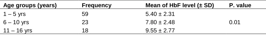
Chi square test showed clinical significant differences of HbF level among age group with P.value = 0.01, Table 1.
The severity of disease was variable in the study population when Hedo scoring was applied; 38 (38%) of the patients was presented with mild feature, while 54 (54%) of the patients presented with moderate disease and only 8 (8%) patients had a severe disease.
The mean of HbF level in patients with mild disease severity was 7.7 ± 2.82% ranging between (2.1% - 13.4%). In patients with moderate disease severity the mean of HbF level was 7.65 ± 2.23% the range was (3.2% - 12.1%), and in severe case the mean was 7.00 ± 1.51% range was (4% - 10%).There was no clinical significant differences of HbF level in different severity groups of the patients (P. value = 0.737) Table 2.
A total of 29/100 (29%) of the patients showed Hb F level ≥ 10%, of them 19/29 (65.5%) were Female and 10/29 (34.5%) were male. The disease severity of the 29 patients was showed in Table 3, of them 15/29 (51.7%) showed moderate disease, while 13/29 (44.8%) presented with a mild disease and only one patients (3.5%) was showed severe disease. The goodness of fit test using Chi square showed that, there is a significant differences of disease severity when HbF level ≥10% with P. value = 0.03.
Table 1. Hemoglobin F levels in different age groups
Age groups (years) Frequency Mean of HbF level (± SD) P. value
1 – 5 yrs 59 5.40 ± 2.31
0.01
6 – 10 yrs 23 7.80 ± 2.48
11 – 16 yrs 18 9.55 ± 2.77
Table 2. HbF level in different severity group of study patients
P. value Mean ± SD
Degree of severity
0.737 7.7 ± 2.8
Mild
7.6 ± 2.2 Moderate
7 ± 1.5 Severe
Table 3. Severity of the disease in 29 patients with HbF≥ 10%
HbF % Gender Mild disease Moderate disease Severe disease P. value
10% Female 1 7 1
0.03
Male 5 3 -
11% Female 5 5 -
Male 1 - -
13% Female - - -
Male 1 - -
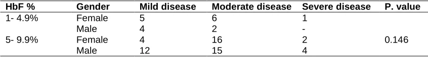
Table 4. Severity of the disease in 71 patients with HbF < 10%
HbF % Gender Mild disease Moderate disease Severe disease P. value
1- 4.9% Female 5 6 1
0.146
Male 4 2 -
5- 9.9% Female 4 16 2
Male 12 15 4
The rest 71/100 (71%) was showed HbF < 10%. Of them 34/71(48%) female and 37/71(52%) were male. The disease severity of the 71 patients was showed in Table 4 above. A total of 25/71 (35%) showed mild disease, while 39/71 (55%) presented with a moderate disease and only 7/71 patients (10%) was showed severe disease. The Chi square test showed that, there was no significant differences of disease severity when HbF level <10% (P.value = 0.146).
4. DISCUSSION
The heterogeneous phenotype in patients with sickle cell disease (SCD) is determined by the interaction of genetic and environmental factors. One of them is Hb F level which influenced on disease manifestation in many studies [13]. The objective of our study was to determine Hb F level and examine the relationship between HbF level and disease severity among Sudanese patients with SCA attending Sudan sickle cell anemia center (SSCAC), Northern Kordofan State, Western Sudan.
The average of HbF level in our population was 7.6% which was lower comparable to other studies in Congo [14], Nigeria [15], and Saudi Arabia [16] while the mean of Hb F levels were 8.8%, 9.5% and 9.1% respectively. This
difference may be due to genetic, environmental or nutritional factors.
The study found that the mean of Hb F level among patients study was slightly higher in female than male, this finding disagree with study done in Nigeria by Kotila et al, where males recorded a higher HbF levels than females, although the study in Nigeria was done among adults [17]. In the other hand, the study showed there is a difference of HbF level according to the age group. Higher levels were found in older age. Many study showed that genetic loci are known to increase HbF levels in adult life [18]. The present study confirmed earlier findings [19].
According to ethnicity, the majority of the present patients belong to Afro-Asiatic tribes followed by Niger-Congo. HbF level was found to be slightly higher in Afro-Asiatic than Niger-Congo. The result was disagree with study done by YASIR A [20]. He found the majority of the patients were Niger – Congo with higher Hb- F level than Afro – Asiatic. The dissimilarity may be due to the increase number of Messeryia and Baggara tribe (one of Afro-Asiatic tribes) in North Kordofan; these tribes represent the most affected tribes.
Arabia have been described as having less severe disease with fewer complications [22] and better survival [23]. Our study showed that the mean of Hb F level in patients with a severe disease was 7%, in patients with a moderate disease was 7.6% and in patients with a mild disease severity was7.8%. there was no statistical significant differences of HbF level in different severity groups of the patients, our study was in accordance with study done by Lena Mpalampa et al. [24], and Konotey et al. [25] they showed that some sicklers had frequent crisis and their HbF levels were below 20%, and those with mild crisis had HbF levels above 20%. Middle East sicklers who have frequent crisis were also found with 30% HbF [26].
One of our study observation was that, Hb F level play a role in a disease severity when it is more than 10% as showed in 29 patients, denotes that HbF level more than 10% represent a higher level with a less severity, this in accordance with many study mentioned high HbF level had no or mild crisis [27]. On the other hand, our study showed no significance differences between HbF and disease severity in 71 patients their Hb F level is less than 10%. Our finding was in agreement with early study suggested that the threshold level of HbF needed to prevent organ damage and acute clinical events was about 10% and 20% [26].
HbF level is important in sickle cell patients' survival, As those Patients who had HbF level more than 2 % had a 10-year probability of survival of 89 %, compared with 53 % among patients with HbF lower than 2 % [28]. It's important to determine HbF level in SCA patients and investigate its association with clinical presentation.
5. CONCLUSION
The study concluded that Hb F level has no effect in severity of the disease among studied sickle cell patients, unless the HbF level more than 10%.
6. LIMITATIONS OF THE STUDY
A limitation of this study was our inability to use the HPLC method to measure HbF level this method was not available in Elobied- Sudan. Secondly, the scoring of the disease severity depended on the patients' relatives data to remember the clinical feature that occurred in the past which may affect our results.
CONSENT
As per international standard or university standard, patient’s written consent has been collected and preserved by the authors.
ETHICAL APPROVAL
As per international standard or university standard, written approval of Ethics committee has been collected and preserved by the authors.
COMPETING INTERESTS
Authors have declared that no competing interests exist.
REFERENCES
1. Fathallah H, Atweh GF. Induction of fetal hemoglobin in the treatment of sickle cell disease. Hematology Am Soc Hematol Educ Program. 2006;58–62.
2. Silva-Pinto AC, Angulo IL, Brunetta DM, Neves FI, Bassi SC, Santis GC, et al. Clinical and hematological effects of hydroxyurea therapy in sickle cell patients: A single-center experience in Brazil. Sao Paulo Med J. 2013;131(4):238–43.
3. Sabah Elzain MM, Hamamy H. The ethnic distribution of sickle cell disease in Sudan. Pan Afr Med J. 2014;18:13.
4. Steinberg MH. Predicting clinical severity in sickle cell anaemia. Br J Haematol. 2005;129(4):465–81.
5. Moumni I, Ben Mustapha M, Ben Manour I, et al. Fetal hemoglobin in Tunisian sickle cell disease patients: Relationship with polymorphic sequences Cis to the B-Globien Gene. Indian J Hematol Blood Transfus. 2016;32(1):114–119.
6. Chaouch L, Moumni I, Ouragini H, et al. rs11886868 and rs4671393 of BCL11A association with HbF level variation and Modulate clinical events among sickle cell anemia patients. Hematology. 2016; 22:1-5.
7. Ofori-Acquah SF, Lalloz MR, Serjeant G, Layton DM. Dominant influence of gamma-globin promoter polymorphisms on fetal haemoglobin expression in sickle cell disease. Cellular and Molecular Biology. 2004;50:35–42.
hemoglobinopathies. Cold Spring Harb. Perspect. Med. 2012;2(10):1–12.
9. Green NS, Barral S. Emerging science of hydroxyurea therapy for pediatric sickle cell disease. Pediatr Res. 2014;75(1-2): 196-204.
10. Steinberg MH, Voskaridou E, Kutlar A, et al. Concordant fetal hemoglobin response to hydroxyurea in siblings with sickle cell disease. Am J Hematol. 2003;72:121–126. 11. Hedo CC, Aken'ova YA, Okpala IE,
Durojaiye AO, Salimonu LS. Acute phase reactants and severity of homozygous sickle cell disease. Journal of Internal Medicine. 1993;233:467-470.
12. Prehu C, Ducrocq R, Godart C, Riou J, Galacteros F. Determination of Hb F levels: The routine methods. Hemoglobin. 1998;22:459–467..
13. Sebastiani P, Nolan VG, Baldwin CT, bad-Grau MM, Wang L, Adewoye AH, et al. A network model to predict the risk of death in sickle cell disease. Blood. 2007;110(7): 2727-35.
14. Mouele R, Galacteros F, Feingold J. Hemoglobin F (HbF) levels in sickle-cell anemia patients homozygous for the Bantu haplotype. Eur J Haematol. 1999;63:136– 137.
15. Fatunde OJ, Scott-Emuakpor AB. Hemoglobin F and A2 in Nigerian children with sickle cell anaemia. J Trop Pediatr. 1993;39:251–252.
16. El-Hazmi MAF. Clinical and Hematological Diversity of Sickle cell disease in Saudi Children. J Trop Pediatr. 1992;38:106– 112.
17. Kotila TR, Fawde OI, Shokunbi WA. Haemoglobin F and clinical severity of sickle cell anaemia among Nigerian adults. Afr J Med Sci. 2000;29(3-4):229–31. 18. Thein SL, Sampietro M, Rohde K,
Rochette J, Weatherall DJ, Lathrop GM, Demenais F. Detection of major gene for hetro cellular hereditary persistence of foetal haemoglobin after accounting for
genetic modifiers. American Journal of Human Genetics. 1994;54:241–228. 19. Dominic Edoh, Charles Antwi- Bosaiko,
and Dominic Amuzu. Fetal hemoglobin during infancy and in sickle cell adults Afr Health Sci. 2006;6(1):51–54.
20. Yasir A, Elshazali W, the effect of hydroxyurea treatment on fetal hemoglobin level and clinical status of Sudanese sickle cell anemia patients. European Academic Research. 2014;1:6.
21. Charache S. Fetal hemoglobin, sickling, and sickle cell disease. Adv Pediatrics 1990;37:1–31.
22. Perrine RP, Pembrey ME, John P, Perrine S, Shoup F. Natural history of sickle cell anemia in Saudi Arabs; A study of 270 subjects. Ann Intern Med. 1978;88:1–6. 23. Platt OS, Brambilla DJ, Rosse WF, Milner
PF, Castro O, Steinberg MH, Klug PP. Mortality in sickle cell disease – life expectancy and risk factors for early death. N Engl J Med. 1994;330:1639–1644. 24. Lena Mpalampa, Christopher M Ndugwa,
Henry Ddungu and Richard Idro, Foetal haemoglobin and disease severity in sickle cell anaemia patients in Kampala, Uganda. BMC Blood Disorders. 2012;12:11.
25. Konotey Ahulu FID. The sickle cell disease patient. 1991;91–108.
26. Omoti CE. The value of foetal hemoglobin level in the management of Nigerian sickle cell anemia patients. Nigeria Post Graguate Medical Journal. 2005;12(3): 149–154.
27. Powars D, Weiss JN, Chan LS, Schroeder WA. Is there a threshold level of fetal hemoglobin that ameliorates morbidity in sickle cell anemia? Blood. 1984;63:921– 926.
28. Silva-Pinto AC, Angulo IL, Brunetta DM, Neves FI, Bassi SC, Santis GC, et al. Clinical and hematological effects of hydroxyurea therapy in sickle cell patients: A single-center experience in Brazil. Sao Paulo Med J. 2013;131(4):238–43.
_________________________________________________________________________________
© 2017 Abdelgadir et al.; This is an Open Access article distributed under the terms of the Creative Commons Attribution License (http://creativecommons.org/licenses/by/4.0), which permits unrestricted use, distribution, and reproduction in any medium, provided the original work is properly cited.
Peer-review history: