Publicly Accessible Penn Dissertations
1-1-2011
The Structural Proteomics of S-Nitrosylation: From
Global Identification to Elucidating Protein
Function Through Structural Bioinformatics
Jennifer L. GreeneUniversity of Pennsylvania, [email protected]
Follow this and additional works at:http://repository.upenn.edu/edissertations
Part of theBiochemistry Commons,Bioinformatics Commons, and theBiophysics Commons
This paper is posted at ScholarlyCommons.http://repository.upenn.edu/edissertations/513
For more information, please [email protected].
Recommended Citation
Greene, Jennifer L., "The Structural Proteomics of S-Nitrosylation: From Global Identification to Elucidating Protein Function Through Structural Bioinformatics" (2011).Publicly Accessible Penn Dissertations. 513.
Abstract
ABSTRACT
THE STRUCTURAL PROTEOMICS OF S-NITROSYLATION: FROM GLOBAL IDENTIFICATION TO ELUCIDATING PROTEIN FUNCTION THROUGH STRUCTURAL BIOINFORMATICS
Jennifer L. Greene
Harry Ischiropoulos, Ph.D.
S-nitrosylation is the covalent addition of nitric oxide to reduced cysteine residues on proteins. It has been well documented that not all proteins are S-nitrosylated and more specifically, not all cysteine residues within an S-nitrosylated protein are modified. Therefore, it is very important to determine how this specificity is derived. Additionally, the mechanism by which nitric oxide can modify cysteines is still unclear. Even with the discovery of functional consequences of S-nitrosylation, there are still large deficits in our understanding and validation that it is a newly identified means of nitric oxide signaling within the body. These gaps in knowledge primarily exist due to a lack of tools necessary for identifying in vivo sites of S-nitrosylation. To this end, complementary mercury-based mass spectrometric approaches were developed for the identification of endogenous S-nitrosoproteomes. This resulted in the identification of 328 SNO-cysteines coordinated to 192 proteins in the mouse liver, 97% of which corresponded to novel targets of S-nitrosylation. Bioinformatic analysis of these targets then revealed that multiple mechanisms of S-nitrosylation may occur in vivo, one of which involving S-nitrosoglutathione (GSNO). To test this hypothesis, the SNO-proteome of mice incapable of metabolizing GSNO was resolved. Quantum mechanics/molecular mechanics calculations coupled with molecular dynamics simulations proposed a novel GSNO-mediated mechanism of transnitrosation. Basic residues in the surrounding cysteine microenvironment were shown to catalyze the formation of protein nitrosocysteine residues. Collectively, these data suggest that the specificity of cysteines targeted for S-nitrosylation is driven by the surrounding protein microenvironment. Additionally, with only 9 structures of S-nitrosylated proteins our present understanding of the structural consequences of S-nitrosylation is limited. Using an in vivo model, attempts were made to correlate changes in enzymatic activity as a function of S-nitrosylation. Normal mode analysis revealed local motions near the site of S-nitrosylation which may alter product release. In summary, this thesis utilized a global proteomic approach to craft a more targeted investigation into the specificity and molecular mechanism of S-nitrosylation.
Degree Type
Dissertation
Degree Name
Doctor of Philosophy (PhD)
Graduate Group
Biochemistry & Molecular Biophysics
Keywords
mass spectrometry, nitric oxide, proteomics, S-nitrosoglutathione, S-nitrosylation
Subject Categories
Biochemistry | Bioinformatics | Biophysics
STRUCTURAL BIOINFORMATICS
Jennifer L. Greene
A DISSERTATION
in
Biochemistry and Molecular Biophysics
Presented to the Faculties of the University of Pennsylvania
in Partial Fulfillment of the Requirements for the
Degree of Doctor of Philosophy
2011
___________________________________
Supervisor of Dissertation: Harry Ischiropoulos, Ph.D., Research Professor of Pediatrics and Pharmacology
___________________________________
Graduate Group Chairperson: Kathryn M. Ferguson, Ph.D., Associate Professor, Physiology
Dissertation Committee
Doron C. Greenbaum, Ph.D., Assistant Professor, Department of Pharmacology
Cecilia Tommos, Ph.D., Assistant Professor, Department of Biochemistry and Biophysics Roland L. Dunbrack, Jr, Ph.D., Adjunct Associate Professor, Department of
Biochemistry and Biophysics
Ian A. Blair, Ph.D., Professor, Department of Pharmacology
Gideon Dreyfuss, Ph.D., Professor, Department of Biochemistry and Biophysics
STRUCTURAL BIOINFORMATICS
COPYRIGHT
2011
“I was leaving the South to fling myself into the unknown. I was taking a part of the South to transplant in alien soil, to see if it could grow differently, if it could drink of new
and cool rains, bend in strange winds, respond to the warmth of other suns, and, perhaps, to bloom.”
-Richard Wright
ACKNOWLEDGMENTS
I would first like to thank the entire Ischiropoulos Laboratory. I joined this lab at a time
when I was unsure about my own future in science. The collaborative spirit and good
nature of everyone in the lab, past and present, helped bring my scientific dreams back to
life. I would like to thank my advisor, Dr. Harry Ischiropoulos, for his constant support
and encouragement. Under his tutelage, I have become a far better scientist than I could
have imagined. I would like to thank Richard Lightfoot, Drs. Margarita Tenopoulou and
Paschalis-Thomas Doulias. Karthik Raju, Kristen Malkus, Marissa Martinez, and past
members: Drs. Elpida Tsika, Todd Greco, and Christie Bruno. I would also like to thank
the members of the Vanderkooi laboratory for being my lab home for my first 3 years
here at Penn. I would like to thank the members of my committee for their advice and
support. I’d also like to thank my friends and family, especially my mother Mel, my sister
Nikoia, and my brother Bernard. And lastly, I’d like to thank my fiancé, Ebenezer.
Without your love, I don’t think I would have made it.
ABSTRACT
THE STRUCTURAL PROTEOMICS OF S-NITROSYLATION: FROM GLOBAL
IDENTIFICATION TO ELUCIDATING PROTEIN FUNCTION THROUGH
STRUCTURAL BIOINFORMATICS
Jennifer L. Greene
Harry Ischiropoulos, Ph.D.
S-nitrosylation is the covalent addition of nitric oxide to reduced cysteine residues
on proteins. It has been well documented that not all proteins are S-nitrosylated and more
specifically, not all cysteine residues within an S-nitrosylated protein are modified.
Therefore, it is very important to determine how this specificity is derived. Additionally,
the mechanism by which nitric oxide can modify cysteines is still unclear. Even with the
discovery of functional consequences of S-nitrosylation, there are still large deficits in
our understanding and validation that it is a newly identified means of nitric oxide
signaling within the body.
These gaps in knowledge primarily exist due to a lack of tools necessary for
identifying in vivo sites of S-nitrosylation. To this end, complementary mercury-based
mass spectrometric approaches were developed for the identification of endogenous
S-nitrosoproteomes. This resulted in the identification of 328 SNO-cysteines coordinated to
192 proteins in the mouse liver, 97% of which corresponded to novel targets of
S-nitrosylation. Bioinformatic analysis of these targets then revealed that multiple
S-nitrosoglutathione (GSNO). To test this hypothesis, the SNO-proteome of mice incapable
of metabolizing GSNO was resolved. Quantum mechanics/molecular mechanics
calculations coupled with molecular dynamics simulations proposed a novel
GSNO-mediated mechanism of transnitrosation. Basic residues in the surrounding cysteine
microenvironment were shown to catalyze the formation of protein S-nitrosocysteine
residues. Collectively, these data suggest that the specificity of cysteines targeted for
S-nitrosylation is driven by the surrounding protein microenvironment.
Additionally, with only 9 structures of S-nitrosylated proteins our present
understanding of the structural consequences of S-nitrosylation is limited. Using an in
vivo model, attempts were made to correlate changes in enzymatic activity as a function
of nitrosylation. Normal mode analysis revealed local motions near the site of
S-nitrosylation which may alter product release. In summary, this thesis utilized a global
proteomic approach to craft a more targeted investigation into the specificity and
TABLE OF CONTENTS
TITLE PAGE………...i
DEDICATION………..iii
ACKNOWLEDGMENTS………...iv
ABSTRACT………...v
TABLE OF CONTENTS………..vii
LIST OF TABLES………..x
LIST OF FIGURES………...xi
CHAPTER ONE: INTRODUCTION……….………..………...1
1.1 Introduction………...1
1.2 Nitric oxide………....………...2
1.2.1 Chemical properties of nitric oxide………...2
1.2.2 Biological synthesis of nitric oxide………...3
1.2.3 Nitric oxide in health and disease………....5
1.2.4 Nitric oxide as a signaling molecule………... 8
1.3 S-nitrosylation as an emerging posttranslational modification…...……...10
1.3.1 Methods for identification of S-nitrosylated cysteines………...11
1.3.2 Alteration of protein activity as a function of S-nitrosylation…...14
1.3.3 Proposed mechanisms of S-nitrosylation formation………..17
1.3.4 Denitrosylation: Reversing the signal………19
1.4 Specificity of cysteines targeted for S-nitrosylation………..22
1.4.1 Cysteine reactivity……… 22
1.5 Structural implications of S-nitrosylation……….26
1.5.1 Insights from the structures of S-nitrosylated proteins………….26
1.5.2 Computational tools for extracting structural information from S-nitrosylated proteins………...…...33
1.6 Rationale and Objectives………..….35
CHAPTER TWO: STRUCTURAL PROFILING OF ENDOGENOUS S-NITROSOCYSTEINE RESIDUES REVEALS UNIQUE FEATURES THAT ACCOMMODATE DIVERSE MECHANISMS FOR PROTEIN S-NITROSYLATION……….….37
2.1 Abstract……….38
2.2 Introduction………...39
2.3 Materials and Methods………..…40
2.4 Results and Discussion………..43
2.5 Supplementary Methods………....61
2.6 Supplemental Figures……….………....70
2.7 Supplementary Tables……….………...74
CHAPTER THREE: ELECTROSTATIC PROPERTIES NAVIGATE GSNO-MEDIATED PROTEIN TRANSNITROSATION………..….…93
3.1 Abstract………...94
3.2 Introduction………...94
3.3 Materials and Methods………..98
3.4 Results………..104
3.5 Discussion………116
CHAPTER FOUR: STRUCTURAL CONSEQUENCES OF S-NITROSYLATION THAT INFLUENCE ACTIVITY IN VIVO……….……..133
4.1 Abstract………....134
4.3 Methods………...…....138
4.4 Results and Discussion………....139
CHAPTER FIVE: SUMMARY AND GENERAL DISCUSSION 5.1 Summary……….….150
5.2 Proteomic techniques are essential for characterization of the endogenous S-nitrosoproteome……….…...150
5.3 Transnitrosation of GSNO is precisely controlled……….………..152
5.4 Implications of this work……….155
LIST OF TABLES
CHAPTER TWO
Table 2.1:Biochemical and biophysical properties of S-nitrosylated and unmodified cysteine residues within the same proteins………52
Supplementary Table 2.1: Endogenously S-nitrososylated proteins in wild type mice liver...74
Supplementary Table 2.2: Endogenously S-nitrosylated proteins in eNOS-/- mouse liver………86
Supplementary Table 2.3: Cysteine residues coordinated to metals………..88
Supplementary Table 2.4: Endogenously S-nitrosylated proteins sensitive to Trx/TrxR-mediated denitrosylation……….………..89
CHAPTER THREE
Table 3.1: Endogenously S-nitrosylated GSNOR-/- unique proteins in liver and
thymus……….119
Table 3.2: GSNOR-/- unique proteins coordinated to PDB structures………127
CHAPTER FOUR
Table 4.1: Calculated bond angles of existing S-nitrosylated cysteines………...143
LIST OF FIGURES
CHAPTER TWO
Figure 2.1: Site-specific identification of S-nitrosocysteine…………..……….46
Figure 2.2: Hydropathy index and pKa values of S-nitrosylated residues………..54
Figure 2.3: Analysis of primary sequence, distribution in secondary structures, and surface accessibility of S-nitrosylated cysteine residues……….……….57
Scheme 2.1: Chemical synthesis of mPEG-biotin compound……….……….62
Figure S2.1: Characterization of protein S-nitrosocysteine………..………...70
Figure S2.2: GeLC-MS/MS analysis of protein S-nitrosocysteine…………..…………71
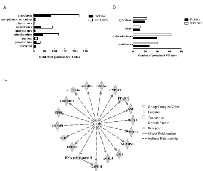
Figure S2.3: Ontological analysis of liver S-nitrosoproteome…………..………...72
Figure S2.4: Distribution of residues flanking unmodified cysteine residues in secondary structures……….………..73
CHAPTER THREE
Figure 3.1:CYG intermediate used in molecular dynamics simulations…………...…103
Figure 3.2: Gene ontology analysis of GSNOR null unique proteome………..105
Figure 3.3: Kernel density plot of S-nitrosylated cysteines from GSNOR-/- unique SNO-proteome………..………106
Figure 3.4: Distribution of secondary structure flanking S-nitrosylated GSNOR-/- unique cysteine residue………108
Figure 3.5: RSA accessibility of GSNOR-/- unique S-nitrosated cysteines...109
Figure 3.6: Predicted pKa values for S-nitrosylated GSNOR-/- unique cysteine…...….110
Figure 3.7: DCCM plots of all four cysteines in 2DGV……….113
Figure 3.8: Distance probability plots of CYG675 intermediate residue………...……113
CHAPTER FOUR
Figure 4.1: Ingenuity pathway analysis (IPA) identifies indirect relationship between leptin and Very long chain acyl-CoA dehydrogenase………135
Figure 4.2: Cysteine238 confirmed as S-nitrosation site after GSNO treatment….….137
Figure 4.3:VLCAD structure 2uxw………..141
Figure 4.4: S-nitrosylation does not induce large scale motions in VLCAD………....145
Figure 4.5: Binding site of trans-Δ2-Palmitenoyl-CoA in VLCAD………..146
Figure 4.6: Higher frequency mode indicates extensive movement of Cys238 upon S-nitrosylation………...….148
CHAPTER ONE
1.1
Introduction
Nitric oxide (•NO) received the distinction of a signaling molecule in physiology when Furchgott, Ignarro, and Murad were awarded the 1998 Nobel Prize in Medicine &
Physiology for their combined efforts in elucidating the role of •NO in vasodilation. It was concluded that •NO produced by endothelial cells can bind to the heme iron of soluble guanylate cyclase (sGC) thereby converting GTP to cGMP and resulting in a
cascade of events leading to smooth muscle relaxation. Because it is well known that •NO can react with many intracellular targets, the mechanism by which •NO selectively reacted with sGC became of significance. Stamler et al. hypothesized that •NO could also propagate its signal by forming stable adducts with protein thiols that would then deliver
nitric oxide to a specific target (Stamler et al. 1992). This study resulted in the first
observation that nitric oxide could react with cysteine residues in proteins to form protein
S-nitrosocysteine.
Over the past 2 decades, hundreds of proteins have been identified as being
S-nitrosylated. S-nitrosylation is defined as the covalent addition of nitric oxide to selective
protein cysteine residues that can regulate protein activity across a variety of organs and
cellular systems. Their cellular functions have ranged from oxidoreductases to ion
channels to histone deacetylases, suggesting that S-nitrosylation appears to be a
widespread biological mechanism of nitric oxide signaling. Nevertheless, fundamental
questions regarding this emerging posttranslational modification (PTM) still remain
this small highly reactive molecule, •NO selectively modifies a specific cysteine residue. The mechanism by which •NO can modify cysteines is still unclear. For other more characterized PTMs such as acetylation (which is of comparable relative size to •NO), specific enzymes are necessary to catalyze its reaction with its target protein. Therefore,
with the unequivocal identification of such enzyme(s) for S-nitrosylation, it is difficult to
ascertain how specificity is achieved. Answers to these pressing questions will expand
our understanding of S-nitrosylation and offer insight as to how to better regulate this
emerging posttranslational modification.
1.2 Nitric oxide
1.2.1 Chemical properties of nitric oxide
Nitric oxide (also known as nitrogen monoxide) is a colorless, diatomic gas
composed of one nitrogen atom and one oxygen atom. It was first described as “nitrous
air” in 1774 by Joseph Priestley in his book titled Experiments and Observations on
Different Kinds of Air, Vol. 1. An uncharged free radical, nitric oxide has one unpaired
electron. With an oil/water partition coefficient ~6.5 (Kow measured at 37°C) and a low
solubility in water, •NO is capable of easily diffusing through hydrophobic environments such as cell membranes (Balbatum et al. 2003). In aqueous solutions, nitric oxide reacts
primarily with oxygen and its half-life is proportional to its concentration.
The half-life of •NO can be altered by its interactions with different targets. Nitric oxide can react with a wide range of molecules to form end products with various
functions. Upon its reaction with molecular oxygen (O2), nitric oxide is oxidized to form
with free radicals such as superoxide (O2-•) to generate the strong oxidant peroxynitrite
(ONOO-). This reaction has been observed to be near diffusion limited suggesting that it is only restricted by the ability of the molecules to come into contact with one another
(Beckman et al. 1990; Koppenol, et al. 2001). The interaction of nitric oxide with
lipid-derived radicals has also been characterized as being a diffusion-limited reaction
(O’Donnell et al. 1997). •NO has been most classically described by its coordination to transition metals such as copper or iron to form M-NO (Wink et al. 1994; Wade & Castro
1990).
In general, the chemical reactivity of •NO is contingent upon its surrounding environment. Therefore, when attempting to understand the role of nitric oxide in
biological processes, it is important to know when and where it is produced within the
body.
1.2.2 Biological synthesis of nitric oxide
In 1916, Mitchell, Shonle, and Grindley first noticed that the amount of nitrates
and nitrites excreted through urine from healthy subjects was more than the amount that
could be ingested from the diet (Mitchell, et al. 1916). Using 15N-labelled nitrate (15NO3
-), Tannenbaum et al. monitored nitrate metabolism and quantified an excess of excreted
nitrates in human urine compared to dietary intake, supporting this 65-year old
observation (Tannenbaum, et al. 1978; Green et al. 1981, PNAS). These experiments
allowed them to conclude that an endogenous metabolic process was responsible for
across different research groups biochemically characterized this mechanism and its
necessary components (Stuehr & Marletta 1985; Hibbs et al. 1987; Iyengar et al. 1987;
Marletta et al. 1988). It was discovered that the radical nitric oxide is an endogenously
synthesized intermediate generated during the process of nitrite and nitrate formation
(Ignarro et al. 1987a; Ignarro et al. 1987b; Palmer et al. 1987; Palmer et al. 1988).
•
NO is synthesized from L-arginine and oxygen by the enzyme nitric oxide
synthase (NOS) (Moncada & Higgs, 1993). Nitric oxide synthases are 150 kiloDaltons
and composed of a homodimer containing an oxygenase domain and a reductase domain
that are linked by a calmodulin recognition site. The oxygenase domain contains binding
sites for the cofactors tetrahydrobiopterin (BH4), protoporphyrin IX haem, and L-arginine
while the reductase domain contains binding sites for the electron carriers FAD & FMN,
and the reductant NADPH (Ghosh & Stuehr 1995). In summary, electrons supplied by
NADPH are shuttled across the reductase domain by FAD and FMN to the heme iron of
the oxygenase domain where they can oxidize L-arginine. A guanidino nitrogen of L
-arginineundergoes a 5-electron oxidation via two mono-oxygenation steps to form •NO and L-citrulline (Marletta et al. 1993; White & Marletta et al. 1992; Mayer et al. 1991;
Bredt & Snyder 1990; Bredt et al. 1991). Binding of calmodulin is required for the flow
of electrons from the reductase domain to the oxygenase domain.
Three nitric oxide synthase isoforms exist: neuronal nitric oxide synthase (nNOS;
NOS1), inducible nitric oxide synthase (iNOS; NOS2), and endothelial nitric oxide
synthase (eNOS; NOS3). They differ in their regulation, quantity, and location of •NO production. Neuronal NOS was the first isoform to be purified and cloned (Bredt &
brain and skeletal muscle (Salter et al. 1991; Nakane et al. 1993). Endothelial NOS is
found in endothelial cells, cardiac myocytes, neurons, and epithelial cells. Typical output
from eNOS and nNOS is in the nM range. iNOS produces a large burst of •NO in response to cytokines and endotoxins across a variety of cell types including neutrophils,
epithelial cells, vascular smooth muscle, hepatocytes and chondrocytes (Xie et al. 1992;
Lincoln et al. 1997). nNOS and eNOS differ from iNOS in their dependency upon
calcium (Ca2+) to produce synthesize •NO (Cho et al. 1992;Garcin et al. 2004; Dudzinski et al. 2007; Venema et al. 1996). The efficiency of calmodulin binding for nNOS and
eNOS is mediated by intracellular calcium levels whereas calmodulin is already tightly
bound to iNOS.
The presence of so many cofactors suggests that nitric oxide synthases are very
highly regulated. Lack of the cofactor BH4 can cause the uncoupling of the eNOS dimer
leading to production of superoxide (Chen et al. 2011). NOSs have also been shown to be
regulated by posttranslational modifications. S-Glutathionylation can also uncouple the
eNOS dimer serving as an autoregulatory mechanism for •NO production (Chen et al. 2010). S-nitrosylation of iNOS has also been shown to destabilize the dimer (Rosenfeld
et al. 2010).
1.2.3 Nitric oxide in health and disease
Nitric oxide has long played an important role in human health and disease, even
when it had not yet been identified as the culprit (Butler et al. 2006). As with most great
scientific discoveries, it all began with an observation. An English surgeon noticed that
by removing blood (Butler et al. 2006). This suggested to him that the remedy for angina
pectoris was somehow related to lowering blood pressure. The surgeon’s friend told him
of how inhaling amyl nitrite helped to lower the blood pressure of his animals and so the
surgeon commenced in using this therapeutic remedy for those suffering from chest pain
(Gamgee et al. 1868; Brunton et al. 1867). Nitroglycerin which contained a nitro group
(-NO2) was also used as a vasodilator over the next century. It would be many years later
before a series of experiments conducted by Moncada, Furchgott, Ignarro & Murad
revealed the identity of nitric oxide as the true vasodilator. This Nobel Prize winning
discovery will be discussed in the following section.
Nitric oxide has been classically identified as having a role in the body’s innate
immune response and neurotransmission. As was previously described, macrophages
were instrumental in revealing the endogenous production of nitric oxide (Hibbs et al.
1987; Stuehr et al. 1989; Marletta et al. 1988; Hibbs, et al. 1989). In response to certain
stimuli, iNOS can produce a burst of the •NO which targets invading pathogens and participates in anti-tumor activities through a variety of mechanisms. Mice lacking the
gene for iNOS are much more susceptible to parasitic infection and replication of bacteria
(Wei et al. 1991; MacMicking et al. 1995). •NO has been shown to impair mitochondrial function by disrupting iron-sulfur centers within oxidoreductases as well as altering
mitochondrial membrane potential (Kroncke et al. 1995; Hibbs et al. 1987; Stuehr et al.
1989). •NO can also react with non-heme iron, resulting in Fe release which might also promote lipid peroxidation and serve as an additional mechanism of cellular damage
While all three isoforms of NOS are found throughout the brain, nitric oxide
produced from neuronal NOS has been the best characterized within the central nervous
system. A few of its important roles involve neuroprotection, neurotoxicity, and synaptic
plasticity. Glutamate binds to the NMDA-receptor which allows calcium (Ca2+) entry into the neurons thereby activating NOS. Once it is produced, •NO acts as a retrograde messenger to strengthen synaptic activity. It has been postulated that this function of
nitric oxide is important during long term potentiation (LTP), an important process
underlying learning and memory in the brain.
Nitric oxide can also serve a neuroprotective function by regulating its own
production. Disulfide bridge formation in the NMDA-receptor channel has been
previously shown to decrease the influx of Ca2+ through the channel (Lei et al. 1992). Nitric oxide can modify these same two cysteines and help promote the formation of this
disulfide bridge (Lipton, Singel, & Stamler, 1994; Lipton & Stamler, 1994). In addition,
•
NO can also inhibit the binding of glutamate to the NMDA receptor, preventing the flow
of Ca2+ through the channel and regulate NO synthesis.
When observing the presence of •NO throughout the central nervous system, peripheral nervous system, and immune system, it is easy to imagine how its dysfunction
might lead to disease. Dysregulation of nitric oxide has been implicated in ischemia
(Mugge et al. 1991; Harrison et al. 1992; Maseri et al. 1991), atherosclerosis (Rubanyi et
al. 1993), septic shock (Thiemermann et al. 1993; Petros et al. 1991), pre-eclampsia
Nitric oxide has also been used as a therapeutic agent as it has been found to play
an important role in wound healing (Frank et al. 2002; Shi et al. 2000) and protection
after ischemic injury (Lima et al. 2009). In fact, researchers Jonathan Stamler and Joseph
Bonaventura hold a United States patent for “Red blood cells loaded with S-nitrosothiol
and its uses”. As the patent abstract states, “loaded red blood cells can be used in methods
of therapy for conditions which are characterized by abnormal O2 metabolism of tissues,
oxygen-related toxicity, abnormal vascular tone, abnormal red blood cell adhesion, or
abnormal O2 delivery by red blood cells.” United States Patent #6153186
(
http://www.wikipatents.com/US-Patent-6153186/red-blood-cells-loaded-with-s-nitrosothiol-and-uses-therefor). Direct inhalation of nitric oxide has been used to treat
preterm infants with bronchopulmonary dysplasia (Kinsella, et al. 2007). As researchers
continue to identify the role of nitric oxide in human physiology, it is even more pressing
to identify the mechanisms by which nitric oxide may exert its function and how these
functions are regulated.
1.2.4 Nitric oxide as a signaling molecule
Although nitrovasodilators such as nitroglycerin were known to alleviate the
symptoms of pectoris angina since 1867, their mechanism of action was not fully
elucidated until over 120 years later. The discovery of nitric oxide as an endogenous
vasodilator came from a series of elegantly designed experiments across multiple
Schultz, 1977). It was also noted that this effect was inhibited by hemoglobin and
myoglobin (Murad, et al. 1978). Shortly thereafter, the Ignarro laboratory reported that
activation of guanylate cyclase by •NO resulted in smooth muscle relaxation (Gruetter, et al. 1979); however, it was still unknown that •NO existed endogenously.
The Furchgott Laboratory monitored the response of the aorta to the cholinergic
agonist carbachol using a protocol which required the isolation of aortic rings; however,
when a new technician prepared helical strips of the aorta instead of rings, he found that
he could no longer induce smooth muscle relaxation. This finding highlighted two
important discoveries: 1) an intact endothelium was necessary for smooth muscle
relaxation and 2) acetylcholine must bind to receptors on the surface of the endothelium
to produce the molecule responsible for smooth muscle relaxation (Furchgott et al. 1980).
This yet identified molecule was later termed “endothelium-derived relaxation factor”
(EDRF).
While both Furchgott and Ignarro independently speculated that EDRF was in
fact •NO, it still remained inconclusive (Moncada et al. 1988). Once concurrent research identified the endogenous synthesis of nitrites and nitrates (Ignarro, et al. 1981; Iyengar,
et al. 1987), this speculation immediately became more concrete. A series of experiments
by Moncada and Ignarro finally revealed that EDRF was nitric oxide (Ignarro, et al.
1987; Ignarro, et al. 1987; Palmer et al. 1987) and it was synthesized from terminal
guanidine nitrogen of L-arginine in vascular endothelial cells (Palmer et al. 1988).
In 1998, Furchgott, Ignarro, and Murad were awarded the Nobel Prize in
Medicine and Physiology for their combined contributions in elucidating the role of nitric
oxide as a “signaling molecule”. This newly identified role for nitric oxide was clinched
with the key discovery that eNOS produced •NO within endothelial cells. Previous work had shown that nitric oxide was produced in response to a stimulus, specifically targeted
to the heme of soluble guanylate cyclase, and necessary for a specific physiological
response, i.e. dephosphorylation of myosin light chain resulting in vasodilation. This new
discovery added that nitric oxide was spatially restricted after its production by eNOS.
This series of events became the canonically described cGMP-dependent pathway
whereby which nitric oxide can act as a second messenger and induce a physiological
response.
1.3
S-nitrosylation
as
an
emerging
posttranslational
modification
Once it was determined that nitric oxide bound to the heme of soluble guanylate
cyclase, it still remained unclear how •NO stably diffused from nitric oxide synthase to its target sGC. As was previously described, •NO can react with a variety of intracellular targets. Nitric oxide derived species are known to react with sulfhydryls and form more
stable adducts of S-nitrosothiols (Oae, et al. 1983). Stamler et al. hypothesized that •NO could prolong its half-life and deliver •NO to targets such as sGC by reacting with reduced thiols on protein cysteine residues (Stamler, et al. 1992). By using both
endogenous and exogenous sources of •NO, they found that several proteins could form S-nitroso adducts, including serum albumin. A series of later experiments by this same
group detected S-nitrosylated serum album circulating in human plasma, suggesting that
In 1996, the Stamler group identified an S-nitrosylated cysteine residue on the
Beta chain of hemoglobin (Cysβ93) (Jia, et al. 1996). Upon oxygenation of hemoglobin,
Cysβ93 was S-nitrosylated (SNO-Hb) in the lung resulting in release of nitric oxide and
delivery of O2 to peripheral tissues through circulation. This suggested that SNO-Hb may
be involved in the normal regulation of blood flow and maintenance of blood pressure.
The role of •NO in vasodilation was thought to be a very well characterized mechanism. Therefore, the identification of a new role for nitric oxide in vasodilation was a bit
shocking, especially since it involved a new role for an enzyme which had also been
heavily scrutinized for decades previously.
Even with the discovery of functional consequences of S-nitrosylation, there are
still large deficits in our understanding and validation that it is a newly identified means
of nitric oxide signaling within the body. The mechanism by which S-nitrosylation occurs
has not conclusively been identified. Additionally, how specificity is derived still remains
unclear. The following sections discuss how recent advances in the identification of
S-nitrosylated proteins have increased our knowledge of this emerging posttranslational
modification.
1.3.1 Methods for identification of S-nitrosylated cysteines
The crux to categorizing any emerging posttranslational modification is to
identify its targets. With recent advances in technology, the development of techniques
for detecting nitrosylated proteins has exponentially enhanced our understanding of
S-nitrosylation and helped to elucidate its ubiquitous role in nitric oxide signaling. Proof of
S-Nitroso bovine serum albumin (Stamler et al. 1992). Stamler et al. utilized UV-Visible
spectroscopy and NMR spectroscopy to show that •NO was bound to the protein. To quantify the amount of SNO present, they developed an assay which lysed the S–NO
bond resulting in liberated •NO which could be detected via chemiluminescence after its reaction with ozone (Stamler et al. 1992; Stamler & Feelisch, 1996). Utilizing Saville
chemistry, mercury (II) chloride (HgCl2) was also used to remove nitric oxide for the
purposes of quantification (Saville et al. 1958; Xu et al. 1998; Mannick et al. 1999). In
the case of hemoglobin, a very well characterized protein, UV-Vis spectroscopy proved
extremely informative in proving that nitric oxide was bound to a thiol and not the heme
(Jia et al. 1996). However, it required its isolation, thereby mandating previous
knowledge of its identity. Therefore, initial identifications of S-nitrosylated proteins did
not allow for a priori hypotheses regarding their existence or identification.
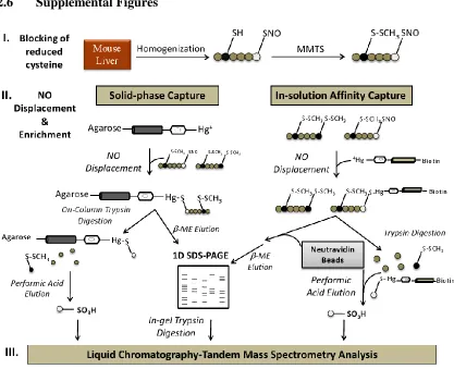
In 2001, Jaffrey et al. published the first method which globally identified
S-nitrosylated proteins contained within complex mixtures such as tissues (Jaffrey et al.
2001). The biotin switch technique (BST) consisted of three main steps: 1) the blocking
of reduced thiols with methyl methanethiosulfonate (MMTS), 2) reduction of
nitrosylated cysteine residues using ascorbate, and 3) biotinylation of previously
S-nitrosylated cysteine residues with N
-[6-(biotinamido)hexyl]-3’-(2’-pyridyldithio)propionamide (biotin–HPDP). Because MMTS is a thiol-specific agent,
ascorbate selectively decomposes nitrosothiols, and biotin-HPDP is a sulfhydryl-specific
agent, the method allows for the specific labeling of S-nitrosylated cysteine residues.
Biotinylated proteins were purified using avidin, then separated and resolved using
bands can be excised and identified by mass spectrometric analysis. While the BST was
the first tool of its kind, concerns still exist regarding its efficiency to identify
S-nitrosylated proteins. The reduction of S-nitrosothiols by ascorbate has been called into
question due to its poor sensitivity and its ability to reduce disulfide bonds (Zhang, et al.
2005; Giustarini et al. 2008). Moreover, a key objective for identifying scores of
S-nitrosylated proteins is to reveal the specificity of cysteines endogenously targeted for
this modification. However, this is impossible to do when identifying the protein without
the specific site of modification. Two papers were published in 2006 which offered
proteomic techniques for identifying sites of S-nitrosylation in complex mixtures (Hao et
al. 2006; Greco et al. 2006). Briefly, the two comparable methods introduced a
trypsinization step before biotinylation which allowed for enrichment of peptides
containing modified cysteines.
Since the development of the BST in 2001, there have been many derivatives
aimed at improving its deficiencies (Sun et al. 2007; Kettenhofen et al. 2008,; Sinha et al.
2010) and development of other promising techniques which directly label and detect
S-nitrosocysteine (Torta et al. 2010; Wang et al. 2008; Faccenda et al. 2010; Zhang et al.
2010). More recently, Benhar et al. developed a quantitative BST method using stable
isotope labeling by amino acids in cell culture (SILAC) (Benhar et al. 2010). The authors
employed their technique to characterize the denitrosylation of SNO-cysteines by
thioredoxin in Jurkat cells. While the above described methods point toward significant
advances in the field of S-nitrosylation, detection of S-nitrosylated proteins is often still
method would be sensitive enough to detect and accurately quantify endogenous
S-nitrosylation events.
1.3.2 Alteration of protein activity as a function of
S-nitrosylation
With the advent of the Biotin Switch Technique and its derivatives, many more
proteins have been identified as S-nitrosylated. For many researchers, S-nitrosylation has
provided the missing link between observing a role of nitric oxide in human physiology
and pinpointing its specific mechanism of action. A comprehensive discussion of all such
proteins would prove to be impossible within the limits of this dissertation; therefore,
several key studies are highlighted which demonstrate how S-nitrosylation can alter a
wide range of protein functions and further extend the role of nitric oxide in human
health and disease.
Nitric oxide has long been known to play an important role in neurophysiology. In
2004, the Lipton group identified a role for S-nitrosylation in sporadic Parkinson’s
disease (Yao et al. 2004). S-nitrosylation of the E3 ubiquitin ligase parkin in SH-SY5Y
cells led to an increase in its activity and ultimately resulted in its own ubiquitination.
Inhibition of parkin can result in a decreased clearance of its substrates and ultimately
lead to cell death. This fact is made even more apparent by the detection of S-nitrosylated
parkin in human brains of those with sporadic Parkinson’s disease. Later studies
involving the S-nitrosylation of glyceraldehyde-3-phosphate dehydrogenase (GAPDH)
led to an additional role of nitric oxide in regulating E3 ubiquitin ligase activity. It has
al. 1997; Ishitani, et al. 1998; Dastoor, et al. 2001). Because GAPDH lacks a nuclear
localization signal, how GAPDH is able to enter the nucleus has remained a mystery. In
2005, Hara et al. showed that nitric oxide S-nitrosylates GAPDH thereby increasing its
ability to bind to the E3 ubiquitin ligase Siah1, and induces translocation of the complex
into the nucleus (Hara et al. 2005). Once inside, GAPDH promotes the stabilization of
Siah1, allowing for it to carry out its role in apoptosis. This discovery propelled a myriad
of studies which continue to identify how this mechanism is regulated (Sen, et al. 2008;
Sen, et al. 2009; Kornberg, et al. 2010).
In addition to regulating the activity of ubiquitin ligases, S-nitrosylation has also
been shown to modulate other posttranslational modifications such as phosphorylation.
Previous reports have shown that nitric oxide can negatively regulate the signaling
cascade of c-Jun N-terminal kinases also known as JNKs (Park et al. 1996; Lo, et al.
1996; Kim et al. 1997; Wang et al. 1998; Lander et al. 1996; Jun et al. 1999). As a
member of the mitogen activated protein (MAP) kinase family, JNKs are important in the
cellular response to different stresses. They are activated through phosphorylation by
MAP kinases MKK4 and MMK7 and typically inactivated by phosphatases (Ip, et al.
1998). Park et al. demonstrated that endogenous production of •NO by activation of iNOS resulted in suppression of JNK activity in microglia and macrophages (Park et al. 2000).
Using a series of experiments, the authors showed that JNK activity was attenuated by
nitric oxide, more specifically, by a cGMP-independent chemical modification of Cys116
which they identified as S-nitrosylation. Conversely, S-nitrosylation has also been shown
tyrosine kinase c-Src and found that S-nitrosylation of Cys498 resulted in its activation
and ultimately its invasion into cancer cells (Rahman, et al. 2010).
S-nitrosylation has also been shown to play a role in the regulation of DNA
transcription. NF-κB is a transcription factor thought to regulate over 200 genes which
are involved in the inflammatory response (Marshall, et al. 2000; Shishodia, et al. 2004;
Xie, et al. 1994). It is activated by its release from inhibitory proteins within the
cytoplasm and translocation to the nucleus. IκB kinase (IKK) is one of the proteins
responsible for inducing the degradation of these inhibitory proteins thereby activating
NF-κB. Reynaert et al. demonstrated that S-nitrosylation of Cys179 in IKK inhibited its
activity resulting in prolonged inactivation of NF-κB (Reynaert et al. 2004). By inhibiting
endogenous production of •NO in Jurkat cells, the authors were able to again activate IKK. Interestingly, additional evidence has also shown that NF-κB itself is S-nitrosylated,
offering an additional mechanism for by which •NO can regulate its transcriptional activity (Matthews, et al. 1996; delaTorre, et al. 1997).
Major classes of proteins have been found to be S-nitrosylated. Coupled with the
discovery of S-nitrosylation across different types of cells and tissues (Gow et al., 2002),
this underscores the widespread effects that nitric oxide can have across different
biological processes. Nevertheless, it is important to acknowledge that identifying a
protein as being susceptible to nitrosylation does not prove that it is endogenously
S-nitrosylated. Lack of sensitive detection technologies often mandate that proteins are
frequently supplemented with exogenous sources of nitric oxide to aid in their
produced. And as is described in the following sections, the chemistry of a nitric oxide
moiety may influence which cysteines are actually targeted.
1.3.3 Proposed mechanisms of S-nitrosylation formation
Perusing the literature on S-nitrosylation may leave the reader slightly confused
due to the assorted terminology. Several key terms are used to describe this newly
emerging protein modification by nitric oxide. The difference in their definitions stems
from the proposed mechanism by which •NO modifies cysteine residues to form S-nitrosocysteine (Protein-Cys-NO). The most commonly used term is “S-nitrosylation”.
This historically describes the reaction of a nitric oxide radical with a transition metal
(“nitrosylation”) to form a product which then reacts with a protein cysteine thiol (“S
-nitrosylation”) (Stamler, et al. 1992). Another term very commonly used is “nitrosation”
or “transnitrosation”. This term traditionally refers to the addition or transfer of a
nitrosonium ion (NO+) to a thiolate anion (SH-) whereas “trans-S-nitrosation” specifies the transfer to a protein cysteine residue. “S-nitrosylation” is now more routinely used to
encompass any reaction resulting in the addition of a nitric oxide moiety to a protein
cysteine thiol.
There are several proposed mechanisms which can result in the formation of
protein S-nitrosocysteine. The first mechanism describes the direct reaction of an NO
radical with a transition metal. •NO can react with a metal at a heme center such as Fe3+ to form Fe2+ and NO+ (Pacher et al. 2007). The nitrosonium ion is then capable of reacting with the reduced thiol of nearby cysteine residues (Wade & Castro 1990). An
pools of iron in the cell to form dinitrosyliron complexes which can S-nitrosylate cysteine
residues (Bosworth et al. 2009; Boese et al. 1995).
Additional mechanisms for forming S-nitrosocysteine involve the transfer of a
nitrosonium ion to a reduced thiol of a cysteine residue. It has been proposed that
S-nitrosoglutathione (GSNO), a major endogenous S-nitrosothiol, is involved in the
transnitrosation of protein cysteine. Although it can be synthesized within the laboratory
using sodium nitrite and GSH in an acidic environment, the mechanism by which GSNO
forms endogenously still remains unclear (Zhang et al. 2004; Keszler et al. 2009; Gow et
al. 1997; Tullett et al. 2001). It is also possible for S-nitrocysteine residues within
proteins to then react with the reduced thiols of GSH or other protein cysteine residues
and transfer a nitrosonium ion. This is what is referred to as a “protein-assisted”
transnitrosation. This mechanism was observed with the protein thioredoxin (Mitchell et
al. 2005). After its own reaction with GSNO, SNO-thioredoxin (SNO-Trx) can
transnitrosate a select cysteine residue in the enzyme caspase-3. Experiments conducted
by the Marletta group illustrate that SNO-Trx and not GSNO is the favored mechanism
by which this specific cysteine forms S-nitrosocysteine (Mitchell et al. 2007).
The last proposed mechanism of S-nitrosation involves the autoxidation of •NO to form higher oxides that serve as transnitrosating agents. Due to its lipophilic character,
•
NO is known to favor hydrophobic environments (Moller et al. 2007). Therefore, •NO can congregate in hydrophobic regions of the protein where it can also react with O2 and
nitrogen dioxide to form the transnitrosating agent dinitrogen trioxide N2O3 (Nedospasov
There is a diversity of mechanisms which ultimately result in the formation of
protein-SNO, each of which is dependent upon the chemical structure of nitric oxide.
Therefore, being able to identify the chemical species of nitric oxide may help to pinpoint
which mechanism is occurring and may contribute to our understanding of the specificity
of this posttranslational modification.
1.3.4 Denitrosylation: Reversing the signal
To convincingly describe S-nitrosylation as a signaling mechanism of nitric
oxide, it is essential to show the reversibility of the modification. There are two major
endogenous systems which have been identified to influence the stability of
S-nitrosocysteine: the GSH/S-nitosoglutathione/S-nitrosoglutathione reductase system and
the thioredoxin/thioredoxin reductase system. Thioredoxin (Trx) is directly involved in
an enzyme-mediated mechanism of denitrosylation whereas the glutathione system
indirectly mediates nitrosylation through metabolism of GSNO by the enzyme
S-nitrosoglutathione reductase (GSNOR).
In addition to its roles as both a reductant and transnitrosating agent (Mitchell et
al. 2005; Mitchell et al. 2007), thioredoxin has also been shown to serve as a
denitrosylating enzyme. Nikitovic and Holmgren first demonstrated an indirect role for
the thioredoxin system through its metabolism of the low molecular weight thiol GSNO
(Nikitovic et al. 1996). Stoyanovsky and colleagues further demonstrated that the Trx
system was also capable of denitrosating protein-SNO (Sengupta, et al. 2007). The
proposed mechanism involves active site cysteines 32 and 35 (Sengupta, et al. 2007).
and Cys35 then form a disulfide bond allowing NO to then be transferred
intramolecularly to the other cysteine residues (Cys62, Cys69, & Cys72) which have
previously been shown to be S-nitrosylated. For protein denitrosation, the work of
Mitchell & Marletta with caspase-3 may provide insight as to how thioredoxin might
interact with target proteins (Mitchell et al. 2007). E70A/K72A mutations in hTrx
resulted in some loss of contact, suggesting that these charged residues may help
facilitate protein-protein interactions between the denitrosating agent and its targets.
Proteomic studies have also been used to identify proteins which might be potential
denitrosylation targets of thioredoxin (Benhar et al. 2010). Using a model which
overexpressed Trx, recent work done by Fu et al. identified 55 putative protein targets of
thioredoxin in the mouse heart (Fu et al. 2009). Interestingly, previous reports have
identified several of these proteins as being targets of S-nitrosylation.
The thioredoxin system can be regulated through a variety of mechanisms.
Stamler and colleagues first demonstrated that caspase-3 activation was tightly tethered
to its denitrosylation upon induction of the Fas apoptotic pathway (Mannick et al. 1999).
Nearly 10 years later, they were able to show that denitrosylation of caspase-3 was
carried out by mitochondrial thioredoxins (Benhar et al. 2008), suggesting a
stimulus-coupled regulatory mechanism. Recently, Forrester and colleagues have identified
Thioredoxin-interacting protein (Txnip), a protein which can also regulate the
denitrosation activities of thioredoxin (Forrester et al. 2009). And most obviously,
thioredoxin requires the presence of NADPH and thioredoxin reductase to control the
Glutathione has long been known for its essential antioxidant properties in the
cell. Although the specific in vivo mechanism remains elusive, GSH can react with a
nitric oxide moiety to form the transnitrosating agent GSNO (Zhang et al. 2005; Gow et
al. 1997). Once it was determined that GSNO readily forms S-nitrosocysteine
endogenously, understanding how this process was regulated became of great
importance. In 2001, Liu et al. identified GSNO reductase (GSNOR), an endogenous
enzyme which could regulate the intracellular levels of GSNO (Liu et al. 2001). GSNOR
is an endogenous alcohol dehydrogenase (Class III) which can oxidize an alcohol to a
ketone or aldehyde. Previous investigations showed that it was highly specific for GSNO
(Jensen et al. 1998). GSNOR regulates the availability of GSNO by converting it to
oxidized glutathione (GSSG) in the presence of NADPH. The Stamler group generated
knockouts of this enzyme showing increased levels of SNO-hemoglobin as compared to
wildtype (Liu et al. 2004). This suggests that the equilibrium that exists between GSNO
and protein-SNO is disrupted upon removal of GSNOR.
Glutathione itself is also capable of denitrosating protein-cysteine residues
(Romero et al. 2009; Paige et al. 2008). Denitrosation of proteins by GSH was
investigated using a quantitative proteomic method which monitored the stability of
S-nitrosylated cysteine residues (Paige et al. 2008). The authors revealed a subset of
proteins that were insensitive to denitrosation via GSH and even remained modified after
synthesis of •NO was inhibited. Additional experiments with the reductant DTT also revealed no decrease in S-nitrosylation. Only after the proteins were denatured were they
take place to prevent denitrosation from occurring, thereby increasing the longevity of
S-nitrosocysteine.
Investigations continue into identifying additional proteins which might serve as
denitrosating agents or aid in the process of regulating denitrosylation events. To date
studies have shown that other enzymes including protein disulfide isomerase (Sliskovic et
al. 2005), xanthine oxidase (Trujillo et al. 1998), superoxide dismutase (Jourd’heuil et al.
1999; Johnson et al. 2001), and glutathione peroxidase (Hou et al. 1996) may play a role
in the metabolism of S-nitrosothiols.
1.4 Specificity of cysteines targeted for S-nitrosylation
A large gap in knowledge exists regarding the specificity of endogenous
S-nitrosylation. While it has been well documented that not all proteins are S-nitrosylated
and more specifically, not all cysteine residues within an S-nitrosylated protein are
modified, it is very important to determine how this specificity is derived. The best
example for illustrating this phenomenon is the skeletal muscle Ca2+ release channel/ryanodine receptor 1 (RYR1) protein (Sun et al. 2001). With a total of 50
cysteines per subunit, only a single cysteine, Cys3635, has been identified as being
S-nitrosylated (Sun et al. 2003).
1.4.1 Cysteine reactivity
Although cysteine residues are not the most frequently occurring amino acids,
they are among one of the most conserved (Fomenko et al. 2008). Cysteine residues are
bonds. However, with a nucleophilic sulfhydryl side chain and a pKa of 8.3, cysteines are
highly reactive under physiological conditions. Not only are they susceptible to
oxidation, but they can be coordinated to metals, serve as catalytic sites, or sites of
posttranslational modification. Many investigations have been aimed at predicting the
propensity of cysteines to undergo different chemical modifications (Fomenko et al.
2007). Marino et al. utilized the structures of oxidoreductases and the biophysical
properties of their catalytic cysteines to predict similarly reactive cysteine residues
(Marino et al. 2009). They developed an algorithm which took into account the primary
and secondary structures, cysteine accessibility, and reactivity. This algorithm was then
applied to a test set of proteins as well as the entire Saccharomyces cerevisiae proteome.
Their results correctly identified all thiol oxidoreductases in the test set and identified 42
proteins in the S. cerevisiae proteome, 33 of which were known oxidoreductases. This
suggests that the reactivity of cysteines to undergo a specific chemical modification can
be deciphered from its surrounding environment. This was also seen in global
investigations into disulfide bridge formation (Petersen et al. 1999). In addition,
identifying the specificity of cysteines which undergo S-glutathionylation has long been a
subject of interest (Thomas et al. 1995; Giustarini et al. 2005; Tao et al. 2004).
1.4.2 S-nitrosylation and the cysteine microenvironment
A study conducted by Marino and Gladyshev was the first comprehensive
interrogation into the specificity of S-nitrosylation (Marino & Gladyshev 2010). Using a
dataset of S-nitrosylated cysteines (and a randomly chosen set of Cys residues) curated
an attempt to uncover any attributes specific to S-nitrosylation. As was described
previously, a potential mechanism of S-nitrosylation is the autoxidation of •NO which is favored in hydrophobic environments (Liu et al. 1998; Nedaspov et al. 2000; Moller et al.
2007); therefore, cysteines targeted for S-nitrosylation via this mechanism would likely
be present in hydrophobic regions of a protein. Hydrophobic SNO-cysteines were
observed in several proteins including the ryanodine receptor and argininosuccinate
synthetase (Hao et al. 2004; Sun et al. 2001). Using both the flanking amino acid
sequence and an 8 Angstrom radius, Kyte-Doolittle hydropathy indices were calculated
for each SNO-Cys and random Cys. Both approaches revealed no significant difference
between modified and random cysteines, suggesting that hydropathy was not a major
determinant.
Based upon an observation of SNO-Cys93β in hemoglobin, Stamler et al.
hypothesized that the presence of charged residues flanking a cysteine residue may
regulate its nucleophilicity and ultimately target it for S-nitrosylation (Stamler et al.
1997). Using these data, the authors derived a linear consensus motif of
(H,K,R)(C)(hydrophobic)X(D, E) where X can be any amino acid (Stamler et al. 1997).
Perez-Mato et al. later proposed that pairs of charged residues could also play a role in
navigating GSNO-mediated mechanisms of transnitrosation (Perez-Mato et al. 1999).
Many individual S-nitrosylated proteins have since been identified as containing either a
linear or structural acid/base motif within a 6 Å radius (Choi et al 2000; Kim et al. 2002).
Several studies have attempted proteomic investigations into the occurrence of this
acid/base motif, but were limited in their findings due to low numbers (Greco et al. 2006;
exposed charged residues which were thought to help stabilize GSNO and/or facilitate
protein-protein interactions necessary for transnitrosation.
Surface exposure has also been used to evaluate the ability of cysteines to be
nitrosylated. With several proposed nitrosylation mechanisms, the size of the
S-nitrosylating or transnitrosating agent would undoubtedly influence its accessibility to
different cysteines within the protein. A high-resolution structure of tubulin illustrated
this fact when only one of the four cysteines modified endogenously was found to be
surface exposed; however, upon supplementation with GSNO, four additional cysteine
residues were modified (Nogales et al. 1999; Roychowdhury et al. 2000; Kim et al.
2004). Knipp et al. demonstrated that Cys221 of Zn(II)-free dimethylarginase-1
(DDAH-1) was S-nitrosylated via gaseous •NO yet supplementation by S-nitrosohomocysteine only modified solvent-accessible cysteine residues (Knipp et al. 2003). Marino et al.
found used a 1.2 Å probe to calculate that 35% of SNO-cysteines were buried and not
accessible to a •NO molecule.
The investigation by Marino et al. represents a great effort into the global
investigation of the specificity of S-nitrosylation. While the diversity of data initially
appears to be inconclusive for a specific mechanism, the authors themselves suggest that
this may represent that different cysteines are modified by different mechanisms. The
better experiment would be to isolate a specific population or subpopulation of
S-nitrosylated cysteine and characterize its attributes specific to its precise mechanism. An
vivo and therefore represent putative targets but not necessarily the in vivo sites of
S-nitrosylation.
1.5 Structural implications of S-nitrosylation
1.5.1 Insights from the structures of S-nitrosylated proteins
While a host of proteins have been identified as being S-nitrosylated, a current
search of the Protein Data Bank yields only 9 structures of S-nitrosylated proteins, four of
which are related structures of similar proteins. It has been suggested that this obvious
deficit of structures is due not only to the lability of the modification (Derakshan et al.
2008) but also to the sensitivity of the S-NO bond to radiation (Scheiter et al. 2007;
Weischel et al. 2007; Rosenfeld et al. 2010). Therefore, our present understanding of the
structural consequences of S-nitrosylation is limited to these six unique/individual
(non-redundant) proteins.
Not surprisingly, the first published structure of an S-nitrosylated protein was that
of human hemoglobin (SNO-HbA: PDBid 1BUW) (Chan et al. 1998). The Arnone Group
at the University of Iowa determined the X-ray crystal structure at a resolution of 1.8 Å.
Crystals of SNO-HbA were prepared by repeatedly exposing the carbonmonoxy form of
the protein to gaseous NO under anaerobic conditions over a time-course of 10 days. This
resulted in residue Cysteine-93 of both β chains being converted from cysteine to
S-nitrosocysteine (with nitric oxide additionally replacing the four CO ligands bound to the
heme of each tetramer). Of the 6 total cysteines contained within hemoglobin, Cys-93
Previous studies have shown that Cysteine-93 is more reactive than its
unmodified counterparts (Chiancone et al. 1989). Additionally, it has been well
characterized that Cys93 is more susceptible to modification (S-nitrosylation) while
hemoglobin is in its relaxed state rather than tense state. Therefore, it was originally
thought that Cys93 became more surface accessible due to conformational changes
resulting from oxygen binding. Surface accessible calculations proved, however, that the
residue remained buried and only partially exposed when comparing the oxyHbA R
structure, the carbonmonoxyHbA R2 structure, and the deoxyHbA structure (Fermi et al.
1984; Kavanaugh et al. 1992). Hydrogen exchange experiments in conjunction with a
comparison of the B-factors of high resolution hemoglobin structures revealed an
increase in mobility and local unfolding near of COOH-termini of β-subunits (Englander
et al. 1992). Because cysteine-93 is in such close proximity to the COOH-terminus, it
was deduced that this increased mobility upon ligand binding allows for transient
exposure of the residue, thereby making it susceptible/accessible to S-nitrosylation.
Comparison of the carbonmonoxyHbA and SNO-nitrosylHbA structures revealed
an obvious absence of electron density at the COOH-termini of SNO-nitrosylHbA.
Residues Tyr145β and His146β (which are present in the carbonmonoxy form of the
protein) are highly disordered and no longer visible after S-nitrosylation of Cysteine-93.
Examination of carbonmonoxyHbA reveals that Cys93β lies near the end of an α-helix
and is adjacent to Tyr145β. Upon S-nitrosylation, Cys93β clashes with the side chain of
Tyr145β, disrupting the binding pocket that it also shares with Val98β and Pro100β and
causing residues 145-146 to become disordered. While S-nitrosylation of hemoglobin
this study offered the first crystallographic evidence of any structural changes which may
occur as a structural consequence of S-nitrosylation.
The second structure of a S-nitrosylated protein is an isoform of Nω,Nω -dimethyl-L-arginine dimethylaminohydrolase (DDAH-1) which was published nearly 8 years later
(PDBid: 2CI1) (Frey, et al. 2008) [clean up]. A cysteine hydrolase, DDAH is responsible
for the in vivo metabolism of L-Nω-methylarginine (MMA) and L-Nω, Nω -dimethylarginine (ADMA), two well characterized inhibitors of NOS (Ogawa et al.
1989). There are two isoforms of DDAH in mammals, DDAH-1 and DDAH-2 (Leiper et
al. 1999) which share less than 50% sequence homology. DDAH-2 is found primarily in
the heart, placenta, lung, liver, skeletal muscle, kidney, and pancreas (Leiper et al. 1999).
In addition to the liver, kidney, pancreas, and skeletal muscle at very low levels,
DDAH-1 is also detected in the brain (Leiper et al. DDAH-1999). Increases in DDAH-DDAH-1 and neuronal
NOS expressions were found in injured neurons, underscoring the important role of
DDAH-1 in regulation of neuronal nitric oxide generation (Nakagomi et al. 1999).
It was originally discovered that DDAH-1 is inhibited when Zn(II) is coordinated
to the enzyme via two sulfur atoms belonging to cysteine residues (Fundel et al. 1996;
Bogumil et al. 1998; Knipp, et al. 2001). More recently, it has been shown that DDAH-1
is susceptible to S-nitrosylation when Zn(II) is not bound resulting in abatement of its
activity (Knipp et al. 2003). Therefore, S-nitrosylation serves as an additional regulatory
mechanism for inhibiting DDAH-1 activity. Upon incubation with the NO donor DEA
NONOate (2-N,N-diethylamino)-diazenolate-2-oxide)), Knipp et al. identified Cys221
et al. 2003). At the time this study was conducted, there were no available crystal
structures of a mammalian DDAH. Therefore, a crystal structure of DDAH from the
bacterium Pseudomonas aeruginosa (PDBid: 1H70) was used to build a homology model
of DDAH-1 (Murray-Rust et al. 2001).
The homology model suggests a conservation of five (5) ββαβ-modules from
bacterial DDAH to bovine DDAH-1. However, the two structures diverge significantly
when observing the secondary structure near the opening of the active site. An N-terminal
loop closes the active site in the bacterial protein whereas a helix-turn-helix motif exists
in the homology model. The S-nitrosylated form of bovine DDAH-1 was obtained by
incubating the protein with S-nitroso-L-homocysteine before crystallization (Frey et al.
2008). Upon binding of S-nitroso-L-homocysteine, the lid of the protein which opens the
active site adopts a closed conformation. Instead of Cys221 and Cys273 which were
previously identified as being S-nitrosylated, Cysteine-83 is the only residue found to be
modified. This was thought to be an off target effect since cysteine 83 is solvent-exposed
and on the opposite end of the pore, away from the active site. Modification of
cysteine-83 does not appear to alter the overall structure of the protein. It is thought that the major
structural changes observed in the structure are due to substrate binding (SNO-cys) rather
than the posttranslational modification.
Human SNO-thioredoxin was first crystallized by Weischel et al. in 2007 and
then by Hashemy & Holmgren in 2008 (Weischel et al. 2007; Hashemy & Holmgren
2008). Because a comparison of the two resolved structures shows conflicting results,