endocrine progenitors in zebrafish
Kimmel
et al.
R E S E A R C H A R T I C L E
Open Access
Requirement for Pdx1 in specification of latent
endocrine progenitors in zebrafish
Robin A Kimmel
1*, Lucas Onder
2, Armin Wilfinger
1, Elin Ellertsdottir
3and Dirk Meyer
1*Abstract
Background:Insulin-producing beta cells emerge during pancreas development in two sequential waves. Recently described later-forming beta cells in zebrafish show high similarity to second wave mammalian beta cells in developmental capacity. Loss-of-function studies in mouse and zebrafish demonstrated that the homeobox transcription factors Pdx1 and Hb9 are both critical for pancreas and beta cell development and discrete stage-specific requirements for these genes have been uncovered. Previously, exocrine and endocrine cell recovery was shown to follow loss ofpdx1in zebrafish, but the progenitor cells and molecular mechanisms responsible have not been clearly defined. In addition, interactions ofpdx1andhb9in beta cell formation have not been addressed.
Results:To learn more about endocrine progenitor specification, we examined beta cell formation following morpholino-mediated depletion ofpdx1andhb9. We find that after early beta cell reduction, recovery occurs following loss of eitherpdx1orhb9function. Unexpectedly, simultaneous knockdown of bothhb9andpdx1leads to virtually complete and persistent beta cell deficiency. We used aNeuroD:EGFPtransgenic line to examine endocrine cell behaviorin vivo and developed a novel live-imaging technique to document emergence and migration of late-forming endocrine precursors in real time. Our data show that Notch-responsive progenitors for late-arising endocrine cells are predominantly post mitotic and depend onpdx1. By contrast, early-arising
endocrine cells are specified and differentiate independent ofpdx1.
Conclusions:The nearly complete beta cell deficiency after combined loss ofhb9andpdx1suggests functional cooperation, which we clarify as distinct roles in early and late endocrine cell formation. A novel imaging approach permitted visualization of the emergence of late endocrine cells within developing embryos for the first time. We demonstrate apdx1-dependent progenitor population essential for the formation of duct-associated, second wave endocrine cells. We further reveal an unexpectedly low mitotic activity in these progenitor cells, indicating that they are set aside early in development.
Background
In the vertebrate pancreas, the increase in endocrine cells during late and post-embryonic development is caused by de novoprogenitor differentiation and prolif-eration of pre-existing endocrine cells [1]. In mice, the majority of endocrine cells arise after embryonic day 12.5 (e12.5), from cells located in the branched epithe-lium of the dorsal and ventral buds. These second wave endocrine cells delaminate, migrate and cluster into numerous islets [1,2]. In addition, early-forming, first wave endocrine cells of unknown function appear before
epithelial branching at e9.5, but do not contribute to mature islets [1]. Only recently, two waves of endocrine cell development were also observed in zebrafish. Simi-larly, cells with a mature gene expression profile only form during the second wave, while the early cells do not contribute to the later bulk of mature beta cells that produce insulin at high levels [3].
The zebrafish pancreas arises from two progenitor domains called the dorsal bud and ventral bud (DB and VB, respectively), which have distinct differentiation potentials [3,4]. The DB forms before 24 h post fertiliza-tion (hpf), and consists of clustered early endocrine cells called the principal islet. The VB arises from the gut tube after 34 hpf, and these cells migrate to engulf the principal islet [4,5]. Cells of the VB expand posteriorly
* Correspondence: [email protected]; [email protected] 1
Institute of Molecular Biology/CMBI; Leopold-Francis University, Technikerstrasse 25, A-6020 Innsbruck, Austria
Full list of author information is available at the end of the article
to form the pancreatic tail and differentiate into exo-crine cells, duct cells, and late arising (second wave) endocrine cells. The ventral bud-derived ductal system includes the attachment of the pancreas to the gut (extrapancreatic duct (EPD)), and a branching network in the expanding pancreatic tail (intrapancreatic duct (IPD)). Late endocrine cells in zebrafish appear to origi-nate from progenitors located in or adjacent to the duc-tal system and contribute to expansion of the large principal islet and also coalesce to form scattered smal-ler secondary islets [5-8]. The emergence of late endo-crine cells is considered analogous to the second wave of endocrine cell differentiation in mammals [3,5,7].
Pancreas formation requires the function of a highly conserved network of hierarchically expressed transcrip-tion factors. Among them, Pdx1 and Hb9 (also called Mnx1, Hlxb9) play key roles in pancreas and beta cell development, as demonstrated by loss-of-function stu-dies in mice [9,10]. Pdx1 is expressed throughout the early pancreatic progenitor domain and is highly expressed in mature beta cells. Pdx1 is not required for the formation of first wave endocrine cells in mice [11,12], but promotes second wave islet cell formation [13,14].Hb9is expressed in a pattern overlapping with
Pdx1 in the early progenitors of the dorsal and ventral buds [15]. After formation of pancreatic buds, Hb9 is initially downregulated, and is activated again in differ-entiating beta cells [16,17]. Loss of functionHb9-/-mice lack the dorsal bud, while ventral bud-derived islets are smaller and have decreased numbers of incompletely differentiated beta cells [16,17].
In zebrafish, morpholino knockdown approaches have been used to study pancreatic function ofhb9andpdx1
[18-20]. Following morpholino-mediated pdx1 knock-down, embryos showed delayed appearance of both exo-crine and endoexo-crine cells but displayed an almost normal overall structure of the pancreas by 5 days post fertilization (dpf) [18,19]. This reported recovery raises questions about the requirement forpdx1in progenitor specification in zebrafish. hb9 is expressed broadly in the endoderm during early somite stages and becomes restricted to insulin-producing beta cells after onset of endocrine differentiation at 15 hpf. Morpholino knock-down analysis revealed a requirement for hb9 in early beta cells [20]. The coexpression of pdx1 and hb9in beta cell progenitors suggests possible functional inter-actions that have as yet not been assessed.
Differentiation of beta cells from progenitors is coor-dinated with morphogenetic events that include delami-nation of precursors from the ductal epithelium, migration and islet assembly. Real-time imaging of this dynamic process has not been previously achieved, as direct observation is prevented by the deep interior loca-tion of the pancreas. Such studies are feasible in
transparent zebrafish embryos. However, as visual access to the developing gut is obstructed by the large yolk cell, observation of endocrine cell emergence requires the implementation of new imaging approaches.
Regulation of cell fate decisions in pancreatic endo-crine progenitors involves Notch-regulated lateral inhibi-tion that in mouse leads to increasedNgn3expression in a subset of cells within the nascent duct epithelium and induction of proendocrine transcription factors such as
NeuroD [10,21,22]. Although the presumed zebrafish
ngn3ortholog appears not to be functionally equivalent to the mouse gene based on timing and localization of expression [6,23],NeuroDis expressed in early precur-sors that give rise to all endocrine cells in both mouse and zebrafish [24-27]. In mouse, NeuroD was found to have the capacity to drive endocrine differentiation in pancreatic progenitors [28]. NeuroD expression is main-tained in insulin-positive beta cells, while it is downre-gulated in the other endocrine cell types [26].
In this study, we aimed to achieve a better under-standing of beta cell progenitor specification and beha-vior, and the dependence of these processes on pdx1
andhb9. After early reduction of beta cell numbers fol-lowing morpholino-mediated depletion ofhb9or pdx1, there is recovery ofinsulinexpression at late stages. In contrast, loss of both pdx1and hb9 leads to an essen-tially complete and persistent absence of beta cells. A detailed analysis of endocrine precursors in wild-type and morpholino-treated embryos using a NeuroD:EGFP
transgenic line revealed cooperative activity of hb9and
pdx1 in establishing beta cell fate. Importantly, we define a requirement for pdx1in latent, duct-associated, Notch-responsive progenitors responsible for the pro-duction of second wave endocrine cells. These investiga-tions were facilitated by the establishment of new protocols forin vivotimelapse imaging that allow visua-lization of delaminating and migrating endocrine pre-cursors at high resolution.
Results
Beta cell loss following simultaneous knockdown ofhb9
andpdx1
To determine how beta cell differentiation is affected by the depletion ofhb9 andpdx1, we examinedinsulin
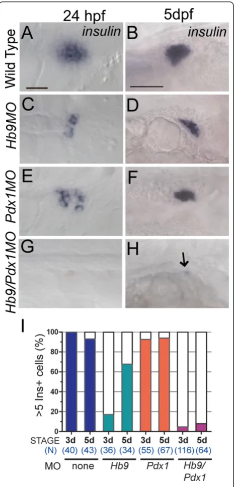
(ins) expression over a time window of 5 days follow-ing morpholino (MO) injection. Consistent with pre-vious studies [18,20,29], hb9 and pdx1 MO-treated embryos (morphants) analyzed by RNA in situ hybridi-zation at 24 hpf had a marked decrease in ins expres-sion (Figure 1C,E, compared to A). As seen in previous analyses ofpdx1morphants, we now show that also in
We next asked the question whether hb9 and pdx1
might cooperate in beta cell formation by coinjecting embryos withhb9andpdx1morpholinos. This resulted in an almost complete lack ofinsexpression at 24 hpf (Figure 1G). This phenotype persisted, and at 5 dpf and 9 dpf, hb9/pdx1 morphants showed virtually no ins
expression (at 5 dpf 92% < 5 inscells, n = 64; Figure 1H,I and at 9 dpf 93% < 5inscells, n = 14).
To exclude that pancreatic development is globally disrupted in the double morphant, we examined expres-sion of the exocrine marker carboxypeptidase A (Cpa). As in the pdx1 morphants [18,19], hb9/pdx1 double morphants show substantial development of exocrine tissue at 4 dpf [see Additional file 1]. We also asked whetherpdx1 expression returns at later stages in mor-pholino-injected embryos, by examining Pdx1 protein in morphants. At 28 hpf and 48 hpf, Pdx1 expression is entirely absent in the developing gut and pancreas of
pdx1 morphants (n = 13 [see Additional file 2A-F]). At 84 hpf during normal development, Pdx1 is strongly expressed in cells of the islet with lower expression in the exocrine pancreas (n = 8 [see Additional file 2G]), while pdx1morphants have low levels of Pdx1 protein in exocrine pancreas and islet at this stage (n = 8 [see Additional file 2H]). Thus, the morphant is not comple-tely devoid of Pdx1 at late stages, but a low level of expression is observed that allows rescue of exocrine development and may account for the recovery of beta cells.
Perturbed endocrine precursor formation inhb9/pdx1
knockdown embryos
Two mechanisms could account for the persistent absence of beta cells in thehb9/pdx1double morphants: endocrine precursors may not be specified, or they may fail to differentiate. To distinguish these possibilities we analyzed green fluorescent protein (GFP) expression in morphant embryos transgenic forTgBAC(NeuroD:EGFP) nl1[30]. These fish express enhanced green fluorescent protein (EGFP) integrated into the NeuroD locus con-tained in a bacterial artificial chromosome (BAC) by homologous recombination. EGFP expression has been reported to recapitulateNeuroDexpression in the ner-vous system. We now confirm that EGFP in TgBAC (NeuroD:EGFP)nl1 embryos is first expressed in dis-persed cells of the prepancreatic endoderm, which migrate posteriorly and cluster to form the principal islet by 24 hpf [see Additional files 3A and 4]. In a sub-set of cells there is coexpression of endocrine hormones [see Additional file 3B-G], recapitulating endogenous
NeuroD expression in islet cell precursors [25,27]. In addition, the stability of EGFP allowed us to trace endo-crine precursors during maturation after the downregu-lation of the endogenousNeuroD.
Figure 1insmRNA expression inhb9andpdx1morphants.In situdetection ofinsmRNA in wild-type embryos(A,B),hb9 morphants(C,D),pdx1morphants(E,F)andhb9/pdx1double morphants(G,H)at 24 h post fertilization (hpf) and 5 days post fertilization (dpf). Inhb9andpdx1morphants the number ofins expressing cells is strongly reduced at 24 hpf as compared to control embryos (A,C,E) but has substantially increased by 5 dpf (D, F). (G,H)insexpression is missing in most double morphants (arrow in H marks a singleinspositive cell). Embryos are shown from ventral (24 hpf) and lateral (5 dpf) view with anterior to the right. Scale bars correspond to 20μm (24 hpf) and 50μm (5 dpf).(I)
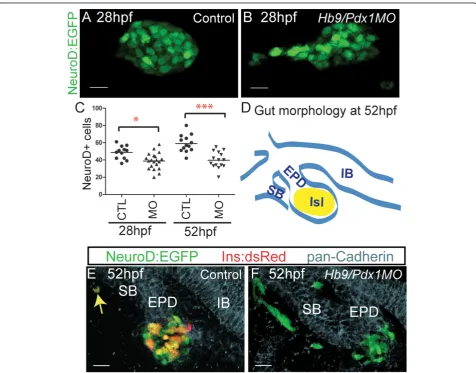
To assess endocrine precursor specification inhb9/ pdx1double morphants, we quantified pancreatic EGFP + cells at 28 hpf, that is, before the emergence of the ventral bud. At 28 hpf, double morphant embryos dis-played a slightly reduced number of EGFP positive cells as compared to controls (Figure 2A-C). In addition, while EGFP+ cells in control embryos formed a compact islet (n = 12), 50% of double morphants (n = 20) showed loosely clustered or even widely separated GFP+ cells (Figure 2B), suggesting disrupted migration. Thus, early endocrine cells were specified in these morphants,
albeit at reduced levels, and the residual endocrine cells displayed defects in migration.
We next examined endocrine cell specification and differentiation at 52 hpf, after the fusion of dorsal and ventral buds. For this analysis we used embryos that were double transgenic for TgBAC(NeuroD:EGFP)nl1
andTg(ins:dsRed)m1018, enabling simultaneous visuali-zation of progenitors and differentiated beta cells by EGFP and dsRed, respectively, and in addition immu-nostained the embryos for pancadherin to delineate gut morphology (Figure 2D-F). In control embryos, the
Figure 2Disrupted precursor specification and differentiation inhb9/pdx1double morphants.(A,B)Projections of confocal image stacks of native enhanced green fluorescent protein (EGFP) expression in control uninjected and inhb9/pdx1morpholino injectedTgBAC(NeuroD:EGFP) nl1embryos at 28 h post fertilization (hpf). In morphants, the clustering of EGFP labeled cells is disrupted (B) and the number of EGFP expressing cells is reduced as compared to controls(C). By 52 hpf,NeuroD:EGFPcell numbers have increased in control embryos, but not changed in the morphants ((C), *P< 0.05, ***P< 0.001 as determined by one-way analysis of variance (ANOVA) with Bonferroni’s post test).(D)
Schematic depicting overall gut morphology in 52 hpf embryos. EPD, extrapancreatic duct; IB, intestinal bulb; ISL, islet; SB, swim bladder.(E,F)
average number of EGFP+ cells increased by 20% between 28 hpf and 52 hpf (from 49 to 59 cells), but these remained essentially constant in number in the double morphants (average 39 cells; Figure 2C). Control embryos had an average of 18 EGFP+/DsRed+ cells (n = 12), compared to double morphants that had few to no DsRed+ cells (0 to 5 EGFP+/DsRed+ cells in 14/14 embryos) (Figure 2E,F). Furthermore, 47% (7/15) of dou-ble morphant embryos showed clusters of EGFP +/DsRed- cells located at a distance from the islet and outside of the pancadherin stained gut epithelium (Fig-ure 2F). Only one ectopic EGFP+ cell was seen in a sin-gle control embryo (n = 12), and this cell also displayed robust DsRed expression (Figure 2E). As DsRed takes 24 h to reach its half maximal fluorescence intensity [31], this likely represents a rare displaced early rather than a late beta cell. In summary, our analyses show that early endocrine cell specification occurred inhb9/pdx1double
morphants, and therefore a deficit in differentiation con-tributes to the absence of ins-expressing cells at later developmental stages. In addition, the formation of sec-ond wave endocrine cells was disrupted, as the number of NeuroD:EGFP cells did not increase after 28 hpf in double morphants.
Loss of late formingNeuroD:EGFPcells inpdx1morphants
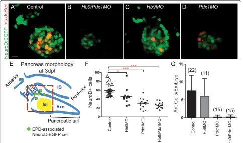
We next analyzed NeuroD:EGFP cells in the islet of morphant embryos at 3 dpf to determine if emergence of second wave endocrine cells was delayed. Strik-ingly, hb9/pdx1 double morphants displayed a persis-tently low number of NeuroD:EGFP+ cells in the principal islet, which even appears reduced relative to earlier time points when only cells in the vicinity of islet and extrapancreatic duct were counted (Figure 3E), and distal cells outside of the gut were not included (Figure 2F).
Figure 3Absence of ventral bud endocrine precursors inhb9/pdx1andpdx1morphant embryos.(A-D)Projections of confocal stacks showing native fluorescence of 3 days post fertilization (dpf)TgBAC(NeuroD:EGFP)nl1;Tg(ins:dsRed)m1018embryos. In uninjected control embryos (A) and inhb9morphants (C), enhanced green fluorescent protein (EGFP)+ cells are found in the islet and in smaller numbers also anterior to the islet. Inhb9/pdx1double morphants (B) andpdx1morphants (D), the number of islet-associated EGFP+ cells is strongly reduced and anterior cells are missing. FewIns:DsRedcells are present inpdx1single morphants at 3 dpf (D), reflecting slow maturation of the DsRed fluorophore.(E)
As Pdx1 is required in second wave endocrine pro-genitors in mouse [13,14], we hypothesized that the defect in late endocrine cell formation in double mor-phants could be due to loss of pdx1only. To assess the individual contributions of pdx1andhb9to late endo-crine cell formation, we quantitated NeuroD cell num-ber in single morphants. At 3 dpf, EGFP+ cells in hb9
morphants were only slightly reduced relative to con-trols, whereas in pdx1morphants EGFP+ cells were reduced by 50%, similar to the number observed in dou-ble morphants (Figure 3A-D, F). In addition, control embryos had EGFP+ cells located anterior to the islet that were absent in double morphant embryos (Figure 3B). These cells occupied a position corresponding to that of previously described late-arising endocrine cells [4,32]. Strikingly, in pdx1 morphants but not in hb9
morphants, the anterior EGFP+ cells were also absent (Figure 3C,D,G). Thus, the formation of late endocrine cells specifically depends on pdx1, while early endocrine cells were specified in the absence of pdx1or hb9 [see Additional file 5].
We also confirmed the persistent deficiency in new endocrine cell formation by examining NeuroD:EGFP+
cells in the principal islet at 5 dpf inpdx1morphants as compared to control embryos, and found thatNeuroD: EGFP cells remained dramatically reduced [see Addi-tional file 6A]. Furthermore, in pdx1morphants (n = 7),
NeuroD:EGFP+ cells in the principal islet expressed
Pdx1 at low levels, as compared to robust expression in controls [see Additional file 6C]. Asins-expressing cells have increased by 3 dpf inpdx1 morphants (Figure 1I), these beta cells originated from early specified, first wave endocrine precursors that differentiate under con-ditions of deficient Pdx1.
Extrapancreatic duct-associatedNeuroD:EGFPcells are migratory endocrine precursors
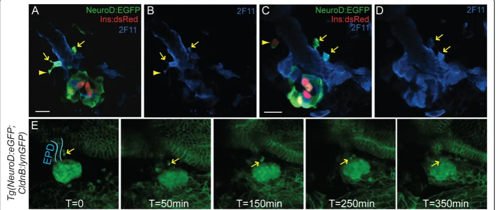
Second wave endocrine cells arise in association with the developing ductal epithelium [10,32]. To precisely localize the site at whichpdx1-dependent anterior endo-crine precursors arise, we immunostained TgBAC(Neu-roD:EGFP)nl1;Tg(ins:dsRed)m1018embryos at 3 dpf for the duct marker 2F11. NeuroD-expressing cells were found along the extrapancreatic duct (EPD), showed coexpression of 2F11, and even appeared to be emerging from the duct, with elongated processes characteristic of delaminating and migrating cells (Figure 4A-D). In sup-port of their identity as endocrine precursors, occasion-ally such anterior NeuroD:EGFP cells expressed low levels of Ins:DsRedand thus correspond to newly differ-entiating beta cells (Figure 4C [see Additional file 7A]). This was confirmed by insulin (Ins) antibody staining [see Additional file 7C].
To document the emergence of these anterior endo-crine cells, we developed a timelapse imaging technique based on a method described for imaging the central
nervous system [33]. The large yolk cell, which normally obstructs the deeply located pancreas, was paralyzed and then extracted to establish a clear ventral view. This procedure did not interfere with morphogenetic pro-cesses. Using this novel method, we performed time-lapse analysis ofTgBAC(NeuroD:EGFP)nl1;Tg(-8.0cldnb: lynEGFP)zf106embryos at 3 dpf. In double transgenic embryos, we recorded active delamination and clustering ofNeuroD:EGFP-positive cells adjacent to the extrapan-creatic duct and migration of cells towards the principal islet (Figure 4E [see Additional file 8]). Thus, our method allows the visualization of dynamic cell beha-viors in the context of the developing organ in real time, and unambiguously demonstrates that new endo-crine cells emerge from the duct epithelium and join the principal islet.
Having established that second wave endocrine pre-cursors emerge from the duct, we analyzed if their absence inpdx1 morphants could be caused by a global duct defect. At 72 hpf, duct development appeared nor-mal in morphant embryos, as demonstrated by a nornor-mal pattern of GFP-labeled duct cells inTg(-3.5nkx2.2a:GFP) ia3 transgenics [34] [see Additional file 7D-G]. Further-more, Tg(-8.0cldnb:lynEGFP)zf106 embryos, in which EGFP outlines membranes of the gut epithelium, revealed normal cellular structure of the extrapancreatic duct in pdx1morphants as compared to controls [see Additional file 7H]. We examined Pdx1 expression in these embryos to address whether Pdx1 might have a
role within the duct as new endocrine cells emerge. Pdx1 was highly expressed throughout the extrapancrea-tic duct and proximal pancreas in control embryos, while expression was very weak inpdx1morphants [see Additional file 7J]. This analysis indicates that depletion of Pdx1 eliminated the ability of duct-associated cells to give rise to endocrine precursors and argues against an overall perturbation of the duct.
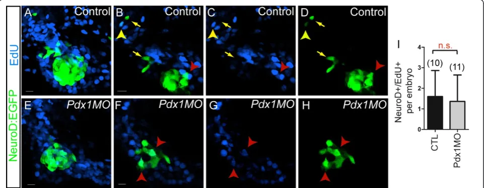
Post-mitotic origin of late emerging endocrine cells
We next asked the question whether late endocrine pre-cursors arise from a mitotically active cell population. We tested this by incubating control and pdx1 mor-phant TgBAC(NeuroD:EGFP)nl1embryos with nucleo-tide analog EdU from 28 hpf to 76 hpf. This extended incubation is expected to label dividing NeuroD+ endo-crine precursors and dividing duct cells whose progeny differentiated into NeuroD-expressing endocrine cells. At 76 hpf, an average of 1.6NeuroD:EGFPcells/embryo in control embryos were EdU positive, and their number in pdx1 morphants was not significantly different (1.4 NeuroD+/EdU+ cells/embryo (Figure 5I). In addition, rare EdU+/NeuroD+ cells were found at the periphery of the islet and distal to the islet, along the extrapan-creatic duct in control embryos (Figure 5A-D). In sum-mary, the NeuroD+/EdU+ double-labeled cells (average 1.6/embryo) cannot account for the appearance of ten additional NeuroD-expressing cells that emerge in embryos between 28 hpf and 76 hpf. Furthermore, most
Figure 5Contribution of cell proliferation to late endocrine cell expansion.TgBAC(NeuroD:EGFP)nl1embryos incubated with EdU from 28 to 76 h post fertilization (hpf), followed by EdU detection and green fluorescent protein (GFP) antibody staining.(A, E)Confocal projections and single plane views(B-D, F-H), showing pancreatic region of control (A-D), andpdx1morphant (E-H) embryos. (B-D) In control embryos, EdU/ NeuroD:enhanced green fluorescent protein (EGFP) colabeled cells are located at the periphery of the islet (red arrowhead) and in the region of the extrapancreatic duct (EPD) (yellow arrowhead). Most islet cells are EdU- (C compared to D) and additional EdU-/NeuroD:EGFP+ cells are found along the EPD (yellow arrows). (F-H) Inpdx1morphants, few EdU/NeuroD:EGFP colabeled cells are found within the islet (red arrowhead).
NeuroD+ cells outside of the principal islet were EdU-, indicating that late endocrine cells appearing after 28 hpf arose from post-mitotic precursors. Thepdx1 mor-phant showed a similar low number of NeuroD+/EdU+ cells, which were all associated with the islet (Figure 5E-H). Our data therefore suggest that late endocrine cells emerge predominantly from a postmitotic cell type.
As proliferating cells were found directly adjacent to the NeuroD:EGFP+ islet, we next used the Tg(ela3l: EGFP)gz2allele that labels exocrine pancreas [35] to define their identity. Incubation of embryos from 28 to 76 hpf with EdU, followed by a 12-h chase, resulted in extensive EdU labeling of the GFP+ exocrine pancreas in control as well as in pdx1 morphant embryos [see Additional file 9]. Therefore, ventral bud cells forming the exocrine pancreas are highly proliferative during this time period, and in pdx1morphants no general prolif-eration defect exists in the pancreas. We conclude that beta cell recovery in pdx1morphants is not accompa-nied by activated proliferation in progenitors or differen-tiated beta cells, but occurs through delayed precursor differentiation.
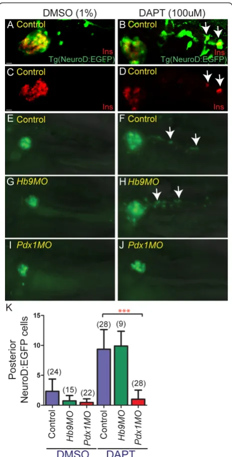
Notch-responsive beta cell progenitors requirepdx1
The zebrafish intrapancreatic duct is a ventral bud deri-vative containing Notch-responsive endocrine progeni-tors that begin to differentiate and assemble into secondary islets after 3 dpf [5,7]. Blocking Notch signal-ing after 3 dpf was previously shown to induce differen-tiation of endocrine cells from duct-associated progenitors [5,7]. We assessed whether the latent endo-crine progenitors associated with the intrapancreatic duct also arise in apdx1-dependent manner.
To visualize the emergence of endocrine cells from the intrapancreatic duct, we first used a Notch inhibitor (N-[N-(3,5-difluorophenacetyl)-L-alanyl]-S-phenylglycine
t-butyl ester (DAPT)) to induce endocrine differentia-tion andNeuroD:EGFPexpression in latent progenitors. In control experiments, treatment of TgBAC(NeuroD: EGFP)nl1embryos with DAPT from 3 to 5 dpf caused an increase of cells in the principal islet and a fourfold increase of GFP labeled cells within the pancreatic tail as compared to vehicle-treated larvae (Figure 6A,E,F,K). Overlapping expression of Ins and GFP following DAPT treatment confirmed that newly-inducedNeuroD:EGFP
cells in the pancreatic tail can differentiate into beta cells (Figure 6B,D).
We next analyzed if these Notch-responsive cells were present inpdx1 andhb9morphants. We found virtually noNeuroD:EGFP induction following DAPT treatment inpdx1morphants, while DAPT treatment inhb9 mor-phants resulted in an increased number of NeuroD: EGFP+cells similar to controls (Figure 6G-K). Immu-nostaining for duct marker 2F11 indicated that
Figure 6Notch-responsive endocrine progenitors are absent in
intrapancreatic duct formation in pdx1morphants was comparable to controls [see Additional file 10A]. Over-all, this indicates that Notch-responsive progenitors for late-forming endocrine cells, that contribute new cells to both principal islet expansion and secondary islet forma-tion, requirepdx1(Figure 7).
Discussion
In this work, we determined that both hb9and pdx1
have roles in early beta cell formation and thatpdx1is distinctly required for second wave endocrine progenitor specification, which generates endocrine cells that expand the principal islet and form secondary islets. Prior studies of pdx1 morphant zebrafish did not
quantitatively assess endocrine cell dynamics, and there-fore the requirement for Pdx1 in formation of progeni-tors for late endocrine cells went unrecognized. Our new imaging approach allowed us to visualize the beha-vior of emerging endocrine cells in real time and define the source of the late endocrine progenitor cells. Specifi-cally, we show that they originate and delaminate from the ductal epithelium in a process that resembles forma-tion of second wave endocrine cells in mammals. Our findings identify a genetic requirement for formation of late endocrine cells, establish the site where the latent postmitotic cell type that gives rise to the endocrine progenitor resides, and describe a novel method to visualize morphogenetic events that occur during islet formation.
Sources of beta cell recovery
Embryos deficient in eitherpdx1orhb9alone display an initial decrease inins expression, followed by restoration of some ins-expressing cells by 5 dpf. We show here that in double morphants such a recovery does not occur, and define pdx1 andhb9dependent steps in the generation and differentiation of two distinct endocrine cell populations arising in the early and late embryo. The early appearance of NeuroD:EGFP-expressing islet cells demonstrated that committed, first wave endocrine cells of the dorsal bud were specified despite the absence of hb9and/or pdx1. By contrast, second wave endocrine cells strictly depend on pdx1. The apparent recovery of beta cells in pdx1 morphants thus results from delayed differentiation of early arising, dorsal bud-derived endocrine precursors in conditions of severely reduced Pdx1. Since we have detected hb9 in ventral bud-derived beta cells (D Mayer, RA Kimmel, unpub-lished results), andHb9knockout causes global beta cell deficits in mouse, delayed beta cell differentiation inhb9
morphants can be explained by restoration of protein function due to dilution of morpholino at the time when late endocrine cells emerge.
Progenitors for late endocrine cell expansion
During organ development, progenitors are often set aside during the early stages to undergo later expansion and differentiation. In prior studies of beta cell prolifera-tion during zebrafish embryogenesis using nucleotide analogs or proliferating cell nuclear antigen (PCNA) antibody, dividing beta cells were detected only very rarely or not at all [3,6,18,36,37]. To analyze and define the proliferative status of cells generating the late-arising endocrine cells, we used a protocol in which embryos were exposed to a long pulse of EdU covering the time window when late endocrine cells start emerging (from 28 to 76 hpf). This captures all cell divisions and also labels cells that proliferate prior to endocrine
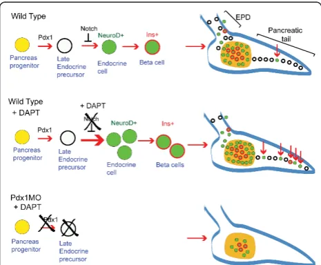
Figure 7Schematic of Notch-responsive progenitors. In wild-type embryos (top), an early pancreatic progenitor (yellow) gives rise to early endocrine cells (green) as well as progenitors for late endocrine cells (white) located in the extrapancreatic duct (EPD) and pancreatic tail. Notch signaling maintains these latent progenitors in an undifferentiated state. Inhibition of Notch signaling by treatment withN-[N-(3,5-difluorophenacetyl)-L-alanyl]-S -phenylglycinet-butyl ester (DAPT) (center) activates differentiation of the latent progenitors, leading to new endocrine cell formation in the principal islet and pancreatic tail (red arrows). Inpdx1 morphants (bottom), early endocrine specification occurs but Notch-responsive progenitors for late endocrine cells are absent or hindered in differentiation, which can be demonstrated by treatment with DAPT.
differentiation. Indeed, due to the prolonged labeling time, we observed a higher proportion of cells that had undergone proliferation as compared to previous stu-dies, indicating that dividing progenitors that gave rise to beta cells as well as the rare beta cell mitoses were revealed. Nevertheless, the low number of EdU+ endo-crine precursors and beta cells that we detected in nor-mal development shows that the vast majority of late arising endocrine cells (appearing between 28 hpf and 76 hpf) have not undergone mitosis during the labeling period. Therefore, these cells differentiate directly from a precursor with low proliferative activity. Similarly, in the pdx1morphant, proliferating beta cells or mitotic progenitors contribute only a minor fraction to the beta cells emerging after 28 hpf.
These studies indicate that, during normal develop-ment, the cell type that gives rise to late endocrine cells represents a subtype of ventral bud cells that is already set aside at 28 hpf, and that slowly divides in contrast to the rapidly dividing cells that form the exocrine pan-creas. Previous analysis showed that the ventral pancrea-tic bud contains ptf1a-expressing cells that form the exocrine pancreas and a distinct population that responds to Notch and gives rise to endocrine and duct cells [5]. Our data indicates that within the non-exo-crine cells, there is a population of non-proliferative cells that comprises latent endocrine progenitors.
Function of Pdx1 in late endocrine cells
A major finding of this work is the requirement for
pdx1in specification of all late forming endocrine cells. Expression ofPdx1 is very dynamic in the developing pancreas and genetic analyses using conditional and hypomorphicPdx1 mutant alleles in mouse have already indicated that Pdx1 is important in various specification and differentiation events [38-41]. In zebrafish, fate-mapping studies indicated that a spatial separation of the future pancreatic DB versus VB progenitors is already established shortly after gastrulation (10 hpf) [42,43]. Medium levels ofpdx1 expression during early somitogenesis (10 to 15 hpf) correlate with VB fate spe-cification [44]. Thus, already these early differences in
pdx1expression could provide permissive or instructive cues for the specification of latent progenitors that later give rise to second wave endocrine cells.
The developing extrapancreatic duct and the proximal pancreas express high levels of Pdx1 from 48 hpf ([32] and this study). In pdx1 morpholino-treated embryos, Pdx1 is diminished at least until 3 dpf, but overall for-mation of the extrapancreatic duct is not affected. An absence of duct-associated progenitors for second wave endocrine cells could result from duct cells being com-mitted to a non-endocrine fate due to an early defi-ciency in Pdx1. Alternatively, deficient Pdx1 in duct
cells might impede the generation of endocrine precur-sors from a latent progenitor. Several lines of research suggests that pancreatic ductal epithelium is heteroge-neous and contains a limited numbers of cells that have the potential for endocrine differentiation [45]. We have here defined a strategy that eliminates endocrine cell formation without affecting duct morphology, which will be useful to help further characterize the latent progeni-tor cell type.
Morphogenesis of late endocrine cells
Our transgenic and timelapse analyses provide new insights into the behavior of late-arising endocrine cells.
NeuroD:EGFP-positive cells associated with the extrapan-creatic duct defined by 2F11 antibody staining exhibited protrusions characteristic of delaminating cells. We devel-oped a method for maintaining embryo viability after yolk removal, which allowed for the first time a clear visualiza-tion of developing late endocrine cells in living embryos. We delineated pancreatic progenitors in the context of the pancreatic duct and digestive system, and recorded migra-tion ofNeuroD:EGFPpositive endocrine progenitors as they emerged from the duct and moved to the principal islet. This method provides the basis for future analysis of molecular regulators of these complex morphogenetic events during pancreatic islet development.
Conclusions
These studies establish thatpdx1 is essential for gener-ating an endocrine cell progenitor population associated with the pancreatic duct in zebrafish. We show that for-mation of latent progenitors in the extrapancreatic and intrapancreatic duct requirespdx1, and provide evidence that they are slowly dividing cells, and are thus distinct from rapidly proliferating cells that form the exocrine tissue. Further, these latent progenitors differentiate in response to Notch inhibition. Our findings open up new approaches for examination of the specification and behavior of endocrine progenitors during zebrafish embryogenesis, in which these processes can now be visualized, and where large-scale chemical and pharma-cologic screening can be applied. This can in turn pro-vide clues as to how developmental programs can be harnessed and reactivated to generate replacement beta cells for more efficient treatment of diabetes.
Methods
Zebrafish transgenic lines
Morpholino injection
The following previously validated morpholinos, targeted against the initiation codon of pdx1 and hb9, respec-tively, were obtained from Gene Tools (Philomath, OR, USA):pdx1MO: 5’ -GATAGTAATGCTCTTCCCGATT-CAT-3’ [18,19]; hb9MO: 5’ -TTTTTAGATTTCTC-CATCTGGCCCA-3’[20].
Each morpholino was prepared at a concentration of 1.5 mM. This solution was diluted to 750 μM in water and 2 to 8 ng of morpholino in a volume of approxi-mately 1.5 nl was injected into one-cell stage embryos. In generating double morphants, the concentration of individual morpholinos was halved to keep the total amount of morpholino injected the same as for single morphants.
In situhybridization
Whole mountin situ hybridization was performed as described [47]. Digoxigenin antisense probes (Roche, Vienna, Austria) were synthesized forinsulin[48]. As pre-cise cell counting in older embryos becomes difficult with increasing cell numbers in a tight cluster, embryos with cell count > 5 were grouped together for quantitation.
Antibody staining
Embryos were fixed in 4% paraformaldehyde (PFA) at 4°C (2 h to overnight, depending on stage) followed by protei-nase K (Carl Roth, Karlsruhe, Germany) digestion (10μg/ ml in phosphate-buffered saline (PBS)), post fixation in 4% PFA, and incubation in blocking buffer containing 1% bovine serum albumin (BSA, AppliChem, Darmstadt, Ger-many), 2% Sheep serum (Sigma, Vienna, Austria), 1% Tri-ton, and 1% dimethylsulfoxide (DMSO). Primary antibody was diluted in blocking buffer followed by overnight incu-bation at 4°C. Embryos were washed in PBS/BSA/Triton, and incubated overnight at 4°C in secondary antibody diluted 1:1,000 in blocking buffer, followed by extensive washing in PBST (PBS + 0.1% Tween 20). Primary antisera and dilutions were: guinea pig anti-insulin (1:200, Dako, Vienna, Austria), rabbit anti-glucagon (1:200, Dako), rabbit anti-pan-cadherin (1:500, Sigma), mouse anti-2F11 (1:100, generous gift from Julian Lewis, London Research Insti-tute, UK), rabbit anti-somatostatin (1:200, Dako), rabbit anti-carboxypeptidase A (1:200, Chemicon, Vienna, Aus-tria), mouse anti-GFP (1:200, Roche), rabbit anti-GFP (1:200, Torrey Pines Biolabs, East Orange, NJ, USA), gui-nea pig polyclonal anti-Pdx1 (1:200, generous gift from Chris Wright, Vanderbilt University, TN, USA). Alexa-conjugated secondary antibodies (1:1,000 dilution) were from Invitrogen (Lofer, Austria).
Cell labeling with EdU
EdU staining was performed as previously described [37]. In brief, reagents for the Click-iT EdU Alexa Fluor
647 Assay (Invitrogen) were prepared according to man-ufacturer’s protocol. Embryos were incubated in 0.5 mM EdU/0.4% DMSO in egg water (0.3 g/L Red Sea Coral Pro Salt in reverse osmosis H2O) followed by fixation in
4% PFA. Embryos were then washed in PBST, dehy-drated with a methanol wash, and incubated in metha-nol at -20°C for 2 h. For the detection assay, larvae were rehydrated through a methanol series into PBST and manually deyolked. The larvae were treated with protei-nase K (10μg/ml) for 45 min, refixed in 4% PFA for 20 min, washed in PBST, and permeabilized in 1% DMSO/ 0.5% Triton/PBS. Incubation in Click-iT reaction cock-tail was performed for 2 h at room temperature fol-lowed by rinses in PBST. Following the EdU detection reaction, larvae were incubated in blocking buffer and immunostaining was performed as described above.
Microscopy
Tg(ins:dsRed)m1018 and TgBAC(NeuroD:EGFP)nl1
embryos for direct fluorescence imaging were fixed in 4% PFA at room temperature for 1 to 2 h (48 hpf and younger) or 2 to 3 h (older than 48 hpf), rinsed three times with PBST followed by manual yolk removal. Transgene and antibody immunostaining were imaged with a Zeiss LSM5 Exciter (Carl Zeiss, Vienna, Austria) confocal laser microscope, using a 40 × water immer-sion objective. Stacks of optical sections were recorded with a z-step ranging from 1 to 2 μm. Larvae for live imaging were anesthetized using 1 × Tricaine mesylate (0.003%, Sigma), immobilized in low melt agarose (1.5%, Biozym, Vienna, Austria) and imaged on a Leica DM6000B microscope (Leica Microsystems, Vienna, Austria) equipped with a SPOT-RT3 digital camera (Diagnostic Instruments, Inc., Sterling Heights, MI, USA), using a 20 × water objective. Fluorescence stack and differential interference contrast (DIC) images were captured using Visiview software (Visitron Systems, Puchheim, Germany). Fluorescence brightfield compo-site images were prepared with ImageJ (http://rsbweb. nih.gov/ij).
Confocal timelapse analyses
(1 × final, Invitrogen), glucose (10 mM final, Appli-Chem) and 1 × Tricaine mesylate. Embryos were imaged for 5 to 10 h in a temperature-controlled room at 28°C. Images were collected every 5 min, a total z-stack of 60 to 70μm was acquired with a slice interval of 2.5 to 3.5 μM. Images were captured using a 40 × water objective on a Zeiss LSM Exciter5 equipped with Zen 2008 soft-ware (Carl Zeiss), followed by processing and assembly of still images and videos using LSM Image Browser (Carl Zeiss) and ImageJ.
Quantitative image analysis
NeuroD cell numbers in 24 hpf to 3 dpf embryos were quantitated using Imaris 7.1.1 software (Bitplane, Zurich, Switzerland). The spot detection algorithm was applied to three-dimensional reconstructions of samples to iden-tify cells using a spot size of 3 to 4 μm. Spots were fil-tered, confirmed and edited by visual inspection, and cell counts transferred to Prism (GraphPad Software, La Jolla, CA, USA) for statistical analyses. For proliferation analyses, spot detection was similarly performed for EdU+ and NeuroD+ cells, double labeling of cells was determined using the ‘colocalize spots’ function of Imaris with a threshold value of 3μm.
DAPT treatment
DAPT (Sigma) was prepared as a 10 mM stock solution in DMSO. Larvae were incubated in DAPT diluted to 100 μM in egg water starting at 3 dpf for 48 h. The DAPT solution was replaced after 24 h. The embryos were then rinsed in egg water and incubated an addi-tional 12 h. Control embryos were incubated in 1% DMSO in egg water.
Additional material
Additional file 1: Exocrine pancreas development inhb9/pdx1
double morphants. Confocal image projection of the pancreas of 4 days post fertilization (dpf) embryos immunostained for
carboxypeptidase A (Cpa) and insulin (Ins).hb9/pdx1double morphant embryos(B)with severely reduced Ins (insets,(A), compared to B), show Cpa expression comparable to control embryos (A). All are in
ventrolateral view. Scale bar = 30μM.
Additional file 2: Pdx1 protein expression inpdx1morphants. Confocal projections of control andpdx1morpholino-injected embryos at 28 h post fertilization (hpf)(A-C)and 48 hpf(D-F), immunostained for Pdx1. (A-C) Control embryos express Pdx1 in the developing gut and pancreas. (D-F)pdx1morphant embryos have no detectable Pdx1 expression. (B,E) Overlay of bright field with confocal projection of Pdx1 antibody staining, (C,F) single channel showing Pdx1 antibody. Dashed circle (E) indicates islet as determined from bright field images. Non-specific labeling by this antibody of somites lateral to the gut (asterisks) has been previously described [49]. Ventral view. Scale bar = 10μM. Confocal projections(G,H)and single plane views(I-L)of control and pdx1morphants at 84 hpf, immunostained for Pdx1 and
carboxypeptidase A (Cpa) to indicate the exocrine pancreas. Control embryos show robust Pdx1 expression in the islet and weaker expression in the exocrine pancreas (G,I,K).pdx1morphant embryos have low-level
Pdx1 expression in the islet and exocrine pancreas (H,J,L). Lateral view, anterior to left. Scale bar = 30μM.
Additional file 3: NeuroD: enhanced green fluorescent protein (EGFP) cells are endocrine precursors.(A)TgBAC(NeuroD:EGFP)nl1 embryos, mounted dorsal side up, imaged by confocal timelapse microscopy starting at 19 h post fertilization (hpf). Images were captured every 15 min. Asterisks indicate fixed points of the embryo determined from the bright field image. Anterior is to the left.(B-G)Confocal image projection of 24 hpfTgBAC(NeuroD:EGFP)nl1embryo immunostained for green fluorescent protein (GFP) and insulin (Ins) (B-D) and GFP and somatostatin (Sst) (E-G), showing overlap of NeuroD:EGFP expression with islet hormones in a subset of cells. All are ventral view. Scale bar = 15μM.
Additional file 4: Migration of early endocrine cells. Confocal timelapse imaging ofTgBAC(NeuroD:EGFP)nl1embryo beginning at 19 h post fertilization (hpf). Confocal stacks were collected every 15 min. Dorsal view, anterior to the left. Still images from this file were used in Additional file 3A.
Additional file 5: Additional table 1. NeuroD: enhanced green fluorescent protein (EGFP) cells at 28 h post fertilization (hpf) in control, hb9- andpdx1- morphants.
Additional file 6: Larval NeuroD:EGFP and Pdx1 expression. Confocal projections(A,D)and single plane views(B,C,E,F)of control (A-C) and pdx1morpholino injected (D-F)TgBAC(NeuroD:EGFP)nl1embryos at 5 days post fertilization (dpf), immunostained for Pdx1 and green fluorescent protein (GFP). (A-C) In control embryos, robust Pdx1 expression in the islet overlaps with GFP. (D-F)pdx1morphant embryos have reduced Pdx1 expression and GFP+ cells in the islet as compared to control. Lateral view, anterior to left. Scale bar = 10μM.
Additional file 7: Beta cell differentiation and duct development.(A, B)Single plane, ventral view ofTgBAC(NeuroD:EGFP)nl1;Tg(ins:dsRed) m1018control embryo at 72 h post fertilization (hpf). With the red channel overexposed, anterior enhanced green fluorescent protein (EGFP)+ cells with low levels of InsDsRed can be detected (arrow).(C)
Confocal projection ofTgBAC(NeuroD:EGFP)nl1embryo at 72 hpf immunostained for green fluorescent protein (GFP) and insulin (Ins). The Ins signal is overexposed to show low-expressing cells anterior to the principal islet. Scale bar = 10μM.(D-G)Confocal projection of 84 hpfTg (-3.5nkx2.2a:GFP)ia3embryos immunostained for GFP and Ins. GFP expression delineates developing duct in control (D,F), andpdx1 morphant (E,G) embryos. EPD, extrapancreatic duct; IPD, intrapancreatic duct. Ventral view. Scale bar = 15μM.(H-K)Single plane views of 72 hpf Tg(-8.0cldnb:lynEGFP)zf106embryos immunostained for Pdx1 and GFP. Control embryos show robust Pdx1 expression in the proximal pancreas and EPD (H,J), as compared to weak expression inpdx1morphants (I,K). (J) and (K) represent the red channel only from (H) and (I). Ventral view, anterior to left. Scale bar = 10μM.
Additional file 8: NeuroD:enhanced green fluorescent protein (EGFP) cells migrate from duct to islet. Timelapse confocal series of a 3 days post fertilization (dpf)TgBAC(NeuroD:EGFP)nl1;Tg(-8.0cldnb:lynEGFP) zf106embryo imaged for 6 h from a ventral view with yolk removed. Images were collected every 5 min. Migrating cells are indicated by arrows in the initial frames. Still images from this file were used in Figure 4E.
Additional file 9: Proliferation during exocrine pancreas formation. Tg(ela3l:EGFP)gz2[35] embryos labeled with EdU from 24 to 72 h post fertilization (hpf), and fixed at 84 hpf. EdU detection (blue) was followed by antibody staining to label Elastase (Ela)-expressing exocrine cells (Ela: green fluorescent protein (GFP), green) and beta cells (insulin (Ins), red).
(A,C)Three-dimensional confocal projections showing composite of Ela: GFP, EdU and Ins in control (A), andpdx1morphant embryos (C) at 84 hpf. EdU labels exocrine pancreas extensively in control and morphants.
Additional file 10: Larval duct morphology. Confocal projection of 6 days post fertilization (dpf)TgBAC(NeuroD:EGFP)nl1embryos
immunostained for green fluorescent protein (GFP) and 2F11. In control embryos(A), 2F11 positive cells surround GFP+ islet cells and extend into the pancreatic tail. Single NeuroD+ cells can be found in the pancreas tail (arrow).(B)pdx1morphant embryos, with fewer GFP+ cells in the islet, have similar 2F11 expression around the islet and in the pancreatic tail (arrows). Lateral view. Scale bar = 30μM.
Acknowledgements
We thank Wolfgang Driever for general support during the initial phase of this project and for theTg(ins:dsRed)m1018fish line, Julian Lewis for the 2F11 antibody, Teresa Nicolson for theTgBAC(NeuroD:EGFP)nl1fish line, Petra Haas and Darren Gilmour for theTg(-8.0cldnb:lynEGFP)zf106fish line, and Chris Wright for the Pdx1 antibody. We are grateful to Mayra Eduardoff for technical assistance, Tanja Walsen, Sonja Toechterle and Dzenana Tufegdzic for transgenic zebrafish care, and Pia Aanstad and Carmen Birchmeier for critical reading of the manuscript and helpful discussions. This work was funded by the University of Freiburg and Austrian Science Fund (FWF) grants P20492 and M1150 to DM and RAK, respectively.
Author details
1Institute of Molecular Biology/CMBI; Leopold-Francis University,
Technikerstrasse 25, A-6020 Innsbruck, Austria.2Institute of Immunobiology, LFA, Kantonsspital St. Gallen, Rorschacherstrasse 95, CH-6900 St. Gallen, Switzerland.3Growth and Development, Biozentrum, University of Basel, Klingelbergstrasse 50/70, CH-4056 Basel, Switzerland.
Authors’contributions
RAK and DM designed the research. EE, RAK, LO, AW and DM performed research and analyzed data. RAK and DM wrote the paper. All authors read and approved the final manuscript.
Competing interests
The authors declare that they have no competing interests.
Received: 12 September 2011 Accepted: 31 October 2011 Published: 31 October 2011
References
1. Guney MA, Gannon M:Pancreas cell fate.Birth Defects Res C Embryo Today 2009,87:232-248.
2. Solar M, Cardalda C, Houbracken I, Martin M, Maestro MA, De Medts N, Xu X, Grau V, Heimberg H, Bouwens L, Ferrer J:Pancreatic exocrine duct cells give rise to insulin-producing beta cells during embryogenesis but not after birth.Dev Cell2009,17:849-860.
3. Hesselson D, Anderson RM, Beinat M, Stainier DY:Distinct populations of quiescent and proliferative pancreatic beta-cells identified by HOTcre mediated labeling.Proc Natl Acad Sci USA2009,106:14896-14901. 4. Field HA, Dong PD, Beis D, Stainier DY:Formation of the digestive system
in zebrafish. II. Pancreas morphogenesis.Dev Biol2003,261:197-208. 5. Wang Y, Rovira M, Yusuff S, Parsons MJ:Genetic inducible fate mapping in
larval zebrafish reveals origins of adult insulin-producing {beta}-cells. Development2011,138:609-617.
6. Moro E, Gnugge L, Braghetta P, Bortolussi M, Argenton F:Analysis of beta cell proliferation dynamics in zebrafish.Dev Biol2009,332:299-308. 7. Parsons MJ, Pisharath H, Yusuff S, Moore JC, Siekmann AF, Lawson N, Leach SD:Notch-responsive cells initiate the secondary transition in larval zebrafish pancreas.Mech Dev2009,126:898-912.
8. Chen S, Li C, Yuan G, Xie F:Anatomical and histological observation on the pancreas in adult zebrafish.Pancreas2007,34:120-125.
9. Bonal C, Herrera PL:Genes controlling pancreas ontogeny.Int J Dev Biol 2008,52:823-835.
10. Pan FC, Wright C:Pancreas organogenesis: from bud to plexus to gland. Dev Dyn2011,240:530-565.
11. Offield MF, Jetton TL, Labosky PA, Ray M, Stein RW, Magnuson MA, Hogan BL, Wright CV:PDX-1 is required for pancreatic outgrowth and differentiation of the rostral duodenum.Development1996,122:983-995.
12. Ahlgren U, Jonsson J, Edlund H:The morphogenesis of the pancreatic mesenchyme is uncoupled from that of the pancreatic epithelium in IPF1/PDX1-deficient mice.Development1996,122:1409-1416. 13. Oliver-Krasinski JM, Kasner MT, Yang J, Crutchlow MF, Rustgi AK,
Kaestner KH, Stoffers DA:The diabetes gene Pdx1 regulates the transcriptional network of pancreatic endocrine progenitor cells in mice. J Clin Invest2009,119:1888-1898.
14. Burlison JS, Long Q, Fujitani Y, Wright CV, Magnuson MA:Pdx-1 and Ptf1a concurrently determine fate specification of pancreatic multipotent progenitor cells.Dev Biol2008,316:74-86.
15. Murtaugh LC:Pancreas and beta-cell development: from the actual to the possible.Development2007,134:427-438.
16. Harrison KA, Thaler J, Pfaff SL, Gu H, Kehrl JH:Pancreas dorsal lobe agenesis and abnormal islets of Langerhans in Hlxb9-deficient mice.Nat Genet1999,23:71-75.
17. Li H, Arber S, Jessell TM, Edlund H:Selective agenesis of the dorsal pancreas in mice lacking homeobox gene Hlxb9.Nat Genet1999, 23:67-70.
18. Yee NS, Yusuff S, Pack M:Zebrafish pdx1 morphant displays defects in pancreas development and digestive organ chirality, and potentially identifies a multipotent pancreas progenitor cell.Genesis2001, 30:137-140.
19. Lin JW, Biankin AV, Horb ME, Ghosh B, Prasad NB, Yee NS, Pack MA, Leach SD:Differential requirement for ptf1a in endocrine and exocrine lineages of developing zebrafish pancreas.Dev Biol2004,270:474-486. 20. Wendik B, Maier E, Meyer D:Zebrafish mnx genes in endocrine and
exocrine pancreas formation.Dev Biol2004,268:372-383.
21. Apelqvist A, Li H, Sommer L, Beatus P, Anderson DJ, Honjo T, Hrabe de Angelis M, Lendahl U, Edlund H:Notch signalling controls pancreatic cell differentiation.Nature1999,400:877-881.
22. Kim W, Shin YK, Kim BJ, Egan JM:Notch signaling in pancreatic endocrine cell and diabetes.Biochem Biophys Res Commun2010,392:247-251. 23. Zecchin E, Filippi A, Biemar F, Tiso N, Pauls S, Ellertsdottir E, Gnugge L,
Bortolussi M, Driever W, Argenton F:Distinct delta and jagged genes control sequential segregation of pancreatic cell types from precursor pools in zebrafish.Dev Biol2007,301:192-204.
24. Gradwohl G, Dierich A, LeMeur M, Guillemot F:neurogenin3 is required for the development of the four endocrine cell lineages of the pancreas. Proc Natl Acad Sci USA2000,97:1607-1611.
25. Naya FJ, Huang HP, Qiu Y, Mutoh H, DeMayo FJ, Leiter AB, Tsai MJ: Diabetes, defective pancreatic morphogenesis, and abnormal enteroendocrine differentiation in BETA2/neuroD-deficient mice.Genes Dev1997,11:2323-2334.
26. Itkin-Ansari P, Marcora E, Geron I, Tyrberg B, Demeterco C, Hao E, Padilla C, Ratineau C, Leiter A, Lee JE, Levine F:NeuroD1 in the endocrine pancreas: localization and dual function as an activator and repressor.Dev Dyn 2005,233:946-953.
27. Soyer J, Flasse L, Raffelsberger W, Beucher A, Orvain C, Peers B, Ravassard P, Vermot J, Voz ML, Mellitzer G, Gradwohl G:Rfx6 is an Ngn3-dependent winged helix transcription factor required for pancreatic islet cell development.Development2010,137:203-212.
28. Schwitzgebel VM, Scheel DW, Conners JR, Kalamaras J, Lee JE, Anderson DJ, Sussel L, Johnson JD, German MS:Expression of neurogenin3 reveals an islet cell precursor population in the pancreas.Development2000, 127:3533-3542.
29. Huang H, Liu N, Lin S:Pdx-1 knockdown reduces insulin promoter activity in zebrafish.Genesis2001,30:134-136.
30. Obholzer N, Wolfson S, Trapani JG, Mo W, Nechiporuk A, Busch-Nentwich E, Seiler C, Sidi S, Söllner C, Duncan RN, Boehland A, Nicolson T:Vesicular glutamate transporter 3 is required for synaptic transmission in zebrafish hair cells.J Neurosci2008,28:2110-2118.
31. Baird GS, Zacharias DA, Tsien RY:Biochemistry, mutagenesis, and oligomerization of DsRed, a red fluorescent protein from coral.Proc Natl Acad Sci USA2000,97:11984-11989.
32. Dong PD, Munson CA, Norton W, Crosnier C, Pan X, Gong Z, Neumann CJ, Stainier DY:Fgf10 regulates hepatopancreatic ductal system patterning and differentiation.Nat Genet2007,39:397-402.
34. Pauls S, Zecchin E, Tiso N, Bortolussi M, Argenton F:Function and regulation of zebrafish nkx2.2a during development of pancreatic islet and ducts.Dev Biol2007,304:875-890.
35. Wan H, Korzh S, Li Z, Mudumana SP, Korzh V, Jiang YJ, Lin S, Gong Z: Analyses of pancreas development by generation of gfp transgenic zebrafish using an exocrine pancreas-specific elastaseA gene promoter. Exp Cell Res2006,312:1526-1539.
36. Pisharath H, Rhee JM, Swanson MA, Leach SD, Parsons MJ:Targeted ablation of beta cells in the embryonic zebrafish pancreas usingE. coli nitroreductase.Mech Dev2007,124:218-229.
37. Kimmel RA, Meyer D:Molecular regulation of pancreas development in zebrafish.Methods Cell Biol2010,100:261-280.
38. Gannon M, Ables ET, Crawford L, Lowe D, Offield MF, Magnuson MA, Wright CV:pdx-1 function is specifically required in embryonic beta cells to generate appropriate numbers of endocrine cell types and maintain glucose homeostasis.Dev Biol2008,314:406-417.
39. Ahlgren U, Jonsson J, Jonsson L, Simu K, Edlund H:beta-cell-specific inactivation of the mouse Ipf1/Pdx1 gene results in loss of the beta-cell phenotype and maturity onset diabetes.Genes Dev1998,12:1763-1768. 40. Hale MA, Kagami H, Shi L, Holland AM, Elsasser HP, Hammer RE,
MacDonald RJ:The homeodomain protein PDX1 is required at mid-pancreatic development for the formation of the exocrine pancreas.Dev Biol2005,286:225-237.
41. Fujitani Y, Fujitani S, Boyer DF, Gannon M, Kawaguchi Y, Ray M, Shiota M, Stein RW, Magnuson MA, Wright CV:Targeted deletion of a cis-regulatory region reveals differential gene dosage requirements for Pdx1 in foregut organ differentiation and pancreas formation.Genes Dev2006, 20:253-266.
42. Ward AB, Warga RM, Prince VE:Origin of the zebrafish endocrine and exocrine pancreas.Dev Dyn2007,236:1558-1569.
43. Chung WS, Stainier DY:Intra-endodermal interactions are required for pancreatic beta cell induction.Dev Cell2008,14:582-593.
44. Chung WS, Shin CH, Stainier DY:Bmp2 signaling regulates the hepatic versus pancreatic fate decision.Dev Cell2008,15:738-748.
45. Kopp JL, Dubois CL, Schaffer AE, Hao E, Shih HP, Seymour PA, Ma J, Sander M:Sox9+ ductal cells are multipotent progenitors throughout development but do not produce new endocrine cells in the normal or injured adult pancreas.Development2011,138:653-665.
46. Haas P, Gilmour D:Chemokine signaling mediates self-organizing tissue migration in the zebrafish lateral line.Dev Cell2006,10:673-680. 47. Thisse C, Thisse B:High-resolutionin situhybridization to whole-mount
zebrafish embryos.Nat Protoc2008,3:59-69.
48. Argenton F, Zecchin E, Bortolussi M:Early appearance of pancreatic hormone-expressing cells in the zebrafish embryo.Mech Dev1999, 87:217-221.
49. Shin D, Lee Y, Poss KD, Stainier DY:Restriction of hepatic competence by Fgf signaling.Development2011,138:1339-1348.
doi:10.1186/1741-7007-9-75
Cite this article as:Kimmelet al.:Requirement for Pdx1 in specification of latent endocrine progenitors in zebrafish.BMC Biology20119:75.
Submit your next manuscript to BioMed Central and take full advantage of:
• Convenient online submission
• Thorough peer review
• No space constraints or color figure charges
• Immediate publication on acceptance
• Inclusion in PubMed, CAS, Scopus and Google Scholar
• Research which is freely available for redistribution