Mehmet Kivanc Erdem
1, A, B, Gamze Yurdakan
2, C, Emine Yilmaz-Sipahi
3, A–FThe Effects of Ketamine, Midazolam
and Ketamine/Xylazine on Acute Lung Injury
Induced by α-Naphthylthiourea in Rats*
1 Department of Medical Pharmacology, Health Science Institute, Bulent Ecevit University, Turkey 2 Department of Pathology, Faculty of Medicine, Bulent Ecevit University, Turkey
3 Department of Pharmacology, Faculty of Medicine, Bulent Ecevit University, Turkey
A – research concept and design; B – collection and/or assembly of data; C – data analysis and interpretation;
D – writing the article; E – critical revision of the article; F – final approval of article; G – other
Abstract
Objectives. Ketamine is a drug used in human and veterinary medicine, primarily for the induction and maintenance of general anesthesia, analgesia (particularly in emergency medicine), and treatment of bronchospasm. Midazolam is the preferred drug in intensive care units for sedation and anesthesia. Ketamine/xylazine combination is used as an anesthetic agent in veterinary medicine and experimental animals. Aside from anaesthetic properties, these agents can cause physiologic and metabolic alterations and modulate and improve the inflammatory responses. The objective of the present study was to investigate the effects of ketamine, midazolam, and veterinary and experimen-tally used ketamine/xylazine combination in acute lung injury induced by α-naphthylthiourea (ANTU).
Material and Methods. ANTU was injected intraperitoneally (i.p.) in rats at the dose of 10 mg/kg. Ketamine (15, and 50 mg/kg, i.p.), midazolam (2 and 4 mg/kg, i.p.), and ketamine/xylazine (50/10 mg/kg, i.p.) administered to rats 30 min prior to ANTU. Four hours later, the lung weight/body weight (LW/BW) ratio and pleural effusion (PE) were measured. Histopathological changes were documented in each lung tissue, including intra-alveolar hemorrhage, alveolar edema and inflammation. The severity of the lung injury was scored (0–3).
Results. Ketamine, midazolam and ketamine/xylazine had a significant prophylactic effect on pleural effusion formation at all doses and significantly reduced pleural effusion. Ketamine caused a significant reduction of inflam-mation, hemorrhage and edema scoring and midazolam (2 mg/kg) and ketamine/xylazine caused a significant reduction of inflammation and edema scoring.
Conclusions. It can be concluded that ketamine and midazolam may attenuate lung injuries induced by ANTU. In addition to their anesthetic or sedative properties, the prophylactic effects of these agents on lung tissue damage will contribute to the treatment of intensive care unit diseases including acute lung injury. Similarly, the effects of these agents on lung pathophysiology should be considered in experimental applications (Adv Clin Exp Med 2014, 23, 3, 343–351).
Key words: acute lung injury, pulmonary edema, pleural effusion, anaesthetic agents, a-naphthylthiourea.
Adv Clin Exp Med 2014, 23, 3, 343–351 ISSN 1899–5276
ORIGINAL PAPERS
© Copyright by Wroclaw Medical University
Acute lung injury (ALI) and its more severe form, acute respiratory distress syndrome (ARDS), are both defined by acute onset of respiratory failure due to a variety of direct and indirect injuries of the lung. ALI/ARDS is a major cause of death and cost in intensive care units (ICU). However, pharma-cological interventions have revealed little benefit in those patients, with the mortality rate remaining
as high as 40–50% [1]. An increase in capillary per-meability and pulmonary edema play a critical role in the development of ALI/ARDS. Pulmonary in-flammation and protein rich alveolar edema leads to acute respiratory failure in these patients [1–4]. However, the exact mechanisms of lung injury, time courses, inflammatory pathways and cell re-pair processes are not well-understood [1].
Many anaesthetic agents modulate the lung in-flammatory responses [5–7]. Aside from its anaes-thetic properties, these agents have been shown to induce physiological changes that suggest a power-ful anti-inflammatory effect [6, 8, 9]. Ketamine and midazolam are preferred drugs in intensive care units for sedation and anesthesia. The anti-inflam-matory effects of these agents have been demonstrat-ed in previous experimental animal models [8–10]; however, there are no findings about the effects of these agents on ANTU induced pulmonary edema and pleural effusion. The rodenticide ANTU causes acute pulmonary vascular injury in animals [11]. Gross manifestations of this injury include increased lung weight due to pulmonary edema and pleural ef-fusion [12], resulting from injuries to the endothe-lial cells and pneumocytes in the lung [13]. Because the effects of ANTU are rather specifically directed at the lungs, use of this agent has become a popular method of investigating the physiological changes of ALI. In this study the effects of the clinically used in-travenous anesthetics ketamine and midazolam, and veterinary and experimentally used ketamine/xyla-zine tested on ALI model induced by ANTU in rats. We hypothesized that ketamine, midazolam and ketamine/xylazine could attenuate pulmonary tissue damage/pleural effusion and ketamine and midazol-am usage in the ICU may prevent/decrease the de-velopment of ALI/ARDS.
Material and Methods
Experimental Animals
and Materials
The experiment was carried out on Wistar al-bino male rats weighing 200–240 g obtained from the Animal Laboratory of Bulent Ecevit University. They were housed under standard laboratory con-ditions with a 12-h light/dark cycle and were al-lowed free access to food and water. The proce-dures and protocols of the study were in accord with our institutional guideline, which is accor-dance to the “Guide for the Care and Use of Labo-ratory Animals (US National Institutes of Health)”. Approval for the experiments was obtained from the Bulent Ecevit University Animal Experiments Local Ethics Committee.
Animal Model/Assessment
of Acute Lung Injury
During the experiment, the animals were placed in separate cages and kept at room temperature (22°C). ANTU (suspended in olive oil at 4 mg/mL)
was injected intraperitoneally (i.p.) at a dose of 10 mg/kg. When injected into rats, ANTU produc-es pulmonary edema, as indicated by an increase in the lung weight/body weight ratio (LW/BW) and pleural effusion (PE), reaching a maximum with-in 4 h. The solvent control group received the same volume of olive oil alone. Four hours later, the an-imals were anesthetized with thiopental sodium (50 mg/kg i.p.) and exsanguinated by cutting the abdominal aorta. The thorax was opened, and any PE was collected carefully by suction and measured volumetrically. Care was also taken to eliminate blood contamination of the PE. The lungs were re-moved, all surrounding tissues were dissected and weighed with an analytical balance and used for histological scoring (0–3). The volume of PE (mil-liliters) and the LW/BW and pleural effusion/body weight (PE/BW) ratios were calculated.
Experimental Protocol
The animals were divided into 8 groups. There were 8 animals in each group. The groups are de-scribed in Table 1. All of the drugs were prepared daily. ANTU (suspended in olive oil at 4 mg/mL) was injected intraperitoneally (i.p.) at a dose of 10 mg/kg. The solvent control group received the same volume of olive oil alone. Ketamine (15 and 50 mg/kg, i.p.), midazolam (2 and 4 mg/kg, i.p.), and ketamine/xylazine (50/10 mg/kg, i.p.) carried out to rats 30 min prior to ANTU. Four hours after ANTU injection, PE was collected and the lungs were removed.
Table 1. Study groups are listed below
Groups Chemicals
1 control
2 olive oil i.p.
3 ANTU (10 mg/kg) i.p.
4 ANTU + Ketamine (15 mg/kg)a i.p./i.p.
5 ANTU + Ketamine (50 mg/kg)a i.p./i.p.
6 ANTU + Midazolam (2 mg/kg)a i.p./i.p.
7 ANTU + Midazolam (4 mg/kg)a i.p./i.p.
8 ANTU + Ketamine (50 mg/kg)a
+ xylazine (10 mg/kg) a
i.p./i.p.
a All these treatments were made 30 min before ANTU injection.
Histological Examination
concentrations of ethanol, cleared in xylene and embedded in paraffin. All lung lobes were used for the histological examination. At least eight tis-sue sections, 10 µm thick, were obtained and then stained with hematoxylin and eosin. A patholo-gist performed a blind examination of the sec-tions under a light microscope. All histopathologi-cal changes were documented in each lung tissue, including intra-alveolar hemorrhage, alveolar ede-ma and inflamede-mation. Alveolar edeede-ma (swelling of the alveolar wall), and intra-alveolar hemorrhage were scored on a scale from 0 to 3, where 0 = ab-sence of pathology (< 5% of maximum patholo-gy), 1 = mild (< 10%), 2 = moderate (15–20%), and 3 = severe (20–25%) [14]. Leukocyte infiltration was evaluated to determinate the severity of alve-olar inflammation. Each section was divided into 10 subsections, and leukocyte infiltration was ex-amined in each of the subsections at a magnifica-tion of ×400 with the following scale: 0 = no extra-vascular leukocytes; 1 = < 10 leukocytes; 2 = 10–45 leukocytes; 3 = > 45 leukocytes. The average of the numbers was used for comparison [15].
Statistical Analysis of Results
Results were expressed as mean ± SEM. Com-parisons between groups were made using One-Way ANOVA, followed by a Turkey test in case of significance. P < 0.05 was accepted as significant. Statistical analysis was performed using SPSS for Windows 13.0 (SPSS Inc., Chicago, IL, USA).Chemicals
The following chemicals were used in this study: α-Naphthylthiourea (Interchim) was sus-pended in olive oil (4 mg/mL) and was a gift from Dr. E. Schillinger, Schering AG, Berlin, Germa-ny. Olive oil was purchased from Sigma (St. Louis, MO, USA). Ketamine was purchased from Pfizer, midazolam was purchased from Roche, xylazine hydrochlorid was purchased from Alfasan Inter-national B.V. (3440 AB, Holland).
Results
Effect of ANTU
on Pulmonary Tissue
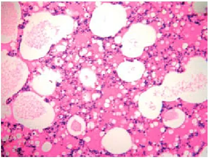
In the histopathological examination, ANTU triggered a severe pulmonary injury mainly docu-mented as an interstitial and intra-alveolar edema, alveolar wall destruction and interstitial inflamma-tory cell infiltration (Fig. 1B). There were no dif-ferences in histology and LW/BW ratio between
control (Fig. 1A) and olive oil-treated (solvent control) rats.
A significant lung edema was developed after 4 h i.p. injection of ANTU at the dose of 10 mg/kg as indicated by an increase in the LW/BW ra-tio and PE when compared with control rats. The LW/BW ratio was measured as 106.9 ± 9.9 × 10⁻⁴
for ANTU-treated rats while it was found to be 54.2 ± 2.4 × 10⁻⁴ for control rats (p < 0.05) (Fig. 2A).
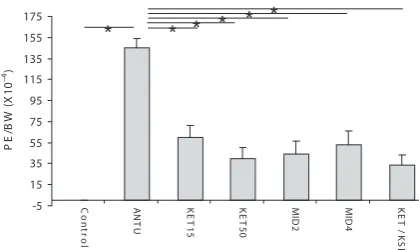
PE was measured as 3.2 ± 0.2 mL in ANTU-treat-ed rats, while no PE was observANTU-treat-ed in control rats (p < 0.05) (Fig. 2 BC).
The Effects of Ketamine
on ANTU-Induced ALI
Ketamine alone has no deleterious effect on healthy lung parameters. On the other hand, ket-amine had a prophylactic effect on ANTU-induced PE formation at all doses and significantly reduced PE and PE/BW ratio (p < 0.05) (Fig. 2 BC).
Ketamine 15 and 50 mg/kg groups caused a sig-nificant reduction of inflammation, hemorrhage
Fig. 1A. Normal histological appearance of olive oil-treated rat lungs (H&E ×10)
and edema formation according to the histopath-ological scoring (p < 0.05) (Fig. 3). Inflammation and hemorrhage results of 15 and 50 mg/kg groups were close to the control group. The best improve-ment in ANTU-induce lung damage was seen in ketamine 15 mg/kg group for inflammation, and ketamine 50 mg/kg group for edema and hemor-rhage compared to all groups. Ketamine at the dos-es of 15 and 50 mg/kg demonstrated no significant prophylactic effect on LW/BW ratio (Fig. 2A).
The Effects of Midazolam
on ANTU-Induced Acute
Lung Injury
Midazolam alone has no deleterious effect on healthy lung parameters. Midazolam in doses of 2 and 4 mg/kg significantly reduced PE and PE/ /BW ratio and had a prophylactic effect on ANTU-induced PE formation in all doses (p < 0.05) (Fig. 2 BC).
In the histopathological examination, midazol-am 2 mg/kg group caused a significant reduction of inflammation and edema formation according to the histopathological scoring (p < 0.05) (Fig. 4). Midazolam 4 mg/kg group caused a reduction of all scores but these are not significantly different from the ANTU group at the current dose and the lowest histopathological protection on ANTU- -induce lung damage was seen in the midazolam group. Midazolam at the doses of 2 and 4 mg/kg demonstrated no significant prophylactic effect on LW/BW ratio (Fig. 2A).
Fig. 2A. Figure represents the calculated results of acute lung injury induced by α-naphthylthiourea and alterations by ketamine (KET) (15, and 50 mg/kg), midazolam (MID) (2 and 4 mg/kg), and ketamine/ /xylazine (KET/-KSI) (50/10 mg/kg), as evaluated by the changes of lung weight/body weight (LW/BW) ratio (×10–4), * P < 0.05. (A), pleural effusion (PE) (mL)
Fig. 2B. Pleural effusion/body weight (PE/BW) ratio (×10–4)
Fig. 2C. Each column shows the mean value of eight experiments, vertical bars on the columns represent SEM 0 20 40 60 80 100 120 140 C o n tr o l A N T U K E T 15 K E T 50 M ID 2 M ID 4 K E T / K S I L W /B W ( x1 0 –4)
*
*
*
*
*
*
*
0 0.5 1 1.5 2 2.5 3 3.5 4 C o n tr o l A N T U K E T 15 K E T 50 M ID 2 M ID 4 K E T / K S I P E ( m L) -5 15 35 55 75 95 115 135 155 175 C o n tr o l A N T U K E T 15 K E T 50 M ID 2 M ID 4 K E T / K S I P E /B W ( X 10 –4) * * * * * *Fig. 3. Histopathological examination after pre-treat-ment with ketamine 50 mg/kg (H & E ×4)
The Effects of Ketamine/
/Xylazine on ANTU-Induced
Acute Lung Injury
Ketamine/xylazine (50/10 mg/kg) adminis-tration prior to ANTU significantly reduced PE and PE/BW ratio and had a prophylactic effect on ANTU-induced PE formation (p < 0.05) (Fig. 2 BC). Maximum protective effect on PE formation was seen in ketamine/xylazine (50/10 mg/kg) group in all groups. On the other hand, xylazine in-crease the protective effect of ketamine (50 mg/kg) (0.9 ± 0.2 mL) on pleural effusion in the ketamine/ xylazine group (0.7 ± 0.2 mL) but this is not signif-icantly different at the current dose (Fig. 2 BC).
In the histopathological examination, ket-amine/xylazine caused a significant reduction of inflammation and edema formation according to the histopathological scoring (p < 0.05) (Fig. 5). Nevertheless, histopathological scores seemed to be lower but not significantly different in the ketamine (50 mg/kg) group than the ketamine/
/xylazine (50/10 mg/kg) group. Ketamine/xylazine demonstrated no significant prophylactic effect on LW/BW ratio (Fig. 2A).
Discussion
Results of the present work have demonstrat-ed for the first time that ketamine, midazolam and ketamine/xylazine attenuate the pulmonary tissue damage and pleural effusion induced by ANTU. All of these agents significantly prevent the develop-ment of pleural effusion. The maximum decrease in pleural effusion was observed in the ketamine/ /xylazine group. On the other hand, although these anesthetic agents caused a statistically significant reduction of histopathological scores, there was no significant lowering effect on LW/BW ratio at the current doses.
ANTU is a chemical agent that produces an inflammatory reaction leading to pulmonary edema, secondary to permeability changes in the lung microvasculature. The capillary endothe-lial cell damage in the lung is the primary rea-son for ANTU toxicity. Injury to the endotheli-um leads to an eventual loss of the endothelial barrier. This leads to interstitial and alveolar ede-ma. Pulmonary edema and pleural exudate for-mation start approximately 1 h after administra-tion of ANTU, reaching a maximum at 2–4 h, and then either resolve or cause death [6, 7, 11, 12, 16–19]. The mechanism of pleural effusion for-mation in the ANTU model is drainage of lung interstitial liquid, through the visceral pleura, into the pleural space. This occurs when the in-terstitial lung space is saturated with fluid [20]. Thus, the amount of pleural effusion is close-ly relevant to pulmonary edema development. It has been speculated that various vasoactive sub-stances originating from pulmonary vascular bed
Table 2. Histological scoring are listed below (0–3)
Groups Inflammation Edema Hemorrhage
Control 0 0 0
ANTU (10 mg/kg) 2.4 ± 0.2a 2.4 ± 0.2 a 1.3 ± 0.2a
Ketamine (15 mg/kg) 0.4 ± 0.2b 0.9 ± 0.1 b 0.4 ± 0.2 b
Ketamine (50 mg/kg) 0.5 ± 0.2 b 0.6 ± 0.2 b 0.3 ± 0.2b
Midazolam (2 mg/kg) 1.5 ± 0.2 bcd 1.5 ± 0.2 bd 0.8 ± 0.2
Midazolam (4 mg/kg) 2 ± 0.3cd 1.9 ± 0.1cd 1.1 ± 0.2cd
Ketamine + xylazine 0.9 ± 0.1be 1 ± 0.3 be 0.9 ± 0.2
a p < 0.05 vs. control; b p < 0.05 vs. ANTU; c p < 0.05 vs. Ketamine (15 mg/kg);d p < 0.05 vs. Ketamine (50 mg/kg);e p < 0.05 vs. Midazolam (4 mg/kg).
and airways may contribute to the lung injury in-duced by ANTU [6, 16–18, 21, 22].
Glutamate is an essential amino acid and a trans-mitter in the mammalian nervous system. N-meth-yl-D-aspartate (NMDA), α-amino-3-hydroxy-5- -methyl-4-isoxazole-propionate (AMPA), kainate and metabotropic receptors are activated by gluta-mate. In recent years the increasing number of in-vestigators have demonstrated the involvement of glutamate and glutamate signaling in non-neuro-nal tissues, including bone osteoblasts and osteo-clasts, keratinocytes, megakaryocytes, pancreatic islet cells, the lung, the liver, the heart, kidney cells, adrenal tissue, and taste buds [23]. Although the NMDA receptor subtypes have been characterized in terms of their functional roles in the central ner-vous system (CNS) and their relationship to neu-ronal excitotoxicity in neurological disorders, lit-tle or nothing is known about the physiologic and pathophysiologic significance of these receptors in the respiratory system. NMDA can induce ex-citotoxicity in the lung, in the form of an acute, high-permeability pulmonary edema, which was prevented by the non-competitive NMDA recep-tor antagonist MK-801, and that glutamate may be linked to the pathogenesis of bronchial asthma and airway inflammation [24].
The intravenous dissociative anesthetic, NMDA receptor antagonist ketamine has many pharmacological properties, including analgesic, anesthetic and sympathomimetic effects [25]. Ket-amine is a drug used in human and veterinary med-icine, primarily for the induction and maintenance of general anesthesia, usually in combination with a sedative. Other uses include sedation in intensive care, analgesia (particularly in emergency medi-cine), and treatment of bronchospasm [26]. Ow-ing to its ability to induce relaxation of bronchial smooth muscle, ketamine is recommended as an optimizing anesthetic for asthmatic patients, and has been clinically used to treat bronchospasms, asthma exacerbation and status asthmaticus [27]. On the other hand, ketamine’s role in neuroanes-thesia is limited because of its association with in-creased intracranial pressure and other side effects such as psychotropic properties, tachycardia and hypertension [26]. Recent years studies have shown that ketamine plays a protective role against lung injury, via its anti-inflammatory properties [8–10]. Under ketamine anesthesia, neurogenic pulmo-nary edema is less pronounced in a rat model of spinal cord injury, and the mortality of severe-ly burnt rats is reduced [28]. Yang et al. demon-strated that ketamine, only at a supra-anesthetic dosage, could inhibit endotoxin-induced pulmo-nary inflammation in vivo [9]. Ketamine has been shown to attenuate symptoms of endotoxemia in
a lipopolysaccharide-induced rat model of sepsis, by reducing nuclear factor kappa B (NF-kappa B) activity and tumor necrosis factor-alpha (TNF-α) production [29], and decreasing the expression of iNOS in various rat tissues [30]. Furthermore, Yu et al. found that ketamine at sub-anesthetic doses also suppress the production of inflammatory cy-tokines on lung tissue such as TNF-α and inter-leukin-6 (IL-6), attenuate NF-kappa B activity, and inhibit toll-like receptor 2 (TLR2) and TLR4 ex-pression in polymicrobial sepsis [8]. In addition, ketamine has weak suppressive effect on reactive oxygen species (ROS) production by neutrophils and modulate the stimulated adhesion molecule expression [31].
system. Our results may support earlier findings concerning the anti-inflammatory effects of ket-amine on inflammation.
Ketamine and xylazine are commonly used in combination as an anesthetic agent in experimental animal models. Ketamine has many known anti-in-flammatory actions. In contrast to ketamine, xyla-zine, an alpha2-adrenergic agonist, has been shown
to have proinflammatory effects. Xylazine has been shown to induce pulmonary edema and to increase LPS-induced mortality in rats. Furthermore, dur-ing endotoxemia, xylazine worsens mortality and decreases levels of the anti-inflammatory cytokine IL-10 [33, 34]. On the other hand, ketamine/xyla-zine attenuates lipopolysaccharide (LPS)-induced TNF-α protein levels and iNOS transcription [35] in a variety of tissues. We suppose that there is an interaction between several different pathways that work together to attenuate inflammation as shown in ketamine/xylazine group. In the present study we found that ketamine/xylazine caused a signif-icant reduction of inflammation and edema for-mation but these scores seemed to be higher but not significantly different than the ketamine alone (50 mg/kg) group. These results suggest that xyla-zine did not show an additive effect on the protec-tive effect of ketamine alone. On the contrary, xyl-azine reversed the protection effect of ketamine on hemorrhage. Moreover, these results concur with those obtained by other investigators which have demonstrated the negative effects of xylazine on lung tissue [33, 34].
In the present study it was found for the first time that ketamine/xylazine has a prophylactic ef-fect in ANTU-induced PE. Maximum protective effect on PE formation was seen in ketamine/xy-lazine group in all groups; however, the amount of PE did not significantly differ between ketamine and ketamine/xylazine groups. Experimental re-sults show that similar to other anesthetic agent, xylazine is not an inert agent and may change the data in animal experiments. Therefore, when using these agents in veterinary or experimental studies, these effects should be kept in mind.
Among the benzodiazepines, midazolam, wa-ter-soluble benzodiazepine is the most widely used anxiolytic and sedative drug for short proce-dures and in intensive care. Midazolam interferes the synthesis of NO and TNF-α generated by acti-vated microglia cells, blood monocytes, and mast
cells, suggesting an inhibitory action on proin-flammatory mediators. We have shown that mid-azolam at the doses of 2 and 4 mg/kg reduced in-flammation and edema scoring and PE in ANTU induced ALI. The protective effect of midazolam was more pronounced and significant at 2 mg/kg dose (p < 0.05). Our results concur with those ob-tained by other investigators which have demon-strated the modulatory effects of benzodiazepine receptors in immune/inflammatory process [36]. Recent evidence indicates that midazolam sig-nificantly inhibited TNF-α induced vascular cell adhesion molecule-1 and monocyte adhesion in human umbilical vein endothelial cells [37]. Meanwhile, midazolam not only modulates the production of cytokines, but also decreases the generation of NO, inhibits the function of neutro-phils, suppresses the expression of LPS-stimulat-ed iNOS, cyclooxygenase-2 and superoxide anion production [36, 38]. On the other hand, some ear-lier works have shown that midazolam does not have the protective effect on hemodynamics and organ dysfunction in the endotoxemic rats [39]. The controversy in literature may be partly due to the use of different inflammation models, doses and animals. These results demonstrate the need for the use of different tests and doses in the eval-uation of the effects of midazolam in tissue injury as well as ALI.
The results of the present study indicate that ANTU induced ALI is reduced by i.p. injection of ketamine, midazolam and ketamine/xylazine. Acute inhibitory effects of these anesthetic agents on fluid accumulation were more effective in the pleural cavity than the interstitial compartment in this ALI model. We can conclude that anesthetic agents may affect and attenuate lung injury. The precise mechanisms underlying the inhibitory ef-fect are still unknown but we can suggest that the effects of anesthetic agents on immune/inflamma-tory reactions might be important. Our results may corroborate and support earlier findings concern-ing the protective effects of ketamine and midazol-am on tissue injury. Along with the literature, these results suggest that, aside from anesthetic proper-ties, many anesthetic agents can cause physiologic and metabolic alterations and modulate and im-prove the lung inflammatory responses and these effects of anesthetic agents should be considered in clinical and experimental applications.
References
[1] Jain R and DalNogare A: Pharmacological therapy for acute respiratory distress syndrome. Mayo Clin Proc 2006, 81, 205–212.
[2] Matthay MA, Zimmerman GA, Esmon C, Bhattacharya J, Coller B, Doerschuk CM: Future research directions in acute lung injury: summary of a National Heart, Lung, and Blood Institute Working Group. Am J Respir Crit Care Med 2003, 167, 1027–1035.
[3] Bellingan GJ: The pulmonary physician in critical care *6: The pathogenesis of ALI/ARDS. Thorax 2002, 57, 540–546.
[4] Monahan LJ: Acute respiratory distress syndrome. Curr Probl Pediatr Adolesc Health Care 2013, 43, 278–284. [5] Hanci V, Yurdakan G, Yurtlu S, Turan IO, Sipahi EY: Protective effect of dexmedetomidine in a rat model of
alpha-naphthylthiourea-induced acute lung injury. J Surg Res 2012, 1–7.
[6] Comert M, Sipahi EY, Ustun H, Isikdemir F, Numanoglu G, Barut F: Morphine modulates inducible nitric oxide synthase expression and reduces pulmonary oedema induced by alpha-naphthylthiourea. Eur J Pharmacol2005, 511, 183–189.
[7] Sipahi E, Ustun H, Niyazi Ayoglu F: Acute effects of pentobarbital, thiopental and urethane on lung oedema induced by alpha-naphthythiourea (ANTU). Pharmacol Res 2002, 45, 235–239.
[8] Yu M, Shao D, Yang R, Feng X, Zhu S, Xu J: Effects of ketamine on pulmonary inflammatory responses and survival in rats exposed to polymicrobial sepsis. J Pharm Pharm Sci 2007, 10, 434–442.
[9] Yang J, Li W, Duan M, Zhou Z, Lin N, Wang Z: Large dose ketamine inhibits lipopolysaccharide-induced acute lung injury in rats. Inflamm Res 2005, 54, 133–137.
[10] Zhang X, Feng J, Zhu P, Zhao Z: Ketamine inhibits calcium elevation and hydroxyl radical and nitric oxide pro-duction in lipopolysaccharide-stimulated NR8383 alveolar macrophages. Inflammation 2013, 36, 1094–1100. [11] Scott AM, Powell GM, Upshall DG, Curtis CG: Pulmonary toxicity of thioureas in the rat. Environ Health
Perspect 1990, 85, 43–50.
[12] Richter CP: The physiology and cytology of pulmonary edema and pleural effusion produced in rats by alpha- -naphthyl thiourea (ANTU). J Thorac Surg 1952, 23, 66–91.
[13] Cunningham AL, Hurley JV: Alpha-naphthyl-thiourea-induced pulmonary oedema in the rat: a topographical and electron-microscope study. J Pathol 1972, 106, 25–35.
[14] Torre D, Minoja G, Maraggia D, Chiaranda M, Tambini R, Speranza F: Effect of recombinant IL-1 beta and recombinant gamma interferon on septic acute lung injury in mice. Chest 1994, 105, 1241–1245.
[15] Calikoglu M, Tamer L, Sucu N, Coskun B, Ercan B, Gul A: The effects of caffeic acid phenetyl ester on tissue damage in lung after hindlimb ischemia-reperfusion. Pharmacol Res 2003, 48, 397–403.
[16] Sipahi EY, Ozel Tekin I, Comert M, Barut F, Ustun H, Sipahi TH:Oxidized low-density lipoproteins accumulate in rat lung after experimental lung edema induced by alpha-naphthylthiourea (ANTU). Pharmacol Res 2004, 50, 585–591.
[17] Sipahi E, Hodoglugil U, Ercan ZS, Türker RK:Acute effect of endothelin-1 on lung oedema induced by alpha-naphthylthiourea (ANTU). Pharmacol Res 1996, 33, 375–378.
[18] Sipahi E, Hodoglugil U, Ustun H, Zengil H, Türker RK, Ercan ZS: An unexpected interaction between NG-nitro-L-arginine methyl ester and L-arginine in alpha-naphthylthiourea-induced pulmonary oedema in rats. Eur J Pharmacol 1997, 321, 45–51.
[19] Meyrick B, Miller J, Reid L: Pulmonary edema induced by ANTU or by high or low oxygen concentrations in rat—an electron microscopic study. Br J Exp Pathol 1972, 53, 347–358.
[20] Wiener-Kronish JP and Broaddus VC: Interrelationship of pleural and pulmonary interstitial liquid. Ann Rev Physiol 1993, 55, 209–226.
[21] Pankhania JJ, Bakhle YS: Effect of pulmonary oedema induced by alpha-naphthylthiourea on synthesis of cyclo-oxygenase products in rat isolated lungs. Prostaglandins 1985, 30, 37–49.
[22] Hardwick SJ, Skamarauskas JT, Smith LL, Upshall DG, Cohen GM: Protection of rats against the effects of alpha-naphthylthiuorea (ANTU) by elevation of non-protein sulphydryl levels. Biochem Pharmacol 1991, 42, 1203–1208.
[23] Rzeski W, Ikonomidou C, Turski L: Glutamate antagonists limit tumor growth. Biochem Pharmacol 2002, 64, 1195–1200.
[24] Said SI, Berisha HI, Pakbaz H: Excitotoxicity in the lung: N-methyl-D-aspartate-induced, nitric oxide-dependent, pulmonary edema is attenuated by vasoactive intestinal peptide and by inhibitors of poly (ADP-ribose) poly-merase. Proc Natl Acad Sci USA 1996, 93, 4688–4692.
[25] Zhu MM, Qian YN, Zhu W, Xu YM, Rong HB, Ding ZN: Protective effects of ketamine on allergen-induced airway inflammatory injure and high airway reactivity in asthma: Experiment with rats. Zhonghua Yi Xue Za Zhi 2007, 87, 1308–1313.
[26] Bowles ED and Gold ME: Rethinking the paradigm: evaluation of ketamine as a neurosurgical anesthetic. AANA J 2012, 80, 445–452.
[27] Allen JY, Macias CG: The efficacy of ketamine in pediatric emergency department patients who present with acute severe asthma. Ann Emerg Med 2005, 46, 43–50.
[29] Sun J, Li F, Chen J, Xu J: Effect of ketamine on NF-kappa B activity and TNF-alpha production in endotoxin-treated rats. Ann Clin Lab Sci 2004, 34, 181–186.
[30] Helmer KS, Cui Y, Dewan A, Mercer DW:Ketamine/xylazine attenuates LPS-induced iNOS expression in various rat tissues. J Surg Res 2003, 112, 70–78.
[31] Weigand MA, Schmidt H, Zhao Q, Plaschke K, Martin E, Bardenheuer HJ:Ketamine modulates the stimulated adhesion molecule expression on human neutrophils in vitro. Anesth Analg 2000, 90, 206–212.
[32] Gokcinar D, Ergin V, Cumaoglu A, Menevse A, Aricioglu A: Effects of ketamine, propofol, and ketofol on pro-inflammatory cytokines and markers of oxidative stress in a rat model of endotoxemia-induced acute lung injury. Acta Biochim Pol 2013, 60, 451–456.
[33] Fessler HE, Otterbein L, Chung HS, Choi AM: Alpha-2 adrenoceptor blockade protects rats against lipopolysac-charide. Am J Respir Crit Care Med 1996, 154, 1689–1693.
[34] Szelenyi J, Kiss JP, Puskas E, Selmeczy Z, Szelényi M, Vizi ES:Opposite role of alpha2- and beta-adrenoceptors in the modulation of interleukin-10 production in endotoxaemic mice. Neuroreport 2000, 11, 3565–3568. [35] Helmer KS, Cui Y, Chang L, Dewan A, Mercer DW:Effects of ketamine/xylazine on expression of tumor necrosis
factor-alpha, inducible nitric oxide synthase, and cyclo-oxygenase-2 in rat gastric mucosa during endotoxemia. Shock 2003, 20, 63–69.
[36] Zavala F, Taupin V, Descamps-Latscha B:In vivo treatment with benzodiazepines inhibits murine phagocyte oxidative metabolism and production of interleukin 1, tumor necrosis factor and interleukin-6. J Pharmacol Exp Ther 1990, 255, 442–450.
[37] Joo HK, Oh SC, Cho EJ, Park KS, Lee JY, Lee EJ: Midazolam inhibits tumor necrosis factor-alpha-induced endo-thelial activation: involvement of the peripheral benzodiazepine receptor. Anesthesiology 2009, 110, 106–112. [38] Kim SN, Son SC, Lee SM, Kim CS, Yoo DG, Lee SK: Midazolam inhibits proinflammatory mediators in the
lipopolysaccharide-activated macrophage. Anesthesiology 2006, 105, 105–110.
[39] Tsao CM, Wu CC, Liaw WJ, Ho ST:Effects of midazolam on organ dysfunction in rats with endotoxemia induced by lipopolysaccharide. Acta Anaesthesiol Taiwan 2009, 47, 10–16.
Address for correspondence:
Emine Yilmaz-Sipahi Bulent Ecevit University
Department of Pharmacology, Medical School 67600 Kozlu–Zonguldak
Turkey
Tel.: + 90 372 261 32 12 E-mail: [email protected]
Conflict of interest: None declared