KINEMATIC ANALYSIS OF POSTURAL CHANGES IN BIPEDAL STANCE
AT APPLICATION OF STIMULUS FROM EXTERNAL ENVIRONMENT
AND MODIFICATION OF VISUAL SCENE IN PATIENTS
WITH ANTERIOR CRUCIATE LIGAMENT RECONSTRUCTION
Petra Hlavačková, Miroslav Janura
Faculty of Physical Culture, Palacký University, Olomouc, Czech Republic
Submitted in August, 2007 Tears and rupture of the anterior cruciate ligament (ACL), which has an important function in knee joint stability, are very common and more so in sports. ACL injury can be managed in two ways. One alternative pertains to conserva-tive therapy whereas the second variant is surgical intervention (reconstruction). The number of ACL reconstructions has risen recently. The purpose of this study was to analyze the postural changes in bipedal stance at application of a stimulus from external environment and the resultant modifi cation in the visual scene in patients with ACL recon-struction. The examined group consisted of 25 subjects – 11 patients with ACL reconstruction and 14 healthy adults. The external stimulus was realized by striking a fl ying ball; the modifi cation of visual scene was obtained using special glasses. The 3D videography method was applied to evaluate postural changes. The most diff erences during three various monitored measurements were found for kinematic parameters of the lower limbs and the trunk in each group (both patients and healthy subjects). Statistically signifi cant diff erences between the group of patients and healthy subjects (between both groups)were obtained for comparison of measurements with an unmodifi ed visual scene and measurements with a modifi ed visual scene, especially for the elbow and shoulder joints.
Keywords: Anterior cruciate ligament (ACL), proprioception, visual modifi ed scene, 3D videography.
INTRODUCTION
The anterior cruciate ligament (ACL) plays a key role in assuring stability of the knee joint. Aff erent in-formation from ACL mechanoreceptors signifi cantly participates in facilitating dynamic stability of the knee joint.This somatosensory information, as well as infor-mation from the visual and vestibular receptors, contrib-utes to the maintenance and control of bipedal stance, posture and balance in a particular position during movement. A considerable modifi cation of the sensory information, disturbance of the functional stability of the knee and impairment of postural control can occur due to traumatic ACL injury or ACL reconstruction (Harrison et al., 1994; Hoff man et al., 1999; Bonfi rm et al., 2003; Liu-Ambrose et al., 2003).Impaired postural control measured by balance in the case of single limb stancehas been reported after acute and chronic ACL injuries, as well as after ACL reconstructions(Lysholm et al., 1998; Hoff man et al., 1999; Denti et al., 2000; Henriksson et al., 2001).
Vision is particularly important in stabilizing posture under more challenging conditions or if the propriocep-tive information from the lower limbs is reduced (ankle, knee injuries, amputees, etc.) (Lord & Menz, 2000).
Okuda et al. (2005) investigated the type of role played by vision in patients with LCA insufficiency during maintenance of postural stability. Notably, there was no signifi cant diff erence between the injured and uninjured legs regarding postural sway during one leg standing with the eyes open, but the amount of postural sway increased signifi cantly with the eyes closed. Pursuant to these results it can be inferred that vision appears to be dominant in compensating for reduced and/or com-promised contribution by the injured (or reconstructed) ACL especially under more challenging conditions.
Modifi cation of the visual scene and contemporary application of stimulus from an external environment can culminate in higher demands on balancing ability, especially as regards the lower limbs and the trunk.Our interest was focused on the question of whether the po-tential defi cit in proprioception is manifested by postural changes under simple motor test conditions (hitting of a ball on arms raised forward) and how the visual sys-tem reacts in order to compensate for this defi cit.
SUBJECTS
A total of 25 individuals agreed to participate in this study. Fourteen healthy individuals who reported no history of signifi cant orthopaedic knee injury or bal-ance related disorders served as the control group. The control group comprised 7 females and 7 males (age 24.9 ± 3.5 years, body weight 70.6 ± 9.4 kg, height 174.4 ± 10.3 cm). For the ACL reconstructed group, the mean age was 25.4 ± 6.9 years, weight was 70.5 ± 10.7 kg and the height was 174.2 ± 9.5 cm. The 4 females and 7 males had undergone reconstruction of the ACL with patellar tendon graft (7 patients) or hamstring tendon graft (4 patients). The average post surgery period was 51 days.
METHODS
Special hardware Olympus Eye trek FMD-700 (Fig. 1) was applied to modify the visual scene in our research.This optic system has OSR (Optical Super Resolution) for high quality on screen display equivalent to 720 000 pixels. The advantage of this special optical lens system is the possibility of connecting to various ex-ternal devices (for example connection with a personal computer, television, DVD player, etc.). Connection to the video camera was also incorporated during our measurement.
Fig. 1
Special hardware Olympus Eye trek FMD-700
The experiment used two diff erent visual conditions (scenes) with respect to the form of visual perception.
The fi rst part of the measurement took place with full visual control, without any restriction as to special hard-ware and with real visual perception.This was followed by the next phase where we modifi ed visual perception by using special optical glasses, Eye trek FMD-700.
Stimulus from the external environment was applied by reason of major “dynamization” of the situation (by reason of creating a more complicated situation). The impulse was applied by a basketball of variable weight depending on the weight of the subject and from diff er-ent heights. The weight of the ball was set up so that its momentum at the moment of striking the board con-formed to 7% of the weight of the subject. To determine the solid weight (ball + weight) for the given proband we applied the law of conservation of mechanical energy. In the event of zero level potential energy at the bump site applies mt gh = ½mt l2ω2, where mtis the weight of
the hanging body/sphere, g = 9.81 m · s–2, h determines the sphere height before activation with respect to the sphere height after the bump, ω its angular speed and a l is the suspension length. If ν = ωl = √2gh applies, then the conditions for the dynamic of the solid at the moment of the bump is mtν = 0.07mp(where mp is the
proband’s weight) we gain for h = 0.5 m the relation mt = 0.0220mp.
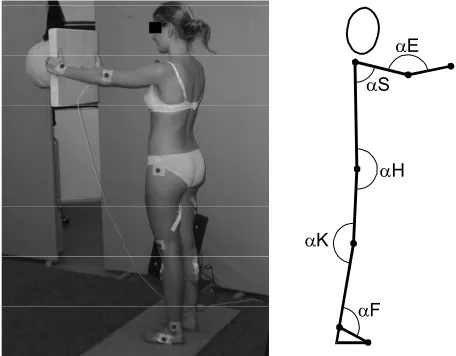
The main task of each subject was to cushion the striking ball with the aid of a light board made of lami-nated polystyrene, which was held by the subject’s raised arms (Fig. 2). A total of 7 attempts were made. The fi rst three measurements were without any visual modifi ca-tion. The other four measurements were conducted with special glasses, whereas the fl ying ball was caught by the experimenter near the impact site (already out of the visual fi eld of the subject) during third trials with simulated stimulus.
Fig. 2
An illustration of measurement without modifi cation to the visual scene and the evaluated angle parameters
The 3D videographic method was used to evaluate the basic kinematic parameters. Special marks indicat-ing projection of the specifi c anatomic structures on the body were assigned on the left side of the body of
αF αK
αH αS
each subject. These eight structures comprised: the tra-gus, the acromion, the lateral humerus epicondyl, the capitatum, the major femoral trochanter, the lateral femoral epicondyle, the lateral malleolus, and the ca-pitulum ossis metatarsi quinti. Each subject was fi lmed by two video cameras, followed by processing of the video record, resulting in kinematic parameters of 3D movement evaluation.
The changes in angle magnitudein the sagittal plane at intervals 0–1600 ms were analyzed. The time 0 ms was characterized as the moment when the experiment-er let go of the ball and 600 ms as the impact of the ball on the board. Later, we evaluated the interval 1000 ms following impact. We noted the time run of changes of these angles: metatarsus – shank, shank – thigh, thigh – trunk, trunk – arm, and arm – forearm (Fig. 2). STATISTICAL ANALYSIS
Data obtained by kinematic analysis were statisti-cally processed by the programme Statistica, version 6.0.
The non parametric unpaired test (Mann-Whitney) was used for comparing the two diff erent groups (patients and healthy subjects). Specifi cation of diff erences be-tween the angles in a single group during various meas-urements was performed by using the non parametric sign test.
RESULTS AND DISCUSSION
Comparison of the kinematic parameters of a single group of healthy subjects
Signifi cant diff erences in single kinematic param-eters (excepting the angle shank – thigh of healthy subjects) when comparing measurement results with an unmodifi ed visual scene and measurements taken at a modifi ed visual scene with real stimulus were not found for healthy subjects not even after ACL recon-struction (TABLE 1).
TABLE 1
Comparison of kinematic parameters on the lower limbs of the group of healthy subjects
Aver N Aver I Aver II Comparison of diff erences
Metatarsus – shank αF AV SD AV SD AV SD N × I N × II I × II
Max. (°) 138.50 1.78 138.01 1.73 137.82 1.72 6/8 8/6 9/5
Min. (°) 136.68 1.81 135.62 1.73 134.14 1.74 9/5 13/1 ** 12/2 *
Dif. (°) 1.83 0.15 2.39 0.22 3.69 0.25 3/11 1/13 ** 1/13 **
Time max. (ms) 0.36 0.11 0.18 0.14 0.38 0.12 7/7 8/6 6/8
Time min. (ms) 0.03 0.09 0.15 0.10 0.36 0.11 4/10 3/11 4/10
Aver 1 (°) 137.40 1.80 136.91 1.76 135.92 1.71 9/5 11/3 11/2 *
Aver 2 (°) 137.64 1.79 136.94 1.72 135.81 1.74 8/6 11/3 12/1 **
Shank – thigh αK AV SD AV SD AV SD N × I N × II I × II
Max. (°) 171.24 1.34 170.91 1.35 172.47 1.34 7/7 4/10 0/14 **
Min. (°) 168.73 1.28 166.87 1.37 170.13 1.34 13/1 ** 3/11 0/14 **
Dif. (°) 2.52 0.35 4.04 0.34 2.34 0.23 2/12 * 6/8 13/1 **
Time max. (ms) –0.12 0.12 –0.06 0.10 0.45 0.07 6/8 1/12 ** 3/11
Time min. (ms) 0.28 0.05 0.30 0.03 0.00 0.13 2/10 * 11/3 11/3
Aver 1 (°) 170.88 1.33 170.62 1.36 170.68 1.35 6/8 6/8 6/7
Aver 2 (°) 169.85 1.30 168.91 1.37 171.56 1.34 11/3 2/12 * 0/13 **
Legend to TABLE 1, 2, 3 and Fig. 3, 4, 5
AV = average * = p < 0.05
SD = standard deviation ** = p < 0.01
N = measurements without any modifi cation of the visual scene (non-modifi cation) I = measurements with modifi cation of the visual scene with a real stimulus II = measurements with modifi cation of the visual scene with a simulated stimulus Max. = maximal amplitude of angle in time period 0–1600 ms
Min. = minimal amplitude of angle in time period 0–1600 ms
Dif. = diff erence between maximal and minimal amplitude of angle in time period 0–1600 ms Time max. = time of achievement of maximal amplitude of angle in time period 0–1600 ms Time min. = time of achievement of minimal amplitude of angle in time period 0–1600 ms Aver 1 = verage magnitude of angle in time period 0–600 ms (before the stroke of a ball) Aver 2 = average magnitude of angle in time period 600–1600 ms (after the stroke of a ball)
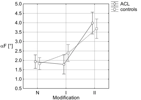
Fig. 3
Graphic comparison of the diff erence between the maxi-mal and minimaxi-mal degrees of the metatarsus-shank angle (αF)at single measurements (modifi cation)
ACL controls
N I II
0.5 1.0 1.5 2.0 2.5 3.0 3.5 4.0 4.5 5.0
αF [°]
Modification
TABLE 2
Comparison of kinematic parameters on the lower limbs of the group of patients
Aver N Aver I Aver II Comparison of diff erences
Metatarsus – shank αF AV SD AV SD AV SD N × I N × II I × II
Max. (°) 138.83 2.01 138.82 1.95 138.36 1.94 7/4 6/5 9/2
Min. (°) 136.90 2.04 137.04 1.95 134.38 1.96 4/7 10/1 * 11/0 **
Dif. (°) 1.93 0.17 1.78 0.25 3.98 0.28 5/6 0/11 ** 0/11 **
Time max. (ms) 0.34 0.13 0.37 0.16 0.27 0.13 4/7 6/5 4/6
Time min. (ms) –0.02 0.10 0.15 0.12 0.29 0.13 5/6 5/6 4/7
Aver 1 (°) 137.68 2.03 137.20 1.98 136.55 1.93 4/7 10/1 * 10/1 *
Aver 2 (°) 138.03 2.02 137.10 1.94 136.28 1.97 7/4 10/1 * 9/2
Shank – thigh αK AV SD AV SD AV SD N × I N × II I × II
Max. (°) 172.57 1.51 173.30 1.52 174.80 1.51 3/8 0/11 ** 0/11 **
Min. (°) 170.55 1.44 169.67 1.55 172.91 1.52 9/2 0/11 ** 0/11 **
Dif. (°) 2.02 0.39 3.63 0.38 1.88 0.26 2/9 6/5 10/1 *
Time max. (ms) 0.10 0.13 –0.20 0.11 0.53 0.08 7/4 2/9 0/11 **
Time min. (ms) 0.22 0.05 0.27 0.03 0.17 0.15 2/8 7/4 7/4
Aver 1 (°) 172.16 1.50 173.08 1.53 173.42 1.52 1/10 * 1/10 * 3/8
Aver 2 (°) 171.63 1.47 171.52 1.55 173.97 1.51 7/4 0/11 ** 0/11 **
Fig. 4
Graphic comparison of the time of achievement of the maximal value of the arm – forearm angle (αE) in single measurements (time of stroke – 0 ms on Y-axis)
Time
Modification
ACL controls
N I II
-0.6 -0.5 -0.4 -0.3 0.2 0.1 0.0 0.1 0.2 0.3 0.4 The number of significant differences increased
when a simulated stimulus was applied. Perhaps the best illustration of these changes is provided by the dif-ference between the maximum and minimum degrees of angle. Most of the diff erences were ascertained for
Comparison of kinematic parameters and measurements between both groups
a) Measurements without modifi cation to visual scene Statistically signifi cant diff erences were found at the only angle (arm – forearm) during measurement without any modifi cation to the visual scene between patients and healthy subjects (TABLE 3). The time of achieve-ment of maximal magnitude of the arm – forearm angle and the average magnitude of this angle in the time pe-riod 600–1600 ms (after the stroke of a ball) were men-tioned as observed parameters. As is illustrated in Fig. 4, it can be inferred that the value of the maximum angle at the elbow is achieved in patients after striking the ball, whereas in healthy subjects this is seen before such a hit. The value of the angle at the elbow in time period 600–1600 ms is greater in the group of patients. b) Measurements with modifi cation to the visual scene
with a real stimulus
More statistically signifi cant diff erences were found during measurement with a modifi ed visual scene and at the application of a real stimulus in comparison to measurements without special glasses. A possible in-terpretation thus could be that maintaining postural stability is rather more diffi cult when the visual scene is changed. Additionally, altered visual function means higher demands on proprioception and vestibular in-formation.More marked diff erences were evidenced with the arm – forearm angle.Diff erences at the level of statistically signifi cant p < 0.05 were shown for the minimal magnitude of the shank – thigh angle and the time of achievement of the minimal magnitude of the trunk – arm angle.
c) Measurements during a modifi ed visual scene with a simulated stimulus
Measurements carried out during a modifi ed visual scene and a simulated stimulus represented an unex-pected situation for each subject. In comparison with the other two measurements mentioned above, where
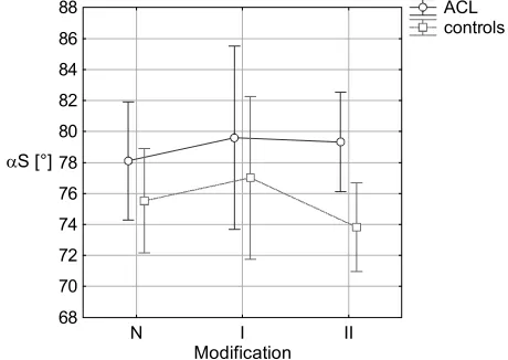
the arm – forearm angle manifested itself as a signifi cant diff erence, the trunk – arm angle parameters (Fig. 5) showed similar signifi cance. The reaction of subjects to simulated stimuli was more complex (global) as far as the particular segments of body and their connec-tions are concerned, and also more individual – from the strategical aspect.
Fig. 5
Graphic comparison of the minimal magnitude of the trunk – arm angle (αS)at single measurements (modi-fi cation)
According to the results mentioned above it is evi-dent that applying special glasses did not have a strong infl uence on the motor task. Major diff erences occurred during changes between real and expected situations. It should then be generally true that measurements with-out any modifi cation to the visual scene be regarded as “posturally simplest” rather than “more challeng-ing” following modifi cation of a visual scene with a real stimulus, and the attempt with a simulated stimulus was relatively specifi c.
Thehigher average values of most angles were regis-tered for patients in contrast to healthy subjects. How-ever, the question is how we can interpret and judge TABLE 3
The values of test standards for kinematic parameters of the upper limbs during comparison between healthy subjects and patients
ANGLE Attemp Max. Min. Dif. Time max. Time min. Aver 1 Aver 2
Trunk – arm αS N 0.602 0.985 0.164 0.712 0.137 0.876 0.766
Arm – forearm αE N 1.478 1.916 –1.642 2.600 ** –0.082 1.369 2.026 *
Trunk – arm I 1.095 1.204 –0.493 1.724 –2.546 * 0.931 1.040
Arm – forearm I 2.245 * 2.573 ** –1.752 0.684 –1.177 2.190 * 2.464 *
Trunk – arm II 1.916 2.245 * –1.040 1.013 –0.438 1.861 2.245 *
Arm – forearm II 1.861 2.464 * –1.150 0.164 –1.177 2.080 * 2.683 **
αS [°]
Modification
ACL controls
N I II
these fi ndings with regard to postural stability. Greater tendencies to extension in almost the majority of the tested joints demonstrated a higher value of the magni-tude of particular joints in patients. Many of the joints (knee, elbow) are locked in extension. The stability of the knee joint is highest at full extension (Kapandji, 1991). Therefore, it is rather easier to stabilize the joints only in these extension positions and total propriocep-tion control is thus not indicated.It also manifests itself in the form of smaller demands being made on muscle coordination.
It is, however, possible that proprioceptive changes following ACL injury can occur without aff ecting any control of standing balance, as suggested by Birming-ham et al. (2001) – possibly through compensation by means of visual, vestibular and somatosensory input from receptors in other structures and joints. Correc-tions of postural sway rendered at the hip and trunk can play a compensatory role intended for maintaining balance, too (Allum et al., 1998).
We supposed that disturbed interpretation as well as the processing of proprioceptive information will be shown in patients with ACL reconstruction, which will manifest in postural changes. Our hypothesis was proven to be true only partially.
One of the reasons here can be the fact that a small group of 25 subjects was investigated. The applied ex-ternal stimulus could be insuffi cient and nither was the power of stroke strong. Therefore, the subjects were able to cushion the strike of the ball at the upper part of the body, eventually balancing some postural sways by using ankle strategy. The tested task was not as demand-ing since not only healthy subjects but also the patients were able to compensate their potential proprioceptive defi cit very well.Alternation of knee joint propriocep-tion wasn’t manifested and didn’t infl uence essentially the postural control of the lower limbs.
Generally, good health, muscle power and motor abilities could play a substantial role in patients as well. However, the possibility of minimal propriocep-tive defi cit cannot be excluded in instances such as in cases of its measurement following optimally selected rehabilitation.
CONCLUSIONS
From the stated results it follows that postural de-mands in a given situation change with modifi cation to the visual scene. Similar tendencies of (in) postural changes were noted in both groups. The most statistical-ly signifi cant diff erences between both groups (between the group of patients and healthy subjects) were ob-tained for comparison of measurements with the modi-fi ed visual scenes, especially for the elbow and shoulder
joints.It is not so apparent that the changes observed by us are related to a potential proprioception defi cit at the knee joint. The supposed varied postural strategies were not confi rmed for the lower limbs.
Acknowledgments
This study was carried out within the research project granted by the Ministry of Education, Youth and Sports “Physical activity and inactivity of inhabit-ants of the Czech Republic, no: 6198959221”.
REFERENCES
Allum, J. H. J., Bloem, B. R., Carpenter, M. G., Hul-liger, M., & Hadders-Algra, M. (1998). Propriocep-tive control of posture: A review of new concepts. Gait & Posture, 8, 214–242.
Birmingham, T. B., Kramer, J. F., Kirkley, A., Inglis, J. T., Spaulding, S. J., & Vandervoort, A. A. (2001). Knee bracing after ACL reconstruction: Eff ects on postural control and proprioception. Medicine & Sci-ence in Sports & Exercise, 33, 1253–1258.
Bonfi rm, T. R., Jansen Paccola, C. A., & Barela, J. A. (2003). Proprioceptive and behaviour impairments in individuals with anterior cruciate ligament recon-structed knees. Archives of Physical Medicine and Re-habilitation, 84(8), 1217–1223.
Denti, M., Randelli, P., Lo Vetere, D., Moioli, M., Bagnoli, I., & Cawley, P. W. (2000). Motor control performance in the lower extremity: Normals vs. anterior cruciate ligament reconstructed knee 5–8 years from the index surgery. Knee Surgery, Sports Traumatology, Arthroscopy, 8, 296–300.
Harrison, E. L., Duenkel, N., Dunlop, R., & Russel, G. (1994). Evaluation of single leg standing following anterior cruciate ligament surgery and rehabilitation. Physical Therapy, 74, 245–252.
Henriksson, M., Ledin, T., & Good, L. (2001). Postural control after anterior cruciate ligament reconstruc-tion and funcreconstruc-tional rehabilitareconstruc-tion. The American Journal of Sports Medicine, 29, 359–366.
Hoff man, M., Schrader, J., & Koceja, D. (1999). An investigation of postural control in postoperative anterior cruciate ligament reconstruction patients. Journal of Athletic Training, 34(2), 130–136.
Kapandji, A. I. (1991). The physiology of the joints: Lower limb. Edinburgh: Churchill Livingstone.
Lord, S. R., & Menz, H. B. (2000). Visual contributions to postural stability in older adults. Gerontology, 46, 306–310.
Lysholm, M., Ledin, T., Ödkvist, L. M., & Good., L. (1998). Postural control: A comparison between pa-tients with chronic anterior cruciate ligament insuffi -ciency and healthy individuals. Scandinavian Journal of Medicine & Science in Sports, 8, 432–438. Okuda, K., Abe, N., Katayma, Y., Senda, M., Kurda,
T., & Inoue, H. (2005). Eff ect of vision on postural sway in anterior cruciate ligament injured knees. Journal of Orthopaedic Science, 10, 277–283.
KINEMATICKÁ ANALÝZA POSTURÁLNÍCH ZMĚN V BIPEDÁLNÍM STOJI PŘI APLIKACI
PODNĚTU ZE ZEVNÍHO PROSTŘEDÍ A MODIFIKACI VIZUÁLNÍ SCÉNY U PACIENTŮ PO PLASTICE PŘEDNÍHO ZKŘÍŽENÉHO VAZU
(Souhrn anglického textu)
Tato studie se zabývá změnami postury při modifi -kované zrakové scéně a při aplikaci podnětu ze zevního prostředí u zdravých jedinců a u pacientů po plastice předního zkříženého vazu (LCA) za nezměněné a
modi-fi kované vizuální scény. Do vyšetřovaného souboru bylo zahrnuto 11 pacientů po plastice LCA a 14 zdravých jedinců. Modifi kace vizuální scény bylo dosaženo pro-střednictvím speciálního optického systému Olympus – Eye trek FMD-700. Zevní podnět byl realizován pomocí nárazu letícího míče. K hodnocení posturálních změn jsme využili kinematickou analýzu. U pacientů po rekon-strukci LCA i u zdravých jedinců jsme nejvíce diferencí v jednotlivých způsobech provedení nalezli pro kinema-tické parametry na dolních končetinách a trupu. U obou skupin probandů jsme zaznamenali podobné tendence k posturálním změnám. Při porovnávání kinematických parametrů mezi skupinou pacientů a zdravých jedinců jsme nalezli rozdíly v poloze horních končetin. Pro dolní končetiny a trup (s výjimkou minimální velikosti úhlu bérec – stehno při modifi kované scéně s reálným pod-nětem) nejsou rozdíly mezi oběma skupinami statisticky významné.
Klíčová slova: přední zkřížený vaz (LCA), propriocepce, modifi kace vizuální scény, 3D videografi cká metoda.
Contact: