International Journal of Research in AYUSH and
Pharmaceutical Sciences
Website: http://ijraps.in 282
Research Article
PREBIOTIC POTENTIAL OF STARCH OF
TINOSPORA
CORDIFOLIA
, A COMPONENT
OF SOMAVIT®: AN IN VITRO STUDY ON GROWTH MODULATION OF
BIFIDOBACTERIUM AND LACTOBACILLUS STRAINS
Shruti L Samant
1, Hasmukh I Gandhi
2, Nishith J Desai
3, Namrata M Koli
4*
1
Associate Professor, Department of Microbiology, Bhavan's College, Andheri, Mumbai; Technical
Manager, Bhavan's Research Centre, Andheri, Mumbai, India.
2
Director, Millennium Herbal Care Ltd. Mumbai, India.
3
Manager-Technical and Operations, Bhavan's Research Centre, Andheri, Mumbai, India.
*
4Manager-Medical Affairs, Millennium Herbal Care Ltd. Mumbai, India.
Keywords:
Bifidobacterium
bifidum; Gut microbiota;
Lactobacillus
plantarum; Prebiotic; Somavit®; Starch of
Tinospora cordifolia
ABSTRACT
Prebiotic is a substrate that is selectively utilized by host microorganisms conferring a health benefit. The present study aimed to investigate the prebiotic potential of starch of Tinospora cordifolia (5% w/v) based on in vitro growth stimulation of Lactobacillus plantarum and Bifidobacterium bifidum, using individual strains and consortia. Bacterial cultures were incubated under anaerobic conditions with starch of Tinospora cordifolia (test) and with distilled water as the control. At various contact time points (0, 1, 48 and 96 hours), small aliquots were sampled. The organisms were plated on specific growth media under anaerobic conditions for Bifidobacterium and microaerophilic for
Lactobacillus, and total viable count was determined by standard plate count. Starch of Tinospora cordifolia promoted growth of both bacterial strains, singly and in consortia. The mean log colony forming units (CFU) ± standard deviation of
Lactobacillus in test was significantly higher in comparison to control (8.09±0.07
vs 7.06±0.03, p<0.05) at 48 hours incubation, thereafter the growth of
Lactobacillus plateaued through 96 hours. CFUs for Bifidobacterium in test were 8.52±0.28 at 48 hours and 8.74±0.14 at 96 hours incubation, both significantly higher (p<0.005) when compared to control. Bifidobacterium in test exhibited 90-fold relative increase in growth when compared to control. Lactobacillus being more robust showed an immediate increase in growth, while Bifidobacterium
demonstrated a delayed but sustained growth, which extended over a period of time. These findings suggest that Starch of Tinospora cordifolia may enhance the gastrointestinal health of the host through modulation of overall composition of gut microbiota.
INTRODUCTION
The human gastrointestinal tract harbors a diverse range of resident microorganisms, termed gut microbiota, which play a crucial role in health and disease of the host by maintaining immune and metabolic homeostasis, integrity of mucosal gut barrier and protecting against pathogens. 1-3Antibiotics, pathogens, dietary variations, exercise, hygiene practices, stress, depression, smoking, vaccinations, or several environmental factors can
vitro study on growth modulation of Bifidobacterium and Lactobacillus strains
IJRAPS | September 2018 | Vol 2 | Issue 9 283
inflammatory diseases like rheumatoid arthritis and psoriasis to neurodevelopmental illness.7-10 The beneficial effects of the gut microbial genera,
particularly Bacteroides, Eubacterium,
Ruminococcus, Bifidobacterium and Lactobacillus, have been noted and modulation of gutmicroflora for growth of these beneficial microorganisms offers a promising therapeutic strategy in illness borne from the gutdysbiosis or “leaky gut”.11
Antibiotics are generally a choice of therapy for the treatment for gastrointestinal infections, however, increased antibiotic resistance and antibiotic-associated alteration in native gut microbiota are major concerns.12, 13 Probiotics are live microorganisms, which when administered in adequate amounts, confer a health benefit on the host.14Probiotics are bacterial supplements recommended for a variety of clinical reasons, including stimulating the immune system, protecting against cardiovascular or metabolic diseases, enhancing post-infectious health, and improving bowel function. Probiotics are effective only if they are in their viable forms and resistant to gastric acid, bile, and pancreatic enzymes.15
On the contrary, the concept of prebiotics is based on the strategy to improve health by restoring or favorably modulating one’s own gut microbiota composition. According to the current International Scientific Association for Probiotics and Prebiotics (ISAPP) consensus panel, prebiotic is defined as a substrate that is selectively utilized by host microorganisms conferring a health benefit.16 Prebiotics are digestible, non-absorbable, and resistant to gastric acids and hydrolytic enzymes. They are fermented by gut microbiota and cause selective proliferation of the beneficial bacteria that already exist in the gut hence offering the benefits they inadvertently do. The effective combinations of antibiotic or probiotic with prebiotic supplement can produce sustainable changes in gut microbiota and can benefit the human host.
About 40-90% of gut microorganisms are difficult to culturein the in vitro settings,17hence most of the in vitro studies related to the effects of prebiotics on the gut microbiota have been focused on the stimulation of Bifidobacterium and
Lactobacillus.18-20Prebiotics are typically complex carbohydrates or dietary fibers, containing fructooligosaccharides, inulin and galactooligo-saccharides, which encourage growth of these beneficial gut bacteria. These bacteria express carbohydrate-active enzymes, which facilitate fermentation of complex carbohydrates to generate
short chain fatty acids (SCFAs) such as acetate, propionate and butyrate, which help in regulating immune system and inflammatory responses, and maintaining gut mucosal integrity.
Tinospora cordifolia, an herbaceous vine (family:Menispermaceae) indigenous to the tropical areas of India, Myanmar and Sri Lanka, is widely used in ethnomedicine to treat various ailments
related to immune-related diseases. An
arabinogalactan is a highly branched, high molecular weight, purified polysaccharide isolated from stems of Tinospora cordifolia. Arabinogalactan serves as a non-digestible substrate for beneficial gut habitant bacteria assisting in their repopulation and is thus considered to be responsible for immunomodulatory activity.21,22 However, these ethnopharmacological uses are mainly with limited or without scientific evidence. A polyherbal formulation, commercially available as Somavit® (Millennium Herbal Care Ltd.) contains starch of
Tinospora cordifolia as its key ingredient, and is recommended for its prebiotic activity. Thus, the present study aimed to investigate the prebiotic effect of starch of Tinospora cordifolia, based on the potential to stimulate the growth of Lactobacillus plantarum and Bifidobacterium bifidum in an in vitro setting.
MATERIAL AND METHODS Sample preparation
Test samples were prepared by weighing 0.75 gm of starch of Tinospora cordifolia and dispensing in 15 mL of differential reinforced clostridial medium (DRCM) broth to achieve 5% w/v concentration in each test tube, while control samples were prepared in a similar manner by replacing starch of
Tinospora cordifolia with 0.75 mL of deionized distilled water.
Bacterial strains and growth conditions
Two standard test organisms, Lactobacillus plantarum (American Type Culture Collection [ATCC] 8014) and Bifidobacterium bifidum (ATCC 11863) were selected as representative resident gut microflora. The Lactobacillus and Bifidobacterium
cultures were maintained in glycerol stocks at -80ºC and subcultured on specified growth agar medium.
Bifidobacterium strain was grown on DRCM agar under anaerobic conditions at 37ºC±1ºC for 5 days and Lactobacillus strain was grown on de Man, Rogosa and Sharpe (MRS) agar 30ºC±1ºC for 48 hours under microaerophilic conditions. Cell suspensions of Lactobacillus and Bifidobacterium
Website: http://ijraps.in 284 Quantitative determination of growth in
bacterial count: prebiotic effect
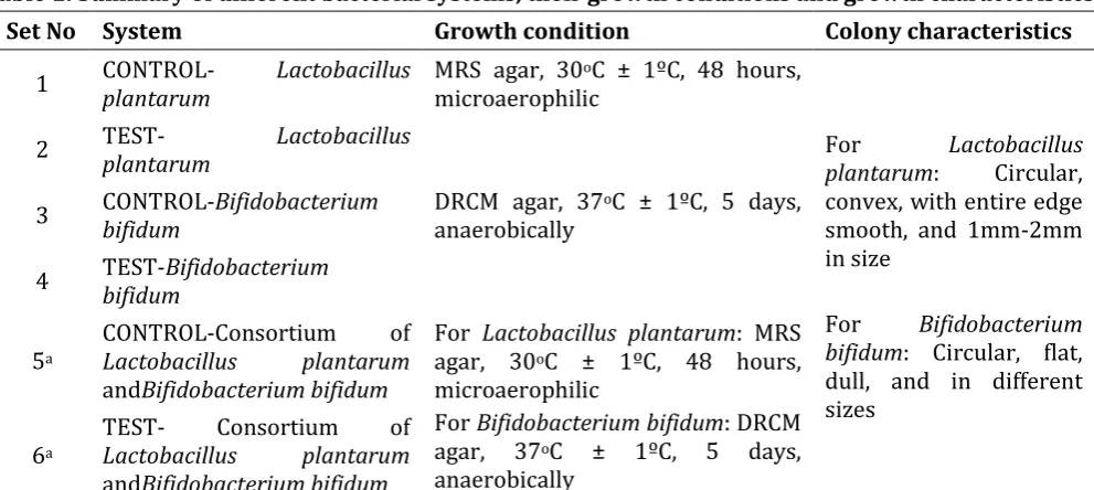
Three systems of each, test and control (Table 1), (all in triplicates) were prepared to evaluate the effect of 5%w/v starch of Tinospora cordifolia
(test)on the individual bacterial strains of
Bifidobacterium and Lactobacillus, and in consortia of both these strains. Bacterial suspension was added to the respective system, homogenously mixed and the viable counts of the test cultures were immediately determined which represented the 0 hour counts. Thereafter, all the systems were incubated under anaerobic conditions at 37ºC ± 1ºC for up to 96 hours and sampled at the above defined contact times.
Growth promotion by starch of Tinospora cordifolia was evaluated by determining the bacterial population using 10-fold serial dilutions and standard platecount method. At all contact times, including the 0 hour sample, 1 mL of inoculated sample from both test and control systems were analyzed by carrying out 10-fold serial dilution up to 10-8 and plating each dilution on specific growth agar medium using spread plate technique. All the plates were incubated at conditions specified for optimum growth of test strains used in the study (Table 1), and bacterial colonies were enumerated (Figure 1).
Statistical analyses
Descriptive results were presented as mean ± standard deviation (SD). Paired t-test was applied to the data using Microsoft office excel, the p-values <0.05 were considered as significant.
Results
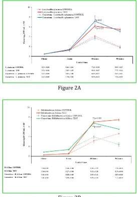
Starch of Tinospora cordifolia (5%w/v) showed greater than 1 log difference in mean CFU/mL of
Lactobacillus plantarum and Bifidobacterium bifidum following 48 hours incubation. The mean log colony count ± standard deviation (SD) of
Lactobacillus was 8.09±0.07 in test, which was significantly higher (p<0.05) when compared to control (7.06±0.03) at 48 hours, thereafter the growth of Lactobacillus plateaued in both the systems through 96 hours. The mean log colony count of Bifidobacterium in test was 8.52±0.28 at 48 hours and 8.74±0.14 at 96 hours, which were significantly higher (p<0.005) when compared to control. Furthermore, Bifidobacterium continued to grow in contact with starch of Tinospora cordifolia
even after 96 hours of incubation (Figure 2).
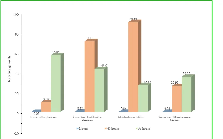
Relative difference in bacterial populations between the control and the test system was calculated to demonstrate the effect of starch of
Tinospora cordifolia on the growth of test strains at
a given contact time. Bifidobacterium bifidumin contact with starch of Tinospora cordifolia, at the contact time of 48 hour exhibited 90 percent relative increase in growth when compared with control, while in the consortium system,
Lactobacillus plantarume xhibited better growth. The relative growth of Lactobacillus plantarum at the end of 48 hour contact time with starch of
Tinospora cordifolia was found to be 70 percent as compared to 26 percent in its control system (Figure 3).
DISCUSSION
Probiotics and prebiotics are extensively studied to understand their influence and role on the gut microbiota.23Probiotics colonize the human intestine transiently which necessitates frequent dosing to maintain biologically significant numbers of probiotic strains. Also, there are variations across the gut microbiome of the host that would either resist or facilitate the colonization intended by the probiotic.24 The clinical outcome of probiotic varies from one person to another since the number of viable bacteria populating or colonizing the intestine depends on several factors other than dose; including type of probiotic formulation, co-administration of food or milk, and the host’s gastric pH, intestinal motility, adherence to intestinal epithelium and prior composition of gut microbiome. This was revealed in studies where the fecal recovery and biopsy microbiome analyses of the host were not equivalent after administration of probiotic.
In contrast, prebiotics are non-digestible, non-absorbable substrates that serve as food for existing gut microbiome towards a sustained healthy composition. Although probiotics are recommended by clinicians in a wide range of diseases, a polyherbal prebiotic like Somavit® might provide a host-centric delivery approach. In the present study, starch of Tinospora cordifolia, the principle ingredient of Somavit® was studied for its role as a prebiotic. Majority of studies have focused on pure oligosaccharides, including inulin, fructooligosaccharides, galactooligosaccharides, xylooligosaccharides, starch etc. for their prebiotic potential.20, 25 The prebiotic potential of blueberry, pomegranate, almond skin, and red grape extracts on Lactobacillus and Bifidobacterium species have been assessed.26-29 Furthermore, a study has reported the prebiotic effects of Tulsi, Ginger, and Black pepper using cultures of Lactobacillus rhamnosus GG and Bifidobacterium infantis.30
vitro study on growth modulation of Bifidobacterium and Lactobacillus strains
IJRAPS | September 2018 | Vol 2 | Issue 9 285
Bifidobacterium bifidum and Lactobacillus plantarum, singly and their consortia. The use of consortia of Lactobacillus and Bifidobacterium for assessment of prebiotic potential supports the symbiotic behavior of these cultures. The entire contact study was conducted under anaerobic conditions to mimic the growth conditions in their natural ecosystem. The viable growth was assessed under anaerobic conditions for Bifidobacterium and microaerophilic conditions for Lactobacillus. Furthermore, since the Ayurvedic preparation tends to sediment to the base of the liquid medium, the entire study was conducted in a controlled liquid interface to facilitate uniform contact of the prebiotic. The study explored comprehensively the impact of starch of Tinospora cordifolia on the response of bacterial population studied individually and in consortium for the period of 1hour, 48hours and 96hours, which aligns with the time required for invitro multiplication and visualization of growth outcome by the chosen bacterial strains and the maximum time period that the prebiotic may remain in the gut.
This study inferred that the beneficial roles of both these organisms possibly proceed in a deferred manner; Lactobacillus being more robust exhibited a rapid growth in bacterial population, while Bifidobacterium displayed a delayed growth. Growth of Lactobacillus promotes enhancement of
Bifidobacterium, allowing it to sustain for a longer duration in the human gut. The distinctive observation was the increase in count of
Bifidobacterium even after 96 hours of contact time. These observations indicated the sustenance of these beneficial bacterial populations, which may help in reducing the dosing frequency of the prebiotic. These results suggest that the growth modulation of these bacterial populations with starch of Tinospora cordifolia could offer the desired benefits over an extended period of time.
CONCLUSION
In conclusion, starch of Tinospora cordifolia, a key
component of Somavit® demonstrated a
considerable growth stimulatory activity by increasing the population of beneficial gut microorganisms, Bifidobacterium bifidum and
Lactobacillus plantarum in vitro and thus, confirmed a high prebiotic potential for this polyherbal formulation. Furthermore, a sustained growth modulation offered could probably reduce the dosing frequency. However, in order to substantiate the present in vitro data, further clinical studies to establish the prebiotic effect of starch of Tinospora cordifolia are warranted.
Acknowledgments: Akshada Deshpande, PhD provided writing assistance and Sangita Patil, PhD, ISMPP CMPPTM (both from Siro Clinpharm Pvt Ltd, India) provided editorial assistance.
The in vitro prebiotic activity of starch of Tinospora cordifolia was conducted at Bhavan’s Research Centre Microbiology, NABL (ISO, IEC 17025:2005) accredited laboratory.
Supported by: This work was supported by funding from Millennium Herbal Care Ltd, Mumbai, India.
Author contributions: Gandhi HI conceptualized and formulated Somavit SGC. Koli NM had primary role in study design along with Desai NJ and Samant SL. Desai NJ and Samant SL were involved in conceptualizing, executing, summarizing and inferencing the research work.
All authors met ICMJE criteria and all those who fulfilled those criteria are listed as authors. All authors had access to the study data and made the final decision about where to publish these data and approved submission to this journal.
Conflict of Interest: No potential conflict of interest was reported by the authors
REFERENCES
1. Kamada N, Seo SU, Chen GY, Nunez G. Role of the gut microbiota in immunity and inflammatory disease. Nat Rev Immunol 2013; 13(5): 321-35. doi:10.1038/nri3430.
2. Thursby E, Juge N. Introduction to the human gut microbiota. Biochem J 2017; 474(11): 1823-36. doi:10.1042/bcj20160510.
3. Roberfroid M, Gibson GR, Hoyles L, McCartney AL, Rastall R, Rowland I, Wolvers D, Watzl B, Szajewska H, Stahl B, et al. Prebiotic effects: metabolic and health benefits. Br J Nutr 2010; 104 Suppl 2: S1-63. doi:10.1017/s0007114510003363.
4. Allen JM, Mailing LJ, Niemiro GM, Moore R, Cook MD, White BA, Holscher HD, Woods JA. Exercise Alters Gut Microbiota Composition and Function in Lean and Obese Humans. Med Sci Sports Exerc 2018; 50(4): 747-57. doi:10.1249/mss.0000000000001495.
5. David LA, Maurice CF, Carmody RN, Gootenberg DB, Button JE, Wolfe BE, Ling AV, Devlin AS, Varma Y, Fischbach MA, et al. Diet rapidly and reproducibly alters the human gut microbiome. Nature 2014; 505(7484): 559-63. doi:10.1038/nature12820.
Website: http://ijraps.in 286
administration on the human intestinal microbiota. Isme j 2007; 1(1): 56-66. doi:10.1038/ismej.2007.3.
7. Breban M. Gut microbiota and inflammatory joint diseases. Joint Bone Spine 2016; 83(6): 645-9. doi:10.1016/j.jbspin.2016.04.005. 8. Cani PD, Delzenne NM. The role of the gut
microbiota in energy metabolism and metabolic disease. Curr Pharm Des 2009;
15(13): 1546-58.
doi:10.2174/138161209788168164.
9. Foster JA, McVey Neufeld KA. Gut-brain axis: how the microbiome influences anxiety and depression. Trends Neurosci 2013; 36(5): 305-12. doi:10.1016/j.tins.2013.01.005.
10.Sekirov I, Russell SL, Antunes LC, Finlay BB. Gut microbiota in health and disease. Physiol
Rev 2010; 90(3): 859-904.
doi:10.1152/physrev.00045.2009.
11.Louis P, Flint H, Michel C. How to manipulate the microbiota: prebiotics. In Microbiota of the human body. Springer, Cham; 2016. 119-42. 12.Martinez JL. Antibiotics and antibiotic
resistance genes in natural environments.
Science 2008; 321(5887): 365-7.
doi:10.1126/science.1159483.
13.Willing BP, Russell SL, Finlay BB. Shifting the balance: antibiotic effects on host-microbiota mutualism. Nat Rev Microbiol 2011; 9(4): 233-43. doi:10.1038/nrmicro2536.
14.Hill C, Guarner F, Reid G, Gibson GR, Merenstein DJ, Pot B, Morelli L, Canani RB, Flint HJ, Salminen S, et al. Expert consensus document. The International Scientific Association for Probiotics and Prebiotics consensus statement on the scope and appropriate use of the term probiotic. Nat Rev Gastroenterol Hepatol 2014; 11(8): 506-14. doi:10.1038/nrgastro.2014.66.
15.Boyle RJ, Robins-Browne RM, Tang ML. Probiotic use in clinical practice: what are the risks? Am J Clin Nutr 2006; 83(6): 1256-64; quiz 446-7. doi:10.1093/ajcn/83.6.1256. 16.Gibson GR, Hutkins R, Sanders ME, Prescott
SL, Reimer RA, Salminen SJ, Scott K, Stanton C, Swanson KS, Cani PD, et al. Expert consensus document: The International Scientific Association for Probiotics and Prebiotics (ISAPP) consensus statement on the definition and scope of prebiotics. Nat Rev Gastroenterol Hepatol 2017; 14(8): 491-502. doi:10.1038/nrgastro.2017.75.
17.Gong J, Yang C. Advances in the methods for studying gut microbiota and their relevance to the research of dietary fiber functions. Food Research International 2012; 48(2): 916-29. doi:10.1016/j.foodres.2011.12.027.
18.Bouhnik Y, Flourie B, D'Agay-Abensour L, Pochart P, Gramet G, Durand M, Rambaud JC.
Administration of
transgalacto-oligosaccharides increases fecal bifidobacteria and modifies colonic fermentation metabolism in healthy humans. J Nutr 1997; 127(3): 444-8. doi:10.1093/jn/127.3.444.
19.Gibson GR, Beatty ER, Wang X, Cummings JH. Selective stimulation of bifidobacteria in the human colon by oligofructose and inulin. Gastroenterology 1995; 108(4): 975-82. 20.Li Z, Summanen PH, Komoriya T, Finegold SM.
In vitro study of the prebiotic
xylooligosaccharide (XOS) on the growth of Bifidobacterium spp and Lactobacillus spp. Int J Food Sci Nutr 2015; 66(8): 919-22. doi:10.3109/09637486.2015.1064869.
21.Aranha I, Clement F, Venkatesh YP. Immunostimulatory properties of the major protein from the stem of the Ayurvedic medicinal herb, guduchi (Tinospora cordifolia). J Ethnopharmacol 2012; 139(2): 366-72. doi:10.1016/j.jep.2011.11.013.
22.Sharma U, Bala M, Saini R, Verma PK, Kumar N, Singh B, Munshi RK, Bhalerao S. Polysaccharide enriched immunomodulatory fractions from Tinospora cordifolia (Willd) miers ax hook. f. & Thoms. Indian J Exp Biol 2012; 50(9): 612-7.
23.Williams MD, Ha CY, Ciorba MA. Probiotics as therapy in gastroenterology: a study of physician opinions and recommendations. J Clin Gastroenterol 2010; 44(9): 631-6. doi:10.1097/MCG.0b013e3181d47f5b.
24.Steer T, Carpenter H, Tuohy K, Gibson GR. Perspectives on the role of the human gut microbiota and its modulation by pro- and prebiotics. Nutr Res Rev 2000; 13(2): 229-54. doi:10.1079/095442200108729089.
25.Scott KP, Martin JC, Duncan SH, Flint HJ. Prebiotic stimulation of human colonic
butyrate-producing bacteria and
bifidobacteria, in vitro. FEMS Microbiol Ecol 2014; 87(1): 30-40. doi:10.1111/1574-6941.12186.
vitro study on growth modulation of Bifidobacterium and Lactobacillus strains
IJRAPS | September 2018 | Vol 2 | Issue 9 287
intestinal microbiota. Int J Food Microbiol
2010; 140(2-3): 175-82.
doi:10.1016/j.ijfoodmicro.2010.03.038.
27.Mandalari G, Faulks RM, Bisignano C, Waldron KW, Narbad A, Wickham MS. In vitro evaluation of the prebiotic properties of almond skins (Amygdalus communis L.). FEMS Microbiol Lett 2010; 304(2): 116-22. doi:10.1111/j.1574-6968.2010.01898.x. 28.Molan AL, Lila MA, Mawson J, De S. In vitro
and in vivo evaluation of the prebiotic activity of water-soluble blueberry extracts. World Journal of Microbiology and Biotechnology 2009; 25(7): 1243-9. doi:10.1007/s11274-009-0011-9.
29.Pozuelo MJ, Agis-Torres A, Hervert-Hernandez D, Elvira Lopez-Oliva M, Munoz-Martinez E, Rotger R, Goni I. Grape antioxidant dietary fiber stimulates Lactobacillus growth in rat cecum. J Food Sci 2012; 77(2): H59-62. doi:10.1111/j.1750-3841.2011.02520.x.
30.Babu KN, Hemalatha R, Satyanarayana U, Shujauddin M, Himaja N, Bhaskarachary K, Kumar BD. Phytochemicals, polyphenols, prebiotic effect of Ocimum sanctum, Zingiber officinale, Piper nigrum extracts. Journal of
Herbal Medicine 2018; 13: 42-51.
doi:10.1016/j.hermed.2018.05.001.
Table 1: Summary of different bacterial systems, their growth conditions and growth characteristics
Set No System Growth condition Colony characteristics
1 CONTROL-plantarum Lactobacillus MRS agar, 30microaerophilic oC ± 1ºC, 48 hours,
For Lactobacillus plantarum: Circular, convex, with entire edge smooth, and 1mm-2mm in size
For Bifidobacterium bifidum: Circular, flat, dull, and in different sizes
2 TEST-plantarum Lactobacillus
3 CONTROL-bifidum Bifidobacterium DRCM agar, 37anaerobically oC ± 1ºC, 5 days,
4 TEST-bifidumBifidobacterium
5a
CONTROL-Consortium of
Lactobacillus plantarum
andBifidobacterium bifidum
For Lactobacillus plantarum: MRS agar, 30oC ± 1ºC, 48 hours, microaerophilic
For Bifidobacterium bifidum: DRCM agar, 37oC ± 1ºC, 5 days, anaerobically
6a
TEST- Consortium of
Lactobacillus plantarum
andBifidobacterium bifidum
aConsortium systems were incubated in both the growth conditions separately for enumeration of
Lactobacillus and Bifidobacterium colonies. TEST (containing 5% w/v starch of Tinospora cordifolia) and CONTROL (containing distilled water) systems were inoculated with cell suspensions of test strains of cell density of 1 x 10⁵ to 3 x 10⁵. Abbreviations: MRS agar: de Man, Rogosa and Sharpe agar; DRCM: Differential reinforced clostridial medium.
Figure legends
Figure 1: Representative plates of growth of (A) Lactobacillus plantarum on MRS agar and (B) Bifidobacterium bifidum on DRCM agar at sampling point of 48 hour
Website: http://ijraps.in 288 Figure 2: Growth of bacterial strains (A) Lactobacillus plantarum (B) Bifidobacterium bifidum, expressed as average bacterial count in terms of Mean log10 CFU/mL with increase in contact time
with Test (5%w/v Tinospora cordifolia) and Control (5%w/v distilled water)
Test: 5% w/v starch of Tinospora cordifolia; Control: 5% w/v Distilled water. Abbreviations: B.bifidum: Bifidobacterium bifidum; CFU: colony forming units; L.plantarum: Lactobacillus plantarum; SD: standard deviation
Figure 2A
vitro study on growth modulation of Bifidobacterium and Lactobacillus strains
IJRAPS | September 2018 | Vol 2 | Issue 9 289 Figure 3: Relative percent increase in bacterial growth when in contact with the starch of Tinospora cordifolia as compared to control
Cite this article as:
Shruti L Samant, Hasmukh I Gandhi, Nishith J Desai, Namrata M Koli. Prebiotic potential of starch of Tinospora cordifolia,a component of Somavit®: An in vitro study on growth modulation of Bifidobacterium and Lactobacillus strains. International Journal of Research in AYUSH and Pharmaceutical Sciences, 2018;2(8):282-289.
Source of support: Nil, Conflict of interest: None Declared
*Address for correspondence Dr. Namrata Koli
Manager-Medical Affairs, Millennium Herbal Care Ltd., 12-B, Nirmal 241/242, Backbay Reclamation, Nariman Point, Mumbai 400021, India.