Scholarship@Western
Scholarship@Western
Electronic Thesis and Dissertation Repository
8-22-2012 12:00 AM
Optimizing Physical Function Following Distal Radius Fracture
Optimizing Physical Function Following Distal Radius Fracture
Siamak Bashardoust Tajali The University of Western Ontario
Supervisor
Dr Joy C MacDermid
The University of Western Ontario
Graduate Program in Health and Rehabilitation Sciences
A thesis submitted in partial fulfillment of the requirements for the degree in Doctor of Philosophy
© Siamak Bashardoust Tajali 2012
Follow this and additional works at: https://ir.lib.uwo.ca/etd Part of the Physical Therapy Commons
Recommended Citation Recommended Citation
Bashardoust Tajali, Siamak, "Optimizing Physical Function Following Distal Radius Fracture" (2012). Electronic Thesis and Dissertation Repository. 827.
https://ir.lib.uwo.ca/etd/827
This Dissertation/Thesis is brought to you for free and open access by Scholarship@Western. It has been accepted for inclusion in Electronic Thesis and Dissertation Repository by an authorized administrator of
OPTIMIZING PHYSICAL FUNCTION FOLLOWING
DISTAL RADIUS FRACTURE
(Spine title: Function Following Distal Radius Fracture)
(Thesis format: Integrated-Article)
By
Siamak Bashardoust Tajali
Graduate Program in Health and Rehabilitation Science (Physical Therapy)
A thesis submitted in partial fulfillment
of the requirements for the degree of
Doctor of Philosophy
The School of Graduate and Postdoctoral Studies
The University of Western Ontario
London, Ontario, Canada
CERTIFICATE OF EXAMINATION
Supervisor
______________________________ Dr. Joy C. MacDermid
Supervisory Committee ______________________________
Dr. Pamela Houghton
______________________________ Dr. Ruby Grewal
Examiners
______________________________ Dr. Trevor Birmingham
______________________________ Dr. Jeff Dixon
______________________________ Dr. Norma J. MacIntyre ______________________________
Dr. Dave Walton
The thesis by
Siamak Bashardoust Tajali
entitled:
Optimizing Physical Function Following Distal Radius Fracture
is accepted in partial fulfillment of the requirements for the degree of
Doctor of Philosophy
Date__________________________ _______________________________ Chair of the Thesis Examination Board
Distal Radius Fracture (DRF) is one of the most frequent of all human bone fractures. Wrist and/or finger range of motion (ROM) and grip strength are standard outcome measures used by clinicians to evaluate recovery after a hand injury. ROM is considered to be an important component of joint mobility and relates to measures of functional impairment and disability. Impaired wrist and hand ROM are related to a decrease in grip strength, grasp ability, fine manipulation, and hand function. The relationship between ROM and other physical impairments as they relate to patient-rated outcomes after DRF have not been well identified.
The thesis includes three studies. The first study (Chapter 2) is a systematic review and meta analysis of existing literature on the effects of laser irradiation on bone regeneration, suggesting that low power laser can enhance biomechanical indicators of bone during fracture healing in animal models. The second study (Chapter 3) explores the intra-rater, inter-rater, and inter-instrument reliability and construct validity of two digital electro goniometers to measure active wrist and active/passive index finger ROM in patients with limited wrist and/or hand. The results of this study demonstrate that digital goniometry is highly reliable for all measures across occasions, raters and instruments. The moderate correlation between individual joint motions and patient-rated self-reported function suggests that joint motion impairments contribute to functional disability. The third study (Chapter 4) has a specific focus on the relationship between physical impairment outcome measures and patient-rated wrist pain and function in early and late stages after distal radius fracture. Wrist flexion, extension, supination, pronation, grip strength, age and gender, were found to contribute significantly with wrist pain and function. Good wrist arc of motions (close to normal) and moderate grip strength must be recovered to have optimal wrist functional outcomes after distal radius fracture. The thesis concludes with a discussion of the next steps required toward understanding effective mechanisms to promote bone healing and earlier function after DRF, which may lead to more effective patient-centered treatment protocols. Keywords: Bone Healing,
Distal Radius Fracture, Physical Impairment, Patient-Rated Wrist Evaluation.
This thesis contains material from one published manuscript (Chapter 2), and two submitted manuscript (Chapters 3 and 4). All studies in this thesis were conceived, performed, analyzed, interpreted and written by me with invaluable input, guidance and advice from my supervisor Dr. Joy C. MacDermid. There are other important collaborators which must be recognized because of their roles in various aspects of this thesis.
Advisory Committee
Dr. Ruby Grewal provided guidance on the design of the research program overall, as well as specific guidance and support for methods, interpretation of results and revisions of the manuscripts for Chapters 2, 3 and 4.
Dr. Pamela Houghton provided guidance on the design of the research program overall, as well as specific guidance and support for methods of systematic review and meta analysis, interpretation of results and revision of the manuscript for Chapter 2.
Other Collaborators
Chris Young was employed as a research assistant under Dr. MacDermid at the time of completing the reliability study in Chapter 3. He helped to recruit appropriate patients, performed physical impairment measures as an independent rater during the reliability study and reviewed the final manuscript of Chapter 3.
Kate Kelly was employed as a research assistant under Dr. MacDermid at the time of completing all Chapters. She helped to recruit appropriate patients and provided useful suggestions to complete the ethics application for Chapter 3.
I would like to dedicate my thesis
To my lovely mother, who has been a source of never ending encouragement, love, and kindness to me throughout my life.
To my beloved father, who taught me the values of humanity, gave me the courage to dream and the will to achieve. He is not with me in this important moment, but he is/will be alive always in my heart and my soul.
To my brother and sisters, who gave me their kindness and extensive support in this important time of my life.
First of all, I would like to acknowledge my supervisor, Dr. Joy C. MacDermid. This is not just for her magnificent knowledge and expertise, but because of her valuable support, great patience and kind consideration to help in all stages of my program and development as an independent researcher. Over the last five years, Joy challenged me to be better, supported me to move forward, and encouraged me to strive for excellence. Joy, thank you so much for everything.
I must express special acknowledgements to my advisors, Dr. Ruby Grewal and Dr. Pamela Houghton; their inputs into my thesis and education program were invaluable. Dr. Grewal provided many critical constructive comments which helped me to prepare the manuscripts, specifically in Chapters 3 and 4. Dr. Houghton helped me to expand my knowledge of systematic review and meta analysis. Her invaluable suggestions and comments helped me to design, appraise and write two beneficial reference manuscripts as a part of my thesis (Chapter 2) and also comprehensive process. Ruby and Pam, I would like to express my deep appreciation for all your great work.
Dr. Trevor Birmingham and Dr. Jeff Dixon helped me to have better understanding of the concepts of physical outcome measures and fracture healing. Their invaluable knowledge and guidance helped me to grow as an independent researcher.
Dr. Tom Overend and Dr. Dave Walton were always available for any help when I needed them. They are truly magnificent friends whom anybody may wish to have.
Dr. Bert Carron helped me to expand my understanding of meta analysis and critical appraisal.
I would like to express my appreciation to all office administrators at the Departments of Health and Rehabilitation Sciences and Physical Therapy, who were available anytime for help during my program. Special thanks of Ms Nancy Inchley, Ms Cathy Collins, Ms Amber Trent and Ms Donna Beer.
The Ontario Graduate Scholarship, the Joint Motion Program (JuMP) of the Canadian Institutes of Health Research and Graduate Funding at the University of Western Ontario. The department and the lab mates at the Hand and Upper Limb Centre (HULC)-St Joseph Hospital provided their valuable feedback and great suggestions through the monthly meetings.
Last but not least, the volunteer participants gave their time to complete three sets of data collection. It was not possible to complete the clinical parts of this study without their valuable participation.
Preliminary Pages
Certificate of Examination ………..……….…………... ii
Abstract and Keywords ………....……...… iii
Co-Authorship Statement ………..………..….………... iv
Dedication ………...……….…….... v
Acknowledgements ………...…..….… vi
Table of Contents ………...….…….. viii
List of Tables ……….. xiv
List of Figures ………. xvi
List of Appendices ………...……….…….. xviii
List of Abbreviations ………...…... xix
Chapter 1: Introduction and Background 1.1 Bone Injury and Fracture in Upper Extremity – Distal Radius Fracture ……..…...… 2
1.2 Epidemiology and Prevalence – Distal Radius Fracture …...………..………… 3
1.3 Bone Healing in Upper Extremity after Distal Radius Fracture …………..………… 5
1.3.1 Endochondral Bone Ossification ………..……….. 6
1.3.2 Intramembraneous Bone Ossification ………..…... 8
1.4 Physical Modalities and Bone Healing ……….. 10
1.5 Function, Structure, Activity and Participation in Distal Radius Fracture ………… 10
1.5.1 Measurement of Physical Impairments in Distal Radius Fracture ……….... 12
1.5.1.2 Grip and Pinch Strength ………...….. 13
1.5.1.3 Pain ………...………...………. 14
1.5.1.4 Radiographic Values ………..…... 15
1.5.2 Measurement of Function after Distal Radius Fracture ………. 16
1.5.2.1 Patient-Rated Wrist Evaluation (PRWE) ………...………… 16
1.5.2.2 Disabilities of the Arm, Shoulder and Hand (DASH) …….…..…… 17
1.5.2.3 Short Form-36 Health Survey (SF-36 Health Survey) ……..….…... 17
1.5.2.4 Michigan Hand outcome Questionnaire (MHQ) ……….……..….... 17
1.6 Reliability ……….. 18
1.6.1 Measurement Errors ………. 18
1.6.2 Measurement of Reliability ……….. 19
1.6.2.1 Relative Reliability ………...……… 19
1.6.2.2 Absolute Reliability ………..………….……... 20
1.6.3 Types of Reliability ………..…….………... 20
1.6.3.1 Test-Retest Reliability ………...………... 20
1.6.3.2 Rater Reliability ………..….…. 20
1.6.3.2.1 Intrarater Reliability ……….……….….… 20
1.6.3.2.2 Interrater Reliability ………..…….… 21
1.6.3.3 Internal Consistency ………..….…………....….. 22
1.8 Thesis Purpose ……….….. 23
1.9 Overview of Chapters ……….…... 24
1.10 References ……….…... 25
Chapter 2: Effects of Low Power Laser Irradiation on Bone Healing in Animal Models: A Meta Analysis 2.1 Summary ………..……….………. 40
2.2 Introduction ……….…... 40
2.3 Methods ……….……. 42
2.3.1 Study Design ……… 42
2.3.2 Data Extraction ……… 45
2.3.3 Statistical Analysis ………... 46
2.4 Results ……… 48
2.4.1 Description of Studies ……….. 48
2.4.2 Outcome Measures ………....……... 52
2.4.3 Calculation of effect size ………...……….. 52
2.4.4 Testing for Homogeneity of Variance ………. 53
2.4.5 Merits of Different Published Studies (Variables) ……….. 53
2.4.6 Fail Safe N and the Number of Treatment Sessions ……… 54
2.5 Discussion ……….. 55
2.7 Conclusions ………... 57
2.8 Additional Files ……….……… 57
2.9 References ……….…… 58
Chapter 3: Reliability and Validity of Electro-Goniometric Range of Motion Measurements in Patients with Wrist and Hand Limitations 3.1 Summary ……….….. 66
3.2 Introduction ……….…….. 67
3.3 Methods ……….… 70
3.3.1 Study Design ……….... 70
3.3.2 Participants ……….….. 70
3.3.3 Raters and Instruments ……….… 71
3.3.4 Statistical Analysis ………... 75
3.4 Results ……….….. 77
3.5 Discussion ……….…… 88
3.6 Study Limitations ……….……. 91
3.7 Conclusion ……… 91
3.8 Disclosure ………. 92
3.9 References ……… 92
4.1 Summary ……….……... 97
4.2 Introduction ……….. 98
4.3 Methods ………...……. 100
4.3.1 Study Design ………...… 100
4.3.2 Participants ……….………...…….. 100
4.3.3 Outcome Measurements ……….………. 101
4.3.3.1 Physical Impairment ………... 101
4.3.3.2 Self-reported Pain and Disability ………...……...…….. 102
4.3.4 Statistical Analysis ………..…… 102
4.4 Results ………... 104
4.5 Discussion ……… 114
4.6 Study Limitations ……….… 117
4.7 Conclusion ………...… 118
4.8 Disclosure ……… 118
4.9 References ……… 118
Chapter 5: General Discussion and Future Direction 5.1 Overview of Thesis ………... 125
5.2 Implications of Thesis Findings on Practice, Policy and Future Research …….…. 126
5.2.1 Low-power Laser Irradiation ………. 126
5.2.2 Range of Motion Measurement ………. 127
5.2.3 Physical Impairment Outcome Measures and Pain and Function ……. 128
5.4 Recommendations for Future Studies ……….……. 131
5.5 References ……….... 132
Appendices ………..….. 134
Curriculum Vitae ……….…… 150
Table 2.1: Mean maximum force (SD), effect size and quality score of included studies. …...………...………..……... 45
Table 2.2: The biomechanical bone properties (dependent variables) of included studies. ……….……. 46
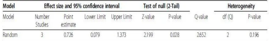
Table 2.3: Computed random effect size, CI 95% and Q value (Heterogeneity test). ……….…... 46
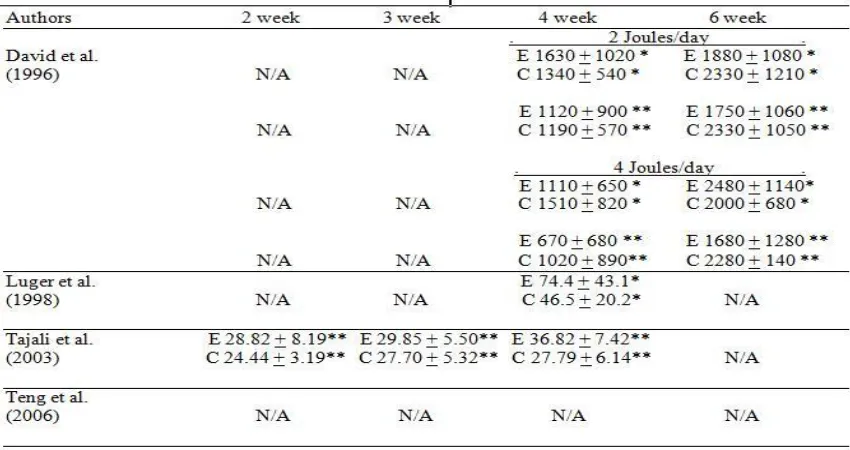
Table 2.4: Maximum force (Mean + SD) 2, 3, 4 or 6 weeks after fracture or surgery. ………...….……... 50
Table 2.5: Study characteristics of selected articles on effects of He-Ne low power laser irradiation on bone healing. ………...………....……….. 50
Table 3.1: Mean of range of motion measures (raters/occasions) and analysis of variances summary for the digital goniometers. ……….. 78
Table 3.2: Factorial Analysis of variance for main effects (rater and instrument) and interaction effects (rater × instrument) for the range of motion measures. ………….… 80
Table 3.3: Intrarater (test-retest) reliability values for NK and J-Tech digital goniometers. ……….…………. 81
Table 3.4: Interrater (between raters) reliability values for NK and J-Tech digital
goniometers. ………. 82
Table 3.5: Inter instrument reliability values for NK and J-Tech digital goniometers. .. 83
goniometers. ………...……….. 84
Table 3.7: Pearson’s r correlation coefficient between the range of motion measures of the digital goniometers (NK, J-Tech), patient-rated wrist evaluation and short version of the disability of the arm, shoulder and hand scales. ………..…….. 85
Table 4.1: Pearson’s r correlation coefficient between the ROM measurements and patient-rated wrist evaluation scores. ………. 105
Table 4.2: Adjusted R2 and regression analysis to identify contributors of patient-rated wrist evaluation total scores at three and twelve months after fracture. ……… 107
Table 4.3: The discriminators’ characteristics for wrist pain and function on injured hand one year after distal radius fracture. ………..…. 109
Table 4.4: The discriminators’ characteristics for wrist pain and function based on physical impairments injured/uninjured percentages one year after distal radius fracture. ………..….. 110
Figure 1.1: Bone healing after a fracture. ………..……… 9
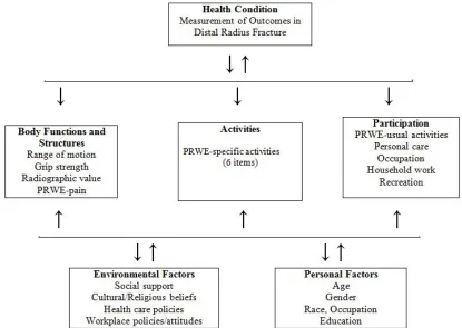
Figure 1.2: Framework of the ICF which is composed on patient’s function and disability (based on patient rated wrist pain and function evaluation) and the contextual factors that
impact the overall health after distal radius fracture. ………..………… 12
Figure 1.3: Traditional and digital goniometers can be used for range of motion measurement. ………... 13
Figure 1.4: Grip Dynamometer. ………..…. 14
Figure 1.5: Samples of Visual Analogue Scale (VAS). ………..………. 15
Figure 2.1: Flow diagram for identification the eligible animal studies evaluating effects of low power laser irradiation on bone healing based on bone biomechanical properties. ……….………. 43
Figure 2.2: The forest plot of the random effects model based on bone biomechanical properties (force maximum) changes 4 weeks after bone injury. ….………... 54
Figure 3.1: NK and J-Tech goniometer instruments. ……….. 72
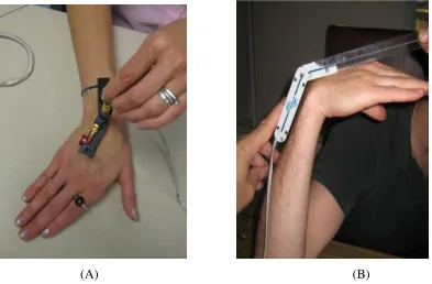
Figure 3.2: A) NK goniometer placement for active ulnar deviation measure. B) J-Tech goniometer placement for active wrist flexion measure. ………. 73
Figure 3.3: Diagram of the study design. ………. 74
(horizontal axis) of radial deviation ROM measures by NK and J-Tech: (A) rater one, (B) rater two. ………...………..……. 86
Figure 3.5: The Bland and Altman plots of mean differences (vertical axis) versus means (horizontal axis) of active ROM measures for PIP index flexion by two raters; (A) NK goniometer, (B) J-Tech goniometer. ……… 87
Figure 3.6: The Bland and Altman plot of mean differences (vertical axis) versus means (horizontal axis) of torques of passive index flexion ROMs by two raters (NK instrument). ……….. 88
Figure 4.1: Receiver operating characteristic curve using arcs of motion to distinguish between good and suboptimal functional outcomes one year after distal radius fracture. ………...………... 111
Figure 4.2: Receiver operating characteristic curve using the grip strength of injured hand and injured/uninjured percentage to distinguish between good and suboptimal functional outcome one year after distal radius fracture. ………...…. 111
Figure 4.3: Receiver operating characteristic curve using the grip strength of injured hand and injured/uninjured percentage to distinguish between good and suboptimal functional outcome one year after distal radius fracture. A) Participants equal or over 65 years old. B) Participants less than 65 years old. ……….……...….. 112
Figure 4.4: Receiver operating characteristic curve using the grip strength of injured hand and injured/uninjured percentage to distinguish between good and suboptimal functional outcome one year after distal radius fracture. A) Female. B) Male. ……….. 113
Appendix A: The Patient Rated Wrist Evaluation (PRWE) Questionnaire. ……….…. 135
Appendix B: The Disabilities of the Arm, Shoulder and Hand (DASH). ………….…. 138
Appendix C: The Short Version of the Disabilities of the Arm, Shoulder and Hand (Quick
DASH). ………... 142
Appendix D: The Quality of Animal/Tissue Research Scale. ………... 145
Appendix E: Ethics Approval Forms. ……… 147
AUC Area Under Curve
CI Confidence Interval
CMA Comprehensive Meta Analysis
DASH Disabilities of the Arm, Shoulder and Hand
DRF Distal Radius Fracture
HULC Hand and Upper Limb Centre
ICC Intraclass Correlation Coefficient
J-Tech J-Tech Electrogoniometer
LoA Limit of Agreement
MDC Minimal Detectable Change
NK NK Hand Assessment Joint Motion (NK Electrogoniometer)
PIP Proximal Inter Phalangeal
PRWE Patient-Rated Wrist Evaluation
QATRS Quality of Animal/Tissue Research Scale
ROC Receiver Operating Characteristic
ROM Range of Motion
RR Relative Risk
SD Standard Deviation
SEM Standard Error of Measurement
CHAPTER 1
1.1 Bone Injury and Fracture in Upper Extremity – Distal Radius fracture
A bone fracture is a complete or incomplete break in the continuity of a bone.1 A fracture can be the result of high force impact or stress, or as a result of certain medical conditions that weaken the bones, such as osteoporosis or cancer.1 Approximately 5.6 million bone fractures occur yearly in the United States.2 Pain, tenderness, bleeding, bruising, tingling, numbness, loss of pulse, loss of sensation, weakness, instability, deformity, paralysis and loss of function are common signs and symptoms of bone fractures.3 Anatomical classifications may discriminate fractures subtypes based on the involved parts of the body, such as head or arm fractures, which can be followed with more specific localization. There are a number of fracture classifications based on various criteria.1
radius fracture.6,11,12 Although nonunion in distal radius fracture is rare (0.2%)10, these fractures can sometime result in permanent pain and impairment, and should not be considered as a minor injury.11-14
1.2 Epidemiology and Prevalence
Although the descriptive epidemiology is well understood and researchers have actively investigated the risk factors, there are relatively little epidemiologic data available for upper extremity fractures.15 The data for extremities fracture in industrialized countries indicate they occur at the most proximal and the most distal ends of the extremities, with the highest incidence being among the elderly.15 Fractures of proximal humerus and distal forearm in adults are common in upper extremities, while hip and ankle fractures are dominant in lower extremities.15,16 Fractures occur at higher rates in women, including upper extremity fractures.15 Blacks of either gender have lower risk for these fractures as compared with other ethnicities.17 The risk of fracture correlates well with age.18 Different studies reported similar prevalence and incidence rates for upper extremity fractures based on age, gender, ethnicity, geographical location, and other factors.19,20 For instance, the incidence of childhood fractures in Malmo - Sweden among 8682 cases between 1950 and 1979 showed that boys in all age groups had higher upper extremity fracture rates than girls (62% vs. 38%).21 However, the incidence and gender ratio changes with increasing age.18,19,21
The National Hospital Ambulatory Medical Care Survey25 indicated that there were approximately 644,985 fractures of the distal radius in 1998 in the United States. The epidemiologic studies 27,28 have reported that the incidence of distal radius fracture increases in both genders with advancing age which occurs frequently because of falls.
The distribution of distal radius fracture peaks in three populations: children ages 5-14, men under age 50, and women over the age of 40.29 Among patients older than 60, the rate of distal radius fracture is seven times higher in women than that in men.30
has recently increased in younger people, since they engage more often in high energy sport activities.46,48,49
1.3 Bone Healing in Upper Extremity after Distal Radius Fracture
Bone healing after fracture is an important homeostatic process, and depends on specialized cell activation and bone immobility during the repair process.50,51 Bone repair is an essential process for reconstitution of skeletal integrity after trauma or skeletal surgery.50 Fracture reduction and fixation are prerequisites for optimal bone healing; however, a variety of other factors, such as age, nutrition, and medical co-morbidities, influence the healing process.52,53 In general, fracture healing is initiated by a sequence of inflammation followed by repair, and ends up with remodeling, thereby restoring the bone to its original state.54,55 Once the damaged cells and matrix have been replaced during the repair phase, a prolonged remodeling phase follows.56 Although the components of healing are similar in almost all fractures, the amount and quality of bone repair may vary based on type of cancellous or cortical bone, the extent of injured soft tissue around the fracture, and other factors which will be discussed below.
1.3.1 Endochondral Bone Ossification
Bone fracture damages cells, blood vessels, matrix, and the surrounding soft tissues, such as the periosteum and muscles, leading to hemorrhage and hematoma within the medullary canal, between the fracture ends and the elevated periosteum. The hematoma is considered as the first step in the repair process, and loss of hematoma leads to impaired fracture healing process.57-59 Damage of the bone blood vessels leads to malnutrition and death of osteocytes. Severe damage in the periosteum, bone marrow, and the surrounding soft tissue may contribute to tissue necrosis at the fracture site. Inflammatory mediators released from platelets and injured cells cause blood vessel dilation, which leak plasma into the fracture area, and produce acute edema in the fracture site.58,59
Hematoma, surrounding periosteal and soft tissues that contain blood vessels may facilitate the initial stages of repair.58,59 Open fractures and the treatment of fractures by open reduction disrupt hematoma formation and may slow down the repair process. The reason why hematoma formation affects fracture healing is still unclear; however, it is believed that hematoma provides a fibrinous scaffold that facilitates migration of certain cells to initiate the repair process. More importantly, growth factors, such as platelet-derived growth factors (PDGF) and transforming growth factors beta (TGF-β) and other proteins, are released by platelets and injured cells in the hematoma. These factors have an important role early in the healing process, including cell migration and proliferation, and the synthesis of new tissue matrix.58,60
tissues, while others migrate to the fracture site with blood vessels. Angiogenesis provides a large source of undifferentiated mesenchymal stem cells which differentiate into different cell types. In addition, these undifferentiated mesenchymal stem cells produce bone morphogenic protein (BMP), which is an important growth factor for the differentiation process.64 Periosteal cells of the cambium layer (i.e., inner layer of periosteum) have an especially prominent role in the healing process and form the earliest bone material. This role is more visible in children and young people because the periosteum is thicker and more cellular. The periosteum becomes thinner with increasing age and its contribution to fracture healing becomes less apparent.64 Osteoblasts from the endosteal surface also participate in bone formation. Most cells responsible for osteogenesis appear in the fracture site within the granulation tissue that replaces the fracture hematoma.63,64
radiographs show trabecular and cortical bone crossing the fracture site. However, even at this stage, healing is not complete yet. The new bone is weaker than normal bone; however, it gradually gains strength during the remodeling phase.
Remodeling begins with replacement of the woven bone by lamellar bone, and resorption of excessive callus. The new bone tissue at the fracture site moves toward rigid stability by progressing through calcified cartilage, woven bone, and finally lamellar bone. The important and functional consequence of remodeling is an increase in mechanical stability. The remodeling phase may continue for years after clinical and radiographic bone union.67 It is notable that the bone density at the fracture site may be decreased years after the fracture, even after a successful fracture healing.68,69 The reason for this density deficiency unclear but it should be considered that a fracture may cause persistent changes in the tissues and function.70
1.3.2 Intramembraneous Bone Ossification
When the fracture site is rigid and stable (by internal or external fixation), fracture healing occurs with less callus formation. This type of fracture healing is refered to intramembraneous bone ossification or primary bone healing, indicating that the healing process occurs without the formation and replacement of callus.71 In the presence of full contact between the fracture ends, lamellar bone can form directly across the fracture line by generation of new osteons.72
site without the formation of callus. In many impacted epiphyseal, metaphyseal, and vertebral fractures, both cancellous and cortical bone surfaces provide ample stability to establish primary bone healing where the bone surfaces are in direct contact.73 Figure 1.1 represents the stages of bone healing after a fracture.
1.4 Physical Modalities and Bone Healing
Over the past 50 years, researchers have looked into various physical and biologic methods to develop new ways of enhancing fracture healing.74 Early work on physical agents as mediators of bone healing was performed by Yasuda, Noguchi and Sata who studied the electrical stimulation effects on bone healing in the mid 1950s.50,75 In subsequent years, other researchers have studied effects of variety of physical modalities as potential mediators of bone healing. The physical agents include mechanical stimulation76, electromagnetic fields2, capacitive-coupled electrical stimulation77,78, direct current50,79, microcurrent80, low intensity pulsed ultrasound81-84, and laser radiation85,86. With increasing influence of lasers in different medical specialities in 1970s, the researchers focused on potential effectiveness of this new physical agent on bone healing.87-89 Although, in recent years, clinicians have recognized the importance of these non invasive physical modalities on healing of different connective tissues, there is still controversy on the characteristics and effectiveness of these physical agents.84,85,89
1.5 Function, Structure, Activity and Participation after Distal Radius Fracture
The goal of any type of treatment of the upper extremity is to restore function not only in the affected site, but also in the entire upper extremity and the body.90 Performing accurate and complete physical examination is the first step of a successful treatment plan, regardless of the type of injury. Function is the most important key in the treatment plan. An injury in a small finger could severely affect life of a piano player. The upper extremity is considered an integrated system that enables the person to do most complicated tasks; from throwing a ball to producing a fine work of art.90
Disability and Health (ICF), which led to changes in the measurement of health outcomes, specifically the evaluation of disability and handicap.92,94 Complications within these domains are called impairment, activity limitation, and participation restriction.95 Using this model, researchers have been able to evaluate how well the existing outcome measures assess the overall health concept associated with specific conditions.94,96-100
Function is a broad concept, beyond physical function and mobility.93 Based on the ICF framework, function is an umbrella that covers body functions, structures, activities and participations.92 Function is not a fixed state for all individuals. Rather, it must be considered as the result of dynamic interaction between health, environment and personal characteristics.93 Full function is achieved if there are no health-related complications, including any problem with function, structure, activities and participations.92 For instance, effective treatment of a patient with distal radius fracture successfully prevents structural impairment, decreases pain and stiffness, and enables the person to perform full range of activities and participation in social events. Conversely, disability, which is a negative concept for function, is achieved if there is structural impairment, pain, stiffness, limitations of activities or participation, despite the treatment.92 The relationship between impairment, activity limitation and participation restriction is bidirectional and can be affected by environmental factors, such as social or healthcare support and other personal factors, such as age, gender, weight, height or ethnicity.93,94
tasks, self-care, recreational, and social activities.103,105,106 Results of these studies suggest that despite surgical and rehabilitation treatment care after distal radius fracture, patients continue to have difficulty with work, self-care, sports, and leisure activities.103,105,106 Figure 1.2 represents framework of the ICF which is composed on patient’s function and disability and the contextual factors that impact overall health after distal radius fracture.
Figure 1.2: Framework of the ICF which is composed of patient’s function and disability (based on patient-rated wrist pain and function evaluation) and the contextual factors that
impact overall health after distal radius fracture.
1.5.1 Measurement of Physical Impairments after Distal Radius Fracture
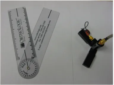
1.5.1.1 Range of Motion: Testing ROM, as a clinical measure for impairment, is an accepted method of musculoskeletal assessment, recommended by the American Medical Association.109 This method faces some challenges and controversies against the relevance of mobility deficits with functional loss. ROM can be measured by traditional manual or advanced electro digital goniometers.110,111 (Figure 1.3)
Figure 1.3: Traditional (left) and digital (right) goniometer can be used for range of motion measurement.
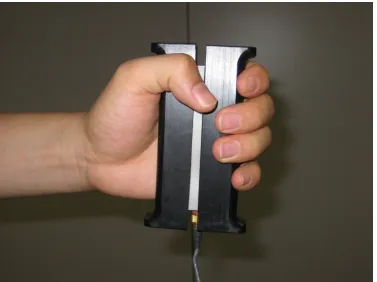
pinch strength is used to measure delicate hand function and grabbing something like a paper or plate.110,112 Grip or other relevant measure of hand strength can be assessed by a dynamometer.112,113 (Figure 1.4).
Figure 1.4:Grip Dynamometer
word in each group that best describes his/her pain and leaves out descriptions that are not applicable. Next, patient is asked to go back and circle the three words in groups 1 to 10 that most likely explain his/her pain response. Then, patient is asked to choose two words in groups 11 to 15, and one word in groups 16 to 20. The first 10 groups of words are somatic (describing what the pain feels like), groups 11-15 are affective, group 16 is evaluative, and groups space 17-20 are miscellaneous.119 Using this method, patient provides seven words to describe both the quality and intensity of pain.
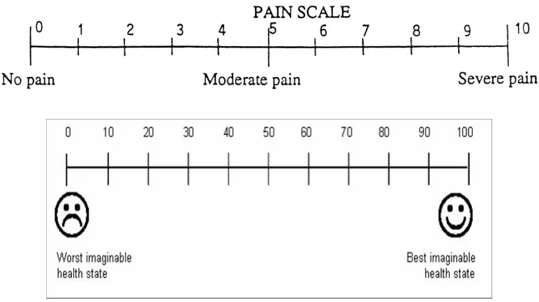
Figure 1.5: Samples of Visual Analogue Scale (VAS)
measures between 0 to 22 degrees (mean 11-14.5 degrees). The dorsal tilt greater than 20 degrees leads to significant transfer load onto the ulna, which will be following with pain and limited grip strength.121
1.5.2 Measurement of Function after Distal Radius Fracture
Traditionally, distal radius fracture outcomes have concentrated on impairment measures that were often called “objective functional measures” to evaluate outcomes following injury. However, in recent years, clinicians have recognized the importance of patient-reported outcome measures to assess functional status and health-related quality of life.122-124 As a result, the number of studies that have evaluated treatment effectiveness from the patient’s perspective are progressively increasing.
1.5.2.2 Disabilities of the Arm, Shoulder and Hand (DASH): This questionnaire is a administered region-specific outcome instrument developed as a measure of self-rated upper extremity disability and symptoms. The DASH consists mainly of a 30-item disability/symptom scale, scored 0 (no disability) to 100 (most severe disability). The DASH addresses difficulty in performing various physical activities that require upper extremity function (21 items); symptoms of pain, activity-related pain, tingling, weakness and stiffness (5 items), or impact of disability and symptoms on social activities, work, sleep and psychological issues (4 items). A shorter version called the Quick DASH is also available. Both tools are valid, reliable and responsive and can be used for clinical and/or research purposes. However, because the full DASH Outcome Measure provides greater precision, it may be the best choice for clinicians who wish to monitor arm pain and function in individual patients.128,129 (See Appendices B and C)
1.5.2.3 SF-36 Health Survey: The Short Form 36 Health Survey (SF-36) is a self-assessed functional outcome measure to assess quality of life. The SF-36 is a broad health-related outcome measure, which includes eight scales and two summary scores. 42-45
The original SF-36 came out from the Medical Outcome Study (MOS, http://www.rand.org/health/surveys_tools/mos/mos_core_36item.html), but scoring of the general health and pain are different.130,131 Each scale of SF-36 is directly transformed into a 0-100, assuming equal weight for each question. The eight sections represent various domains of health including: vitality, physical function, physical role, bodily pain, general health perceptions, emotional role, social role, and mental health. The physical and mental health summary scores represent two main dimensions of health. These scores are calculated in a 3-step process, which involves weighting, transforming and aggregating the subscale scores to compute summary scores for a typical US population. The SF-36 method of health measures separates physical and mental health, providing a more complete concept for overall health. 130-133
function, activities of daily living (ADLs), pain, work performance, aesthetics, and patient satisfaction.135 Each domain is scored from 0 to 100, with 0 being the worst score and 100 being the best. Conversely, 0 in pain domain indicates no pain and higher scores indicate more pain. All domains are assessed for each hand separately (exception of work). There is no adjustment scoring for hand dominance. This validated survey may be used for overall hand function, activity daily living, pain, work performance, aesthetics, and patient satisfaction.134-137
Difficulties in the ICF domains of activity and participation are able to explain a significant portion of physical health.94,96 Post-fracture treatment and outcome measurements should extend beyond physical impairment to provide a comprehensive effective treatment to patients with distal radius fracture.
1.6 Reliability
A major concern for all clinical measurements is to what extent the data are accurate and meaningful. Reliability is the first prerequisite to insure measures are useful for clinical decision making. Reliability is the extent to which a measurement is consistent and free from errors.138 A reliable measure has two important characteristics: a) it must provide consistent values with small errors of measurement, and b) it must be capable of differentiating among the subjects to whom the measurements are applied.139 Both consistency and ability to differentiate among the objects of measurement are prerequisites to a reliable measure.
1.6.1 Measurement Errors
systematic and random. Systematic errors occur consistently in one direction and overestimate or underestimate the true scores. Systematic errors are predictable errors of measurement.141 These errors can be considered a consistent “bias” in the measurement. Incorrect marking of a tape measure is a simple example for systematic errors. Random errors occur inconsistently due to chance and affect the true scores in an unpredictable way. There is no specific direction for random errors and they can lead to an increase or decrease the true scores. Lack of attention, non standardized methods, mechanical inaccuracy or simple mistakes are examples of simple reasons for random errors. As random errors decrease, the observed scores approach the true scores and the measurement is more reliable. Fatigue, learning, instrument limitations can result in random errors.
1.6.2 Measurement of Reliability
The measure of the reliability is often summarized in two methods. Relative and absolute reliabilities.
1.6.2.1 Relative Reliability
The relative reliability represents a measure’s ability to distinguish among clients.142 The relative reliability is defined as the ratio of true variance to observed or total variance which includes true variance plus error variance.140 The relative reliability coefficient is intraclass correlation coefficient which may vary from 0 to 1, with higher values represent higher reliability.
True variance
Variance Between Client Relative Reliability Coefficient = --- = ---
Observed (Total) Variance Between + Within
1.6.2.2 Absolute Reliability
The absolute reliability is the second method to represent reliability. The absolute reliability expresses the measurement error in the same units. The standard error of measurement (SEM) is used to quantify the absolute reliability of a measure.140 In general, statisticians have reported a single score of SEM for a measure. But, some researchers believe that the amount of absolute reliability (SEM) varies based on the client’s condition and must be reported as the conditional standard error of measurement (CSEM).141
. . SEM =
√
Within Client Variance1.6.3 Types of Reliability
There are three general approaches to reliability measurement: Test-retest reliability, rater reliability, and internal consistency.138
1.6.3.1 Test-Retest Reliability
Test-retest reliability is based on parallel assessments of clients on different occasions.140 This type of reliability is used to establish that an instrument is capable to measure a variable with consistency.138 In test-retest reliability one variable is subjected to the identical test on two different occasions, while all test conditions are kept in a consistent situation. The rater must consider that many variables change naturally over time. So, if the responses are labile over time, test-retest reliability may not be possible.138 The intraclass correlation coefficient (ICC) is the preferred statistical method to measure test-retest reliability, as it reflects both correlation and agreement.140
1.6.3.2 Rater Reliability
instrument to measure a variable, such as blood pressure or muscle testing. As a result, the individual who performs the rating must be consistent in the application of criteria for scoring responses. Data cannot be interpreted with confidence unless the raters who collect the data are reliable. The rater reliability should be documented as a part of the research protocol; and it is also critical for confidence in clinical decision making in practice.138,141
1.6.3.2.1 Intrarater Reliability
Intrarater reliability refers to consistency of data recorded by one rater across two or more occasions.138 Some researchers assume that the experienced raters can simply perform all measurements with high level of reliability. But, it should be considered that expertise may not always match with the level of precision which is necessary for the measurement. The statistical measurement of reliability strengthens the research conclusion, and also prevents critiques about the measurement accuracy.
The rater bias is considered in intrarater reliability assessment. When assessing this, it should be considered that raters can be influenced by their memory from the first measurement results. The most effective way to control this type of error is to blind raters from the first measurement scores. However, this technique may not work in many cases where the clinical measurements are observational. For instance, it is not possible to blind a clinician to measure function or gait procedures.138 Measuring range of motion on two different occasions by one rater is an example for intrarater reliability.
1.6.3.2.2 Interrater Reliability
rater’s measurements. For instance, range of motion may stretch the joint structure and change the results of the second rater’s measurements. Interrater reliability allows the researcher to know that the measurements obtained by one rater represent the true scores, and therefore, the results can be interpreted with greater confidence.138,143,144 Simultaneous (approximate) ROM measuring by two raters is an example for interrater reliability. The statistical method to evaluate intra and interrater reliability is the ICC model 2 or 3, depending on whether the raters are representative of other similar raters (model 2) or no generalization is considered (model 3).144
1.6.3.3 Internal Consistency
Internal consistency reliability is based on parallel assessments of clients at an instant time.139 In other words, internal consistency reflects the extent to which items measure various aspects of the same characteristic. This form of reliability mostly associates with questionnaires, but it is also applied to multi-item performance tests. The most important approach to measure internal consistency is correlation among all items in a scale.138 For instance, the Short Form 36-item (SF-36) health status measure has eight subscales (See 1.4.2 Measurement of Function after Distal Radius Fracture). Each of these subscales has been evaluated for internal consistency.145 The statistic method mostly used for internal consistency is Cronbach’s coefficient alpha.146 The Cronbach’s coefficient alpha evaluates homogeneity, suggesting the extent that the items in a scale are measuring the same construct.146
1.7 Summary of the Limitations in Knowledge
Further, since the devices are used by an evaluator, it is not clear if torque is consistently applied by different raters. Finally, since joint motion and grip strength are key indicators of joint status and muscle function following fracture, it is important to know to what extent these must be restored to maximize functional outcomes.
1.8 Thesis Purpose
The overall purpose of this thesis was to inform our understanding of optimizing recovery following fracture; with a focus on distal radius fracture.
The main purpose of this study was followed by the secondary goals:
1) To perform a systematic review and meta analysis of a physical modality (low level laser), which may potentially affect fracture healing.
2) To estimate reliability and validity of physical impairment (range of motion) measures by computerized digital electro goniometers.
3) To examine the consistency of torque which apply by different raters for physical impairment (range of motion) measures.
4) To clarify relationship between physical impairment outcome measures (range of motion, grip strength) and pain and function at different time points in recovery after distal radius fracture.
5) To determine the contribution of physical impairment outcome measures (range of motion, grip strength) and demographic variables to pain and function at early and late stages after distal radius fracture.
6) To identify levels of physical impairments (range of motion and grip strength), which are necessary to achieve optimal functional outcomes after distal radius fracture.
7) To identify risk of suboptimal function in patients with good physical impairments outcome measures (range of motion and grip strength) after distal radius fracture.
1.9 Overview of Chapters
There were a few studies that focused on bone healing and stimulatory effect of physical modalities on the distal radius fracture.82,147-150 Therefore, we decided to broaden our search strategy to examine the effects of physical modalities on bone healing in general and not specific to the location/type of fracture. We were aware of the fact that bone healing process follows a similar pattern in the skeletal system.151 The details of the effective methods used to facilitate bone healing are also applicable to distal radius fracture.
In chapter 2, we initiated a systematic review and meta-analysis of the newest physical modalities, Low Level Laser, which may impact bone healing in fractures of animal models, since there were no published data available on human bone healing treated with laser irradiation and considering that bone healing process is similar in vertebrates.151 Although there is still insufficient evidence to establish optimal dosage, the results appear to be sufficient evidence of improved bone healing in animal models to warrant clinical trials evaluating the role of low-level laser irradiation on human bone healing. Please see chapter 2 for details.
Chapter 3 addresses reliability and validity aspects of physical impairment measurements, in term of range of motion, in patients with wrist and hand limitations. The intrarater, interrater and inter instruments reliability of two digital goniometric instruments (NK and J-Tech) were evaluated in this chapter. Moreover, the relationship between joint motion impairments obtained by digital goniometry and functional disability were studied in this chapter. The quick disability of arm, shoulder and hand (quick DASH) and patient-rated wrist evaluation (PRWE) self-reported pain and function questionnaires were used to identify functional disability of the patients with wrist and hand limitations. Please see chapter 3 for details.
fracture. We identified the levels of physical impairments that discriminated the functional outcomes at early and late stages of recovery after distal radius fracture. We also studied the relationship of physical impairment outcome measures and patient-rated wrist pain and function after distal radius fracture. Finally, we identified the levels of range of motion and grip strength which were necessary to achieve optimal function after distal radius fracture. Please see chapter 4 for details.
The final chapter (Chapter 5) presents a general conclusion and discussion of the above studies, including the most important findings, and provides recommendations to be considered in future studies. In summary, this thesis attempts to lead the reader through evidence-based and clinical approaches to items stimulate healing, examine the reliability of range of motion measures by electro goniometers, define the physical impairments and their contribution, identify the clinical discriminators of functional outcomes, and identify the levels of physical impairment measures required for optimal function after distal radius fracture. The findings that form the head of the results in this study are just a branch of the research road that must follow to establish the methods to stimulate healing process and function after distal radius fracture.
1.10 References
1. Dirschl DR, Cannada LK. Classification of Fractures. In: Bucholz RW, Heckman JD, Court-Brown C. Rockwood and Green’s Fractures in Adults. 6th ed. Philadelphia: Lippincott Williams and Wilkins, 2006.
2. Trock DH. Electromagnetic fields and magnets: Investigational treatment for musculoskeletal disorders. Rheum Dis Clin North Am 2000; 26:51-62
3. Safron MR, Zachazewski J, Stone DA. Instructions for Sports Medicine Patients. 2nd ed. Philadelphia: Elsevier Saunders, 2012.
5. Young BT, Rayan GM. Outcome following non operative treatment of displaced distal radius fractures in low demand patients older than 60 years. J Hand Surg Am 2000; 25: 19-28.
6. Turner RG, Faber KJ, Athwal GS. Complications of distal radius fractures. Hand Clin 2010; 26: 85-96.
7. Becton JL, Colborn GL, Goodrich JA. Use of an external fixator to treat comminuted fractures of distal radius: report of a technique. Am J Ortho 1998; 27: 619-23.
8. Chapman DR, Bennet JB, Bryan WJ, Tullos HS. Complications of distal radius fractures: pins and plaster treatment. J Hand Surg [Am] 1982; 7: 509-12.
9. McKay SD, MacDermid JC, Roth JH, Richards RS. Assessment of complications of distal radius fractures and development of a comparison checklist. J Hand Surg [Am] 2001; 26(5): 916-22.
10. Bacron RW, Kurtzke JF. Colle’s fracture. A study of two thousand cases from the New York State Workmen’s Compensation Board. J Bone Joint Surg [Am] 1953; 35: 643-58.
11. Cooney WP, Dobyns JH, Linscheid RL. Complications of Colles’ fractures. J Bone Joint Surg 1980; 62A:613–9.
12. McQueen MM, Caspers J. Colles’ fracture: does the anatomical result affect the final function? J Bone Joint Surg 1988; 70B: 649–51.
13. Tsukazaki T, Iwasaki K. Ulnar wrist pain after Colles’ fracture. 109 fractures followed for 4 years. Acta Orthop Scand 1993;64: 462–4.
14. Beaule´ PE, Dervin GF, Giachino AA, Rody K, Grabowski J, Fazekas A. Self-reported disability following distal radius fractures: the influence of hand dominance. J Hand Surg 2000;
25A: 476–82.
15. Baron JA, Barrett JA, Karagas MR. The epidemiology of peripheral fractures. Bone 1996; 18(3): 209S-213S.
16. Jacobsen SJ, Goldberg J, Miles TP, Brody JA, Stiers W, Rimm A. Hip fractures incidence among the old and very old. A population based study of 745,435 cases. Am J Public Health 1990; 80: 871-3.
18. Worlock P, Stower M. Fracture patterns in Nothingham children. J Pediatr Orthop 1986; 6: 656-60.
19. Lauritzen JB, Schwartz P, Lund B, McNair P, Transbol I. Changing incidence and residual lifetime risk of common osteoporosis related fractures. Osteoporos Znt 1993; 3: 127-32.
20. Nguyen T, Sambrook P, Kelly P, Jones O, Codes Freund J, Eisman J. Prediction of osteoporotic fractures by instability and bone density. BMJ 1993; 307: 1111-5.
21. Landin LA. Fracture patterns in children. Analysis of 8682 fractures with special reference to incidence, etiology and secular changes in a Swedish urban popultaion 195-1979. Acta Orthop Scand Suppl 1983; 202 : 1-109.
22.Baron JA, Barrett J, Malenka D, Fisher E, Kniffin W, Bubolz T, Tosteson T. Racial differences in vracture risk. Epidemiology 1994; 5: 42-7.
23.Jones G, Nguyen T, Sambrook PN, Kelly PJ, Gilbert C, Eisman JA. Sympthomatic fracture incidence in elderly and women: The Dubbo Osoteoporosis Epidemiology Study (DOES). Osteoporosis Int 1994; 4: 277-82.
24. Baron JA, Karagas M, Barrett J, Kniffin W, Malenka D, Mayor M, Keller RB. Basic epidemiology fractures of the upper and lower limb among americans over 65. Epidemiology 1996;7(6):612-8.
25. Ruch Ds. Fractures of the Distal Radius and Ulna. In: Bucholz RW, Heckman JD, Court-Brown C. Rockwood and Green’s Fractures in Adults. 6th ed. Philadelphia: Lippincott Williams and Wilkins, 2006.
26. Fernandez DL, Jupiter JB. Fractures of the Distal Radius. A Practical Approach to Management. 2nd ed. New York, Springer, 2002.
27. Bengener U, Johnell O. Increasing incidence of forearm fractures. A comparison of epidemiologic patterns 25 years apart Acta Orthop Scand 1985; 56: 158-60.
28. Hagino H, Yamamoto K, Ohshiro H, Nakamura T, Kishimoto H, Nose T. Changing incidence of hip, distal radius, and proximal humerus fractures in Tottori Prefecture, Japan. Bone 1999;24:265-70.
29. Court-Brown CM, Caesar B. Epidemiology of adult fractures: a review. Injury 2006; 37:691-7.
31. Lindau TR, Aspenberg P, Arner M, Redlundh-Johnell I, Hagberg L. Fractures of the distal forearm in young adults. An epidemiologic description of 341 patients. Acta Orthop Scand 1999; 70: 124-8.
32. Cuenca J, Martinez AA, Herrera A, Domingo J. The incidence of distal forearm fractures in Zaragoza (Spain). Chir Main 2003;22:211-5
33. Miller SW, Evans JG. Fractures of the distal forearm in Newcastle: an epidemiological survey. Age Ageing 1985;14:155-8.
34. Jiuliano JA, Jupiter J. Distal Radius Fracture. In: Trumble TE, Budoff JE, Cornwall R. Hand, Elbow, Shoulder. Core Knowledge in Orthopaedics. Philadelphia, Mosby Elsevier, 2006, 84-101.
35. Jupiter JB. Current concepts review: fractures of the distal end of the radius. J Bone Joint Surg 1991; 73A: 461-9.
36. Mensforth RP, Latimer BM. Hamann-Todd Collection aging studies: osteoporosis fracture syndrome. Am J Phys Anthropol 1989;80:461-79.
37. Nguyen TV, Center JR, Sambrook PN, Eisman JA. Risk factors for proximal humerus, forearm, and wrist fractures in elderly men and women: the Dubbo Osteoporosis Epidemiology Study. Am J Epidemiol 2001;153:587-95.
38. Masud T, Jordan D, Hosking DJ. Distal forearm fracture history in an older community dwelling population: the Nottingham Community Osteoporosis (NOCOS) study. Age Ageing 2001;30:255-8.
39. Krolner B, Tondervold E, Toft B. Bone mass of the axial and the appendicular skeleton in women with Colles’ fracture: its relation to physical activity. Clin Physiol 1982; 2: 147-57.
40. Lamke B, Sjoberg HE, Sylven M. Bone mineral content in women with Colles’ fracture: effects of calcium supplementation. Acta Orthop Scand 1988; 49: 143-6.
41. Hove LM, Fjeldsgaard K, Reitan R, Sorensen FK. Fractures of the distal radius in a Nowegian city. Scand J Plast Reconstr Surg Hand Surg 1995; 29: 263-7.
43. Kirschen AJ, Cape RDJ, Hayes KC, Spencer JD. Postrual sway and cardiovascular parameters associated with falls in the elderly. J Clin ExpRes Gerontol 1984; 6: 291-307.
44. Lester GE, Anderson JJ, Tylavsky FA. Update on the use of distal radial bone density measurements in prediction of hip and Colles' fracture risk. J Orthop Res 1990; 8:220-6.
45. Mallmin H, Ljunghall S, Naessen T. Colles' fracture associated with reduced bone mineral content. Photon densitometry in 74 patients with matched controls. Acta Orthop Scand 1992;63:552-4.
46. Mallmin H, Ljunghall S, Persson I, Bergstrom R. Risk factors for fractures of the distal forearm: a population-based case-control study. Osteoporos Int 1994; 4: 298-304.
47. Gardsell P, Johnell O, Nilsson BE. Predicting fractures in women by using forearm bone densitometry. Calcif Tissue Int 1989;44:235-42.
48. Lawson GM, Hajducka C, MacQueen MM. Sports fractures of the distal radius. Epidemiology and outcome. Injury 1995;26:33-6.
49. Schmalholz A. Epidemiology of distal radius fracture in Stockholm 1981-82. Acta Orthop Scand 1988;59:701-3.
50. Paterson D. Treatment of nonunion with a constant direct current: A totally implantable system. Orthop Clin North Am 1984; 15: 47-59.
51. Childs SG. Stimulators of bone healing. Biologic and biomechanical. Orthop Nurs 2003; 22: 421-8.
52. Orwoll ES. Toward an expanded understanding of the role of the periosteum in skeletal health. J Bone Miner Res 2003; 18: 949-54.
53. Saleh M. The principles of non-union management. Orthofix External Fixation in Trauma and Orthopaedics London: SpringerDe Bastiani G, Apley AG, Goldberg A 2000, 523-36.
55. Buckwalter JA, Einhorn TA, Marsh JL. Bone and Joint Healing. In Bucholz RW, Heckman JD, Court-Brown C. Rockwood and Green’s Fractures in Adults. 6th ed. Philadelphia: Lippincott Williams and Wilkins, 2006, 297-311.
56. Leung KS, Sher AH, Lam TSW, Leung PC. Energy metabolism in fracture healing. J Bone Joint Surg 1989; 71B: 657-60.
57. Mark H, Penington A, Nannmark U, Morrison W, Messina A. Microvascular invasion during endochondral ossification in experimental fractures in rats. Bone 2004; 35: 535-42.
58. Gruber R, Koch H, Doll BA, Tegtmeier F, Einhorn TA, Hollinger JO. Fracture healing in elderly patients. Exp Gerontol 2006; 41: 1080-93.
59. Grundnes O, Reikeras O. The importance of the hematoma for fracture healing in rats. Acat Orthop Scand 1993; 64: 340-2.
60. Wildemann B, Schmidmaier G, Brenner N, Huning M, Stange R, Haas NP, Raschke M. Quantification, localization, and expression of IGF-I and TGF-beta during growth factor-stimulated fracture healing. Calcif Tissue Int 2004; 74: 388-97.
61. Brighton CT, Hunt RM. Early histologic and ultrastructural changes in microvessels of periosteal callus. J Orthop Trauma 1997; 71: 244-53.
62. Buckwalter JA, Glimcher MJ, Cooper RR, Recker R. Bone biology. Part I. Structure, blood supply, cells, matrix, and mineralization. J Bone Joint Surg 1995; 77A: 1256-75.
63. Buckwalter JA, Glimcher MM, Cooper RR, Recker R. Bone biology. Part II. Formation, form, modeling, and remodeling. J Bone Joint Surg 1995; 77A: 1276-89.
64. Anderson HC, Hodges PT, Agieilera XM, Missana L, Moylan PE. Bone morphogenetic protein (BMP) localization in developing human and rat growth plate methaphyses, epiphysis, and articular cartilage. J Histochem Cytochem 2000; 48: 1493-1502.
66. Aro HT, Wippermann BW, Hodgson SF, Wahner HW, Lewallen DG, Chao EY.
Prediction of properties of fracture callus by measurement of mineral density using microbone densitometry. J Bone Joint Surg 1989; 71A: 1020-30.
67. McKibbin B. The biology of fracture healing in long bones. J Bone Jt Surg 1978; 60B: 150-62.
68. Marsh JL, Buckwalter JA, Evarts CM. Nonunion, delayed union, malunion, and avascular necrosis. In: Epps CH, ed. Complications in Orthopaedic Surgery. Philadelphia: Lippincott, 1994: 183-211.
69. Wiel HE, Lips P, Nauta J, Patka P, Haarman HJ, Teule GJ. Loss of bone in the proximal part of the femur following unstable fractures of the leg. J Bone Joint Surg 1994; 76A: 230-6.
70. Jansen NW, Roosendaal G, Bijlsma JW, Degroot J, Lafeber. Exposure of human cartilage tissue to low concentrations of blood for a short period of time leads to prolonged cartilage damage: an in vitro study. Arthritis Rheum 2007; 56(1): 199-207.
71. Ross MH, Kaye GI, Pawlina W. Histology: A Text and Atlas. 4th ed. Philadelphia, Lippincott Williams & Wilkins, 2003, 180-213.
72. Lee CA, Einhorn TA. The Bone Organ System; Form and Function. In: Marcus R, Feldman D, Kelsey J. Fundamentals of Osteoporosis. 2nd ed. Amsterdam : Elsevier Academic Press, 2010, 3-20.
73. Zuscik MJ, O’Keefe RJ. Skeletal Healing. In: Rosen CJ, Compston JE, Lian JB. Primer on the Metabolic Bone Diseases and Disorders of Mineral Metabolism. 7th ed. Philadelphia, John Wiley & Son, 2008, 61-65.
74. Malizos KN, Hantes ME, Protopappas V, Papachristos A. Low intensity pulsed ultrasound for bone healing: An overview. Injury 2006; 37S:(suppl 1)S56-62.
75. Singh S, Saha S. Electrical properties of bone. Clinical Orthopedic and Related Research 1984, 186:249-71.
76. Goodship AE, Kennwright J. The influence of induced micro movement upon the healing of experimental tibial fractures. J Bone Joint Surg 1985; 67B:650-5.
78. Benazzo F, Mosconi M, Beccarisi G. Use of capacitive coupled electric fields in stress fractures in athletes. Clin Orthop Relat Res 1995;310:145-9.
79. Cundy PJ, Paterson DC. A ten year review of treatment of delayed union and nonunion with an implanted bone growth stimulation. Clin Orthop Relat Res 1990;259:216-22.
80. Gersh MR. Microcurrent electrical stimulation: Putting it in perspective. Clin Manage 1987;9:51-4.
81. Heckman JD, Ryaby JP, McCabe J, Frey JJ, Kilcoyne. Acceleration of tibial fracture healing by non-invasive, low intensity pulsed ultrasound. J Bone Joint Surg 1994; 76:26-34.
82. Kristiansen TK, Ryaby JP, McCabe J, Frey JJ, Roe LR. Accelerated healing of distal radial fractures with the use of specific, low intensity ultrasound. J Bone Joint Surg 1997;79:961-73.
83. Schortinghuis J, Stegenga B, Raghoebar GM, Bont LGM. Ultrasound stimulation of Maxillofacial bone healing. Crit Rev Oral Biol Med 2003;14:63-74.
84. Bashardoust Tajali S, Houghton P, MacDermid JC, Grewal R. Effects of low intensity pulsed ultrasound therapy on fracture healing. A systematic review and meta analysis. Am J Phys Med Rehab 2012;91:349-67.
85. Gordjestani M, Dermaut L, Thierens H. Infrared laser and bone metabolism: A pilot study. Int J Oral Maxillofac Surg 1994;23:54-6.
86. Bashardoust Tajali S, Ebrahimi E, Kazemi S, Bayat M, Hosaynian M, Azordegan F, Kamali M, Azari A. Effects of He-Ne laser irradiation on osteosynthesis. Osteo Synth Trauma Care 2003; 11:S17-20.
87. Abergel RP, Meeker CA, Lam TS, Dwyer RM, Lesavoy MA, Uitto J. Control of connective tissue metabolism by lasers: recent developments and future prospects. American Academy of Dermatology 1984, 11(6):1142-50.
89. Yamada K. Biological effects of low power laser irradiation on clonal osteoblastic cells (MC3T3-E1). The Journal of the Japanese Orthopedic Association 1991, 65(9):101-14.
90. Trumble TE, Budoff JE, Cornwall R. Hand, Elbow and Shoulder. Philadelphia, Mosbey, 2006.
91. World Health Organization. International Classification of Impairments, Disabilities, and Handicaps. A manual of classification relating to the consequences of disease. Geneva: WHO, 1980.
92. World Health Organization. International Classification of Functioning, Disability and Health: ICF. Geneva: WHO, 2001:1–30.
93. Stucki G, Kostanjsek N, Ustun B, Cieza A. ICF-based classification and measurement of functioning. Eur J Phys Rehabil Med 2008;44: 315–28.
94. Cieza A, Stucki G. The International Classification of Functioning Disability and Health: its development process and content validity. Eur J Phys Rehabil Med 2008;44:303–13.
95. Cieza A, Brockow T, Ewert T, Amman E, Kollerits B, Chatterji S, Ustun TB, Stuki G. Linking health status measurements to the International Classification of Functioning, Disability, and Health. J Rehabil Med 2002; 34:205–10.
96. Harris JE, MacDermid JC, Roth J. The International Classification of Functioning as an explanatory model of health after distal radius fracture: a cohort study. Health Qual Life Outcomes 2005; 3:73– 81.
97. Cieza A, Geyh S, Chatterji S, Kostanjsek N, Ustun B, Stucki G. ICF linking rules: an update based on lessons learned. J Rehabil Med 2005;37:212–8.
98. Stucki A, Borchers M, Stucki G, Cieza A, Amann E, Ruof J. Content comparison of health status measures for obesity based on the International Classification of Functioning, Disability, and Health. Int J Obes 2006;30:1791–9.