Understanding space by moving through
it:
Neural networks of motion- and space
processing in humans
Magdalena Graciela Wutte
Understanding space by moving through
it:
Neural networks of motion- and space
processing in humans
Magdalena Graciela Wutte
Dissertation
of the Graduate School of Systemic Neurosciences
of Ludwig–Maximilians–Universit¨
at
M¨
unchen
submitted by
Magdalena Graciela Wutte
Contents
Abstract vii
1 General Introduction 1
1.1 Processing of motion and movement in the brain . . . 4
1.1.1 Movement processing in sensorimotor systems . . . 4
1.1.2 Visual motion processing . . . 7
1.2 Processing of space in the brain . . . 10
1.3 The medial temporal lobe: linking movement to space . . . 14
1.3.1 Activity during locomotion . . . 15
1.3.2 The medial temporal lobe and goal-directed movement . . . 15
1.4 Designs for fMRI . . . 18
1.4.1 Mental simulation as a tool to study action and perception . . . 19
1.4.2 Multimodality in the PPA: comparing different subject groups . . . 20
1.4.3 Analytical tools for exploring neural inter-individual differences . . 21
1.5 Aim of this thesis . . . 24
2 Research Articles 27 2.1 Networks of self-motion . . . 28
2.2 Multimodality in PPA . . . 42
2.3 Inter-individual differences in hMT+ . . . 60
3 General Discussion 73 3.1 Characterization of self-motion circuits . . . 73
3.2 Extracting spatial information from movement . . . 76
3.3 Category-specific regions reflect perceptual differences . . . 79
3.4 Physiological conclusion . . . 80
3.5 Novelties in paradigm and analysis . . . 81
3.5.1 Using blindfolded training to prepare for body imagery . . . 81
3.5.2 Pattern classification to investigate inter-individual differences . . . 84
3.5.3 Variability of the BOLD signal . . . 85
Bibliography 87
List of Publications 105
Acknowledgements 106
Abstract
Humans explore the world by moving in it, whether moving their whole body as
dur-ing walkdur-ing or drivdur-ing a car, or movdur-ing their arm to explore the immediate environment.
During movement, self-motion cues arise from the sensorimotor system comprising
vestibu-lar, proprioceptive, visual and motor cues, which provide information about direction and speed of the movement. Such cues allow the body to keep track of its location while it
moves through space. Sensorimotor signals providing self-motion information can therefore
serve as a source for spatial processing in the brain. This thesis is an inquiry into human
brain systems of movement and motion processing in a number of different sensory and
motor modalities using functional magnetic resonance imaging (fMRI). By characterizing
connections between these systems and the spatial representation system in the brain, this
thesis investigated how humans understand space by moving through it.
In the first study of this thesis, the recollection networks of whole-body movement
were explored. Brain activation was measured during the retrieval of active and passive
self-motion and retrieval of observing another person performing these tasks. Primary
sensorimotor areas dominated the recollection network of active movement, while higher
association areas in parietal and mid-occipital cortex were recruited during the recollection of passive transport. Common to both self-motion conditions were bilateral activations in
the posterior medial temporal lobe (MTL). No MTL activations were observed during
rec-ollection of movement observation. Considering that on a behavioral level, both active and
passive self-motion provide sufficient information for spatial estimations, the common
ac-tivation in MTL might represent the common physiological substrate for such estimations.
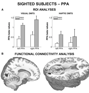
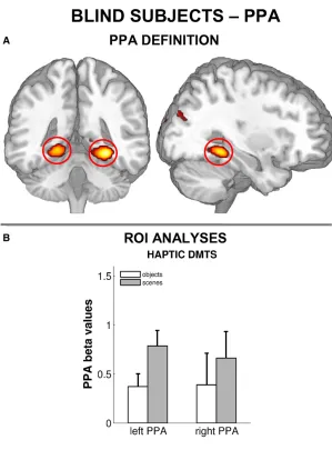
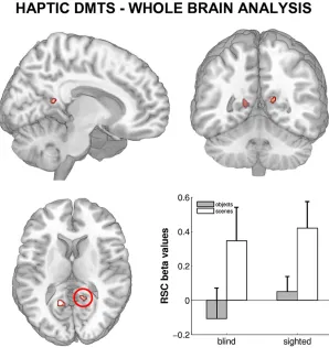
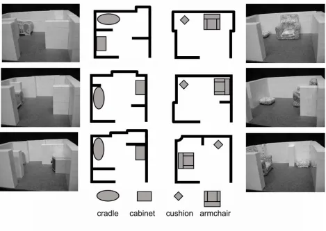
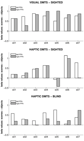
The second study investigated processing in the ’parahippocampal place area’ (PPA), a
region in the posterior MTL, during haptic exploration of spatial layout. The PPA in known
to respond strongly to visuo-spatial layout. The study explored if this region is processing
visuo-spatial layout specifically or spatial layout in general, independent from the encoding
sensory modality. In both a cohort of sighted and blind participants, activation patterns
in PPA were measured while participants haptically explored the spatial layout of model
scenes or the shape of information-matched objects. Both in sighted and blind individuals, PPA activity was greater during layout exploration than during object-shape exploration.
While PPA activity in the sighted could also be caused by a transformation of haptic
no increase in connectivity between the visual cortex and the PPA were observed, which
would be expected if visual imagery took place. Secondly, blind participates, who cannot
resort to visual imagery, showed the same pattern of PPA activity. Together, these results
suggest that the PPA processes spatial layout information independent from the encoding
modality.
The third and last study addressed error accumulation in motion processing on dif-ferent levels of the visual system. Using novel analysis methods of fMRI data, possible
links between physiological properties in hMT+ and V1 and inter-individual differences
in perceptual performance were explored. A correlation between noise characteristics and
performance score was found in hMT+ but not V1. Better performance correlated with
greater signal variability in hMT+. Though neurophysiological variability is traditionally
seen as detrimental for behavioral accuracy, the results of this thesis contribute to the
in-creasing evidence which suggests the opposite: that more efficient processing under certain
circumstances can be related to more noise in neurophysiological signals.
In summary, the results of this doctoral thesis contribute to our current understanding of motion and movement processing in the brain and its interface with spatial processing
networks. The posterior MTL appears to be a key region for both self-motion and spatial
processing. The results further indicate that physiological characteristics on the level
of category-specific processing but not primary encoding reflect behavioral judgments on
motion. This thesis also makes methodological contributions to the field of neuroimaging:
it was found that the analysis of signal variability is a good gauge for analysing
inter-individual physiological differences, while superior head-movement correction techniques
1
General Introduction
Space is a fundamental organization principle of the world as we know it. As such, it has
occupied philosophy and natural science throughout history. When talking about space, a
distinction can be made between ’physical space’ and ’psychological space’. The two are
accurately defined in the scientific treatise “The hippocampus as a cognitive map” (O’Keefe and Nadel, 1978) which is one of the seminal works for neuroscientific research on space
processing. O’Keefe and Nadel (1978)’s definition is based on Kantian theory: ’physical
space’ is defined as a physically measurable property of the external world which we are
not necessarily aware of, and ’psychological space’ as the space perceived and represented
by organisms. In other words, psychological space is a result of the organism’s attempt
to infer the state of physical space. Psychological space is the one the organism acts in
and upon which it bases its goal-directed behavior. The distinction between psychological
and physical space becomes obvious when looking at the discrepancy between the two.
Examples of such discrepancy are size, orientation or motion illusions, in the case of the visual system, and systematic misjudgment of walked distances, in the case of the
sen-sorimotor system (Mittelstaedt and Mittelstaedt, 2001). Both originate from systematic
errors which occur when perception fails to map the environment correctly. Another
impor-tant aspect that differentiates physical and psychological space is that while the former is
generally thought of being absolute, the existence of inter-individual differences in spatial
competence suggests that the latter is relative and differs between individuals (Wolbers
and Hegarty, 2010).
The physical space within arms or walking reach can be perceived by an organism
not only as a passive observer, but also by interacting with it. In the last decades,
re-sults from both behavioral and neurophysiological research have provided evidence that
interacting with the environment (physical space) plays a pivotal role in forming spatial
representations (psychological space) (Newcombe, 2000). One type of interaction between
an organism and the environment is its exploration by moving through it. Theories on the importance of movement for the formation of spatial representations emerged early
in history. As O’Keefe and Nadel (1978) describe in their historical overview of spatial
move-ment in the construction of spatial representations. He saw a primacy for what he called
’tactile’ sensory information for forming spatial knowledge, which could serve as the basis
for visual space (Berkley’s definition of tactile information included proprioceptive
feed-back and would likely be considered haptic information by current standards (Loomis,
1986)). Poincar´e (1854-1912) extended Berkeley’s arguments by adding that bothreal and
represented movements might be used in spatial processing. Poincar´e thereby expanded
the contributors to the formation of spatial representations beyond tactile sensing, to also
include mental processing. More recently, Piaget (1896-1980) incorporated movement into
his developmental theories as essential, initial access an infant has to the external world.
Endowed from birth with a limited set of sensorimotor schemes an infant acts upon the
world, deriving concepts about space by learning the fundamental principles it encounters.
Nowadays the exact correspondence between movement and the formation of spatial
rep-resentations is still poorly understood, nonetheless there are numerous empirical evidences
both from behavioral and neurophysiological studies suggesting that a link between the two
exists. Based on these evidences, some recently developed theories give a pivotal role to movement in the formation of spatial representations. According to those, self-motion cues
arising during goal-directed movement are an important source of direction and distance
information which are continuously calibrated against perceptual or memorized
informa-tion on landmarks and geometry to update our own posiinforma-tion in space (Byrne et al., 2007;
Whitlock et al., 2008).
How organisms extract and represent spatial information from the environment is a key
question in neuroscientific research. In particular, research on the neurophysiological
foun-dations of spatial behavior focuses on the description of the brain areas which are involved
in extracting and representing spatial information. Central questions are: Which neural
networks underly spatial perception and representation? How is spatial knowledge pro-cessed, how is it integrated? Can a spatial representation system be localized in the brain?
Are such neural spatial representations based on hardwired neurophysiological modules or
do they develop flexibly during the encounter of the environment? In the context of this
thesis, it is important to clarify the use of the term representation, since it has a long
history and has slightly different meanings across various disciplines (biology, psychology,
philosophy): representation can refer to both the abstract mental cognitive symbol of the
external world, as well as its neurophysiological foundation. To distinguish between the
two in the context of this thesis, the term spatial representation will be used to indicate
rep-3
resentation system in the brain will be used to indicate the physical location of the brain
areas involved in forming, processing and storing spatial knowledge.
This thesis is an inquiry into the many movement and motion processing systems in the
human brain, and how these systems interface with brain systems which process the space
within arms or walking reach, to contribute to neural spatial representations in reference to
our own body. Functional magnetic resonance imaging (fMRI) was used to investigate and characterize such brain systems. The starting point for this investigation are the identified
neurophysiological correlates of spatial processing which form the ’spatial’ circuitry of the
brain in humans and animals: these correlates include structures of the medial temporal
lobe, the ’head-direction system’ along the Papez’s circuit, the retrosplenial complex and
the posterior parietal cortex. The interaction between these areas seems to constitute
a spatial representation system in the brain (Byrne et al., 2007; Whitlock et al., 2008).
What is still not determined is how motion and movement processing systems interface
with this spatial circuitry. To further our understanding of this issue, the first project
of this thesis explored the sensorimotor systems involved in mental simulation of active and passive whole-body motion. A modulation of the medial temporal lobe induced by
different sensorimotor experiences was tested (2.1). The medial temporal lobe remained
the focus also of the second project, which investigated the multimodal nature of spatial
processing in the parahippocampal place area (2.2). While this region is known to process
visuo-spatial layout, this study explored if it is also activated by spatial layout perceived by
hand movement, i.e. haptic exploration. The focus of the final project was shifted entirely
on the motion system. The study explored on which level of visual motion processing errors
start to accumulate. This was investigated by comparing behavioral and physiological
inter-individual differences in the visual system (2.3).
The following section will describe the neurophysiology of movement and motion pro-cessing in the locomotor and the visual system (1.1), two systems investigated during this
thesis. This will be followed by an outline of the current understanding of how space is
represented in the brain (1.2), and how motion information might contribute to form
neu-ral spatial representations (1.3). Finally, advances in fMRI methodology will be presented
along with a description on how these were used in this thesis to address specific questions
1.1
Processing of motion and movement in the brain
The focus of this thesis was on systems of motion and movement processing, and the
fol-lowing section will introduce the neural foundation of such systems. Movement is processed
in parallel by multiple sensory systems: as all sensory systems encode information both
in space and in time, each of them can provide motion-related information. Besides the
sensory systems, also the motor system is intimately connected to motion, as activity in it
precedes the perception of self-generated motion. Predictions on our self-motion can
there-fore be based on the motor-sequences programmed in the brain. This idea has lead to the
suggestion of a general ’principle of reafference’, which implies that motor circuits provide
a copy (motor efference copy) of the signals they send out to the muscles, which allows the system to predict the consequential sensory feedback (see Cullen (2004) for a review).
The sources of motion information are thereby more numerous during active exploration
(active sensing) compared to when the environment is perceived immobile (passive
sens-ing). In other words, while senses such as vision can provide motion information already
during passive sensing (as e.g. while driving in a car, or navigating in a virtual reality
environment), this information is complemented by signals of the motor systems during
self-generated movement. One of the projects of this thesis characterized active sensing
of motion, by comparing the neural networks processing sensorimotor information during
locomotion and passive transport (see 2.1). Another project investigated activity in the visual system during passive motion perception (see 2.3). The following section will
there-fore introduce the current knowledge of cerebral sensorimotor processing of locomotion and
the cerebral system of visual motion processing. It will further outline first connections
between the processing of movement and space, which will be dealt with in greater detail
in section 1.2 and 1.3.
1.1.1
Movement processing in sensorimotor systems
The system of locomotor control
To move their body towards a goal, animals use locomotion, self-generate rhythmic
alter-nated movements such as swimming, flying or walking. The locomotor program which is
most commonly executed to reach a target in humans is gait. The basic stepping pattern of gait is highly automated and is generated on relatively low levels of the nervous
sys-tem. Walking towards a goal in an ever-changing environment however is controlled by a
1.1 Processing of motion and movement in the brain 5
This reflects that many computations are necessary during goal-directed walking, such
as representing the goal, motor planning, motor plan selection, motor execution, and
ad-justment of the resulted motor act according to sensory feedback and internal motor
effer-ence copies. Due to the close interaction between motor output and sensory feedback, the
functional unit which controls movement is in general considered a ’sensorimotor system’
rather than a pure motor system. The following paragraphs will however focus on the mo-tor component of this sensorimomo-tor system, as a detailed description of the somatosensory,
proprioceptive and visual feedback loops contributing to locomotor control would exceed
the scope of this introduction.
To structure the cerebral system underlying locomotor control, different brain areas are
in general assigned to a functional hierarchy of control levels (the following subdivision is based on Bear et al. (2001c)): 1) The lowest control level, responsible for execution, relies
on neural circuits in the spinal chord and the brainstem; 2) The middle level, concerned
with the control of the sequence of muscle contractions, relies on the primary motor cortex
and the cerebellum; 3) The highest control level, concerned with the goal and strategy of
a movement, involves association areas in the frontal and parietal cortex and the basal
ganglia.
Most neurophysiological knowledge of locomotor control on the lowest and middle level
stems from experiments in the cat. From these studies it is known that the basic rhythm
and the initiation of walking arises from pattern-generators in the spinal cord and the
brain stem (Grillner and Wall´en, 1985; Mori et al., 2001; Garcia-Rill and Skinner, 1987).
Brain areas such as the primary motor cortex and the cerebellum on the middle level of
locomotor control are not necessary to induce this basic stepping pattern, nonetheless, they
reverberated this pattern in their rhythmic neural activity (Kandel et al., 2000b). Beyond
this, the middle control level comes into play when the stepping patterns get more
com-plicated and have to be adapted to avoidance of obstacles (Armstrong, 1988; Garcia-Rill, 1986). While the basic neural circuits of the lowest and middle level of locomotor control
remain preserved in humans, clinical and experimental studies show that the functional
significance of the middle control level has become more pronounced in the evolutionary
transition from quadrupedal to bipedal locomotion (Nielsen, 2003; Snijders et al., 2007;
Fukuyama et al., 1997; Miyai et al., 2001). In particular the increasing significance of the
large and monosynaptic cortico-spinal tract in comparison to the rubrospinal tract deriving
from the brainstem reflects this functional reorganization.
of the movement and the developing the movement strategy to best achieve this goal. At
this level, theories today envisage the formation of a mental body schema, which comprises
an internal representation of the body, its current position in space and its spatial relation
to the environment. The knowledge of these relations is a necessary prerequisite to plan
a goal-directed movement. The highest level of locomotor control can be investigated in
humans with neuroimaging by using mental imagery, the mental simulation of locomotion without actual execution (see e.g. Bakker et al. (2008); Iseki et al. (2008); Jahn et al.
(2004, 2008); la Foug`ere et al. (2010)). Mental simulation of walking has been shown to
involve areas implicated in motor planning like the premotor cortex, the supplementary
motor complex, parts of the parietal cortex, the basal ganglia and the parahippocampal
cortex (Iseki et al., 2008; Jahn et al., 2004, 2008; la Foug`ere et al., 2010).
Sensorimotor systems and spatial processing
As mentioned before, activity in the sensorimotor system reliably informs the organism
of its own movement and thereby contributes to self-motion perception. Sensorimotor
signals can therefore serve as a source for spatial computations based on self-motion cues.
In fact, studies on rodent navigation have shown that sensorimotor cues deriving from
locomotion crucially modulate neurophysiological signals involved in spatial encoding of
the environment (Czurk´o et al., 1999; Ekstrom et al., 2003; Save et al., 1998). It has been
further shown that motor/proprioceptive signals deriving from locomotion have a stronger
influence on neurophysiological signals of space encoding than other self-motion cues such
as vestibular signals or optic flow (Terrazas et al., 2005).
Beyond the formation of spatial representations based on self-motion, clinical findings
show that also the highest and more abstract level of motor control is linked to spatial
processing. The severe effects of lesions in the parietal cortex demonstrate that this region is
crucial for goal directed movement as well as for the processing of personal (concerning the
own body) and extrapersonal space (space beyond the own body). Lesions of the parietal
cortex lead to disturbances of the body schema, such as the confusion between different
body parts in oneself and others (Bear et al., 2001b; Kandel et al., 2000a). In its most severe
form a parietal lesion can lead to spatial hemineglect, a neuropsychological syndrome in
which patients are unaware of the contra-lesional half of personal and extrapersonal space (Coslett, 1998; Husain and Nachev, 2007; Pavani et al., 2003). This neglect is not only
perceptual but also representational, which was shown in the classical study by Bisiach and
1.1 Processing of motion and movement in the brain 7
they failed to describe details on the contra-lesional but not the ipsi-lesional side, depending
on the imagined perspective. These neuropsychological phenomena provide evidence for
the close connection between the neural sensorimotor representation of the body and the
neural representation of personal and extrapersonal space.
Besides these physiological evidences, also results from behavioral studies suggest that
the sensorimotor system contributes to the encoding of space. Several studies in sighted
and blind humans have shown that accurate direction and distance estimations can be
based exclusively on sensorimotor cues (Frissen et al., 2011; Klatzky et al., 2008; Loomis
et al., 2001; Mittelstaedt and Mittelstaedt, 2001; Siegle et al., 2009). Surprisingly, such
estimations remain in large part accurate even during passive transport to a target, during
which only vestibular and somatosensory information are available (Isra¨el et al., 1997;
Frissen et al., 2011; Mittelstaedt and Mittelstaedt, 2001). It remains a subject of debate, on which mental processes spatial estimation ability during active and passive movement
is based. Some argue that such spatial estimations can be based on the extraction of
self-motion information from the sensorimotor system, suggesting a perceptual foundation
(Isra¨el et al., 1997; Frissen et al., 2011). Others stress the importance of an inner simulation
of the body moving through space based on prior experience, suggesting an (additional)
cognitive foundation (Seidman, 2008; Wertheim et al., 2001; Yong et al., 2007).
Neurophysiological data might help to clarify the relative contribution of the perceptual
and cognitive processes to this spatial estimation ability. To contribute to the scarce body
of neurophysiological data on the topic, the first project of this thesis investigated neural
networks representing active and passive self-motion experience through space (see 2.1).
1.1.2
Visual motion processing
The visual system processes many aspects of the world around us, and one of these aspects
is motion. While basic attributes of visual stimuli are analyzed already in the thalamus
and the primary visual cortex (V1 or striate cortex), aspects like shape or motion are
specifically processed in areas of the ’extrastriate cortex’, a term summarizing visual areas
beyond the striate cortex, along the temporal and parietal lobe. Extrastriatal areas are
organized in two anatomically and functionally separate streams. While the ventral stream
extends towards the inferior temporal lobe, the dorsal stream projects towards the posterior parietal cortex (PPC). Areas along these streams exhibit different functional properties:
while areas along the ventral stream process object informations like shape and color, areas
et al., 2001a).
Particularly important for motion processing in the dorsal stream is a functionally
well-defined region first described in non-human primates as area MT1 (Allman and Kaas
(1971) in owl monkey, Dubner and Zeki (1971) in macaque). While most of what we know
about this region has been first described in primate animal models, neuroimaging studies
in humans meanwhile suggest that its organization resembles closely the organization in the human brain. MT receives its major input from V1, and is thought to do essential
integration, segmentation and structure computations based on visual motion (see Born
and Bradley (2005) for a review). It then projects these computations further to several
other motion sensitive areas in the posterior parietal cortex (PPC) such as the medial
superior temporal area (MST) and the ventral intraparietal area (VIP). These PPC areas
have been shown to extract heading information from optic flow (Bremmer et al., 2002;
Britten, 2008; Logan and Duffy, 2006) and have been suggested to integrate visual motion
information with motion cues from other sensory modalities (Duhamel et al., 1998). Further
prominent MT projections go to areas processing eye-movement. On the cellular level, it has been found that the majority of neurons in MT are selective for direction and speed
of visual motion (see Born and Bradley (2005) for a review). Single cell recordings have
also shown a direct link between neural activity and perception, as neurometric functions
reliably predict psychometric functions for direction sensitivity within individual monkeys
(Britten et al., 1992).
A functionally equivalent region to MT in the human cortex was first identified based
on a clinical finding: a patient who suffered brain damage was unable to perceive visual
motion, while other aspects of vision were preserved. The lesions were located in the
lateral temporo-occipital cortex (Zihl et al., 1983). The sensitivity of this region to motion
specifically was later confirmed by a neuroimaging study, which compared cortical blood-flow patterns to a motion stimulus in PET and fMRI (Watson et al., 1993). Following this
first study, hMT+ can meanwhile be reliably identified with functional neuroimaging at the
intersection of the ascending limb of the inferior temporal sulcus and the lateral occipital
sulcus (Dumoulin et al., 2000). In parallel with the primate nomenclature, this
motion-sensitive region has been named human MT (hMT), and as this region is difficult to separate
from human MST with neuroimaging methods, most studies refer to the combination of
hMT and hMST as hMT+ (the human motion complex). hMT+ properties from basic
sensory encoding up to perceptual decision making have meanwhile been characterized
1.1 Processing of motion and movement in the brain 9
by neuroimaging studies (see e.g. Huk et al. (2002); Morrone et al. (2000); Muckli et al.
(2002); Rees et al. (2000); Smith et al. (2006)). In parallel to primate data, some recent
neuroimaging studies provide evidence for direction-selective neuronal subpopulations in
hMT+ (Kamitani and Tong, 2005, 2006).
The perception of visual motion is the basis for the detection of optic flow during
self-motion. While optic flow is only one among many self-motion cues, behavioral findings in humans however show that spatial estimations of distance and direction can be base solely
on this visual information (Warren et al., 1989; Wolbers et al., 2007). fMRI studies in
humans have shown that hMT+ is activated during the perception of optic flow (Diekmann
et al., 2009; Kov´acs et al., 2008; Wolbers et al., 2007). This region could therefore contribute
essential visual motion information to brain areas involved in self-motion integration. In
non-human primates, strong connections between MT and VIP in the PPC suggest that
visual motion information is forwarded to this region. A human equivalent of area VIP
has been described (Bremmer et al., 2001), which makes this pathway also plausible in
humans. Another candidate for the integration of self-motion cues is the medial temporal lobe (MTL). Areas in the MTL have been implicated in the extraction of spatial information
from the environment, based on the integration of self-motion cues from different sensory
modalities in rodents (Moser et al., 2008) (see also 1.3). These areas might have a similar
function in humans, as recent neuroimaging studies in humans show that MTL areas are
active during navigation in virtual reality environments, during which the only source of
self-motion information is optic flow (Caplan et al., 2003; Cornwell et al., 2008; Ekstrom
et al., 2005; Wolbers et al., 2007). In particular the study by Wolbers et al. (2007) suggests
that self-motion information from optic flow is sufficient to trigger MTL activity: using an
impoverished virtual environment in which distance and direcion information could only
be inferred from optic flow, this study shows that the hippocampus was coactivated with hMT+ and the medial frontal cortex during a spatial estimation task.
This short overview shows that hMT+/MT has been studied extensively in humans
and monkeys on different levels. The fact that its response properties are well understood
in primates, and that it has been shown to be closely linked to perception and behavior
makes hMT+ an ideal candidate to explore new questions on motion perception. Because
of this, we focused on this region to explore physiological correlates of inter-individual
1.2
Processing of space in the brain
During exploration, our body acquires information about the world via our senses, which
provide information on the external state of the world, as well as feedback on our interaction
with it. In the brain, all these informations converge and based on present and past sensorimotor informations, representations of the world are formed which can be used to
control behavior pro- and reactively. Spatial representation for example serve to control
planning and execution of goal-directed movement.
All senses map the same 3-dimensional world and spatial representations are thought
to form drawing on multiple sensory modalities (Klatzky et al., 2003; Loomis et al., 1998;
Loomis, 2007). Visual and auditory information can provide information about
environ-mental features such as geometry or position of landmarks. In addition, spatial knowledge
can be extracted from self-motion cues arising during bodily movement. Self-motion cues
comprise optic flow processed in the visual system, acceleration and rotation signals pro-cessed in the vestibular system, proprioceptive cues from muscles, joints and tendons and
motor efference copies deriving from the motor system (Cullen, 2004). During whole-body
movement, integration of self-motion cues over time can serve to track the own position in
reference to a starting point or a goal, a computation called path integration (Etienne and
Jeffery, 2004). Movement of the upper extremities can provide spatial information within
arms reach: haptic exploration can be used to understand spatial layout of the immediate
environment (Giudice, 2009; Giudice et al., 2011; Loomis, 1986).
These examples show that experiencing space does not fit into the classical
action-perception scheme, which describes gaining knowledge of the environment as action-perception and moving in the environment as action (Hurley, 1998). Rather, acquiring spatial
knowl-edge about the environment is based on both action- and perception-systems. Indeed,
matches between spatial estimations based on visual or sensorimotor cues provide
evi-dence that spatial information highly overlaps between these systems. It has for example
been shown that learning spatial layouts haptically or visually resulted in similar spatial
updating performance during intra- and inter-modal trials (Giudice, 2009). And the
find-ing that humans can blindfoldedly walk to a previously seen target with high accuracy
shows that visual distance cues can be converted into a spatially equivalent motor output
(Klatzky et al., 2008; Mittelstaedt and Mittelstaedt, 2001).
This flexibility in conversion is remarkable if one considers that all sensory and
mo-tor cues are initially processed in different peripheral sensory recepmo-tors or momo-tor effecmo-tors.
1.2 Processing of space in the brain 11
resolution and accuracy: some studies show modality specific distortion of spatial
infor-mation if only one modality is available, like distance compression for auditory or haptic
perception (Loomis et al., 1998; Abravanel, 1971). Additionally, all senses encode spatial
information in different coordinate systems: visual information enters the system in retinal
coordinates, vestibular information is organized in reference to the head and motor
effer-ence copies are organized in refereffer-ence to the respective effector (i.e eye-centered if coming from eye-movements, body-centered if coming from limb-movements). Finally, spatial
in-formation arriving in these different coordinate systems has to be re-transformed into the
coordinates of the specific effectors for goal-directed motor output.
To explain how spatial information of different resolution and encoded in different
coordinates can produce a unitary space for perception and action, it has been suggested
that the brain integrates input from multiple sensory modalities into an universal, amodal
spatial representation. This integration might comprise a conversion from the respective
body-centered coordinate systems (egocentric representations) to an abstract coding of
space relative to the environment (allocentric representation). Such a representation could
serve as an unbiased way to store incoming spatial information before it is re-transformed
into the respective motor-coordinate system for goal-directed action (Byrne et al., 2007;
Whitlock et al., 2008). Such a spatial representation might also serve to maintain stable spatial behavior when only insufficient or ambiguous sensory information is available, such
as during navigation to an unseen target. It has been suggested that spatial behavior in
this case relies on spatial memory, on which mental simulations of spatial relationships can
be based (Byrne et al., 2007).
Therefore a spatial representation system in the brain is expected to comprise 1) neural
systems which extract spatial information from sensory and motor cues and integrate
them, 2) systems which convert spatial information from different coordinate systems to
an allocentric spatial representation, 3) memory systems which store and retrieve such
information and can simulate spatial relations, and finally, 4) systems which transform
spatial information into egocentric coordinates for goal-directed motor output. Consistent
with this multitude of functions, empirical findings suggest a distributed neural network
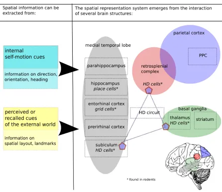
underlying spatial representation (see Figure 1.1). A central role in this network is assigned
to structures of the medial temporal lobe (MTL), which are assumed to construct a neural allocentric representation of space by integrating information on environmental and
self-motion cues (Byrne et al., 2007; Moser et al., 2008; Whitlock et al., 2008). A prominent role
in multiple egocentric coordinate systems (Byrne et al., 2007; Sack, 2009; Whitlock et al.,
2008). Furthermore, it has been suggested that a transformation circuit comprising the
retrosplenial complex, the posterior parietal cortex (PPC) and the head-direction system
distributed along the Papez’s circuit translates between neural egocentric and allocentric
representations (Byrne et al., 2007; Whitlock et al., 2008). The following section will
1.2 Processing of space in the brain 13
1.3
The medial temporal lobe: linking movement to
space
The most likely candidates to mediate between neural movement processing and neural
space processing are brain structures which exhibit both space- and movement-sensitive
responses. Several such structures can be found in the medial temporal lobe (MTL), which
it therefore the prime region of interest for research on goal-directed movement through
space, also known as navigation. The medial lobe has as a folded structure: located
along this fold from dorso-medial to ventro-lateral are the hippocampus, the subiculum,
the rhinal sulcus with the entorhinal and the perirhinal cortex, and the parahippocampal
cortex (see Figure 1.2). In rodents, the same structures have been identified, as the basic circuitry of the MTL is highly conserved in mammals (Manns and Eichenbaum, 2006).
This conservation allows to revert to the vast literature on MTL properties in rodents
1.3 The medial temporal lobe: linking movement to space 15
when forming physiological hypothesis in humans.
1.3.1
Activity during locomotion
Since the seventies it is known that self-movement modulates MTL structures in mammals.
The first evidence for a relation between the hippocampus and movement stems from electroencephalographic studies in rats showing that hippocampal theta rhythm (6-10 Hz
rhythmical activity) accompanies locomotion, but not behavior such as body grooming or
face washing (Vanderwolf, 1969; Coenen, 1975). It has also been shown that hippocampal
lesions result in movement deficits in rodents (Bast and Feldon, 2003). Closer analysis of
neurophysiological signals has further revealed that the spectral power of the theta rhythm
(Czurk´o et al., 1999) and hippocampal population firing rate (McNaughton et al., 1983;
Czurk´o et al., 1999; Ekstrom et al., 2001) depend on running speed. Recently, it has also
been shown that the theta rhythm is parametrically modulated by the amount of
self-motion cues available (Terrazas et al., 2005). On the cellular level, it has been shown that inhibitory interneurons in the hippocampus are sensitive to properties of the movement
signal like acceleration or velocity (Buzs´aki, 2002). It has been further found that firing
characteristics of pyramidal neurons in the hippocampus are modulated by the amount of
self-motion cues available (Terrazas et al., 2005).
Also in humans the MTL is activated during locomotion. Neuroimaging studies have
shown that MTL structures are active during walking on a treadmill (la Foug`ere et al.,
2010; Fukuyama et al., 1997) and during mental simulation of locomotion (la Foug`ere et al.,
2010; Jahn et al., 2004, 2008; Malouin et al., 2003; Sacco et al., 2006; Iseki et al., 2008).
These results suggest that a relation exists between MTL activity and locomotion.
Regarding the nature of this relation, most empirical evidence nowadays support the view
that activity in MTL structures during movement does not primarily reflect locomotor
control, but rather reflects spatial processing due to navigational demand. Processes in
the hippocampus and the parahippocampus might contribute to solve questions such as:
Where am I going? Where am I coming from? Where am I now?
1.3.2
The medial temporal lobe and goal-directed movement
The understanding of spatial processing in the MTL began with the crucial discovery of
’place cells’ in the rodent hippocampus, neurons which show modulation by the absolute
a neurophysiological foundation of a ’cognitive map’ (i.e. neural allocentric representation
of space) exists in mammals (O’Keefe and Dostrovsky, 1971). Over the following 40 years
of research, further ’spatial’ cells were found in rodent MTL structures. One example are
’grid-cells’ in the medial entorhinal cortex: these cells exhibit multiple firing fields which
tessellate the environment in a grid-like pattern (Hafting et al., 2005). Another example
are ’head-direction-cells’, distributed along the Papez’s circuit2, which code the heading
direction of the animal (see Taube (2007) for a review). It has been suggested that a neural
representation of navigable space could result from the interaction between populations of
these three cell types (McNaughton et al., 2006; Moser et al., 2008).
Far less is known about navigation processing in the human brain, however more and
more results point to MTL structures also in humans. Activity in MTL structures is
fre-quently observed in neuroimaging studies during mental navigation (Ghaem et al., 1997;
Rosenbaum et al., 2004) or navigation with a joystick through virtual environments (Caplan
et al., 2003; Cornwell et al., 2008). And further paralleling results obtained in rodents,
recent electrophysiological and neuroimaging studies report evidence for place cells and grid cells in the human hippocampus and the entorhinal cortex (Ekstrom et al., 2003;
Doeller et al., 2010). Furthermore, structural and functional properties of MTL areas
have been shown to correlate with navigational skill. On a structural level, significantly
larger posterior hippocampi have been described in London taxi drivers, a profession with
high navigational demand, compared to a group of control subjects (Maguire et al., 2000).
On a functional level, neuroimaging studies showed that hippocampal and
parahippocam-pal activation patterns correlate with individual performance on navigation tasks (Janzen
et al., 2008; Wolbers et al., 2007). Also clinical reports of patients with hippocampal or
parahippocampal lesions support the view that these regions are involved in spatial
pro-cessing. Patients with such lesions show impaired performance on spatial tasks such as route learning and navigation (Barrash et al., 2000; Glikmann-Johnston et al., 2008).
Most knowledge on spatial processing in the human brain however comes from research
using static spatial stimuli. A region in the posterior parahippocampus has been
identi-fied as being specifically sensitive to information about the 3D structure of space. This
region was originally discovered in a neuroimaging study comparing brain activation while
viewing photographs of landscapes to brain activation while viewing photographs of
ob-jects (Epstein and Kanwisher, 1998). Due to its higher response to ’place’ rather than
’object’ photographs, it was given the name ’parahippocampal place area’ (PPA). Further
1.3 The medial temporal lobe: linking movement to space 17
characterization of this region revealed that it responds to outdoor and indoor scenes, to
familiar and unfamiliar scenes, to real and artificial scenes, that it responds to far-scenes
as well as desktop environments, that it is viewpoint invariant and that it depends on the
background elements defining the geometry of a landscape rather than on discrete objects
contained in the scene (Epstein et al., 1999; Epstein, 2005; Epstein et al., 2007). Common
to all stimuli was the spatial layout which could be extracted, which lead to the suggestion that the PPA is selectively processing the visuo-spatial structure of a scene (the ’spatial
layout hypothesis’) (Epstein, 2008).
Despite the thorough characterization of the PPA with a multitude of static
spatial stimuli, it remains unclear if it is the spatial content of the stimulus or its
visuo-spatial structure which drives PPA activity. If this region is selective for visuo-spatial
com-putations in general, the source of the spatial information might not depend on visual
stimulation, but could also arise from the integration of self-motion cues. To clarify this
question, the second project of this thesis compared PPA activity during haptic exploration
1.4
Designs for fMRI: Studying brain processing of
movement and space in immobile participants
In the last twenty years, the use of magnetic resonance imaging (MRI) in behavioral and
cognitive research has increased exponentially. This is mainly due to the discovery that
hemodynamic changes which accompany neural activity can be captured with MRI (first described by Ogawa et al. (1990) in rats; Ogawa et al. (1992) in humans). This technique
allowed for the first time to study functional changes in the human brain non-invasively.
The most used imaging method within thisfunctional form of MRI (fMRI) is based on the
’blood-oxygen-level-dependent’ (BOLD) signal. By making MR images sensitive to
disrup-tions in the magnetic field, bloodflow in the brain, which has magnetic properties itself,
can be used to track neurophysiological activity (Amaro and Barker, 2006; Nair, 2005).
The BOLD signal originates from neurovascular coupling: when synaptic activity rises in a
particular brain region, energy is needed for transmitter release and re-uptake, which leads
to a rise of metabolic rate and oxygen consumption. Oxygen-rich blood streaming into the activated region changes the local gradient of oxygenated and deoxygenated hemoglobin,
which changes the magnetic properties in a confined region (oxyhemoglobin has
param-agnetic, deoxyhemoglobin has diamagnetic properties). This local magnetic changes can
be picked up by specific scanning sequences used in fMRI (Nair, 2005). While the exact
mechanisms linking neuronal activity to hemodynamics remain to be explored, it has been
shown that the BOLD signal correlates with neural population signals like the local field
potential in primates (Logothetis et al., 2001). Another study using optogenetics in rodents
shows that it is excitatory circuits specifically which evoke a positive BOLD signal (Lee
et al., 2010).
A major challenge when using fMRI to describe neural correlates of a behavior or a cognitive function is the translation of behavioral experiments into a design appropriate
for the scanner, or as the neuroscientist Melvyn Goodale puts it: ’fMRI is like trying to
assemble a ship in a bottle - every which way you try to move you encounter a constraint’3.
Such constraints are obvious for research on movement and spatial abilities: how to design
experiments addressing brain processing of movement through space, during which
par-ticipants lie stock-still in a 60 cm diameter tube? This thesis therefore explored different
study designs with the aim to choose the right design and analytical tools to address the
1.4 Designs for fMRI 19
specific scientific question at hand. In the following I will lay out three scientific questions
of this thesis and the selected study designs and analytical tools used to answer them.
Those scientific questions were: 1) Which are the brain networks involved in active and
passive whole-body movements? We used an approach of mental movement simulation.
2) Is the parahippocampal place area a visuo-spatial region, or does it respond to
spa-tial layout independently of input-modality? We used an approach of comparing subject groups with different spatial experiences (blind and sighted people). 3) Do inter-individual
differences in performance on visual motion tasks have a physiological correlate in hMT+?
In the course of addressing this question, we explored novel analytic tools which answer
physiological questions beyond mere localization.
1.4.1
Mental simulation as a tool to study action and perception
The first project of this thesis aimed to investigate the overlap between brain networks
activated during locomotion and passive transport through space. As actual movement is
not possible during fMRI, a study design which addressed the brain networks activated during recall of movement was chosen. Subjects experienced specific whole-body motion
sequences and recalled these experiences in a subsequent fMRI session. How can this
tell us something about the actual networks during perceiving whole-body motion under
natural conditions? Multiple lines of evidence suggest that mental simulation of experiences
activates brain areas involved in execution and perception of those experiences. Evidence
comes from comparisons of real and mentally simulated perceptions (Goldberg et al., 2006;
Kosslyn et al., 1999; Slotnick, 2004; Weinberger, 2004), from real and simulated limb- and
whole-body movements (Deiber et al., 1998; Filimon et al., 2007; Hanakawa et al., 2008;
Lacourse et al., 2005; la Foug`ere et al., 2010; Miyai et al., 2001; Porro et al., 1996; Stippich et al., 2002), and simulations of complex actions such as playing piano (Meister et al.,
2004), peeling a banana, using a razor (Ruby and Decety, 2001) or navigating through a
town (Ghaem et al., 1997; Rosenbaum et al., 2004). Another line of evidence comes from
the successful use of mental simulation in brain computer interfaces, which has promising
outlooks for the field of neuroprosthetics (Pfurtscheller et al., 2006). More evidence comes
from the success of imagery in motor rehabilitation (Langhorne et al., 2009). Building
on this notion, more and more studies investigate whole-body movements like gait with
mental simulation protocols in fMRI (Bakker et al., 2008; Jahn et al., 2004, 2008, 2009;
Iseki et al., 2008; Sacco et al., 2006; Wang et al., 2008).
percep-tion challenges a basic theme with prevailed in experimental and cognitive psychology for
decades: that mental processes can be divided into perception, cognition and action (also
referred to as ’the sandwich view’, see (Hurley, 1998)). To the contrary, more and more
theories develop which try to unify action, perception and, in part, cognition. One such
theory is the ’mental simulation theory of motor cognition’, brought forward by Jeannerod
(2001). He arguments that action consists not only of an overt stage, observable on the outside when we grasp something or walk somewhere, but includes also the covert stage of
intending actions, imagining actions, recognizing tools, learning by observation and
under-standing the behavior of other people (Jeannerod, 2001). He hypothesizes that the motor
system is part of a simulation network which is activated during these covert actions.
An-other theory, which still goes a step further and tries to unify action, perception AND
cognitive processes like memory and planning, is the ’perceptual symbol theory’ brought
forward by Barsalou (1999). This theory describes mental simulation of information stored
in the neural sensorimotor units as the underlying process for both imagery and memory
(Barsalou, 1999, 2003, 2008). Barsalou calls his point of view ’grounded cognition’ and asserts that mental simulation, drawing on the same brain areas used for perception and
action, provides an essential form of computation in the brain and is the basis for many
cognitive processes such as memory, spatial cognition, perception-action coordination and
interpreting action intentions of other agents (Barsalou, 2008).
The use of mental simulations of action thereby makes it possible to investigate the
neural foundation of spatial and movement processing during whole-body motion with
fMRI.
1.4.2
Multimodality in the PPA: comparing different subject
groups
The next scientific question concerned a specific physiological hypothesis about the
prop-erties of the ’parahippocampal place area’ (PPA), which has been described in the fMRI
literature as a category-specific region for visuo-spatial layout (Epstein et al., 1999). In
neuroimaging, the term ’category-specific region’ has been used for brain regions which
are described to respond specifically to a complex stimulus such as the fusiform-face area
(FFA) to photographs of faces (Kanwisher et al., 1997), the hMT+ to visual motion or the lateral occipital complex (LOC) to object-shape (Malach et al., 1995). Recently,
neu-roimaging studies in blind people found that many of these regions process multimodal
1.4 Designs for fMRI 21
faces (Goyal et al., 2006), the hMT+ has been shown to respond to tactile and auditory
information (Poirier et al., 2006; Ricciardi et al., 2007; Wolbers et al., 2011a), and the LOC
to process object information learned by haptic exploration (Mahon et al., 2009). If these
findings capture a general property of category specific regions, the PPA is also likely to
respond to spatial layout independent of the modality of the input.
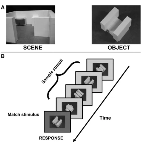
The experiment we designed to address this question compared activity in PPA during visual and haptic perception of spatial layout, contrasted with the perception of objects.
To test if the PPA is a true multimodal region, we had to clarify if a possible activation
of PPA by haptic input was due to haptic information per se, or due to visual recoding.
As expanded on in section 1.4.1, mental simulation plays an important role in perception
and cognition, and haptic perception of spatial layout could have lead to visual mental
imagery of the spatial layout, which is known to activate PPA (O’Craven and Kanwisher,
2000). We approached this possible confound by including a subject group which is not able
to do recoding based on visual experience: blind people. Special subject groups provide
behavioral and brain research with invaluable new insights into behavioral capabilities and brain functions. The study of specific sensory deprivations like the loss of the visual sense
have taught us for example the multimodal nature of spatial perception and representation:
while space is mostly considered through our visual access to it, spatial capabilities in blind
people clearly show that spatial understanding and representation can be built up through
auditory, haptic and self-motion input (Thinus-Blanc and Gaunet, 1997; Loomis et al.,
2001; Loomis, 2007).
By comparing PPA activity during haptic exploration of spatial layout in both sighted
and blind people, we could address the question of multimodality in the parahippocampus
and, at the same time, could distinguish between its activation due to haptic perception
or visual recoding.
1.4.3
Analytical tools for exploring neural inter-individual
dif-ferences
The last project of this thesis explored physiological correlates of inter-individual
differ-ences in visual motion perception using a psychophysical direction discrimination task.
Specifically, the study investigated on which stage of visual processing individual differ-ences in psychophysical threshold would be reflected in the neural activation pattern by
comparing the early stage of visual processing in V1 and the more complex processing level
Based on results obtained in electrophysiological studies in monkeys, we hypothesized
that activity in hMT+ correlates with individual direction discrimination thresholds. Such
monkey studies have found evidence that psychophysical thresholds correlate with
sharp-ness of direction coding in MT. Specifically, it was found that a broadening in tuning curves
of direction-selective neurons accompanies worsening of directional judgment during aging
(Liang et al., 2010).
Based on such findings, we tested whether individual direction discrimination
thresh-olds were correlated to the specific pattern of direction-selective neuronal sub-populations in hMT+. As such differences between individuals would not be detectable in our datasets
using the conventional voxel-based fMRI analysis, we used a multivariate approach
(pat-tern classification). While the conventional voxel-based approach based on the general
linear model cannot separate different direction-selective populations at the current limit
of fMRI resolution, the method of pattern classification can detect signal biases within
voxels. As such, this method can tell us if a set of voxels contains more or less
infor-mation about motion direction. Differences in the amount of inforinfor-mation about direction
contained in hMT+ between individuals might indicate differences in the underlying
func-tional physiology. Such differences might in turn be related to inter-individual variability in discrimination acuity.
Another analysis we performed was to characterize the variability of the hMT+ BOLD
signal during perception of motion. It was tested whether such variability correlates with
individual discrimination thresholds.
The principles of pattern classification and the analysis of BOLD signal variability will
be introduced shortly in the following sections.
Multivariate analysis
In contrast to the wide-spread use of univariate analytical methods based on the general
linear model, multivariate methods which use machine learning algorithms to train
classi-fiers have been introduced to the field of fMRI only recently (see O’Toole et al. (2007) for
a historical review). Instead of dealing with each voxel independently, such methods treat
the dataset as a whole (therefore the term multivariate). Pereira et al. (2009) describes
in his methodological review on machine learning classifiers and fMRI the principles and single steps of the methodology as follows: classifiers can be understood as functions, which
relate the features in an example dataset to a class which this dataset belongs to. As
1.4 Designs for fMRI 23
types of stimuli presented to the subject. A classifier has to be first trained on a subsample
of the dataset, to learn the relationship between features and classes. Subsequently, its
ability to classify an unknown dataset is tested. If the classifier succeeds in classifying
this unknown dataset correctly, this means that this dataset contains information about
the variable of interest. How well a classifier performs is usually measured as its accuracy
(percent correct classification).
In our experiment, we tested whether a classifier could distinguish the activation in
hMT+ or V1 as a result of seeing visual motion in four different directions. The possibility
of decoding direction information from the visual system has been shown before (Kamitani and Tong, 2006). By determining the classification accuracy of motion direction based on
V1 or hMT+ signals we aimed to identify on which level of visual processing behavioral
performance is reflected in brain physiology.
Measuring neural noise: BOLD signal variability
The second analysis we used to characterize inter-individual differences in hMT+
process-ing considered the ’noise’ of the BOLD signal. The term ’noise’ traditionally derives from
the field of engineering and has been used to describe undesirable fluctuations,
obscur-ing meanobscur-ingful information in communication technology (McDonnell and Abbott, 2009).
Following the general trend of considering the brain as a communication system, with neu-rons and brain regions communicating with each other, variability in nervous signals has
also here be termed ’neural noise’. In parallel to its use in engineering, neural noise has
been considered to disturb potentially smooth information transfer in the brain, and has
been described to be increased in mental diseases such as schizophrenia (Winterer et al.,
2006). However, some new ideas on neural noise have been recently expressed which
as-sert that variability of nervous signals could under some circumstances have a functional
significance. The brain is a variable physiological system, processing variable
environmen-tal information, and some phenomena in the brain have meanwhile be described in which
signal variability can serve to amplify the signal in a thresholded system (McDonnell and Abbott, 2009). These phenomena have been summarized under the term ’stochastic
reso-nance’ (McDonnell and Abbott, 2009).
Which impact would neural noise have on hemodynamics, in other words, how can noise be detected with fMRI? Several studies now exist which use the measure of variability of the
BOLD signal to estimate such neural noise (Garrett et al., 2010, 2011; Ghosh et al., 2008;
be estimated by calculating the standard-deviation (SD) over blocks, after accounting for
global sources of noise like scanner drifts, head-movement or fluctuations due to heart-beat
and breathing (Garrett et al., 2010, 2011). Others calculate the mean squared successive
difference (MSSD) rather than the SD, to account for different means of the expected signals
(Samanez-Larkin et al., 2010). If the mean varies between different task conditions, the
SD overestimates the variability and therefore the MSSD is a more appropriate measure (Mohr and Nagel, 2010). Furthermore, care must be taken to explain stimulus-induced
fluctuations of the signal, such as switching the stimulus on and off during a block, to not
confuse such variability with endogenous BOLD signal fluctuations. The current study
used the approach of modeling the expected BOLD signal by a generative model and then
estimating the variability based on this model (see 2.3).
1.5
Aim of this thesis
The general goal of this thesis was to explore brain networks of motion and movement processing in a number of different sensory and motor modalities in order to understand
how these are combined to create an internal representation of space. To this end, two
projects explored brain processing of motion during active sensing, on the one hand while
moving the whole-body through space, on the other hand while exploring spatial layout
with the hand. The third project explored visual motion processing during passive sensing.
The first project investigated neural networks underlying self-motion processing during
active and passive whole-body motion (2.1). It was tested whether sensorimotor brain
networks during mental simulation of self-motion experience overlap for locomotion and
passive transport. Retrieval of previously experienced locomotion and passive transport
during fMRI scanning was used to address this question.
The second project investigated processing in the parahippocampal place area (PPA),
which is known to process visuo-spatial layout in humans (2.2). This study explored,
whether PPA is selective only for visuo-spatial layout, or processes spatial layout in
gen-eral, independent of the encoding modality. To this end, a group of sighted and blind
participants haptically explored the spatial layout of model-scenes during fMRI.
Activa-tions in PPA during the haptic condition were compared to activaActiva-tions during a matched
visual task in the sighted participants.
The final project focused on motion processing in the visual system. It explored on
1.5 Aim of this thesis 25
performance (2.3). This study focused on the motion sensitive area hMT+, based on
results from neurophysiological studies in monkeys, which show that neuronal population
codes in its monkey equivalent MT relate to direction discrimination thresholds. Brain
activity during the perception of visual motion in different directions was characterized
with multivariate pattern classification and a measure for variability of the BOLD signal.
It was tested whether such measures correlate with individual thresholds on a direction discrimination task.
As a contribution to the field of brain research on movement through space, the study
designs of this thesis show different ways how to address motion and movement processing
in the brain with fMRI. A central physiological result obtained within this thesis is that
the medial temporal lobe plays a central role both in motion processing and the formation
2
Research Articles
The research conducted in the realm of this thesis is presented in the form of four research
articles. Full papers are included and are preceded with a short description about the
2.1
Networks of self-motion
The following section consists of two research articles:
• Flanagin, V.L.,Wutte, M., Glasauer, S., Jahn, K., 2009. Driving Dreams: Cortical
Activations during Imagined Passive and Active Whole Body Movement. Annual
New York Academy of Science 1164, 372375.
The author of this thesis helped with the data collection, scanning and did the pre-processing analysis of the data. The design of the paradigm, the final analysis and
the writing of the article was done by Virginia Flanagin.
• Wutte, M.G., Glasauer, S., Jahn, K., Flanagin, V.L., 2011. Moving and being
moved: Differences in cerebral activation during recollection of whole-body motion,
Behavioural Brain Research. [Epub ahead of print, doi: 10.1016/j.bbr.2011.09.042]
BASIC AND CLINICAL ASPECTS OF VERTIGO AND DIZZINESS
Driving Dreams
Cortical Activations during Imagined Passive
and Active Whole Body Movement
Virginia L. Flanagin,
a,bMagdalena Wutte,
a,bStefan Glasauer,
a,band Klaus Jahn
aaDepartment of Neurology, Klinikum Grosshadern, Ludwig-Maximilians-University,
Munich, Germany
bBernstein Center for Computational Neuroscience, Ludwig-Maximilians-University,
Munich, Germany
It is unclear how subjects perceive and process self-motion cues in virtual reality en-vironments. Movement could be perceived as passive, akin to riding in a car, or active, such as walking down the street. These two very different types of self-motion were studied here using motor imagery in fMRI. In addition, the relative importance of vi-sual and proprioceptive training cues was examined. Stronger activations were found during proprioceptive motor imagery compared with visual motor imagery, suggesting that proprioceptive signals are important for successful imagined movement. No sig-nificant activations were found during active movement with proprioceptive training. Passive locomotion, however, was correlated with activity in an occipital-parietal and parahippocampal cortical network, which are the same regions found during navigation with virtual reality stimuli.
Key words: motor imagery; passive locomotion; active locomotion; fMRI; factorial design
Introduction
In humans, virtual reality environments are often used to study navigation, particularly in functional MRI. It is unclear what type of movement subjects engage in when navigating through virtual reality environments. Subjects could perceive passive motion, where the sub-ject does not move himself but is moved by another person or object, or the subject could imagine that he is actively walking through the environment. These two types of move-ment may influence distinct cortical networks, due to differences in sensory input. Passive
Address for correspondence: Virginia L. Flanagin Ph.D., Bern-stein Center for Computational Neuroscience, Department of Neuro-logy, Ludwig-Maximilians-University Marchioninistr. 23 81377 Munich, Germany. Voice: +49 (0) 89-7095-4819; fax: +49 (0) 89-7095-4801. [email protected]
movement, for instance, lacks a motor effer-ence copy1 and has reduced proprioceptive
in-put.2 Passive head movements lead to spiking in the vestibular nuclei in primates, which is suppressed during active head movement.3 Ac-tive whole body motion has been studied in fMRI revealing cortical and subcortical centers responsible for locomotion.4,5 However, there are to date no functional MRI studies on pas-sive whole body movement. We therefore tested the difference between passive and active whole body movement with human fMRI.
The scanner configuration and image acqui-sition methods strongly limit the types of stim-ulation that can be tested with functional MRI. The problem of immobility can be partially overcome by using motor imagery instead of real movement. Path length estimation during motor imagery corresponds to path length es-timation during real locomotion, suggesting a
Basic and Clinical Aspects of Vertigo and Dizziness: Ann. N.Y. Acad. Sci. 1164: 372–375 (2009). doi: 10.1111/j.1749-6632.2009.03844.x C 2009 New York Academy of Sciences.