NOTCH SIGNALING IN THE DEVELOPMENT OF THE
INNER EAR
MARK KEITH LANG EDDISON
UNIVERISTY COLLEGE LONDON
AND
IMPERIAL CANCER RESEARCH FUND, LONDON
ICRF supervisor; Dr. Julian H Lewis
A thesis submitted for the degree of Doctor of Philosophy
University of London
ProQuest Number: 10013865
All rights reserved
INFORMATION TO ALL USERS
The quality of this reproduction is dependent upon the quality of the copy submitted.
In the unlikely event that the author did not send a complete manuscript and there are missing pages, these will be noted. Also, if material had to be removed,
a note will indicate the deletion.
uest.
ProQuest 10013865
Published by ProQuest LLC(2016). Copyright of the Dissertation is held by the Author.
All rights reserved.
This work is protected against unauthorized copying under Title 17, United States Code. Microform Edition © ProQuest LLC.
ProQuest LLC
Relative and absolute,
These the two truths are declared to be.
The absolute is not within the reach of intellect, For intellect is grounded in the relative.
Abstract
The sensory patches of the inner ear consist of two types of cell; sensory hair
cells and supporting cells. The pattern is such that supporting cells surround each hair
cell and no two hair cells touch each other. The aim of this study was to uncover the
genetic mechanisms that control the differentiation and patterning of these two cell
types.
The alternating pattern of hair cells and support cells has led to the suggestion
that their differentiation is co-ordinately regulated by cell- cell interactions involving the
Notch signaling pathway. The key players in this pathway are Delta, a ligand, and
Notch, its receptor, mediating a process known as lateral inhibition - a mechanism
which forces neighbouring cells of an initially equivalent group to become different. The
findings in this study show that two Notch ligands Delta 1 and Serrate2 are expressed in
the nascent hair cells and are thought to deliver lateral inhibition to their neighbours,
which become supporting cells. Intriguingly, the supporting cells also express a Notch
ligand. Serrate 1.
To functionally test the role of the Notch signaling pathway in the developing
chick inner ear, retroviral vectors were used to misexpress components of the Notch
signaling pathway. It is shown that a simple lateral inhibition model based on feedback
regulation of the Notch ligands is inadequate to explain the generation and patterning of
the sensory hair cells. The Notch ligand Serrate 1 is regulated by lateral induction and
not lateral inhibition; commitment to become a hair cell is not simply controlled by levels
Acknowledgements
I would like to thank Julian for giving me the opportunity to ‘do’ science and for
his continual understanding and stimulating discussion, and members of his and the
David-lsh lab for providing an enjoyable atmosphere in which to work. A special thanks
also goes to Sally Lowell and Alex Davies for proof reading this work. I would also like
to thank my treasured family and friends for their undying support over the years; to the
supporters of I.C.R.F. who funded me; and finally to Sogyal Rinpoche who taught me
how to survive this mind of mine.
Table of Contents
Page No.
Chapter 1: Introduction 12
1.1 Cell diversification during development... 12
1.2 The development of the chick inner ear... 12
1.3 Lateral inhibition is mediated by the proteins Notch and Delta... 16
1.4 The molecular mechanism of Notch signaling... 17
1.4.1 Structure of Notch and its ligands... 17
1.4.2 Proteolytic processing of Notch... 18
1.4.3 Intracellular transduction of Notch signal... 22
1.5 Notch signaling in Drosophila can direct cell fate by a variety of mechanisms 24 1.5.1 Lateral inhibition... 25
1.5.2 Asymmetric cell divisions... 28
1.5.3 Lateral induction... 31
1.6 Lateral inhibition operates in vertebrate neurogenesis... 34
1.7 Does Notch mediated lateral inhibition operate In the sensory patch? 35 1.8 Comparisons with Drosophila sensory bristles... 36
1.9 Aim and scope of this work... 40
Chapter 2: Materials and Methods 42 2.1 Cryostat sections... 42
2.2 Antibody Staining... 42
2.3 Primary antibodies used and working dilutions... 43
2.4 Sytol 6 staining... 44
2.5 RNA probes... 44
2.6 In situ hybridisation of cryosections... 45
2.8 Chick primary embryonic fibroblast preparation and revival for virus
production... 48
2.9 RCAS virus production... 50
2.10 Replication-defective virus... 51
2.11 Embryo culture and virus injection... 52
2.12 General strategy for analysing RCAS infections... 52
2.13 Electroporation... 53
Chapter 3: Expression patterns of Notch and its ligands during sensory patch development. 54 3.1 Introduction... 54
3.1.1 The timing of hair cell generation... 55
3.2 Results... 57
3.3 Serrate 1 expression foreshadows the development of the sensory patches in the inner ear epithelium... 57
3.4 Deltal expression foreshadows hair-cell differentiation within the Serratel domains... 60
3.5 D eltal is expressed in delaminated neuroblasts, and remains high after neurogenesis has ceased... 66
3.6 Serrate2 expression is seen only in nascent hair cells... 67
3.7 Notchi is expressed widely in the otic epithelium, including all sites of expression of its ligands... 67
3.8 Discussion... 72
3.9 Expression of Notchi and Deltal in the chick inner ear supports a role in lateral inhibition... 72
3.10 Expression of Serratel demarcates the future and actual sensory patches 73 3.11 Serrate-2 is expressed in hair cells... 74
3.12 Expression patterns of Notch and its ligands are similar in other vertebrates 75 3.13 There is mounting evidence that Notch signaling is critical in the production of hair cells... 76
Chapter 4; Functional analysis of Deltal using retroviruses 81
4.1 Introduction... 81
4.2 Results... 84
4.3 Infections with R C A S -D e lta l... 84
4.3.1 Serratel is regulated by lateral induction... 85
4.3.2 Effects on D eltal transcription in Deltal^" expressing cells could not be easily determined... 86
4.3.3 Effects of Deltal^" expression on hair cell production are difficult to decipher... 90
4.3.4 RCAS-Deltal^" infected cells were often seen in the cochleovestibular ganglion... 93
4.4 Ectopic expression of Deltal does not inhibit hair cell production... 93
4.4.1 Ectopic expression of Deltal by a replication defective virus also did not inhibit hair cell production... 98
4.5 GPP reveals that hair cells change shape during their development... 100
4.6 Discussion... 109
4.7 Deltal^" blocks Notch activation but supernumerary hair cells were not detected... 109
4.8 Deltal expression cannot account for the choice between hair cell and supporting cell fate... 112
4.9 Inhibition signaling might depend on an activation of Deltal in the Deltal-expressing cell... 112
4.10 Escape from inhibition might depend on a loss of sensitivity to Notch signaling in the prospective hair cell... 113
4.11 Signaling by Serratel may serve to prevent premature hair cell production 116
5.3.1 Early expression of Numb between E2 and E4... 121
5.3.2 Numb is expressed in delaminated neuroblasts... 126
5.3.3 Numb expression at E6... 126
5.3.4 Numb expression at E7... 130
5.3.5 Numb expression at E10 and El 2 ... 130
5.4 Overexpression of Numb does not produce supernumerary hair cells 135 5.5 Discussion... 144
5.6 Numb expression in the developing sensory patches is dynamic and suggests a role in the production of hair cells... 144
5.7 The loss of basal localisation may have a significant functional consequence 146 5.8 The asymmetric distribution of Numb is governed by different rules in chick, fly and mouse... 146
5.9 C-Numb antibody might cross react with a putative chick Numb-like 147 5.10 Numb overexpression within a sensory patch does not appear to affect hair cell production... 148
5.11 Numb may nevertheless influence cell fate in the chick ear... 150
5.12 Four isoforms of mammalian numb exist... 152
5.13 Conclusion... 153
Chapter 6: Discussion 155 6.1 Notch signaling is central to hair cell production, but the manner in which it operates is complex... 155
6.2 Other factors may also be involved in hair cell production... 157
6.3 Why do hair cells express Deltal ?... 159
6.4 Possible future experiments... 159
6.5 Clinical implications... 162
List of Figures
Figure 1.1: The gross structure of the chick inner ear at E10
Figure 1.2: Sensory cells in the vertebrate inner ear are arranged in a
characteristic fine-grained pattern.
Figure 1.3: Schematic representation of the structure of Drosophila Notch,
Delta and Serrate
Figure 1.4: Schematic representation of the multiple proteolytic cleavages and
associated proteases of Notchi
Figure 1.5: Lateral inhibition with feedback: a simple model of how Delta-Notch
signaling might single out cells from an equivalence group
Figure 1.6: Schematic representation of the cell lineage in a Drosophila sensory
bristle (microchaete)
Figure 1.7: Lateral induction during wing margin formation, modulated by Fringe
Figure 1.8: The sensory patches of the inner ear are reminiscent of the sensory
bristles of Drosophila
Figure 2.1 Figure 3.1 Figure 3.2 Figure 3.3 Figure 3.4 Figure 3.5 Figure 4.1 Figure 4.2 Figure 4.3 Figure 4.4 Figure 4.5 Figure 4.6 Figure 4.7 Figure 4.8
Strucutre of RCAS-Numb
Expression of D eltal and Serratel at E4
Expression of Deltal and Serratel at E7 & E8
Expression ot Deltal and Serratel at E9, E10 & E12
Expression of Serrate2 and Serratel at E7 & E8
Expression of Notchi and Serratel at E7 & E8
The technique of retroviral infection in the developing inner ear
Blocking Notch signaling causes a downregulation of Serratel
RCAS-Deltal does not affect expression of Serratel
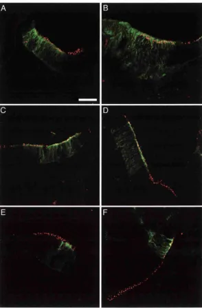
RCAS-Deltal^" often produced an “abutting” phenotype
RCAS-Deltal^" did not affect hair cell population density
Deltal*^" expressing cells often ended up as neurons
RCAS-Deltal in a sensory patch does not inhibit hair cell production
Replication-defective virus expressing Deltal and GFP
Figure 5.1: A new model for Notch signaling during hair cell generation 119
Figure 5.2: Numb expression at E2 and E3 123
Figure 5.3: Numb expression at E4 124
Figure 5.4: Numb is expressed in the delaminated neuroblasts 127
Figure 5.5: Numb expression at E6 129
Figure 5.6: Numb expression at E7 132
Figure 5.7: Numb expression at E10 133
Figure 5.8: Numb expression at El 2 134
Figure 5.9: Overexpression of c-Numb by RCAS 137
Figure 5.10: RCAS-Numb often produced an abutting phenotype 141
Figure 5.11 : Numb overexpression shows a downregulation of Serratel 142
List of Tables
Table 4A: Results of infection with RCAS-Deltal'^". 104
Table 4B: Counts of hair cells in RCAS-Deltal'^" infected and non-infected
regions of sensory patches 105
Table 4C: Results of infection by RCAS-Deltal 106
Table 4D: Counts of hair cells in RCAS-Deltal infected and non-infected
Patches 107
Table 4E: Counts of hair cells in replication-defective-Deltal infected and
non-infected patches 108
Table 5A: Results by infection with RCAS-Numb. 143
Table 5B: Counts of hair cells in RCAS-Numb infected and non-infected
Chapter 1
Introduction
1.1 Cell diversification during development
A central question that pervades developmental biology is how different cell
types arise from a group of initially equivalent cells. Although intracellular events can
play a role in the determination of cell fate, generally speaking a cell’s fate is controlled
by signals received from its environment. A key form of cell-cell signaling required for
proper cell fate determination is the Notch signaling pathway, which has been shown
over the last few years to be as important in vertebrates as it is in Drosophila. In this
study I explore the role Notch signaling has during the development of the inner ear, a
remarkably complex organ but one whose specialised sensory epithelium consists of
relatively few cell types. It therefore provides a relatively simple system to investigate
the rules that govern cell differentiation in vertebrates.
1.2 The development of the chick inner ear
In the chick, the inner ear develops from a thickened epithelial placode next to
the hindbrain at the level of rhombomeres 5 and 6. This placode invaginates into a cup
shape before pinching off from the surface head ectoderm and forming a hollow
epithelial ball, called the otic vesicle. Over the next few days, the vesicle undergoes a
remarkable morphological and functional transformation converting it into a complex
membranous labyrinth. Within this epithelial sheet arise distinct sets of cells constituting
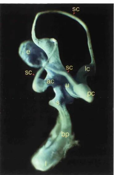
The sensory patches can be divided into two groups, the vestibular patches
responsible for the detection of acceleration and gravity, and the more ventrally located
patch, responsible for hearing. The vestibular patches consist of three cristae, and the
maculae of the utricle, saccule and lagena (at the distal tip of the cochlea). The cristae
are located at the bases of the three semicircular canals, and are involved in the
detection of angular acceleration. The utricular, saccular, and lagenar macula function
to sense gravity and linear acceleration. The ventral auditory patch, called the basilar
papilla, is situated in the banana-shaped cochlea (Figure 1.1).
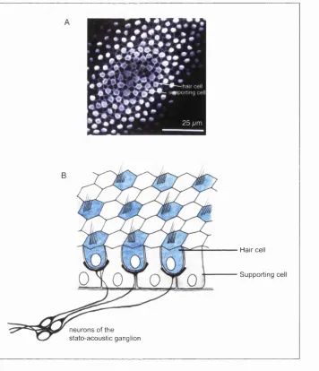
Despite their distinctly different modalities all these sensory patches have
essentially the same structure and consist of the same two epithelial cell types (with
some sub-specialization within each category). Each patch consists of an epithelial
array of supporting cells and mechanosensory hair cells, the sensory transducers. The
hair cells are innervated by neurons that have themselves originated by delamination
from the otic epithelium (reviewed in Fekete, 1996;Torres and Giraldez, 1998; Fritzch et
al., 1998; Fekete 1999). The hair cells and supporting cells are arranged in a fine
grained alternating array such that supporting cells surround each hair cell and no two
hair cells touch each other. In the mature sensory patch, the supporting cells rest on the
basal lamina, while the hair cells rest on the supporting cells; and thin projections from
the supporting cells, extending to the apical surface of the epithelium, separate each
hair cell from neighbouring hair cells (Figure 1.2).
The patterned array of hair cells and supporting cells has lead to the suggestion
that these two cell types are generated through competitive cell-cell interactions. Thus
initially equivalent cells in the presumptive sensory patch compete for the hair cell fate,
and a cell that adopts the hair cell fate delivers an inhibitory signal to its neighbouring
• k
SC
Ic
Figure 1.1: The gross structure of the chick inner ear at E10 (stage 36) and position of its sensory patches. The membranous labyrinth has been revealed by injection of white paint. Dorsal is top, anterior is to the left. The three semicircular canals (sc), utricle (u), saccule (s) and the anterior (ac), lateral (Ic), and posterior cristae (pc) along with the lagenar macula (I) at the tip of the cochlea, constitute the vestibular apparatus. In the banana-shaped cochlea (ventral) is the basilar papilla (bp), responsible for the detection of sound (prepared by R.Massey and J.Lewis,
Hair cell
Supporting cell
neurons of the stato-acoustic ganglion
Figure 1.2: Sensory cells in the vertebrate inner ear are arranged in a characteristic fine-grained pattern.
A) The regular alternating pattern of hair cells and supporting cells in the chick basilar papilla at E10. The hair cells are stained with hair cell antibody (white blobs) and the supporting cells outlined by their cortical actin, stained with fluorescent phalloidin (prepared and photographed by A.Myat).
supporting cells. This mechanism for generating the fine-grained pattern of supporting
cells and hair cells has been termed lateral inhibition (Corwin, 1991; Lewis, 1991).
1.3 Lateral inhibition is mediated by the proteins Notch and Delta
The concept of lateral inhibition was first described in the development of the
fruit fly. Drosophila melanogaster, and is known to involve the genes Notch and Delta,
where Notch acts as the receptor of intracellular signals, and Delta acts as the ligand
(reviewed in Muskavitch, 1994). Both Delta and Notch were originally classified as
‘neurogenic’ genes: loss-of-function mutations in either gene gave rise to abnormalities
of neuroectodermal cell lineages. Homozygous mutants in Delta and Notch resulted in
the expansion (hyperplasia) of the embryonic nervous system, reduction in the
embryonic epidermis and embryonic lethality (Lehmann et al., 1983). In the fly Notch
also has an alternative ligand to Delta, encoded by the closely related gene Serrate
(Fleming et al., 1990). Unlike Delta, Serrate loss of function does not cause a
neurogenic phenotype. Delta and Serrate have different expression patterns and
appear to regulate different developmental decisions (Thomas et al., 1991, Kooh et al
1993). However in some instances they are functionally interchangeable (Gu et al.,
1995; Zeng et al., 1998).
Over the years, it has been discovered that the Notch pathway is central to
many developmental decisions in the fruit fly; in fact hardly any tissue is not affected by
Notch. Moreover the Notch signaling pathway has been conserved during the evolution
of multicellular animals. Homologues of Notch called lin-12 and glp-1 have been found
in the nematode (Yochem et al, 1988; Yochem and Greenwald, 1989), and several
vertebrate Notch genes have also been identified: for example, in chick, mouse,
1993; Franco del Kopan and Weintraub, 1993; Lardelli and Lendahl, 1993; Lardelli et
a!., 1994; Larsson et a!., 1994; Henrique et a!., 1995; Myat, 1995; Westin and Lardelli,
1997).
Notch ligands too have homologues in vertebrates: one Delta gene has been
described so far in the chick (Henrique et al., 1995), whilst in the mouse three
(Bettenhausen et al., 1995; Dunwoodie et al., 1997; Shutter et al., 2000), and in
zebrafish four Delta genes are known (Haddon et al., 1998b). In addition, vertebrates
also possess homologues of the Drosophila Serrate gene (called Jagged in rodents and
humans). In the chick, mouse and rat two Serrate genes have been discovered
(Lindsell et al., 1995; Hayashi et al., 1996; Myat et al., 1996; Shawber et al., 1996a;
Mitsiadis et al., 1997). In vertebrates. Notch malfunction has been shown to disrupt a
wide variety of developmental processes including neurogenesis, somite formation,
angiogenesis, and myogenesis (Shawber et al., 1996b; reviewed in Lewis 1998; Shima
and Mailhos 2000).
1.4 The molecular mechanism of Notch signaling
Before discussing how lateral inhibition is mediated by Notch signaling, I will
review the current thinking on how the Notch signal transduction pathway is thought to
operate. What is described is no doubt an oversimplification of the reality, but it should
give a general understanding of the remarkable way in which Notch activation can elicit
a cellular response.
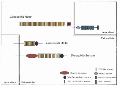
1.4.1 Structure o f Notch and its ligands
species the extracellular region consists of multiple epidermal growth factor (EGF)-like
repeats and three copies of a Lin12/Notch/Glp motif. In the intracellular region Notch
has a region of about 60 amino acids called the RAM 23 domain, six cddO/Ankyrin
repeats and a PEST-containing domain (reviewed in Weinmaster, 1997; Fleming, 1998)
(Figurel.3).
The Notch ligands Delta and Serrate are also single-pass transmembrane
proteins with a series of extracellular EGF-like repeats; however, unlike Notch they all
possess a conserved cysteine-rich motif referred to as the DSL domain (after Delta,
Serrate and Lag-2) which is N-terminal to the EGF repeats. This DSL domain has been
shown to be required for ligand function in invertebrates (Henderson et al., 1994;
Muskavitch, 1994; Tax et al, 1994). Serrate, though structurally related to Delta, differs
in that it is much larger than Delta, with more EGF repeats in the extracellular domain
and a cysteine rich region between the EGF repeats and the transmembrane domain.
Although the cytoplasmic domains of the Notch ligands do not share any significant
homology, they are required for normal ligand functioning, as deletions of the
cytoplasmic domain in either Delta or Serrate have been shown to block Notch
signaling (Chitnis et al., 1995; Sun and Artavanis-Tsakonas; 1996, Henrique et al.,
1997) (Figure 1.3).
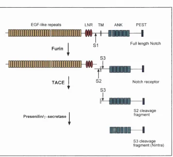
1.4.2 Proteolytic processing of Notch
To function as a receptor, the Notch protein has to be cleaved by a variety of
proteases, both before and after ligand binding (reviewed in Weinmaster, 2000) (Figure
1.4). The Notch receptor that appears at the cell membrane is produced by a proteolytic
cleavage of the primary translational product of the Notch gene. This creates a
heterodimer composed of a large amino-terminal fragment that is entirely extracellular,
Drosophila Notch
IUDDDMO^
Intracellular
DDDIW— Drosophila Delta
-O Ü D D D D D H
Intracellular
Extracellular
Extracellular
Drosophila Serrate
[] EG F-like repeats
Cystine-rich region ( ] ) R A M 23 domain
^ D elta-S errate-L ag2 dom ain | Ankyrin-like repeats
I LNR, Lin-12-Notch repeats | P E S T domain
Figure 1.3: Schematic representation of the structure Drosophila Notch, Delta and Serrate. The same overall arrangement of structural motifs in both ligand and receptor is conserved in other species.
EGF-like repeats LNR TM ANK PEST
-000000— I
T
Full length NotchF u rin
!
33IHH-i
T
I
ODDOOD— I
T A C E ^ S 2 Notch receptor
S3
i ^ D D D D D I
D— I
82 cleavage
Presenilin/y-secretase
I
fragment1
lEIIIII— I
S3 cleavage fragment (Nintra)Figure 1.4: Schematic representation of the multiple proteolytic cleavages and associated proteases of N otchi.
Notch heterodimer is stabilised through non-covalent interactions that are calcium
dependent (Rand et al., 2000). This cleavage is brought about by a furin-like convertase
during export to the cell surface (Logeât et al., 1998).
Ligand binding is thought to cause a conformational change that induces two
further cleavages of the Notch receptor, both in the smaller fragment. The first of these
severs the extracellular region of Notch, while the second releases the intracellular
domain of Notch, allowing the transmission of the Notch signal. The cleavage of the
extracellular region of Notch occurs close to the transmembrane domain and is
achieved by an ADAM (a Disintegrin and Metalloprotease) protease called TACE (Brou
et al., 2000). The cleaved extracellular domain of Notch is thought to be endocytosed
by the ligand-expressing cell, and this endocytosis is critical to Notch receptor activation
(Parks et al., 2000). The gene product Kuzbanian (Kuz), another ADAM protease,
has also been implicated in this Notch cleavage (Pan and Rubin, 1997), but its exact
role is debatable. It has been reported that Notch processing can occur in Kuz- cells,
but not in cells lacking TACE (Mumm et al., 2000), suggesting Kuz maybe functionally
redundant in this cleavage. Moreover, Kuz also has been shown to cleave the Notch
ligand Delta, and this cleavage has been shown to be necessary for signaling (Qi et al.,
1999). It maybe therefore that the original report was wrong, and that the target of Kuz
is not Notch but Delta.
The final cleavage of Notch, which requires the prior TACE cleavage, happens
at a site within the transmembrane domain, near the cytoplasmic face. The current
evidence suggests that this cleavage is performed by a presenilin-dependent
y-secretase or a y-y-secretase activity of presenilin itself. Presenilins were first identified by
mutations that cause early-onset Alzheimer’s disease, where they are involved in
Drosophila presenilin have the same neurogenic phenotypes as mutants with a loss in
Notch activity (Struhl and Greenwald, 1999). Importantly, the production of the
intracellular fragment of Notch from the membrane-bound form is decreased in cells
deficient in presinilini (PS1) activity, but is rescued by overexpression of PS1 (De
Stooper et al., 1999; Song et al., 1999; Ray et al., 1999). Hence the presenilins are
thought to regulate or bring about this final cleavage, which releases the small
intracellular Notch fragment that mediates the next step in the signal transduction
pathway.
1.4.3 Intracellular transduction of Notch signal
According to the current model, Notch signaling activates transcriptional
responses by a translocation of this small Notch fragment (Nintra) to the nucleus, where
it directly interacts with transcription factors. However, evidence for a different model
also exists, and there may be more than one way in which activated Notch can elicit a
response in the cell (reviewed in Artavanis-Tsakonas et al., 1999).
Evidence for a direct role of Nintra in eliciting gene transcription came from the
observation that activated forms of Notch show strong nuclear localisation (Kidd et al.,
1989), and that Nintra contains two potential nuclear localisation sequences that flank
the ankyrin repeats (Lieber et al, 1993; Rebay et al., 1993). However,
immunocytochemical analysis has not detected Notch in the nuclei of developing
animals (Ahmad et al., 1995). More recently it was argued persuasively that only small
amounts of Nintra, below the limit of immunohistochemical detection, are required for in
vivo Notch signaling (Schroeter et al., 1998; Struhl and Adachi, 1998). Using a sensitive
reporter-based assay, nuclear access has been demonstrated to be important for Notch
that targeted it to the membrane or nucleus, Notch activity was respectively either
blocked or potentiated (Struhl and Adachi, 1998).
The transduction of the intracellular Notch signal depends on DNA binding
proteins of the CSL family. This family consists of CBF/RBPJk in mammals. Suppressor
of Hairless (Su(H)) in Drosophila, and LAG-1 in C.elegans. These proteins bind both
Nintra (at the RAM domain and ankyrin repeats) and enhancer regions of Notch target
genes. The CSL protein, in combination with Nintra, forms a functional transcription
factor to stimulate target gene expression in the nucleus (Jarriault et al., 1995; Hsieh et
al., 1996). The activity of Su(H) can be antagonised, in Drosophila at least, through its
interaction with the Hairless protein, a negative regulator of Notch (Brou et al., 1994).
No Hairless homologue has been found in vertebrates.
Whether CSL binds Nintra at the cell surface, and then translocates to the
nucleus, or sits in the nucleus waiting for Nintra to arrive, is debated. In cultured insect
cells the Su(H) protein has been observed to translocate from the cytoplasm to the
nucleus subsequent to Notch activation (Fortini and Artavanis-Tsakonas, 1994; Frise et
al., 1996). However, in vivo, and in vertebrate cells generally, nuclear CSL staining
does not correlate well with Notch activity. For example, RBP-Jk is predominately
nuclear in mouse embryos and no changes in cellular localisation are observed in
Notchi knockout embryos (de la Pompa et al., 1997; Sakai et al., 1995).
The target genes of Notch activation depend on the developmental context.
Perhaps the best studied are the hairy-Wke helix-loop-helix (bHLH) proteins of the
Enhancer-of-SpHt comp\ex {E(spl)-C) in Drosophila (Delidakis and Artavanis-Tsakonas
1992) and their homologues the Has genes in mammals (Sasai et al., 1992; Ishibashi et
al., 1995; Jarriault et al., 1995; Ohtsuka et al., 1999). In Drosophila, Su(H) has been
Schweisguth, 1995). The E(spl)/Hes genes in turn act primarily as transcriptional
repressors, mediating inhibitory effects on cell genes promoting commitment to a
specialised fate, such as the genes of the achaete-scute complex (Oellers et al., 1994;
Tata and Hartly, 1995; Nakao and Campos-Ortega, 1996; Nakamura et al., 2000).
Although Notch activity generally elicits a response through the CSL/Su(H)
pathway, there is mounting evidence that in some developmental decisions Notch can
act independently of Su(H) (Shawber et al., 1996b; Matsuno et al., 1997; Wang et al.,
1997; Ligoxygakis, 1998; Zecchini et al., 1999; Nagel et al., 2000).
In summary Notch signaling in itself does not specify a particular cell fate; rather
it is a general developmental tool, whose signal transduction machinery and
developmental consequences are dependent on the context in which it is operating.
1.5 Notch signaling in Drosophila can direct cell fate by a variety of mechanisms
That Notch signaling can operate in many different developmental contexts is in
part due to proteins that interact with the central components of the pathway to
influence the outcome of a Notch cell-cell signaling event. These proteins can either
have a positive or negative effect on Notch signaling, and act at any point in the
pathway, from ligand binding to signal transduction and transcriptional activity of the
Nintra-CSL protein complex (reviewed in Panin and Irvine, 1998). These modifiers have
made Notch signaling incredibly versatile. Types of cell fate decision that are influenced
by Notch can be classified as lateral inhibition, asymmetric cell division, and inductive
signaling. These three types of Notch signaling are well illustrated in Drosophila, and
similar mechanisms also appear to operate in vertebrates (reviewed in Jan and Jan,
1.5.1 Lateral inhibition
Lateral inhibition, in its purest form is a local contact-mediated cell-cell
interaction through which a population of initially equivalent cells compete for one of two
fates. Cells that win the competition and adopt the primary fate inhibit their neighbours,
forcing them to adopt the alternative or secondary fate. In the absence of lateral
inhibition all of the cells of the equivalence group adopt the primary fate. Initially all cells
express both ligand and receptor. Therefore if the competition is to have winners and
losers, there must be a feedback loop that regulates the expression of the inhibitory
ligand, such that receptor activation as well as inhibiting commitment to the primary fate
also downregulates ligand expression.
As previously discussed, in lateral inhibition mediated by the Delta-Notch cell
cell signaling pathway. Delta is the inhibitory ligand and Notch is the receptor: the
feedback mechanism is one in which Notch activation in a given cell represses Delta
expression in that same cell. In this model, then, initially all cells of the equivalence
group express both Delta and Notch. Subtle differences in the levels of expression of
Delta and Notch, due to random fluctuations, are then amplified through the feedback
mechanism. Thus one cell having higher levels of Delta than its neighbours prevents
them from adopting the primary fate and represses their transcription of Delta. In this
way single isolated cells can adopt a fate different from that of their neighbours (Figure
1.5) (Simpson, 1990).
A well studied and often quoted example of lateral inhibition with feedback is the
isolation and regular spacing of the sensory bristles in Drosophila. The back of the
thorax of a fruit fly, called the notum, is covered in uniformly spaced sensory bristles,
LATERAL INHIBITION
Delta Notch
1°fate T
adopted
\ l
1 ^ 1° fate
_L inhibited
Notch Delta
B
m
Figure 1.5: Lateral inhibition with feedback: a simple model of how Delta-Notch signaling might single out cells from an equivalence group.
A) A cell with slightly higher Delta due to random fluctuations in levels of Delta expression activates Notch on adjacent cells. The consequence of Notch activation is two-fold. Firstly, Notch activation down-regulates the expression of Delta in that cell, preventing it from inhibiting neighbouring cells. Secondly Notch activation prevents the cell from differentiating as the primary fate. Cells with high Delta, and thus low Notch activity, adopt the primary fate.
specification. Loss of either at the time of SOP specification results in excessive
numbers of SOPs, suggesting that Notch signaling acts to repress SOP fate
(Hartenstein and Posakony, 1990; Heitzler and Simpson, 1991; Parks and Muskavitch,
1993). Heitzler and Simpson (1991) found that bristle formation is repressed in wildtype
tissue bordering clones homozygous for hypomorphic Notch alleles. This can be
interpreted as the result of a feedback loop between Notch and Delta, where cells with
reduced Notch function increase in Delta activity and inhibit neighbours from adopting
the SOP fate.
Transcriptional control of the feedback mechanism has been elucidated, and
involves the achaete/scute (ac-sc) complex. The ac/sc genes encode basic helix-loop-
helix (bHLH) proteins that are known to regulate neural development (Ghysen and
Dambly-Chaudière, 1988). The initial expression domain of these genes defines the
equivalence group, or proneural cluster from which the SOP differentiates. During SOP
selection, expression of the ac-sc genes increases in the cell that adopts the SOP fate
and is eventually limited to that cell. This limitation to the SOP cell is due to the increase
in Notch activation in the presumptive epidermal cells surrounding the SOP. Notch
activation via the expression of genes of the Enhancer o f Split complex (Bailey and
Posakony 1995; Leucourtois and Schweisguth, 1995) has been shown to inhibit ac-sc
expression (Kunisch et al., 1994). Furthermore, the ac-sc complex has been shown to
positively regulate the transcription of Delta (Kunisch et al., 1994, Haenlin et al, 1994).
The consequence is a feedback loop that represses Delta expression upon Notch
activation during SOP specification (Heitzler et al., 1996).
As eloquent as this may appear, several observations have been reported that
argue against SOP selection via the simple lateral inhibition model (reviewed in Baker,
1.5.2 Asymmetric cell divisions
In the purest form of lateral inhibition, single cells in an equivalence group are
singled out by random fluctuations in gene expression, for example Notch or Delta. But
it is also possible for the system to be non-random, biased by either external or internal
factors that serve to influence the outcome of Notch signaling in a predictable direction.
A relevant example to this study is the bias of Notch signaling during the development
of the sensory bristles by the asymmetrically inherited cell fate determinant Numb.
The mature sensory bristle, which derives from a single SOP, consists of four
cells with distinct fates: a neuron, and three accessory cells: a sheath cell, a socket cell
and a bristle cell (Figure 1.8). To produce these cell types the sensory organ precursor
cell (SOP), undergoes a stereotypical pattern of asymmetric divisions (Hartenstein and
Posakony, 1989, Posakony, 1994). The SOP cell first divides to generate two
secondary precursor cells, lia and lib. The lia cell then divides to produce two outer
cells, the hair cell and the socket cell. It was thought that the lib cell divides shortly after
the lia division to produce two inner cells, the neuron and the sheath cell. Recently, it
has been demonstrated that the mature sensory bristle actually consists of five cells
(Gho et al., 1999). A fifth cell is produced by the pllb cell. This first divides unequally, to
produce a small migratory glial cell and a daughter cell named plllb. The plllb cell then
divides to produce the neuron and the sheath cell (Figure 1.6).
Studies using temperature sensitive alleles of Notch and Delta show that these
and the other genes of the Notch pathway are required for the proper cell fate
determination of the SOP progeny. A loss of Delta activity, or a loss of Notch function at
the time of the first SOP division produces two Mb cells that both divide to give a neuron
and sheath cell (Parks and Muskavitch, 1993; Guo et al., 1996; Wang et al., 1997). A
SOP lineage leads to the production of fo ur neurons and no accessory cells
(Harteinstein and Posakony, 1990; Parks and Muskavitch, 1993).
Besides cell-cell interactions, a cell intrinsic factor Numb (Uemura et al., 1989),
is also essential for the correct cell fate determination of SOP progeny (reviewed in Jan
and Jan, 1998). Loss of numb causes the Mb cell to be transformed into the lia cell,
whereas overexpression results in the opposite cell fate transformation (Uemura et al.,
1989, Rhyu et al., 1994). Numb is also similarly involved in the subsequent divisions of
the lia and lib cells (after division of the SOP its expression is activated in the plla cell).
Numb is a membrane associated protein that is asymmetrically localised during mitosis
of the SOP cell, and segregates into only the lib cell (Rhyu et al., 1994, Knoblich et al.,
1995). Numb is also segregated in the lib (plllb) division into the cell that differentiates
as the neuron, and Numb is segregated in the lia division into the cell that becomes the
bristle cell (Rhyu et al., 1994, Frise et al., 1996, Wang et al., 1997, Gho and
Schweisguth, 1998). (Figure 1.6).
The effect of Numb is opposite to that of Notch activity, suggesting that it is a
negative regulator. Consistent with this idea, when assayed in cultured cells Numb
inhibits the translocation of Su(H) to the nucleus upon receptor activation (Frise et al.,
1996; Wakamatsu et al.,1999). Furthermore, Numb has been shown to directly bind to
the cytoplasmic tail of Notch (Guo et al., 1996; Wakamatsu et al., 1999). Thus the
presence of Numb in a cell can bias the Notch-mediated cell-cell interaction by
inhibiting Notch activity. This may then lead to an up-regulation of Delta and the
transformation of that cell into a signaling cell, activating Notch in its neighbours that do
pllb
plla
plllb
Socket Bristle Sheath Neuron Glial
1.5.3 Lateral induction
Notch activation can act as an inductive signal as well an inhibitory one. Notch
signaling at the Drosophila wing margin is perhaps the best example of inductive Notch
signaling. It illustrates how Notch activation can, in some contexts, upregulate the
expression of Notch ligand, in contrast to lateral inhibition. It also illustrates how the
efficacy of the different Notch ligands. Delta and Serrate, can be biased through the
Notch modulator. Fringe.
The Drosophila wing develops from one of the imaginai discs. During
development the presumptive wing field can be divided into dorsal and ventral
compartments. The wing margin forms at the interface between the dorsal and ventral
compartments and acts as an organiser of wing development. Notch activity is required
at the wing margin, where it creates the organiser tissue by inducing localised
expression of several genes that affect wing morphogenesis, including vestigial,
wingless and cut (Kim et al., 1995; Rulifson and Blair 1995; Couso et al., 1995; Kim at
al., 1996; Neumann and Cohen, 1996).
Both Delta-Notch signaling and Serrate-Notch signaling are involved in
establishing the wing margin. Serrate is expressed in all of the cells of the dorsal
compartment, and acts as a dorsal to ventral signal (Couso et al., 1995; Diaz-Benjumea
and Cohen, 1995; Kim et al., 1996; de Cells et al., 1996). Delta is expressed at high
levels in ventral cells near the boundary and is principally required to signal to dorsal
cells (Doherty et al., 1996 de Cells et al., 1996). In response to Serrate expressed by
dorsal cells. Notch is activated and Delta is upregulated in ventral cells along the
boundary. Conversely, these high levels of Delta in ventral cells signal back across the
at the dorsal/ventral boundary (de Cells et a!., 1996) (Figure 1.7). This positive
regulatory feedback loop in lateral induction contrasts with the negative feedback loop
observed during lateral inhibition, where Notch activation downregulates the expression
of Delta. Evidence in support of this positive feedback loop comes from ectopic Notch
activation during early wing development, which results in ectopic ligand expression (de
Cells et al., 1997).
Notch is expressed throughout the wing disc. Thus to obtain a localised region
of Notch activation between the dorsal and ventral compartments. Notch ligands must
be prevented from activating Notch in neighbouring cells one within the same
compartment. The gene fringe has been proposed to play such a role. In the wing disc
Fringe is expressed only in the dorsal cells and makes them refractory to Serrate
signaling and more sensitive to Delta signaling (Fleming et al., 1997; Panin et al, 1997).
Thus fringe creates an asymmetry between the two compartments and ensures that
cells expressing Serrate can only signal to the ventral cells and ventral Delta
preferentially activates Notch in the dorsal cells. High concentrations of Delta and
Serrate in the cells adjacent to the wing margin are reported to have a cell-autonomous
dominant negative effect, aiding the restriction of Notch activity to the wing margin
(Micchelli et al, 1997, Klein et al, 1997). The product of another gene, nubbin, is also
involved at the wing disc, and acts to sequester Notch signaling outside the wing
margin. Nubbin is a POD domain protein, expressed throughout the wing disc, which
has been shown to negatively regulate the activation of Notch target genes, raising the
threshold of Notch activity required for their expression (Neumann and Cohen, 1998).
The function of fringe has been recently elucidated (reviewed in Blair, 2000). In
a series of papers. Fringe has been shown to be a glycosyltransferase that glycosylates
the Notch receptor in the Golgi, as it is being exported to the cell surface (Bruckner et
Notch activity
D O R S A L Wing Margin V E N T R A L
Serrate and Fringe
Serrate blocked by Fringe
Delta
Delta potentiated by Fringe
Figure 1.7: Lateral induction during wing margin formation, modulated by Fringe.
2000). Fringe catalyses the addition of GfcNAc (A/-aceytlglucosamine) to an 0-fucose
saccharide that is found on specific extracellular EGF repeats of Notch (Moloney et al.,
2000; Bruckner et al., 2000). It has been proposed that it is this glycoslylation that
makes Notch less sensitive to Serrate ligand but more sensitive to Delta ligand. The
mechanism by which it acts could simply depend on an enhancement of Delta-Notch
binding (Bruckner et al., 2000), though cell culture binding assays argue against this
(Klueg and Muskavitsch, 1999). Alternatively it has been proposed that different
conformational changes affect the efficiency with which the extracellular domain of
Notch is cleaved by the TACE protease (Hicks et al., 2000, Moloney et al., 2000).
1.6 Lateral inhibition operates in vertebrate neurogenesis
The different mechanisms of Notch activation that have been described in the fly
also appear to operate in a similar fashion in vertebrates. Most relevant to this study is
lateral inhibition, which has been demonstrated to operate during neurogenesis in the
central nervous system.
In the neural tube neurons are generated from dividing precursors whose cell
bodies lie close to the lumen; new-born neurons then migrate out into the mantle zone
of the neuroepithelium where they differentiate. Notch 1 is expressed throughout the
proliferative zone: Delta 1 is expressed in the outer part of that zone, in a scattered
subset of cells (Myat et al., 1996). The cells expressing Delta 1 are nascent neurons
(Henrique et al., 1995). The expression of Notch and its ligand Delta thus suggest that
Notch signaling mediates lateral inhibition during neurogenesis. By analogy with
Drosophila the nascent neurons, by expressing Delta 1, are thought to deliver lateral
Although this model has not been tested during secondary neurogenesis, as
described above, it has been tested during primary neurogensis in Xenopus (Chitnis et
al., 1995) and in the zebrafish neural plate (Haddon et al., 1998b). When Deltal mRNA
is injected into the early embryo, so that all cells strongly deliver and receive the
inhibitory signal D eltal, the cells in that region are prevented from differentiating as
neurons. Conversely, when all cells are forced to express a dominant negative form of
Deltal, which blocks Notch signaling, they all escape inhibition and upregulate
endogenous Deltal expression, and an excess of neurons is produced. Similar results
were achieved in the chick retina: here Notch is expressed throughout the retina and
Deltal is expressed in scattered cells. When Deltal is overexpressed in all cells,
through a retrovirus, no neurons are produced in the infected patch. Conversely,
overexpression of a dominant negative form of Deltal results in excessive neuronal
production (Henrique et al., 1997).
It has therefore been proposed that lateral inhibition mediated by Delta-Notch
signaling regulates the choice between remaining as a progenitor and embarking on
differentiation as a neuron in the central nervous system. This inhibition limits the
proportion of cells that differentiate, thereby maintaining a balanced mixture of
progenitors and neurons (Henrique et al., 1997). Furthermore these experiments on
vertebrate neural plate and chick retina are consistent with the simple lateral inhibition
with feedback model. The activation of Notch in a cell inhibits its expression of Deltal
as well as its tendency towards neuronal differentiation.
1.7 Does Notch mediated lateral inhibition operate in the sensory patch?
process of lateral inhibition with feedback, mediated by the Notch signaling pathway.
Initial expression analysis by Anna Myat showed that indeed chick homologues of
Notch, Delta and Serrate are expressed in the ear, suggestive of a role in lateral
inhibition (Myat, 1995).
The most striking evidence that Notch signaling has a central role in the
production of hair cells and supporting cells comes from the zebrafish mutant, mind
bomb (mib). It displays a neurogenic phenotype that affects virtually the entire nervous
system, suggesting that the mutation corresponds to a failure of Notch signaling. For
example in the neural plate of mib, primary neurons are greatly overproduced, and are
contiguous with each other instead of being interspersed among non-neural cells (Jiang
et al., 1996). In accordance with a failure of the Notch signaling pathway, the four delta
genes of zebrafish are all overexpressed (Haddon et al., 1998a). Analysis of the
sensory patches in the mib ear reveals that they consist solely of hair cells, with a
complete absence of supporting cells. These hair cells are produced prematurely and
later die and are extruded from the epithelium (Haddon et al., 1998a). Although the
gene that is mutated in the mib remains elusive, this suggests that the Notch signaling
pathway is essential in inhibiting cells of the sensory patch from all differentiating as
hair cells, and in delaying the production of hair cells until the proper time.
Over the past few years additional evidence has accumulated that Notch
signaling is essential to the correct production of hair cells. This evidence is discussed
in detail in Chapter 3.
1.8 Comparisons with Drosophila sensory bristles
As discussed, lateral inhibition mediated by the Notch signaling pathway, such
during the development of the sensory patches of the inner ear. In fact it has been
argued that these two structures are homologous in structure, in developmental
anatomy and at a molecular level. There are good data to sustain such a claim, further
supporting a central role of Notch signaling in the differentiation of the sensory patch
(Lewis 1991; Adam et al., 1998; Eddison et al., 2000).
First of all, a correspondence can be drawn between the cell types of these
sensory organs. Thus, the sensory neuron of the bristle corresponds to the sensory
(cochleovestibular) neuron of the ear, the socket cell to the supporting cell and the
bristle cell to the hair cell. The neural sheath cell of the bristle, -the sister cell of the
neuron-, however has no obvious counterpart in the sensory patch, as glial cells in the
cochleovestibular ganglion derive from the neural crest and thus have an origin
separate from that of the neurons (see below) (D’Amico-Martel and Noden, 1983). The
similarity between the bristle cell and hair cell is the most impressive: both cells types
have highly characteristic protrusions, containing bundles of actin filaments, and with a
planar polarity corresponding to their directional selectivity as mechanosensors (Tilney
et al., 1996) (Figure 1.8).
The sequences of events during the differentiation of the two types of sensory
organ are also strikingly similar. Firstly, all the cell types, including the neurons, derive
in both cases from the epithelium. Thus the sensory organ precursor cell (SOP) that
subsequently divides to form the five cell types of the bristle, (the neuron, bristle cell,
socket cell, neural sheath cell and the migratory glial cell), has its origin in a layer of
epidermal cells. The SOP divides to give one precursor that migrates from the
epidermis and divides to form the neuron, sheath cell and glial cell, and one that does
not migrate and gives rise to the bristle and socket cell. In the same manner, during the
EAR SENSORY PATCH INSECT MECHANOSENSORY
\ BRISTLE
hair cell
cuticle
socket cell I C
-neural sheath cell supporting cell
neuron neuron
neuroblast, which delaminates from the epithelium, while other epithelial precursors
remain to form the hair and supporting cells (Eddison et al., 2000).
It was such parallels that originally led to the investigation of whether the
development of the fly bristles and the sensory patches in the vertebrate inner ear might
be controlled by homologous systems of genes. As briefly mentioned, and further
elaborated on in this study. Notch and its ligands are expressed during sensory patch
development in a pattern that suggests lateral inhibition is operating, in both neuronal
and hair cell differentiation, as in the fly. Upstream of Notch, as in the fly, vertebrate
‘proneural’ genes are expressed in the sensory patches and are necessary for sensory
differentiation. In the mammalian ear, the key proneural gene appears to be Math1, a
homologue of the Drosophila proneural gene atonal. In mice, Math1 is expressed
throughout the prospective sensory epithelium and is later restricted to the hair cells.
Math1 knockout mice produce no hair cells (Bermingham et al., 1999), and
overexpression of Mathi in cochlear explants can cause ectopic hair cells to be
produced (Zheng and Gao, 2000). In Drosophila, atonal is required for the development
of the chordotonal organs, sensory organs which acts as cuticular stretch receptors and
are closely related to the sensory bristles (Jarman et al., 1993). The atonal gene codes
for a bHLH protein similar both it structure and in its ‘proneural’ function to the bHLH
proteins encoded by the achete/scute {ac/sc) genes, which are responsible for sensory
bristle development. Thus, ectopic expression of the proneural genes ac/sc in areas
normally without bristles leads to the differentiation of ectopic bristles, and loss of ac/sc
expression leads to a loss of bristles (Skeath and Carroll, 1994).
Homologous genes are also employed downstream of Notch. The transcriptional
repressors Hes1 and Hes5, vertebrate homologues of the Drosophila Enhancer o f Split
and Hes1 -I- homozygous mice have recently been reported to show an increase in hair
cell number (Zheng et al., 2000).
Finally, additional molecular similarities lie with the transcription factor Pax-2.
Drosophila-pax2 is expressed initially in all cells of the bristle lineage but is restricted to
the sheath and bristle cell, and is essential for the correct differentiation of the bristle
cell (Kavaler et al., 1999). Vertebrate Pax2 is expressed in the early otic epithelium and
then selectively in hair cells (Riely et al., 1998; Isabelle le Roux, personal
communication). Pax-2 is also required for correct hair cell differentiation in vertebrates.
In Pax-2 knockout mice, the cochlea is missing (Torres et al., 1996) and in the zebrafish
pax2.1 mutant hair cell differentiation is abnormal (Riely et al., 1998).
Such homologies between the insect sensory bristle and the vertebrate inner
ear argue that similar developmental mechanisms control their genesis. Thus the data
from Drosophila provide us with a working hypothesis which one can test in the inner
ear. In particular, it might be expected that Notch-mediated lateral inhibition should
operate in the same way in the differentiation of hair and supporting cells, as it does
during the development of the sensory bristles.
1.9 Aim and scope of this work
The alternating pattern of hair cells and supporting cells in the sensory
epithelium, the phenotype of the zebrafish mind bomb mutant, the expression of Notch
and Delta in these cells, and the strong homologies with the insect sensory bristle, all
point to a mechanism of lateral inhibition mediated by Delta-Notch signaling controlling
the differentiation of these cell types. The aim of this study was to test this hypothesis.
Firstly, I confirm and clarify the expression patterns of C-Notch1 and two of its
in sensory and non-sensory regions. An antibody to Serrate 1 reveals that it is
expressed in all of the cells of the sensory patch, and is later restricted to the supporting
cells. Deltal is expressed in scattered cells throughout the sensory patch. Early on
some of these Deltal expressing cells delaminate from the sensory epithelium and can
be identified as neuroblasts (Adam et al., 1998). The remainder are identified as
presumptive hair cells that migrate to the apical layer upon differentiation. These
expression data implicate Delta-Notch signaling in the singling out of hair cells from the
sensory epithelium by lateral inhibition. The role of Serrate, however, is unclear.
To functionally test the role of Delta in providing the inhibitory signal, I generated
a replication-competent retrovirus to overexpress chick Deltal in the developing inner
ear. Parallel experiments were also conducted with a dominant negative Deltal
construct. Interestingly, overexpression of Deltal in a patch of sensory cells did not
inhibit hair cell production, as the lateral inhibition model would predict. Likewise,
expression of dominant-negative Deltal did not appear to cause the expected
overproduction of hair cells (although, as discussed later, appearances may have been
misleading in this case). What the experiments using dominant negative Deltal did
show clearly was that Serrate 1 expression is regulated by lateral induction, in contrast
to Deltal, whose scattered expression suggests lateral inhibition.
Failure to produce a phenotype with Deltal led me to look into Notch signaling
more deeply. In particular, I examined the expression of the chick homologue of Numb,
a protein that is known to block Notch signaling. I found that Numb expression is
dynamic in the otic epithelium, and that it becomes concentrated in the mature hair
cells, suggesting a role of Numb in hair cell differentiation. Retroviral experiments that
Chapter 2
Materials and Methods
In this chapter I give the detailed protocols of the specific techniques used in the thesis.
2.1 Cryostat sections
Embryos at E6 or later were fixed in 4% formaldehyde/PBSA, usually for 1 hour,
on a rocker. If Serrate 1 staining was to be done on embryos earlier than E6, they were
fixed for SOminutes only. Embryos were rinsed 3 times for 10 minutes in PBS on a
rocker and then embedded in a 1.5% LB agar (Gibco-BRL)/5%sucrose solution.
Embedded embryos were trimmed and the blocks left overnight to equilibrate in 30%
sucrose with 0.1% azide at 4°C. Cryostat sections (15pm) were cut by Jenny Corrigan
on a Reichert-Jung cryomicrotome and transferred to TESPA coated slides. Sections
were air dried for 2 hours at room temperature and stored desiccated with silica gel at -
20°C until needed.
2.2 Antibody Staining
Sections were soaked for 30 minutes in PBS at 37°C to remove surrounding
agar. Primary antibody was diluted in blocking solution and applied to the slide (75pl)
which was then incubated in a humidified chamber overnight at 4°C. After three washes
the section. These were then left in the dark at room temperature for 1hour. The slides
were then washed and mounted in Citifluor, and analysed using confocal microscopy.
2.3 Primary antibodies used and working dilutions
a) Hair cell antigen (MCA)-1:50 (monoclonal) a kind gift of Guy Richardson (Bartolami
et al., 1991).
b) Serratel -1:100 (rabbit polyclonal) a kind gift of Isabelle le Roux, as described in
Adam et al., 1998.
c) Deltal -1:25 (rabbit polyclonal) another kind gift of Isabelle le Roux, as described in
Eddison et al., 2000. At this concentration only retroviral Delta can be detected.
d) Numb -1:300 (affinity purified rabbit polyclonal) a kind gift of Yoshio Wakamatsu
(Wakamatsu et al., 1999).
e) Islet 1/2-1:100 (monoclonal - 39.4D5 Developmental Studies Hybridoma Bank).
f) GFP -1/500 (rabbit polyclonal) (gift from David Shima).
For Numb staining, an additional serial methanol fixation step was performed before the
primary antibody was added:
1x 5min each for 25% methanol/PBS, 50% methanol/PBS, 75% methanol/PBS, 100%
methanol, 75% methanol/PBS, 50% methanol/PBS, 25% methanol/PBS, PBS.
Reagents:
Blocking solution: 3% BSA, 10% Fetal Calf Serum (filter sterilise), 0.1% Triton X-100. PTW: PBS, 0.1% Tween-20.
Secondary antibodies
2.4 Syto16 staining
After the antibody staining protocol was completed, but before mounting, 75pl of
a 1:1000 Syto16 (Molecular Probes S-7578) stain in H2O was added to each slide. Then
the slide was directly mounted using the Prolong antifade kit (Molecular Probes P-
7481).
2.5 RNA probes
Patterns of gene transcription were determined by in situ hybridisation using
DIG RNA antisense probes (Stratagene, RNA transcription kit). Template DNA, usually
in bluescript, was linearised with an appropriate restriction enzyme (see table below)
before phenol-chloroform extraction and ethanol precipitation. The DIG RNA probe was
generated by transcription with the RNA polymerase indicated in the table.
Plasmid Linearise Transcribe
C-Deltal Notl T3
C-Deltal'"^ Stui T3
C-Serrate1 Hind III T7
C-Serrate2 EcoRI T3
C-Notch1 Xhol T3
C-Notch2 EcoRI or Notl T7
C-Numb EcoRI Sp6
To construct a probe corresponding to the intracellular domain of C-Deltal (C-Deltal'^^)
a DNA fragment that codes for just the cytoplasmic domain of D elta l, delineated by
2.6 In situ hybridisation of cryosections
In situ hybridisations were based on a protocol described in Strahle et ai. (1994), but
with various modifications, so the entire protocol is described here.
1. Prepare a Perspex lid with 2 sheets of Whatmann 3MM paper wet with 1x Salts/50 %
formamide.
2. Defrost sections at room temperature for 15min on Whatmann paper.
3. Dilute the probe in Hybridisation Buffer (1/200), and denature at 70°C for 10 min.
Vortex to mix, then quick centrifuge.
4. Add 75pl of probe to each slide (allow for wastage when making up buffer)
The probe may not cover all the sections completely but it does not matter because
agar melts during hybridization.
5. Cover slide with a cover slip (22 x 50mm).
6. Hybridise overnight at 65°C in a sealed Perspex box with pre-wetted filter paper in
IxSalt Solution/50% formamide.
7. Prepare Washing Solution and prewarm to 65°G (1x SSG, 50% formamide (BDH),
0.1% Tween-20).
8. Transfer slides to a metal rack (accommodates 25x slides) and wash them at 65°G
for 15 min in a plastic container with pre-warmed washing solution (~300 ml). Allow the
coverslips to fall off.
9. Wash slides 2 x 30min at 65°G in pre-warmed washing solution.
10. Wash slides 2 x 30min at room temperature in IxT B S T.
1. Dry the slide off around the sections with a paper and encircle with DAKO pen (avoid
drying the sections).
2. Block with blocking reagent (2% Boehringer blocking reagent /20% heat inactivated
sheep serum in TBST) at room temperature for 1 hour.
3. Dilute anti-DIG antibody (1:2000) in blocking reagent and put 75pl on the slide and
cover with a cover slip.
4. Incubate at 4°C overnight or room temperature for 2 hour in a humidified Perspex box
(Put slides onto filter paper soaked in PBS or water).
Histochemistry
1. Wash sections at room temperature in TBST 4-5 times for 1 hour, on rocker.
2. Rinse sections 2 x lOmins in 0.1 M Tris (phS.O), on rocker.
3. Dissolve one tablet of Fast Red in 2ml of 0.1 M Tris (phS.O); filter solution through
0.45pm filter.
4. As for antibody staining, dry the slides off around the sections, put slides onto filter
paper soaked in PBS, add 75pl of Fast-Red solution and cover slip as before.
5. The stain should develop in about an hour: check under fluorescent light.
6. Wash slides in PBS 2 x 5mins, and mount in Citifluor, or alternatively add primary
antibody and proceed with the antibody staining protocol.
Reagents
Hybridisation Buffer: 50% Formamide (FLUKA); IxSalts; 10% dextran sulphate; Yeast RNA (1 mg/ml Sigma R7125); 1x Denharts.
10x Salt: 114g NaCI (1.3X-final concentration), 14.04g Tris HOI, 1.34g Tris Base, 7.8g NaH2PO4.2H20, 7.1g Na2HP04, 100ml 0.5 M EDTA, made up to 1000ml with ddh20. 100x Denharts: 2% bovine serum albumen, 2% Ficoll, 2% polyvinylpyrrolidone.
Dextran Sulphate: Pharmacia Biotech 17-0340-01. 20xSSC: 3M NaCI, 300mM tri-sodium citrate.
TBST: 10 mM Tris (pH 8.0)/150 mM NaCi/0.1 % Tween-20.
Boehringer Mannheim Blocking Reagent: BM 1096176; Made up in MABT (Maieic acid buffer plus 0.1% Tween).
Alkaline phosphatase-conjugated anti-DIG antibody: Boehringer 1093274. Fast-Red Tablets: Boehringer 1496549.
2.7 RCAS viral constructs
2.7.1 RCAS-Deltal and D eltal""
The RCAS(B)-DI1 and RCAS(B)-DI1"" replication-competent retroviral
constructs were as described in Henrique et al. (1997). Briefly, the RCAS(B)-Delta1
construct encodes 628 amino acids, including the whole extracellular domain, the
transmembrane domain and 50 amino acids of the intracellular region. The RCAS(B)-
Deltal*^" construct has a deletion in the intracellular domain, encoding only 13 amino
acids.
In this study the viruses were used at a titre of 5x10^-10® lU/ml. With these two
constructs a total of 184 virus-injected embryos were serially sectioned and analysed.
Results are based on 36 embryos in which I saw informative patches of infection, i.e.
patches that overlapped or touched sensory patches in the ear epithelium.
2.7.2 RCAS-Numb viral construct construction
The full-length chick numb gene was amplified by PCR from a pGEM plasmid
containing full length Numb (pGEMNb13), such that the numb gene wound be flanked
by a 5’Nco1 site and a 3’ EcoRI site for subsequent ligation into the Slax12 adapter