Malaria Journal 2002,
1 x
Methodology
Preparation of pure and intact
Plasmodium falciparum
plasma
membrane vesicles and partial characterisation of the plasma
membrane ATPase
Laurence M Elandalloussi*
1
and Pete J Smith
1,2
Address: 1CCMar, Campus de Gambelas, Universidade do Algarve, 8000-117 Faro, Portugal and 2Department of Pharmacology, University of
Cape Town, Observatory 7925, South Africa
E-mail: Laurence M Elandalloussi* - [email protected]; Pete J Smith - [email protected] *Corresponding author
Abstract
Background: In host erythrocytes, the malaria parasite must contend with ion and drug transport
across three membranes; its own plasma membrane, the parasitophorous membrane and the host plasma membrane. Isolation of pure and intact Plasmodium falciparum plasma membrane would provide a suitable model to elucidate the possible role played by the parasite plasma membrane in ion balance and drug transport.
Results: This study describes a procedure for isolating parasite plasma membrane from P.
falciparum-infected erythrocytes. With this method, the trophozoites released by saponin treatment were cleansed of erythrocyte membranes using anti-erythrocyte antibodies fixed to polystyrene beads. These trophozoites were then biotinylated and the parasite plasma membrane was disrupted by nitrogen cavitation. This process allows the membranes to reform into vesicles. The magnetic streptavidin beads bind specifically to the biotinylated parasite plasma membrane vesicles facilitating their recovery with a magnet. These vesicles can then be easily released from the magnetic beads by treatment with dithiotreithol. The parasite plasma membrane showed optimal ATPase activity at 2 mM ATP and 2 mM Mg2+. It was also found that Ca2+ could not
substitute for Mg2+ ATPase activity in parasite plasma membranes whereas activity was completely
preserved when Mn2+ was used instead of Mg2+. Other nucleoside triphosphates tested were
hydrolysed as efficiently as ATP, while the nucleoside monophosphate AMP was not.
Conclusions: We have described the successful isolation of intact P. falciparum plasma membrane
vesicles free of contaminating organelles and determined the experimental conditions for optimum ATPase activity.
Background
P. falciparum can be obtained by continuous culture of parasitised human erythrocytes [1]. Although extracellu-lar, axenic development of the erythrocyte cycle of P. falci-parum has been obtained [2–4], the number of merozoites
completing the cycle is not sufficient to permit continu-ous extracellular culture. Under the best conditions, only 1% of the merozoites further develops into trophozoites. Therefore, the isolation of trophozoites still relies on their liberation from the host cells.
Published: 26 April 2002
Malaria Journal 2002, 1:6
Received: 22 February 2002 Accepted: 26 April 2002
This article is available from: http://www.malariajournal.com/content/1/1/6
Several methods for the isolation of trophozoites have been developed [5] including: agglutination and lysis by passage through a series of filters [6], glycerol-enhanced haemolysis [7] and sorbitol lysis [8]. However, to prepare intact trophozoites in high yield and of high purity re-mains technically difficult. These methods either affect the integrity of the parasites or fail to remove host materials from the parasites. The most widely used method for free-ing parasites from their host cells is saponin lysis [9], but electron microscopy studies have shown that these para-sites are still trapped within the erythrocyte plasma mem-brane. Released parasites need to be further cleansed of host cell materials, in particular erythrocytes and their ghosts, as well as unlysed infected erythrocytes.
Collecting released parasites is only part of the prepara-tion process. The next step involves obtaining plasmodial constituents from isolated trophozoites. While methods to isolate host cell ghosts from parasitised erythrocytes have been developed [10,8], there is no published meth-od to isolate the parasite plasma membrane. The isolation of the parasite plasma membrane presents technical diffi-culties due to the presence of three closely related
mem-branes (1) the erythrocyte plasma membrane (2) the parasitophorous vacuolar membrane and (3) the parasite plasma membrane, as well as the food vacuole mem-brane. An attempt has been made to prepare membrane vesicles from parasitised erythrocytes [11]. Unfortunately these preparations were found to contain membrane ele-ments of both parasites and host cells.
Although the P. falciparum vacuolar ATPase activity have been characterised [12], the ATPase activity of the parasite plasma membrane has not yet been investigated. To date, investigation of the ATPase activity of the plasma mem-brane has been hampered by the presence of contami-nants in the membrane preparations. Preparation of uncontaminated parasite plasma membranes would rep-resent a major experimental advance for approaching this problem. For this reason, a method was developed to pre-pare pure and intact parasite plasma membranes from P. falciparum infected-erythrocytes. The dependence of P. fal-ciparum plasma membrane ATPase activity on ATP and other nucleotides, divalent cations, time, ATP and Mg2+ concentrations was also evaluated.
Results
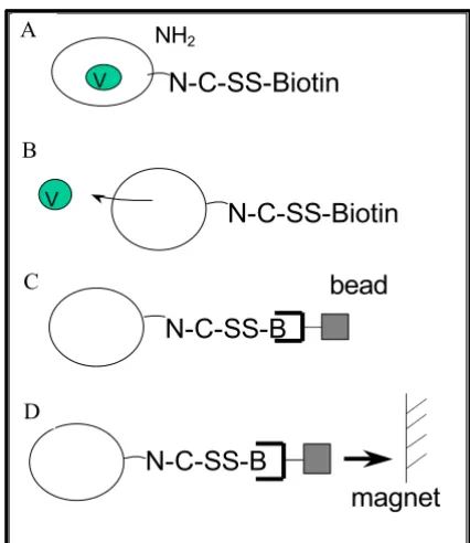
[image:2.612.70.283.86.332.2]Parasites synchronized in the late trophozoite stage were released from erythrocytes by saponin lysis. Removal of the erythrocyte membranes was obtained by immunoaf-finity. For this purpose, polystyrene beads coated with anti-erythrocyte antibodies were incubated with the tro-phozoite preparation. Trotro-phozoites were then biotinylat-ed with NHS-SS-biotin prior to nitrogen cavitation in the presence of protease inhibitors and DnaseI. The resulting biotinylated membranes bound to streptavidin-magnetic beads and were separated from the lysate using a magnet (Figure 1). Membrane vesicle preparations contained 8.2% of the total isolated-trophozoite protein.
The integrity of purified trophozoites was monitored by measuring the uptake of the Trypan blue dye into the par-asite cytoplasm. It was shown that 98% of the tropho-zoites released by saponin lysis did not concentrate the dye, indicating an intact plasma membrane.
Purified P. falciparum plasma membranes examined using transmission electron microscopy were observed to form intact vesicles, although some open membranes could be seen. These vesicles were found to have an average diam-eter of 500 nm. No visible contamination by other mem-branes or organelles was observed, although some liberated hemozoin crystal could be seen (results not shown).
The purity of the parasite plasma membrane preparations was further assayed by the use of enzyme markers of P. fal-ciparum cytosol, the parasite lactate dehydrogenase
Figure 1
Isolation of P. falciparum plasma membrane vesicles. (A) Biotinylation of the trophozoite membrane with a reversible agent. (B) Disruption of the P. falciparum plasma membrane by nitrogen decompression. (C) Binding of the magnetic streptavidin beads to the biotinylated plasma mem-brane. (D) Recovery of the parasite plasma membrane using a magnet.
N-C-SS-B
bead
VNH2
N-C-SS-Biotin
V
N-C-SS-Biotin
N-C-SS-B
magnet
A
B
C
(pLDH) and of human erythrocyte membranes, the ace-tylcholine esterase (AchE). There were no detectable AchE or pLDH activities (less than 0.01 µmol/min/mg protein) in the parasite plasma membrane preparations (Table 1). By comparison, the specific activity of AchE was 2.30 ±
0.15 and 0.05 ± 0.03 µmol/min/mg protein in erythrocyte ghosts and isolated trophozoites (n = 3) respectively, while the specific pLDH activity in isolated trophozoites was 0.14 ± 0.01 µmol/min/mg protein (n = 3).
Time dependence of the ATPase activity of plasma mem-branes isolated from the P. falciparum strain D10 was eval-uated over a time period of 45 minutes. A linear relationship of time versus ATPase activity was found with a correlation coefficient of 0.717 (n = 3) (Figure 2). AT-Pase in plasma membrane preparations required the pres-ence of divalent cations to maintain its activity. ATPase activity was maximal with 2 mM Mg2+ in the presence of 2 mM ATP (Figures 3,4). The ability of 2 mM concentra-tion of Mg2+ and the other divalent cations Mn2+ and Ca2+ to support the ATPase activity of purified plasma membranes isolated from the P. falciparum strain D10 was evaluated, along with no divalent cation addition as a control (Table 2). Maximal ATPase activity was obtained in presence of 2 mM Mg2+. A significant activity (88 ± 33%) was measured when Mg2+ was replaced with Mn2+. When Ca2+ was substituted for Mg2+, only 21 ± 19% of the ATPase activity was retained. Plasma membranes were also capable of hydrolysing other nucleoside triphos-phates as effectively as ATP (Table 3). Only adenosine monophosphate (AMP) was not hydrolysed by the D10 plasma membranes. Purified parasite plasma membranes isolated from the sensitive strains D10 displayed an AT-Pase activity of 3.2 ± 1.7 nmol Pi/min/mg protein.
Discussion
A procedure for isolating parasite plasma membrane from
[image:3.612.52.552.99.177.2]P. falciparum-infected erythrocytes was developed. In this method, trophozoites were released from the erythrocytes by saponin lysis, the remaining erythrocytes membranes being removed by immunoaffinity using anti-erythrocyte antibodies. Previously, erythrocyte monoclonal anti-bodies have been used to remove unlysed erythrocytes and ghost membranes after sorbitol treatment [8]. How-ever, in order to remove most of the erythrocyte ghosts by centrifugation, all the erythrocytes must be lysed by the
Table 1: Marker enzyme activities of isolated erythrocyte ghosts, trophozoites and parasite plasma membranes.
Samples AchE specific activities (µmol/min/mg protein) pLDH specific activities (µmol/min/mg protein)
Erythrocyte ghosts 2.30 ± 0.15 N.D.
Trophozoites 0.05 ± 0.03 0.14 ± 0.01
Parasite plasma membranes No activity No activity
[image:3.612.56.295.278.355.2]Acetylcholine esterase (AchE) and parasite lactate dehydrogenase (pLDH) activities of isolated trophozoites, parasite plasma membranes and/or erythrocyte ghosts were determined as described under materials and methods section. Values are means ± standard deviation of three separate experiments from separate parasite plasma isolations, trophozoite isolations or erythrocyte membrane preparations. N.D.: not determined.
Table 2: Effect of divalent cations on purified D10 plasma mem-branes ATPase activity.
Cations ATPase activity (% of control)
Mg2+ (control) 100
Mn2+ 87.8 ± 33.1
Ca2+ 20.9 ± 19.2
ATPase activity was determined using 2 mM concentration of Mg2+,
Mn2+ and Ca2+ and the ATPase activity in the absence of divalent
cat-ion was subtracted. Values are means of three separate experiments, each performed in quadruplicate ± standard deviations and are expressed as % of control in presence of Mg2+.
Table 3: ATPase activity in the presence of 2 mM concentrations of various nucleotides.
ATPase activity (% of control)
Nucleotide Triphosphate Diphosphate Monophosphate
Adenosine 100 72.6 ± 16.7 -9.2 ± 9.3 Guanosine 120.8 ± 19.6 55.4 ± 17.1 N.D. Cytosine 64.2 ± 7.6 N.D. N.D. Uridine 101.4 ± 55.0 N.D. N.D.
[image:3.612.56.295.471.591.2]detergent, thus allowing isolation of the trophozoites on a larger scale. Sorbitol lyses only the parasitized erythro-cytes, leaving uninfected erythrocytes intact. Saponin, on
the other hand lysed uninfected and infected erythrocytes. For this reason, sorbitol lysis of the trophozoites was not retained in this study. However, because saponin-treat-ment of parasitized erythrocytes has been shown to per-meabilise both the erythrocyte plasma membrane and the parasitophorous membrane [13,14] The exposure of the trophozoites to saponin was very brief to minimize mem-brane damage. Contamination of the plasma memmem-brane vesicle preparations by the parasitophorous membrane, however, could not be discounted since, to our knowl-edge, there is no specific marker for this membrane.
A number of criteria have to be met when isolating subcel-lular fractions for biochemical experiments. The subcellu-lar constituents have to be pure, intact, isolated in high yield and retain their normal physiological capabilities. Evidence for the membrane integrity of saponin-freed par-asites came from studies measuring the rate of incorpora-tion of [14C]-isoleucine into protein [15] and the rate of phosphorylation of the pantothenic acid [16]. In this study, experiments with the trypan blue showed that 98% of the trophozoites released by saponin lysis were capable of maintaining an intact plasma membrane.
[image:4.612.332.522.104.238.2]Nitrogen decompression has been previously shown to be effective for the disruption of cells [17] and was used in this study to disrupt the parasite plasma membranes. A cocktail of protease inhibitors (aprotinin, leupeptin and
Figure 2
Time course of D10 plasma membranes ATPase
activity. ATPase activity (nmol Pi/mg protein) of D10
plasma membranes was determined over a time period of 45 minutes and the activity of the membranes tested without ATP was subtracted. Error bars represent standard devia-tions from means of three separate experiments, each per-formed in quadruplicate.
Figure 3
Dependence of ATPase activity on Mg2+
concentra-tion. ATPase activity of freshly purified D10 plasma
mem-branes expressed as nmol Pi/min/mg protein was measured over Mg2+ concentrations ranging from 0 to 5 mM. ATPase
activity of parasite plasma membranes tested without ATP and absorbance due to ATP hydrolysis was subtracted. Error bars represent standard deviations from means of three sep-arate experiments, each performed in quadruplicate.
0 10 20 30 40 50 0 100 200 300 400 Time (minutes) A T P a s e a c ti v it y ( n m o l P i/ m g p r o te in )
0 1 2 3 4 5 6
0 1 2 3 4 5
Mg2+(mM)
[image:4.612.71.269.106.240.2]A T P a s e a c ti v it y ( n m o l P i/ m in /m g p ro te in ) Figure 4
Dependence of ATPase activity on ATP
concentra-tion. ATPase activity of freshly purified D10 plasma
mem-branes expressed as nmol Pi/min/mg protein was measured over ATP concentrations ranging from 0.5 to 5 mM. ATPase activity of parasite plasma membranes tested without ATP and absorbance due to ATP hydrolysis were subtracted. Error bars represent standard deviations from means of three separate experiments, each performed in quadrupli-cate.
[image:4.612.75.277.373.540.2]phenylmethylsulfonyl fluoride) was used to prevent the degradation of membrane proteins by the proteases re-leased during the nitrogen cavitation step. The presence of DNA presents an additional problem, as the parasites ad-here to the sticky DNA liberated during parasite lysis. Dnase I was, therefore, included in the isolation process to eliminate the DNA liberated during the nitrogen cavita-tion step. The recovery yield of the parasite plasma mem-branes, 8.2% of the total isolated-trophozoite protein, is consistent with the percent protein yield (8–10%) ob-tained for membrane vesicles prepared from cancer cells [17].
Purified P. falciparum examined using transmission elec-tron microscopy were observed to be intact and free of contaminating membranes and organelles, even though a small amount of liberated haemozoin could occasionally be seen in certain fields. Contamination of the plasma membrane vesicles with fragments of food vacuole mem-brane could not be ruled out since nitrogen cavitation is likely to disrupt the food vacuole membrane as well as the plasma membrane. It would therefore had been interest-ing to investigate whether the parasite plasma membrane preparations were contaminated by food vacuole mem-brane components using antibodies to PfCRT. The ab-sence of parasite and erythrocyte protein in the parasite plasma membrane preparation was confirmed by non-de-tectable parasite lactate dehydrogenase and erythrocyte acetylcholine esterase activities. The fact that the mem-branes were bound to a support facilitated their purifica-tion from contaminants and their recovery.
The ATPase activity of parasite plasma membranes (3.2 ± 1.7 nmol Pi/min/mg protein) obtained in this study was found to be of similar magnitude to that of digestive vac-uoles isolated from P. falciparum[18]. For comparison, the specific ATPase activity of plasma membranes prepared from multidrug-resistant cancer cells is 1.15 µmol/min/ mg membrane protein[19].
Conditions required for optimal P. falciparum plasma membrane ATPase activity were determined using the chloroquine-sensitive strain D10. Parasite plasma mem-branes showed optimal ATPase activity at 2 mM Mg2+ and 2 mM ATP. Other divalent cations can be substituted for Mg2+ and maintain the function of a variety of different ATPases [20]. In this study, it was found that Ca2+ could not be substituted for Mg2+ to sustain ATPase activity in parasite plasma membranes, whereas activity was pre-served when Mg2+ was replaced by Mn2+, indicating that the parasite plasma membrane ATPase activity was similar to the vacuolar membrane ATPase activity [18]. However, this contradicts the Choi and Mego [19] study which showed that Ca2+ could support the P. falciparum vacuolar ATPase activity in the absence of Mg2+. Other nucleoside
triphosphates and diphosphates tested were hydrolysed as effectively as ATP. However, the nucleoside monophos-phate AMP was not hydrolysed, suggesting that the para-site plasma membrane ATPase activity is distinguishable from that of membrane-associated alkaline and acid phosphatases [21].
Since the parasite plasma membranes were isolated in the form of intact vesicles the right-side-out, the ATP hydro-lysing regions of the ATPase proteins should be enclosed within the vesicles. This raises the question of whether all of the compounds tested are gaining access to the relevant part of the protein, and whether the differences between the ATPase activities measured in the presence of the ferent agents might reflect, at least to some extent, the dif-ferent ability of these agents to gain access to the interior of the vesicles. Therefore, it could not be excluded that the saturable dependence of ATPase activity on the concentra-tion of ATP in the medium could reflect the saturable transport of ATP into the vesicles, rather than the satura-tion of the ATPase itself. Similarly, the differential ability of divalent cations to support ATPase activity could be the result of a differential transport of these cations into the vesicles.
Conclusions
In conclusion, a method for the isolation and purification of the P. falciparum plasma membrane was developed and optimal conditions for P. falciparum plasma membrane ATPase activity were determined. These purified mem-branes were intact and isolated in a high enough yield to enable a characterisation of their ATPase activity. Thus, plasma membranes cleansed of P. falciparum can be used as a model to establish the role played by ATPases in ion transport. The availability of purified P. falciparum plasma membrane will be of value not only to investigate process-es such as nutrient uptake/efflux, pH regulation and ion balance but also in the understanding of drug transport in this organelle.
Materials and Methods Cell cultures
at-mosphere of 93% nitrogen, 4% carbon dioxide, 3% oxy-gen. Cultures were synchronized by treatment with sorbitol [23].
Trophozoite isolation
Infected erythrocytes were washed twice in 10 volumes of phosphate buffered saline (PBS). The pellet was suspend-ed in 10 volumes of PBS containing 0.05 % (w/v) saponin in order to lyse the erythrocytes. It was then incubated for 2–3 min at room temperature and centrifuged at 1,500 g for 10 min. The resulting pellet was washed twice in PBS and resuspended in 5 volumes of PBS. The protease inhib-itors aprotinin, leupeptin and phenylmethylsulfonylfluo-ride (Boehringer Mannheim) were added to final concentration of 10 µg/ml, 10 µg/ml and 0.1 mM, respec-tively. Unlysed erythrocytes and erythrocyte membranes contained in the trophozoite preparation were adsorbed by immuno-affinity. Anti-human erythrocytes (polyva-lent immunoglobulin (G,A,M) rabbit, Sigma) were dilut-ed 1: 1000 in PBS. 5 g of polystyrene beads (Pierce) were incubated in 20 ml of the above solution for two hours at room temperature with gentle agitation. The beads were washed in PBS and further incubated for one hour with 20 ml 1 % bovine serum albumin in PBS. The beads were then washed again in PBS and incubated with the tropho-zoite preparation for 30 min. The purified trophotropho-zoites were collected and centrifuged at 1,500 g for 10 min.
Trypan blue uptake
The ability of purified trophozoites to exclude trypan blue was monitored by adding 10 µl of Trypan blue (10 mg/ml in distilled water) to 100 µl of trophozoite suspension in PBS (phosphate buffered saline). After five minutes incu-bation at room temperature, the proportion of parasites showing trypan blue accumulation was evaluated using a phase contrast microscope.
Plasma membrane isolation
The biotylination reaction was performed by incubating 250 µl of 1 mg/ml NHS-SS-Biotin (sulfosuccinimidyl 2-[biotin-amido] ethyl-1,3-dithiopropionate) (Pierce) with 500 µl of trophozoites (2 mg of protein) for 30 min at room temperature. The resulting biotinylated tropho-zoites were washed once in PBS, then twice in Vesicle buff-er I (0.25 M sucrose, 1 mM E.D.T.A., 10 mM Tris-HCl pH 7.4). Vesicles were prepared by nitrogen cavitation accord-ing to the method of Lever et al.[24]. The cells were resus-pended in 500 µl vesicle buffer I containing aprotinin, leupeptin and phenylmethylsulfonylfluoride (10 µg/ml, 10 µg/ml and 0.1 mM, respectively) and equilibrated at 4°C under nitrogen pressure at 800 psi for 30 min. The ho-mogenate was then incubated for five min at 37°C with Dnase I (50 µg/ml). The biotinylated vesicles were incu-bated with 100 µl of streptavidin immobilized on iron ox-ide (Sigma) previously washed with PBS for 15 min at
room temperature and then separated using a magnet (Magnetic Cell Sorting, Myltenyi Biotec). The magnetic particles were bound to the side of the tube and washed with 1 ml of 0.25 M sucrose, 10 mM Tris-HCl pH 7.4. The key steps in the purification procedure are shown in Fig-ure 1.
Electron microscopy analysis
Parasite plasma membrane vesicles were fixed with glutar-aldehyde (0.8%)/formglutar-aldehyde (8%) in PBS for one hour at room temperature and washed in PBS, followed by ad-dition of 50 mM NH4Cl for 30 minutes and two washes in PBS. Samples were dehydrated in 70% ethanol and em-bedded in LR White resin (Polyscience Inc.) which was then polymerised at 60°C for 28 hours. Then 1 µm-thick sections were cut using a reichert Ultracut S ultramicro-tome. Sections were mounted on copper grids and stained with 2% uranyl acetate and lead acetate. Samples were viewed on a JEM 200CX transmission electron micro-scope.
Acetylcholine esterase activity
The acetylcholine esterase (AchE) activity was determined by a modification of the method of Ellman et al. [25]. En-zyme assays were carried out with 200 µl of freshly pre-pared 0.1 M sodium phosphate pH 7.5, 0.5 mM 2,2'-dinitro-5,5'-dithiobenzoic acid, 0.6 mM S-acetylthio-choline iodide to which was added 50 µl of a suspension of erythrocyte ghost, trophozoites or parasite plasma membrane, in microtitre 96-well plates. 10 µg of erythro-cyte ghost protein, 3–5 µg of trophozoites protein or 20
µg of parasite plasma membrane protein were used to measure an initial rate. 50 µl of PBS or vesicle buffer was added to the control wells. Wells containing 200 µl H2O and 50 µl of erythrocyte ghosts/ trophozoites/ vesicles sus-pensions were included to subtract any absorbance due to the cells. Changes in absorbance at 405 nm were meas-ured for at least 20 min, using a 7520 Microplate Reader (Cambridge Technology, Inc).
Parasite lactate dehydrogenase activity
Protein determinations
Protein determinations were performed by the method of Lowry et al. [27].
ATPase activity
The ATPase activity was determined using a colorimetric assay adapted from those of Chifflet et al.[28] and Doige
et al.[20]. Enzyme assays were carried out with 50 µg of freshly isolated P. falciparum plasma membranes suspend-ed in 450 µl of reaction buffer (50 mM Tris-HCl, 0.15 M NH4Cl, 2 mM MgCl2, 0.02% NaN3, pH 7.4). To initiate the reaction 50 µl of ATP or the appropriate nucleotide tested in the reaction buffer was added, giving a final con-centration of 2 mM ATP or the appropriate nucleotide. A control containing 50 µg of parasite plasma membranes suspended in 500 µl of reaction buffer was included to subtract any absorbance due to the membranes. Another control consisting of 500 µl reaction buffer containing 2 mM ATP was added to evaluate the ATP hydrolysis. After 45 min. at 37°C, the suspensions were centrifuged at 13,000 rpm for three minutes to pellet the membranes. Aliquots of 100 µl of the resulting supernatant were trans-ferred to the wells of a 96-well microtitre plate. The reac-tion was stopped by addireac-tion of 100 µl of fresly prepared solution A (6% SDS, 3% L-ascorbic acid, 0.5% ammoni-um molybdate in 0.25 M sulfuric acid). After 15 minutes, the phosphoammoniummolybdate complex formed was stabilised by the addition of 100 µl of solution B (2% so-dium citrate, 2% soso-dium arsenite, 2% acetic acid). After 15 minutes, the absorbance at 710 nm was measured us-ing a 7520 Microplate Reader (Cambridge Technology, Inc).
Acknowledgements
This work was supported by the South African Medical Research Council.
References
1. Trager W, Jensen JB: Human malaria parasites in continuous culture.Science 1976, 193:673-675
2. Trager W, Williams J, Gill GS: Extracellular development, in vit-ro, of the erythrocytic cycle of Plasmodium falciparum.
Parasi-tology today 1992, 8(11):384-387
3. Williams JH, Gill GS, Trager W: Effect of erythrocyte membrane on extracellular development of the erythrocytic cycle of Plasmodium falciparum.Proc Natl Acad Sci USA 1995, 92(2): 566-568
4. Trager W, Williams J, Gill GS: Extracellular development of erythrocytic stages of Plasmodium falciparum.Methods in Cell
Science 1996, 18:219-227
5. Hamburger J, Kreier JP: Isolation of malarial parasites and their constituents.In Malaria Immunology and Immunization, New York:
Ac-ademic Press 1980, 3:1-65
6. Heidrich HG, Leutner G: Two types of vesicles from the eryth-rocyte-ghost membrane differing in surface charge. Separa-tion and characterizaSepara-tion by preparative free-flow electrophoresis.Eur J Biochem 1974, 41(1):37-43
7. Wunderlich F, Helwig M, Schillinger G, Vial H, Philippot J, Speth V:
Isolation and characterization of parasites and host cell ghosts from erythrocytes infected with Plasmodium chabaudi.
Mol Biochem Parasitol 1987, 23(2):103-115
8. Hoppe HC, Coetzee J, Louw AI: Plasmodium falciparum : isola-tion of intact and erythrocyte-free trophozoites from sorbi-tol lysates.Parasitology 1992, 104:379-385
9. Christopher SR, Fulton JD: Experiments with isolated malaria parasites (Plasmodium knowlesi) free from red cells.Ann Trop
Med Parasitol 1939, 33:161-170
10. Gruenberg J, Sherman IW: Isolation and characterization of the plasma membrane of human erythrocytes infected with the malarial parasite Plasmodium falciparum.Proc Natl Acad Sci USA
1983, 80:1087-1091
11. Herwaldt BL, Schlesinger PH, Krogstad DJ: Accumulation of chlo-roquine by membrane preparations from Plasmodium falci-parum.Mol Biochem Parasitol 1990, 42(2):257-268
12. Choi I, Mego JL: Purification of Plasmodium falciparum digestive vacuoles and partial characterisation of the vacuolar mem-brane ATPase.Mol Biochem Parasitol 1988, 31(1):71-78
13. Ansorge I, Benting J, Bhakdi S, Lingelbach K: Protein sorting in Plas-modium falciparum-infected red blood cells permeabilized with the pore-forming protein streptolysin O.Biochem J 1996,
315:307-314
14. Ansorge I, Paprotka K, Bhakdi S, Lingelbach K: Permeabilization of the erythrocyte membrane with streptolysin O allows access to the vacuolar membrane of Plasmodium falciparum and a molecular analysis of membrane topology.Mol Biochem Parasi-tol 1997, 84(2):259-261
15. Saliba KJ, Horner HA, Kirk K: Transport and metabolism of the essential vitamin pantothenic acid in human erythrocytes in-fected with the malaria parasite Plasmodium falciparum.J
Biol Chem 1998, 273(17):10190-10195
16. Saliba KJ, Kirk K: pH regulation in the intracellular malaria par-asite, Plasmodium falciparum. H(+) extrusion via a V-type H(+)-ATPase.J Biol Chem 1999, 274(47):33213-33219
17. Cornwell MM, Safa AR, Felsted RI, Gottesman MM, Pastan I: Mem-brane vesicles from multidrug-resistant human cancer cells contain a specific 150 to 170 kDa protein detected by pho-toaffinity labeling.Proc Natl Acad Sci USA 1986, 83:3847-3850 18. Adams B: An investigation of P-type and V-type ATPase
com-ponents and chloroquine sensitivity of digestive vacuoles pu-rified from Plasmodium falciparum.Thesis submitted for doctoral
degree at the University of Cape Town, South Africa 1999
19. Rebbeor JF, Senior AE: Effects of cardiovascular drugs on AT-Pase activity of P-glycoprotein in plasma membranes and in purified reconstituted form. Biochim Biophys Acta 1998,
1369(1):85-93
20. Doige CA, Yu X, Sharom FJ: ATPase activity of partially purified P-glycoprotein from multidrug-resistant chinese hamster ovary cells.Biochim Biophys Acta 1992, 1109(2):149-160
21. Ketcham CM, Baumbach GA, Bazer FW, Roberts RM: The type 5, acid phosphatase from spleen of humans with hairy cell leukemia. Purification, properties, immunological charac-terization, and comparison with porcine uteroferrin. J Biol Chem 1985, 260(9):5768-5776
22. Ekong RM, Robson KJ, Baker DA, Warhurst DC: Transcripts of the multidrug resistance genes in chloroquine-sensitive and chloroquine-resistant Plasmodium falciparum.Parasitology 1993,
106(2):107-115
23. Lambros C, Vanderberg JP: Synchronization of Plasmodium falci-parum erythrocytic stages in culture.J Parasitol 1979, 65(3): 418-420
24. Lever JE: Active amino acid transport in plasma membrane vesicles from Simian virus 40-transformed mouse fibrob-lasts.J Biochem 1977, 252:1990-1997
25. Ellman GL, Courtney KD, Andres V, Featherstone RM: A new and rapid colorimetric determination of acetylcholinesterase ac-tivity.Biochem Pharmacol 1961, 7:88-95
26. Makler MT, Hinrich DJ: Measurement of the lactate dehydroge-nase activity of Plasmodium falciparum as an assessment of parasitemia.Am. J. Trop. Med. Hyg. 1993, 48(2):205-210
27. Lowry OH, Rosebrough NJ, Farr AL, Randall RJ: Protein measure-ment with the folin phenol reagent.J Biol Chem 1951, 193: 265-275