International Journal of Innovative Technology and Exploring Engineering (IJITEE) ISSN: 2278-3075, Volume-8 Issue-10, August 2019
Abstract: Image processing tool is a key role in medical uses, by extracting anatomical features the contouring of the regions from medical images.The discovery of bone sketch from X-ray images has recently introduced a new dimension in the literature as it proves to be an important step of radiological imaging analysis. X-ray images are segmented to study bone skeleton where the image is split into several parts, to examine the fracture of bones, and to plan for treatment before surgery. This report's, purpose is to survey digital image splitting methods. In image segmentation, research analysis is important for categories and it is important to provide an overview of assistive segmentation techniques with highlighting advantage and disadvantage.
Index Terms: Edge Detection, Medical imaging, Medical image segmentation, Wrist X-Ray Processing,
I.INTRODUCTION
Today, Image diagnosis is an excellent tool in medical science. X-ray, computed tomography (CT), Magnetic resonance imaging (MRI) and other imaging modalities chooses an efficient approach to map a subject's anatomy anti-invasively. It became essential to use computers to enhance their processing and interpretation with both increasing medical images. Specifically, computer algorithms are an important part to assist specific radiological functions and automatically automate structures and other areas of interest, such techniques are segmentation based image algorithm and they play important roles in many programs for biomedical imaging, for example, analysis of tissue volume, anatomical structure studies, indigenous diagnostics and operations integrated by computer. The performance of segmentation methods varies widely depending on specific uses and image processing techniques. Every imaging modality has its own characteristics to compete. The region of interest (ROI) must be distinguished from the background when extracting the characteristics in images. During the removal of the features in the images, the areas of interest should be separated from the background (ROI). Removal of facilities from medical imaging is done by splitting technique. Segmentation of images is a difficult task in the area of image processing through segmentation, splitting the image from a series of images, depends entirely on the image's characteristics that are roughly constant in each neighborhood, but from one region to another are variations. X-ray is the
Revised Manuscript Received on August 05, 2019.
Mahima Shanker Pandey, Computer Science and Engineering Department , Institute of Engineering And Technolgy, Sitapur Road Lucknow Uttar Pradesh, India.
Dr.Sudhir Singh Soam, Computer Science and Engineering Department , Institute of Engineering And Technolgy, Sitapur Road Lucknow Uttar Pradesh, India.
Dr.Surya Prakash Tripathi, Computer Science and Engineering Department , Institute of Engineering And Technolgy, Sitapur Road Lucknow Uttar Pradesh, India.
initial stage of medical imaging and therefore, with each of its pros and cons, sundry image methods have been shown through the decades. These include magnetic resonance imaging (MRI), ultrasound (US), atomic imaging, computed tomography single photon emission (SPECT) and positron emission tomography (PET). Reliable algorithm required for depiction of anatomical Skelton. It aims to
I. Extract the characteristics from images in medical imaging.
II.To make a automatic process, which can be handled with same accuracy so result are not affected. III.To get fast and efficient result.
As medical research continues to grow, there is a constant flow of information about a difficult relationship around processes or stages of disease, a current therapeutic objective, and the genome of the being and the associated risk of disease. Medical imaging can now play a main and essential role in global health care systems as it continue to improve health outcomes and cost-effective healthcare diagnosis for virtually all disease and deformity classes. Medical Image Segmentation is a technique for anatomical structure localization and it helps in the study of diagnosis methods and techniques. Forming segments of any biological image has emerged as one of the most used methods by various researchers in last few years. For example, as depicted in Fig. 1(a). This plot represents the number of publications describing technique of Medical Image Segmentation.
Fig 1(a) Growth of Medical Image Segmentation Technique[1]
The second plot (Fig 1(b)) considers publications for a specific imaging modality. The X-ray modality is much more use than MRI and CT for Medical Imaging
Recognition of X-rays bones: Challenges in the
Past, Present and Future
[image:1.595.312.540.507.679.2]Fig 1(b) Comparison of Image Modality[1]
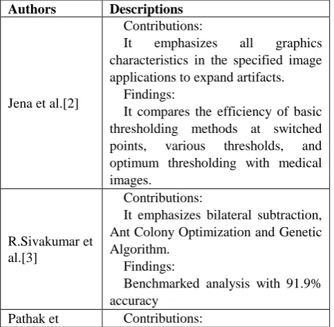
The X-ray image is complex in nature because X-rays have the complexity of noise, bone density variability, inter-patient size variability, etc. Therefore, the X-ray division is a challenging subject. Many segmentation approaches have been used by specialists of radiological images. No universal algorithm for medical images due to the imaging system. From the scenario of X-ray images, several image splitting techniques have been used, some of which are that help in segmenting radiological images of the bones have been compiled to present a comparative survey, Will prove to be a milestone for those looking for medical image segmentation of radiological image. This study provides a comprehensive of existing methods of computer-assisted medical image segmentation. A short analysis of the latest literature's applications and procedures, the scope of this report is a complete explanation of competing methods and the readers are given orientations for further facts. Meanwhile, we emphasis on giving the reader with an inclusion to the different applications for medical imaging segmentation and the different issues that have to be addressed. We just apply to the most frequently used radiological methods for anatomy imaging: X-ray, CT, MRI and ultrasound radiography. However, most concepts also apply to other imaging modalities. Here we will focus on describing some of the key elements in the design of a survey, then highlighting some contribution and finding in the field of medical imaging given as follows.
Authors Descriptions
Jena et al.[2]
Contributions:
It emphasizes all graphics characteristics in the specified image applications to expand artifacts.
Findings:
It compares the efficiency of basic thresholding methods at switched points, various thresholds, and optimum thresholding with medical images.
R.Sivakumar et al.[3]
Contributions:
It emphasizes bilateral subtraction, Ant Colony Optimization and Genetic Algorithm.
Findings:
Benchmarked analysis with 91.9% accuracy
Pathak et Contributions:
al.[4][5]and Kwabwe et al[6]
A multi-level thresholding technique was used to segment images. Based on the ' fuzziness ' index and the entropy index.
Findings:
Edge detection for bone boundaries.
Cristina et al.[7]
Contributions:
Segmentation based on deformable or atlas is considered more appropriate for complex segmentation of medical images.
Findings:
The grouping of classic and complex segmentation techniques is viewed for good and efficient precision.
Alireza Norouzi et al.[8]
Contributions:
Discussed segmentation methods are divided into four classes: regional methods, clustering, classification methods, and hybrid methods.
Findings:
High-time complexities of
clustering and classification approaches are challenging.
Dzung L et al. [9]
Contributions:
New and creative ways of
segmenting regions Findings:
[image:2.595.55.287.48.180.2]Different methods of segmentation such as classifiers approaches to clustering, Markov random field models, artificial neural networks.
Table I List of review articles on Medical Image Processing
The Bone X-Ray challenges:
Analyzing different bone segmentation problems in X-ray images requires studying certain bone image characteristics. X-ray images differ from other methods of medical image processing because it contains areas partially covered by various organs such as flesh, tissues or muscles [10]. Noise: A digital X-ray detector with an avg. of 100 pixels of photons is subjected to an X-ray beam,which is uniform in nature. Some pixels get more X-rays and appear darker, while others seem lighter. This appearance is called noise, these pixels are distributed randomly [11].
Overlapping: In X-ray images, the bone structure covers many tissues and organs.
Vagueness: There may be an X-ray absorption rate equal to the various neighboring tissues within the human body. The findings are in the limbs obscure borders. In an image, the two neighbors create several unclear clauses [12].
[image:2.595.51.293.550.786.2]International Journal of Innovative Technology and Exploring Engineering (IJITEE) ISSN: 2278-3075, Volume-8 Issue-10, August 2019
In a standard patient's, his X-ray image contain thick bones, although patients experiencing osteoporosis have fewer thick bones, which can bring extraordinarily slow bone areas. Even more, different tissue pictures can affect the strength of bone areas too.
Variability of inter-patient shape: The size of different patient’s bones may differ relatively. For good example, female pelvic bone size differs significantly relatively from male patient size. Shoulder bones are relatively narrow compared to men.
[image:3.595.355.497.148.329.2]Imaging pose variability: When taking x-ray images, the situation of the patient can also vary. Because of this, due to different imaging situations, it could show bones stored in various organs.
Fig-2 Image Processing Flow
MEDICAL IMAGE SEGMENTATION: It’s methods have been classified in the following sections on the basis of complexities present in them:
Medical Image Segmentation Technique
The description of all image segmentation methods can be described as optimization issues in which segmentation is required reduces some of the application's defined energy or cost function.
Suppose image v, we desire the segmentation û such that
û =arg min (u,v) (1)
, is the energy function focused on the image v being observed and u is segmentation.
For a suitable is a major challenge in the design algorithms of segmentation due to the wide range of image characteristics like intensity, edges and texture that can be used.
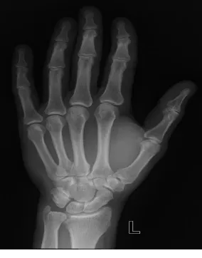
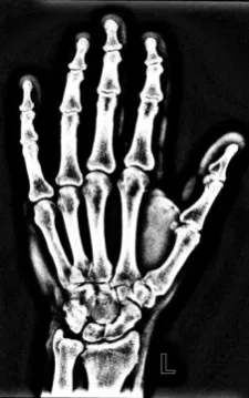
Consider an example of we will consider some segmentation practice as just a real X-ray image displayed shown in fig-3.
[image:3.595.367.481.345.551.2]Fig 3 An image of X-rays
Fig-4 Medical Image Segmentation Approaches Existing Image Segmentation Methods: Methods of segmentation provide the following technique of segmentation: thresholding, methods based on region and methods based on edge.
Thresholding is a kind of segmentation technique that making classes so that similar pixels are forming or pixels are categorized. It selects the correct threshold n T pixels divided into different classes and objects separated from the background. Threshold can be displayed in the following equation.
[image:3.595.58.291.551.687.2]In this equation, T is the threshold ; V(x, y) is the gray point value (x, y) and u(x, y) refers to some local point property such as the neighborhood's average point-centered gray value (x, y).All the pixels that have a gray level more than “t” will be classified as white"1" otherwise it will be black"0". Given an image v(x, y) segmentation is performed as following V (x, y) =Error! Reference source not found. (2) where “t” is the threshold value, It's x and y co-ordinates from the pixel and v(x ,y) represent gray level pixels. Thresholding "t" calculated according to many technique like k-means clustering algorithm. Disadvantage of thresholding method, can be implemented to a single image band or gray image. A study is provided on thresholding techniques [18].
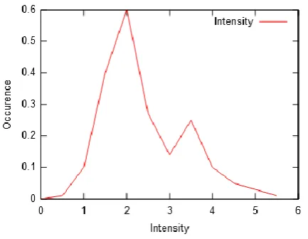
Fig 5 Histogram act with a valley for the value of the threshold
Fig 5 Histogram act with a valley for the value of the threshold
Global Thresholding methods and adaptive thresholding methods are two methods of thresholding [13][17]. One threshold value is choosen for image. Only v(x, y) (gray-level values) depends on global threshold T.
In global thresholding, select the T threshold initial. Segment the image with T in two categories
G1 is all intensity pixels >T G2 is all intensity pixels <=T
Calculate m1 and m2 averages for G1 and G2 pixels Let T = (m1+m2)/ 2 (3)
Global thresholding is bimodal (objects and background). The change in illumination in the image is one factor that affects the performance. The main problem is that only the intensity does not consider any relationship between the pixels.
An image has unequal illumination can transform a perfectly segmental histogram into a histogram that cannot be partitioned effectively by a single global threshold. The main issue in it is that any image that has to be segmented is divided in a number of sub-images based on their threshold value. Calculation of the image intensities every picture element and culling A separate threshold value for each pixel holding a level for different local areas [19] is called Adaptive Thresholding.
In adaptive thresholding all the sub-images incorporates a nearly uni-modal bar chart, and their average grey level was nearer to object than to the background, so it absolutely was classified as object.
Adaptive thresholding is additional complicated than
global thresholding regarding calculations. However, the adaptive method can be used effectively for small area extraction from a background. An implementation of threshold-based segmentation to detect the brest cancer in digital mammography [20].
Weszka et al. [21] also several specified threshold assessment criteria. Palumbo et al. [22] dealt with binarisation issue of documents in comparison with three methods. Sahoo et al[18] conducted a survey nine algorithms of threshold and gave the performance relative. Le,Chung et al.[23] performed a comparative study of five global thresholding methods and several useful performance evaluation criteria have been developed. Glasbey [24] identified the performance relationships and distinctions between 11 histogram-based algorithms supported by scientific statistical study.
Chain-code generation and polygonal approximation: The chain code is a slope representing a form. It’s curve represents the sequences of direction modifications in the curve around neighboring points. Therefore, if the nth point of the curve is in grid position (p, q), the chain element or link relates to the change of position from nth point to (n+1)st point. In an example, going to start at an arbitrary boundary point, the closest counter clockwise boundary point will be to the right (east), preceded by the next north east point and the
next north point, preceded by another north point, and so on. Region-based segmentation use the spatial information
[14], assume that neighboring pixels tend to belong to same intensity. Region-based segmentation algorithm: Region-Growing segmentation and Region Splitting and Merging algorithm.
Region growing algorithm, marks few seed point and then try to go to the region from the seed point.Based on the some similarity of images they go to the region and ensure that a point is included in particular region if the intensity is similar to region . For each border pixel in the field, this process repeats. Regional growing provides many advantages over conventional techniques of segmentation. In noise, it is very stable. There won't be too much background in the areas until the parameters are properly defined. Other techniques, such as border tracking, which produce associated edges, are very unstable. Bone segmentation by region growing technique [25] is acquired in the case of a hand-wrist radiograph.
[image:4.595.322.548.552.831.2]International Journal of Innovative Technology and Exploring Engineering (IJITEE) ISSN: 2278-3075, Volume-8 Issue-10, August 2019
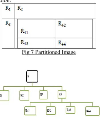
[image:5.595.83.258.127.344.2]Region Splitting and Merging, if the pixel intensities is not similar then partitioned image into four quadrant or four part and check the similarity between pixel in each quadrants, if any of the quadrants find the all the pixels have similar intensities then no need to partition otherwise partition[26]. It requires manual initialization and sensitive noise over segmentation.
Fig 7 Partitioned Image
Fig 8 Corresponding Quad Tree
Edge-based segmentation methods represents methods based on image edge information. This method use edge detecting operators to find an edge in the image, this edge can be viewed as image point to identify image locations of gray level discontinuities, color etc. If the illumination changes rapidly, then at that position there is a high probability of an edge. Table II presents a comparing analysis between different methods of segmentation by providing a segmentation effect and their advantage.
Method Segmentation
Effect
Advantage Pixel Based
Segmentation
Normal More
Complexity Edge Based
Segmentation
Average Average
Complexity Graph Based
Segmentation
Good Very Low
[image:5.595.378.475.607.729.2]Complexity
Table II Comparison of Classification method The following are for edge detection operators: A.Prewitt
B.Sobel C.Roberts
D.Laplacian of Gaussian (LoG) operator.
Many edge detectors are founded on the image utility's local slope. The difference between these operators in practical terms is that they use a different type of filter to understand gradient components and estimate a different method of combining these components.
Prewitt is a discrete operator, which consist two gradient mask detector, Horizontal Gradient Detector (Px) and Vertical Gradient Detector (Py) for detecting the edge. Both operator will superimpose when detecting the edge. So both
edges of the image, be it horizontal or vertical can be detected we can detected.
Vertical Gradient Detector (Py)-
1
0
1
1
0
1
1
0
1
Horizontal Gradient Detector (Px) -
1
1
1
0
0
0
1
1
1
The resulting gradient P=Error! Reference source not found.
(5)
Sobel edge detection is same as Prewitt edge detection. It also uses two kinds of edges in an image
Vertical Direction (Sy
)-1
0
1
2
0
2
1
0
1
Horizontal Direction (Sx) -
1
2
1
0
0
0
1
2
1
Error! Reference source not found.
The resulting gradient
S=Error! Reference source not found. (6)
The mask coefficients are not set it can be set according to need this is a difference with Prewitt operator. When we apply in the upright direction, it acts as a first-order derivative and works out the pixel difference in an edge region.
In this case the contour form of the skull of the hands is not fully seen, some imbalance can be discovered at the tip of the finger bone. Not only the skeleton of the hand detected, but the shape of the hand can also be seen in image.
Roberts Operator carries out the fast calculation 2-D image measurement of special gradients. This highlights areas which corresponds to the edges. It is a 2x2 kernel running over an image to find the edge of the gradient.
The two kernels are
1
0
0
1
and1
0
1
0
The shield edges at -45 ° and the second at 45 ° when converting the image with the first kernel. The fault of Roberts operator is that for very tiny edge structures uses a lesser size filter to identify this edge.
Laplacian of Gaussian (LoG) operator calculates the second spatial image derivative. LoG will initiate zero in the area where the image has a constant intensity, wherever a change takes place in the neighborhood, the LoG will give the darker side a positive response and the lighter side a negative response.
Many advanced edge detectors were suggested in the image processing as Harris detector [27]. The point whose vicinity stands in two different edge directions is called a corner. Harris corner detector not only determines the window that produces large variations but also locates the edges based on the eigenvalues of the Hessian matrix [28].
[image:6.595.306.548.49.182.2] [image:6.595.76.265.407.615.2]Canny corner marking is a method for extracting useful image characteristics. When the image is noisy, it detects the vapid edges. An example of Canny corner marking X-ray segmentation is presented in fig10.
Fig 10 Canny corner marked X-ray image
Table III presents a comparing analysis between different operator for edge detection[29][30] and [31] (Prewitt, Sobel , Roberts, Harris, corner marking) with their pros and cons in following.
Operator Benefits Drawbacks
Classical( Sobel, Prewitt)
Simple, detection and orientation of edges
inaccurate, noise-sensitive Laplacian of
Gaussian(LOG)
Test wider area around the pixel to find the right edge locations
Disorder throughout the corners, curves and where the function of gray intensity varies, Due to the use of the
Lapalacian filter, the edges are not oriented
Gaussian(Canny, Shen-Castan)
Detecting error rate, location and response using probability. Better detection, particularly in noise, increasing the signal-to-noise ratio.
[image:6.595.369.482.490.670.2]Complex information, Crossing false zero, time consuming
Table 3: Some Edge Detectors Advantages and Disadvantages
Pattern Recognition Based: Segmentation proves the pixel classification, so it is viewed as a technique for Pattern Recognition. There are two approaches to pattern recognition: Supervised Classification(Classification) and Unsupervised Classification(Clustering)
Unsupervised Classification method, using split or clustering works by finding hidden attributes in the data. Clustering is a preparing step for classifier and is used for object detection or image tagging. Divide an image using a rectangular grid calculate a feature vector (relative position, distribution of colors, texture, edge orient) for each cell and use K-means to assign each vector as a cluster. The various method includes for unsupervised classification: K-means clustering, Gaussian mixture models, Hidden Markov models and Fuzzy c-means algorithm.
K-means algorithm starts with K, set of x1,x2, .... xn at random places in space. For each individual finding the nearest centroid (ci) assign point x1 to cluster i moving each centroid similarly to the average instances assigned to it. The algorithm continues before the membership of the cluster does not change. An example of segmentation of X-rays using K-means algorithm is presented in fig11.
International Journal of Innovative Technology and Exploring Engineering (IJITEE) ISSN: 2278-3075, Volume-8 Issue-10, August 2019
The issue in analysis of group is that the number of groups must be set a priori [33]. Table IV presents a comparing analysis of speed, calculation complexity, noise reaction and multiple object detection between different methods of segmentation in following.
Degree of Freedom
Threshol d Method
Region Method
Cluster Method
Speed Fast Slow Fast
Calculatio n Complexity
Slow Fast Fast
Noise Reaction
Less Less Modera
te Multiple
Object Detection
Poor Fair Fair
Table IV Result of experimentation for methods of segmentation
Fuzzy C-means is like K-means. Rather than allocating a point to just one cluster, it can have some kind of fuzziness and intersect around two or more clusters [33]. L. Florea et al.[34] proposed a new illusion-based segmentation with the Fuzzy Clustering Algorithm having two objects in X-ray image (femur and tibia).
Supervised Classification is classification based segmentation technique the model is trained on a labeled dataset, so the outcome of sample data can be predicted. Several methods of classification are description in the literature. It could be classified: non-parametric classifier (The nearest-neighbor classifier, The K-nearest neighbor classifier, Parzen window) and parametric classifier (Bayes classifier and maximum likelihood).K-nearest neighbor(KNN) most basic algorithm for regression and classification. In it, each pixel is classified into its most suitable category in k nearest neighbors Consider the weight of the majority of your neighbors. Parzen window is similar as the KNN algorithm. Parzen window takes averages all points at a fixed distance and K-neighbor average a fixed number of points at a distance.
The drawback of supervised classification is only related to scalability function under consideration. The problem occurs when there should be a fragment of uneven intensely corrupt images. It algorithm relies on the selected training samples. Supervised classified technique [35] in which the adaptive fuzzy method was used for lateral skull segmentation. Supervised classification is not effective in segmenting X-ray images because noise is affected by X-ray's intrinsic feature. Deformable models geometric object whose shape as a 2D curve or 3D surface may change with time, model-based techniques using closed parametric curves that deform because of forces to delineate region boundaries. A closed axis or ground must then be located close the required boundary in order to contain the boundary for an object in an image and then be allowed to repeat the rebate process. Internal forces are calculated inside curve or to keep the deformation smooth. Usually, External forces are derived from the image to drive the curve or surface to the important feature that is desired. It has inherited smoothness, more robust to noise. It is good for fitting noisy images where edge
detection or thresholding can produce fragmented object boundaries.
E = Error! Reference source not found. (7) Deformable templates as a tool to segment biological structures and locate them in medical images. A prototype template describes structures and parametric warp mapping that distort the original shape. The motive of a multi-stage, multi-resolution algorithm is to decrease computational complexity and use the time required to achieve the location process. Initially, the algorithm identifies regions of the image with the highest probability of containing objects desired and then examined these regions with increasing resolutions. Active Contour model (ACM) or snakes, doing minimize the energy portion that is cover by the image and portion on which defined by the spline's shape: smoothness and length. The energy is the total of external energy is image energy and energy in the image.
The internal energy (resists stretching and bending) keeps the contour / surface continuity and regularity. The external energy help to attract contour points to appropriate image characteristics..
Level set Methods represents the curve in the form of an implicit surface. Level set methods for X-ray image require a good contour initialization and may therefore segment the regions incorrectly.
Active Shape Models(ASM) enable to automatically recognize with the help of principal component analysis (PCA)to locate the shape in a test image on the basis of training images.
Wavelets method exhibits slow change in trends and oscillations punctuated with transition. This method provides two components: denoised Image component and upscaling of image. In image denoising wavelet transform is used to minimize the noise and in image enhancement a wavelet coefficient mapping function is applied to heighten the contrast of denoised image .
1-level Haar wavelet transformation[36] is used for morphological operators
Partial Differential Equations (PDE) is to find out the object in images. Image is act as a continuous object. In order to perform an object operation on the image, the work is done on pixels and the image is working as a discrete object. Image processing is the result of the repeat of various opaque, which is done on pixels, finally got a constant image i.e. Partial differential equation [38].
The problem associated with MRF models is the selection of parameters that control the intensity of spatial interactions [30].Much more settings can be a disadvantage of intuitive division and significant structural details. Moreover, MRF methods generally require computationally intensive algorithms. Regardless of these losses, MRF is not only widely used for model segmentation classes, but also to model symmetry intensity, which may be in properties of MR images [40] and texture.
Medical image segmentation based on the Atlas are the most often used approaches. Atlas-based methods are segmentation and classification dependent. The method has working in three steps: The first move, registration in which the image is likened to the image of the atlas, the second move is the choice of the atlas, the best tracking of the original image and the third move is the local refinement of the image selected. Atlas-based segmentation is used clinically. It is used to determine object shape and to find morphological differences. Knowledge-based technique: A problem of image processing is that,how to use an image processing library and the automation of understanding the image. Knowledge-based Techniques provides application for a specific problem. How to select the parameter to get features from images. Knowledge-based technique consists of multiple hierarchies for radiograph characteristics using image segmentation. Various modeling methods for pathological changes in anatomy.
Artificial Neural Networks
Artificial neural network is a n array of highly interconnected neurons which trained the network and do the processing.In computer-aided diagnosis, artificial neural network implementations depict computational intelligence in medical imaging [41]. Artificial neural network techniques and algorithms provide Application based on :
(i) How to apply a neural network with a fixed structure and training process to solve a problem of medical imaging. (ii) How to analyze process and characterize medical images through neural networks
(iii) How neural networks could be improved to address issues related to medical imaging.
For training a set of input values for image extract the features the image will be (I) has state variable (V1) either it has image is normal or abnormal (0 or 1) and network predict (Error! Reference source not found.) their value. The trained data set does not include information in unsupervised learning. Unsupervised learning is to minimize or maximize the cost of the training set for all input vectors. Neural networks have many characteristics relative to remaining approaches of computer based learning methods are to be considered. The various network types as well as accessible learning concepts, combined with such a technically unlimited number of possibilities of layer quantities, topology, transfer functions, and number of neurons, make Artificial neural networks, an efficient tools. They can be used with data having any number of outputs and inputs and are followed in various software packages and software suites. Existing expertise can be integrated into their layout and development by manual weight adjustment prior to practice and by enforcing customized constraints on their modification during practice. In addition, after training, neural networks are generally computationally cheap to use, making them suitable for real-time applications where instant output is necessary.
Various network architecture available for medical imaging, Feed-forward (associative) and Feedback (auto-associative) network used for image segmentation.
Deep learning is a technique for identifying optimal representations of features and combining them through the use of neural networks ability to represent and learn. In analysis of medical image, deep learning techniques have made headway [43]. In general, extracting and estimating image characteristics, convolution neural networks have proven effective.
Bio-inspired algorithm (BIAs) involves in medical segmentation techniques. BIAs can be classed in to three types of algorithms: Evolutionary Algorithm (EAs), Swarm Intelligence (SI) and Ecology Inspired Algorithm (EIAs).The well-known stochastic optimization algorithm is the Evolutionary algorithm (EAs) applied to a number of problems arising in image processing. If the images of interest, such as image registration and pattern recognition,are displayed complexity of noise, bone density variability, inter-patient size variability. EAs consist genetic algorithm (GA), Genetic programming, different evolutionary strategy and paddy field algorithm [44]. Genetic Algorithm (GA) simulates the biological evolution learning process by evaluating, crossing and mutating.
Linying et.al [45] has proposed the rule-based production system which is more accurate segmentation result for X-ray image according to complexities such as smoothness, subjective perspectives in the compactness period and compatibility.
II.CONCLUSION:
This talks about various segmentation methods published in the recent research on medical image analysis. We presented a summary of the implementation of each segmentation method, addressing benefits and difficulties. All such segmentation methods could be analysed on the basis of performance, noise sensitivity and computational complexity. We made its classification beginning with the simplest and fastest methods and with each method presented. There are no strict rules or processes to be supported to determine medical image pre-processing is best option for a specific image problem. The result of segmentation depends on factors, color, intensity and image content and it also may be noticed that the decrease image noise, improve visibility and adjust image contrast features. Image classification is efficient and effective method.
REFERENCES 1. www.sciencedirect.com
2. Jena, Shweta, Barnali Sahu, and Alok Kumar Jagadev. "Analysis of Medical X-ray Bone Images Using Image Segmentation." In Intelligent Computing, Communication and Devices, pp. 787-794. Springer India, 2015.
3. R. Sivakumar, M. Karnan, “Intelligent optimization techniques for mammogram Image analysis through bilateral subtraction”, IEEE International Conference of Computational Intelligence and Computing Research, pp.1-4, 2010.
International Journal of Innovative Technology and Exploring Engineering (IJITEE) ISSN: 2278-3075, Volume-8 Issue-10, August 2019
thor-1 Photo 5. Pathak, A and Pal, K ‘Fuzzy grammars in syntactic recognition of
skeletal maturity from x-rays’, IEEE Trans. Syst. Man & Cybern., Vol 16 (1986)
6. Kwabwe, A, Pal, K and King, R A ‘Recognition of bones from x-rays of the hand’, Int. J. Syst. & Sci., Vol 16 No 4 (1985) pp 403-413 7. Cristina Stolojescu-Crisan, Stefan Holban, “A Comparison of
X-Ray Image Segmentation Techniques”, Advances in Electrical and Computer Engineering, Volume 13, Number 3, pp: 85-92, 2013.
8. Alireza Norouzi, Mohd Shafry Mohd Rahim, Ayman Altameem, Tanzila Saba, Abdolvahab Ehsani Rad, Amjad Rehman & Mueen Uddin (2014) “Medical Image Segmentation Methods, Algorithms, and Applications”, IETE Technical Review, 31:3, 199-213.
9. Dzung L. Pham, Chenyang Xu, and Jerry L. Prince, “Current Methods in Medical Image Segmentation”, Annual Review of Biomedical Engineering 2000, Volume 2, pp: 315-337,
10. Rutvi Shah, Priyanka Sharma " Bone Segmentation from X-Ray Images: Challenges and Techniques" Springer Nature Singapore Pte Ltd. 2018, 11. Walter Huda, R. Brad Abrahams "Radiographic Techniques, Contrast,
and Noise in X-Ray Imaging" AJR 2015; 204:W126–W131
12. Vittorio Murino, Enrico Puppo "Image Analysis and Processing — ICIAP 2015" 18th International Conference, Genoa, Italy, September 7-11, 2015, Proceedings, Part 2
13. Rafael C. Gonzalez, Richard E. Woods -"Digital Image Processing" fourth edition, Pearson Education, ISBN: 81-7808-629-8
14. V. Zharkova, S. Ipson, J. Aboudarham and B. Bentley, “Survey of image processing techniques”, EGSO internal deliverable, Report number EGSO 5-D1_F03-20021029, October, 2002, 35p.
15. J. C. Russ. Image Processing Handbook, the Sixth Edition. CRC Press Taylor & Francis Group, 2011.
16. I. N. Bankman. Handbook of Medical Imaging Processing and Analysis. Academic Press, 2000.
17. G. Dougherty. Medical Image Processing Techniques and Applications. Springer, 2011.
18. P.K. Sahoo, S. Soltani, A.K.C. Wong , Y. Chen., A Survey of Thresholding Techniques, Computer Graphics and Image Process., 41(1988) 233-260.
19. Devi, H.K.A., (2006). Thresholding: A Pixel-Level Image Processing Methodology Preprocessing Technique for an OCR System for the Brahmi Script. Ancient Asia. 1, pp.161–165.
20. Aziz Makandar, Bhagirathi Halalli (2015) Threshold Based Segmentation Technique for Mass Detection in Mammography ,Volume 11, Number 6, November 2016,doi: 10.17706/jcp.11.6.472-478 21. J.S. Weszka, A. Rosenfeld, Threshold evaluation techniques, IEEE
Trans. Systems, Man and Cybernetics, SMC-8(8) (1978) 627-629. 22. P.W. Palumbo, P. Swaminathan, S.N. Srihari, Document image
binarization: Evaluation of algorithms, Proc. SPIE Applications ofDigital Image Proc., SPIE Vol. 697, (1986), pp:278-286.
23. S.U. Le, S.Y. Chung, R.H. Park, A Comparative Performance Study of Several Global Thresholding Techniques for Segmentation, Graphical Models and Image Processing, 52 (1990) 171-190 24. C.A. Glasbey, An analysis of histogram-based thresholding algorithms,
Graphical Models and Image Processing, 55 (1993) 532-537. 25. G.K. Manos, A.Y. Cairn, I. W. Rickets and D. Sinclair, “Segmenting
radiographs of the hand and wrist”, Elsevier Computer Methods and Programs in Biomedicine, vol. 43 (3-4), pp.227-237, 1993.
26. Sándor Szénási : Medical Image Segmentation with Split-and-Merge Method
27. C.Harris, M.Stephens. “A Combined Corner and Edge Detector”. 1988 28. D. Feng, “Segmentation of Bone Structures in X-ray Image”, PhD thesis, School of Computing National University of Singapore, under-guidance of Dr. Leow Wee Kheng (Associate Professor), 2006.
29. M. Kulkarni,“X-ray image segmentation using active shape models”, Master's thesis, University of Cape Town, 2008.
30. A. A. Tirodkar, “A Multi-Stage Algorithm for Enhanced X-Ray Image Segmentation”, International Journal of Engineering Science and Technology (IJEST), Vol. 3 No. 9, pp. 7056-7065, 2011.
31. P. Annangi, S. Thiruvenkadam, A. Raja, H. Xu, X. W. Sun, and L. Mao “A region based active contour method for X-ray lung segmentation using prior shape and low level features”, Proc. of the International Symposium on Biomedical Imaging, pp. 892- 895, 2010.
32. B. R. Abidi, J. Liang and M. A. Abidi,"Automatic x-ray image segmentation for threat detection ", Proc. of the Fifth International Conference Computational Intelligence and Multimedia Applications, 2003.
33. B. N. Li, C. K. Chui, S. Chang, and S. H. Ong, “Integrating spatial fuzzy clustering with level set methods for automated medical image
segmentation", Elsevier - Computers in Biology and Medicine, no.10, pp.1-10, 011.
34. L. Florea, C. Florea, C. Vertan and A. Sultana, “Automatic Tools for Diagnosis Support of Total Hip Replacement Follow-up ", Advances in Electrical and Computer Engineering, vol.11, no.4, pp.55- 63, 2011. 35. I. El-Feghi, “X-ray image segmentation using auto adaptive fuzzy index
measure”, Proc. of the 47th Midwest Symposium on Circuits and Systems, vol.3, pp. 499-502, 2004.
36. S. K. Mahendran and S. S. Baboo, “Enhanced automatic X-ray bone image segmentation using wavelets and morphological operators”, Proc. of the International Conference on Information and Electronics Engineering, 2011.
37. E. H. Said, G. Fahmy, D. Nassar, and H.Ammar, “Dental X-ray Image Segmentation”, Proc. of the SPIE, vol. 5404, pp. 409-417, 2004. 38. Bin Zhou, Xiao-Lin Yang, Rui Liu and Wei Wei, 2010. Image
Segmentation with Partial Differential Equations. Information Technology Journal, 9: 1049-1052.
39. S.Z. Li. Markov random field modeling in computer vision. Springer, 1995.
40. K.Held, E.R.Kops, B.J.Krause,W.M.Wells,R.Kikinis,et al. Markov random field segmentation of brain MR images. IEEE T. Med.Imag., 16(6),1997
41. J. Jiang, P. Trundle ,J.Ren "Medical image analysis with artificial neural networks" 2010
42. Alex Krizhevsky, Ilya Sutskever, Geoffrey E. Hinton "ImageNet Classification with Deep Convolutional Neural Networks"
43. Jang Hyung Lee, PhD, Kwang Gi Kim"Applying Deep Learning in Medical Images:The Case of Bone Age Estimation" Healthc Inform Res. 2018 January;24(1):86-92.
44. S. Binitha, S Siva Sathya, “A Survey of Bio inspired Optimization Algorithms”, International Journal of Soft Computing and Engineering (IJSCE), ISSN: 2231-2307, vol.2, Issue 2, May 2012.
45. S. Linying, B .Sharp, and C.C. Chibelushi,"Knowledge-Based Image Understanding: A Rule-Based Production System for X-Ray Segmentation", Proc. of the 4th International Conference on Enterprise Information Systems, vol. 1, pp. 530 - 533, Spain, 2002.
AUTHORSPROFILE
Mahima Shanker Pandey has received his M.Tech from MNNIT, Allahabad. He is research scholar in the department of Computer science at IET, Lucknow. He has published many papers in various international journals. He has presented many papers in reputed conferences and organized many workshops.
Sudhir Singh Soam has received his Ph.D from Dr.A.P.J .A.K.T.U Lucknow.. He has published many papers in various international journals and delivered invited talks in reputed conferences. He has delivered invited talks in many government and non- government organizations. He has supervised many master and doctoral candidates. He is associated as an expert member of various universities. He is a member of various regulatory bodies and panel member of various RDC committees.
Surya.Prakash.Tripathi is Professor at IET, Lucknow. He received Ph.D from Lucknow University.
![Fig 1(a) Growth of Medical Image Segmentation Technique[1]](https://thumb-us.123doks.com/thumbv2/123dok_us/8184639.255969/1.595.312.540.507.679/fig-growth-medical-image-segmentation-technique.webp)