Copyrightq1997, American Society for Microbiology
CD44 Is Not Required for Poliovirus Replication
MICHAEL J. BOUCHARDANDVINCENT R. RACANIELLO*
Department of Microbiology, Columbia University College of Physicians and Surgeons, New York, New York 10032 Received 12 September 1996/Accepted 9 December 1996
The identification of a monoclonal antibody, AF3, which recognizes a single isoform of the cell surface protein CD44 and preferentially blocks binding of serotype 2 poliovirus to HeLa cells, suggested that CD44 might be an accessory molecule to Pvr, the cell receptor for poliovirus, and that it could play a role in the function of the poliovirus receptor site. We show here that only AF3 blocks binding of serotype 2 poliovirus to HeLa cells and, in contrast to a previously published report, that the anti-CD44 monoclonal antibodies A3D8 and IM7 are unable to block binding of poliovirus. To determine whether CD44 is involved in poliovirus infection, we analyzed the replication of all three serotypes of poliovirus in human neuroblastoma cells which lack or express CD44 and in mouse neuroblastoma cells which lack Pgp-1, the mouse homolog of human CD44, and which express Pvr. All three poliovirus serotypes replicate with normal kinetics and to normal levels in the absence or presence of CD44 or in the absence of Pgp-1. Furthermore, the binding affinity constants of all three poliovirus serotypes for Pvr are unaffected by the presence or absence of CD44 in the human neuroblastoma cell line. We conclude that CD44 and Pgp-1 are not required for poliovirus replication and are unlikely to be involved in poliovirus pathogenesis.
Poliovirus, a member of thePicornaviridaeand the causative
agent of poliomyelitis, initiates infection of susceptible human cells by first interacting with the cell surface receptor, Pvr, a member of the immunoglobulin superfamily of proteins (17; for reviews, see references 20 and 30). It is not known if accessory cell surface molecules are required for poliovirus infection of human cells. Although transformation of receptor-negative mouse L cells (17) or Chinese hamster ovary cells (16) with PVR cDNA leads to susceptibility to poliovirus infection, homologs of putative Pvr accessory proteins might be ex-pressed in these cells.
One approach to identify putative Pvr accessory molecules is to isolate monoclonal antibodies (MAbs) that interfere with poliovirus infection but do not recognize Pvr. A MAb, AF3, which was reported to preferentially block binding of serotype 2 poliovirus to HeLa cells, was recently shown to interact with a single isoform of human CD44 (25, 26). The two major species of CD44, CD44E and CD44H, consist of many iso-forms generated by alternative splicing and posttranslational modifications (for reviews, see references 10 and 11). The isoform of CD44 recognized by AF3 belongs to the CD44H class, which has been identified as a lymphocyte homing re-ceptor and a rere-ceptor for hyaluronic acid (28). When expressed in mouse L cells, CD44H does not function as a receptor for poliovirus (25).
While previously published experiments suggested that some anti-CD44 MAbs block binding of serotype 2 poliovirus to
HeLa cells, because the binding assays were performed at 378C
(25, 26) they did not clearly define whether the antibodies affected virus binding to Pvr or subsequent events, including the dramatic conformational alteration of the capsid that
fol-lows its interaction with Pvr at temperatures above 328C, entry
of poliovirus into cells, or uncoating of the viral RNA genome.
By performing binding assays at 48C, a temperature at which
alteration, virus entry, and uncoating do not occur (7), we show
that only MAb AF3 blocks binding of a serotype 2 poliovirus, P2/Lansing, to HeLa cells.
To determine whether CD44H is necessary for poliovirus infection of cells or if its expression influences poliovirus rep-lication, we studied poliovirus growth in cells which express Pvr but lack CD44. All three serotypes of poliovirus replicate as efficiently in CD44-negative human neuroblastoma SK-N-MC cells as in SK-N-MC transformants that express CD44H. Fur-thermore, the binding affinity constants of all three poliovirus serotypes to Pvr were not affected by the presence or absence of CD44 in this cell line. The mouse neuroblastoma cell line
N2A, which does not express Pgp-1, the mouse homolog of
human CD44 (12, 13), supported poliovirus replication when transformed with PVR cDNA. These data demonstrate that neither CD44H nor Pgp-1 is required for poliovirus growth in human or mouse neuroblastoma cell lines.
MATERIALS AND METHODS
Cell lines.SK-N-MC and N2A cells were grown in monolayers in Dulbecco minimal essential medium supplemented with 10% fetal bovine serum, essential amino acids, sodium pyruvate, and glutamine; cell lines transformed with cDNAs were maintained in the same medium supplemented with G418 (Geneticin; GIBCO) (400mg/ml). HeLa S3 cells were grown in suspension cultures in Joklik minimal essential medium containing 5% bovine serum. For growth in mono-layers, HeLa S3 cells were plated in Dulbecco minimal essential medium con-taining 10% bovine serum.
Antibodies.The anti-Pvr MAb 711C has been described previously (18). The anti-CD44 MAb AF3 specifically recognizes the CD44HAF3isoform (25, 26). The anti-CD44 MAbs A3D8 (Sigma), SFF-2 (Biosource), and BBA-10 (R&D Systems) recognize all known isoforms of CD44; MAb IM7 (PharMingen) is directed against Pgp-1, and MAb B159 (PharMingen) is directed against NCAM. IM7 cross-reacts with human CD44 (13, 19).
FACS analysis of cell lines for expression of CD44 or Pgp-1.Expression of surface antigens was determined by fluorescence-activated cell sorter (FACS) analysis as previously described (18).
Generation of transformed cell lines.Stable SK-N-MC CD44 transformants and N2A PVR transformants were generated by electroporation of CD44 and PVR cDNAs, respectively, by using a Bio-Rad Gene Pulser. Both cDNAs were inserted into pcDNA1/NEO (Invitrogen) as described previously (25). Cells were grown on 10-cm-diameter dishes until approximately 50% confluent, treated with trypsin, mixed with 10mg of DNA per 107cells, and pulsed at 0.20 mV and 960 mF. Cells were immediately seeded in plating medium and allowed to recover for 24 h prior to addition of G418 (400mg/ml). After an additional 72 h of growth, single-cell sorting by FACS was used to isolate CD44- or Pvr-expressing cells by using A3D8 and 711C, respectively. FACS analysis with AF3 was also performed on the CD44-expressing SK-N-MC cell lines to ensure expression of CD44HAF3.
* Corresponding author. Mailing address: Department of Microbi-ology, Columbia University College of Physicians & Surgeons, 701 W. 168th St., New York, NY 10032. Phone: (212) 305-5707. Fax: (212) 305-5106. E-mail: [email protected].
2793
on November 9, 2019 by guest
http://jvi.asm.org/
Virus growth and assay.For growth curve analyses performed at high and low multiplicities of infection (MOI), cells were grown to confluence in 3-cm-diam-eter tissue culture plates, medium was aspirated, and virus was added. For high-MOI infections, an inoculum of 10 PFU/cell was used. For low-MOI infec-tions, an inoculum of 0.01 PFU/cell was used. After incubating for 30 min at room temperature to allow virus binding to cells, the plates were washed twice with phosphate-buffered saline (PBS) and growth medium was added. The in-fected cells were incubated at 378C, and at different times postinfection, the cells and medium were frozen and thawed three times and clarified by centrifugation and virus titers in the supernatant were determined by a plaque assay on HeLa cell monolayers.
Polioviruses of all three serotypes, P1/Mahoney, P2/Lansing, and P3/Leon, were derived by transfection of HeLa cells with infectious cDNA clones, and virus stocks were prepared on HeLa cells. Virus titers were determined by plaque assay on HeLa cell monolayers.
Assays of antibody-induced inhibition of radiolabeled virus binding.To test the ability of the anti-CD44 MAbs AF3, IM7, A3D8, BBA-10, and SFF-2 to inhibit virus binding or other early events in the viral life cycle, assays were performed in HeLa S3 cells at both 4 and 378C. The MAb B159, an anti-human NCAM antibody, was used as a negative control. The assays done at 378C were as previously described except that no azide was present in solutions of MAbs (25). For binding assays performed at 48C, HeLa cells were grown in suspension and 53105cells were incubated with 10mg of either MAb or medium for 2 h at 48C. Viral particles (831010) radiolabeled with [35S]methionine and purified by sucrose gradient centrifugation as described previously (18) were then added. Cells were incubated overnight at 48C to allow virus binding to reach steady state (4), and the percent inhibition of viral binding was calculated. For this analysis, cells were pelleted and washed twice with PBS and total virus bound to cells was determined by scintillation counting. All values were related to the samples to which no antibodies were added, and all reactions were performed in duplicate. The binding kinetics were such that less than 10% of the input radiolabeled virus was allowed to bind to the HeLa cells. In all 48C binding assays, azide was present in the stocks of MAbs.
Kddetermination.Saturation binding assays to determine the binding affinity
constant,Kd, were performed as previously described (4), except that the analysis
was done on cells grown in monolayers rather than in suspension. Medium was aspirated, and cells were washed with PBS. Increasing amounts of radiolabeled virus were added, and the volume was adjusted to 500ml with HEPES-buffered growth medium. The plates were incubated for at least 20 h at 48C, and the amount of bound virus was determined as described previously (4).
RESULTS
Blocking of poliovirus binding with anti-CD44 MAbs.The
ability of MAb AF3 to block poliovirus binding was previously
determined at 378C (25, 26), a temperature at which poliovirus
is known to bind to Pvr, undergo a dramatic conformational alteration, enter the cell, and uncoat its genome (14). All these events can occur during the initial 30 min of incubation of virus with HeLa cells used in the previously published assay (5), and any of them might be affected by AF3. To identify the step in the life cycle of poliovirus that is affected by anti-CD44 MAb AF3, we assayed its ability to block type 2 poliovirus binding at
48C. At this temperature, virus binds to Pvr but subsequent
events such as the formation of conformationally altered virus particles, virus entry, and uncoating do not occur (7).
HeLa cells were preincubated separately with the different MAbs for 2 h, and the ability of radiolabeled P2/Lansing to bind to the cells was determined. Only anti-Pvr MAb 711C and
anti-CD44 MAb AF3 blocked virus binding at 48C (Fig. 1).
Anti-CD44 MAb A3D8 and anti-Pgp-1 MAb IM7 (which cross-reacts with CD44), previously reported to block binding
of type 2 poliovirus to HeLa cells at 378C, did not prevent
binding at 48C. Surprisingly, A3D8 and IM7 consistently
en-hanced virus binding. The anti-CD44 MAbs BBA-10 and SFF-2 did not block binding of P2/Lansing to HeLa cells at
48C, while the anti-NCAM antibody B159, which was used as a
negative control, slightly inhibited binding. The significance of the latter observation is unclear but might result from steric hindrance of virus attachment.
To determine whether the differences we observed with the published data could be attributed to the different
tempera-tures used, we also performed the blocking assays at 378C. In
addition, azide-free MAbs IM7 and A3D8 were used; in the
previous work, azide was present in the antibody preparations. Because azide inhibits the ability of cells to produce ATP (29), preincubation of antibody preparations containing azide with
HeLa cells at 378C for 2 h prior to addition of radiolabeled
virus could result in the shutoff of cellular functions required for entry of poliovirus into cells. Virus which attaches to Pvr at
378C would be converted to altered particles and then slough
from the cell surface; an assay of cell-associated radiolabeled virus would show a decrease in counts relative to the negative control and give the appearance of an inhibitory affect of the
MAb. At 378C, only anti-Pvr MAb 711C and anti-CD44 MAb
AF3 blocked P2/Lansing binding to HeLa cells (Fig. 1). When azide was present in the preparation of A3D8, inhibition of poliovirus binding was observed, although not to the extent
reported previously (25). Furthermore, even at 378C, A3D8
enhanced binding of poliovirus to cells (Fig. 1).
We conclude that anti-CD44 MAb AF3 blocks P2/Lansing binding to HeLa cells, while the other anti-CD44 MAbs tested do not inhibit this interaction, in contrast to previously pub-lished findings.
Poliovirus growth in SK-N-MC cells lacking and expressing
CD44.SK-N-MC cells have been reported to lack CD44
tran-FIG. 1. Percent inhibition of poliovirus P2/Lansing binding to HeLa cells by MAbs at 4 and 378C. Percent inhibition was calculated as 10021003(cpm bound to cells with MAb/cpm bound to cells with no MAb). The standard deviations, calculated from four determinations, are shown (error bars). Sodium azide (0.1%) (NaN3) was present in the MAb stocks indicated; for the control without MAb with sodium azide, the final concentration of azide was identical to that in samples incubated with MAbs.
on November 9, 2019 by guest
http://jvi.asm.org/
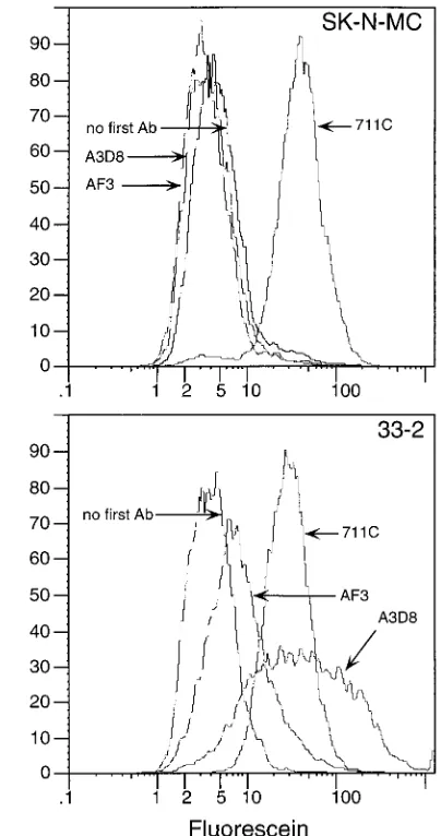
scripts (27). FACS analysis with anti-CD44 MAbs AF3 and A3D8 confirmed the absence of cell surface CD44 in these cells (Fig. 2). SK-N-MC cell lines expressing CD44 were se-lected by FACS after transformation with the cDNA of CD44. These cells were positive for CD44 expression when analyzed by FACS with both A3D8 and AF3 (Fig. 2). Two cell lines generated in this manner, 13-7 and 33-2, were used for subse-quent analyses.
To determine whether CD44H is required for poliovirus replication, CD44-positive and -negative SK-N-MC cells were infected with poliovirus and virus yields were determined at different times postinfection. The replication of each of the three serotypes of poliovirus in the presence of CD44H was indistinguishable from that in the absence of CD44H, both at high and low MOI (Fig. 3).
Poliovirus growth in N2A cells expressing Pvr.Because
ex-pression of PVR in mouse cell lines or in transgenic mice leads to susceptibility to poliovirus infection (15, 17, 21), it was of interest to determine whether the mouse homolog of CD44, known as Pgp-1, is required for poliovirus infection. The
mouse neuroblastoma cell line N2A does not express Pgp-1
(Fig. 4) or Pvr (2). To determine whether N2A cells can
sup-port poliovirus replication, N2A cell lines expressing Pvr were
selected by FACS using the anti-Pvr MAb 711C after transfor-mation with the cDNA of Pvr. Growth curve analyses demon-strated that none of the three serotypes of poliovirus can
rep-licate in N2A cells lacking Pvr, while all three serotypes were
able to replicate in N2A cells which express Pvr (Fig. 5),
dem-onstrating that Pgp-1 is not required for poliovirus replication
in N2A cells.
Determination ofKdof poliovirus for Pvr in SK-N-MC cells
lacking and expressing CD44.The binding affinity constant of
poliovirus for Pvr might be affected by CD44 without changing
viral growth kinetics. Therefore, we determined theKds of all
three poliovirus serotypes for Pvr expressed on SK-N-MC
which lack or express CD44. TheKdof P1/Mahoney was
iden-tical in cells lacking or expressing CD44 (Table 1) and was
similar to the Kd previously published for P1/Mahoney on
HeLa cells (4). TheKds for P2/Lansing and P3/Leon were also
identical in SK-N-MC cells which express or lack CD44 (Table 1). These findings demonstrate that CD44H does not influence the affinity of the virus-receptor interaction.
DISCUSSION
The identification of a MAb, AF3, which recognizes a single isoform of CD44H and is able to block infection of HeLa cells by poliovirus, suggested that CD44H might be associated with Pvr in the cell membrane and could be required for poliovirus-Pvr interactions (25, 26). To determine whether CD44H is required for poliovirus infection, we analyzed viral growth in
two neuroblastoma cell lines, SK-N-MC and N2A, which do
not express any detectable isoforms of CD44 or Pgp-1, respec-tively. The absence or presence of CD44H or Pgp-1 had no effect on the replication of any of the three serotypes of po-liovirus nor on the binding affinity constant of popo-liovirus for its receptor. In addition, the absence or presence of CD44 also had no effect on the replication of the three Sabin poliovirus vaccine strains (2). These findings demonstrate that CD44 is not required for replication of poliovirus in cell culture.
The results of blocking assays carried out at 378C
demon-strate that MAb AF3 reduces the amount of poliovirus P2/ Lansing associated with HeLa cells. Although these results were previously interpreted to mean that AF3 specifically blocks the binding of serotype 2 poliovirus to HeLa cells, it was also suggested that one effect of AF3 might be to prevent interactions between CD44 and Pvr required for poliovirus cell entry (25). The ability of poliovirus to enter cells could be blocked by AF3, and virus particles remaining on the cell
surface would convert to altered particles at 378C and slough
off the cell. Alternatively, the interaction of AF3 with CD44 could increase the ability of Pvr to convert bound virus to altered particles. The result of either AF3-induced effect would be a reduction in the amount of cell-associated virus. By
per-forming binding assays at 48C, a temperature at which
alter-ation, virus entry, and uncoating do not occur, we showed that AF3 blocks binding of a serotype 2 poliovirus, P2/Lansing, to
HeLa cells. Interestingly, at 378C, AF3 decreased the amount
of cell-associated virus to a greater extent than at 48C (Fig. 1),
suggesting that this antibody may also affect postbinding events.
No anti-CD44 antibody other than AF3 was able to block
binding of poliovirus to cells, at 4 or 378C, in contrast to
previously published findings that two other anti-CD44 MAbs,
A3D8 and IM7, blocked poliovirus binding at 378C (25). It is
[image:3.612.84.286.70.453.2]likely that the presence of sodium azide in the preparations of A3D8 and IM7 used in the previous study accounted for their
FIG. 2. Flow cytometric analysis of CD44 and Pvr expression on SK-N-MC cells (top panel) and SK-N-MC transformants expressing CD44 (bottom panel) without first antibody (Ab), with anti-CD44 MAbs A3D8 and AF3, and with anti-Pvr MAb 711C.
on November 9, 2019 by guest
http://jvi.asm.org/
ability to decrease the amount of cell-associated poliovirus.
When the blocking assay was performed at 378C, azide alone
decreased the amount of cell-associated poliovirus, while re-moving azide from A3D8 relieved its inhibitory properties (Fig. 1). IM7, a MAb to Pgp-1, the mouse homolog of human CD44, which cross-reacts with human CD44 (13, 19), also was
unable to block binding of P2/Lansing at 4 or 378C in the
absence of azide. We also tested the ability of A3D8 and IM7 antibodies to inhibit poliovirus plaque formation on HeLa cells. As expected from the results of binding assays performed
at 378C, neither antibody was able to inhibit plaque formation
of P2/Lansing on HeLa cells, whereas under identical condi-tions, the anti-Pvr MAb, 711C, completely blocked poliovirus plaque formation (2). In contrast to previously published re-sults, AF3 did not inhibit poliovirus plaque formation on HeLa cells (2). We are unsure why our results differ from earlier studies (26); however, the following observations, taken to-gether, are consistent with the ability of AF3 to block virus binding but not plaque formation: (i) CD44 is not required for
poliovirus infection, (ii) the number of CD44HAF3molecules
on the surface of HeLa cells is 24 times less than that of Pvr
molecules (24), and (iii) L cells with significantly lower levels of Pvr expression than HeLa cells show normal growth kinetics for poliovirus (18). Because AF3 is unable to completely block P2/Lansing binding to HeLa cells, under the conditions of a plaque assay, poliovirus would still interact with available Pvr, resulting in a productive infection.
[image:4.612.110.506.68.460.2]Based on the findings reported here, the simplest model for the effect of AF3 on poliovirus binding is that Pvr and CD44 are associated either directly or through accessory proteins and that AF3 reacts with CD44 near the virus-receptor interaction site, sterically hindering virus binding. The serotype-specific inhibition of binding by AF3 on HeLa cells is consistent with suggestions that the three serotypes of poliovirus interact with Pvr at slightly different contact points (3, 9). Although we have not been able to demonstrate coimmunoprecipitation of CD44H and Pvr from HeLa cells, using mild detergent and low salt concentrations (2), the two proteins might interact weakly, and chemical cross-linking may be needed to detect their as-sociation. The interaction between Pvr and CD44 might differ among cell lines, as suggested by our finding that AF3 did not block P2/Lansing binding to SK-N-MC cells expressing CD44
FIG. 3. One-step growth curve analysis of poliovirus P1/Mahoney, P2/Lansing, and P3/Leon at 378C in SK-N-MC cells, which lack CD44, and cell lines 13-7 and 33-2, which are SK-N-MC cells that express CD44. Total virus production at different times postinfection was determined by plaque assay on HeLa cells. Results from infections done at high MOI (10 PFU/cell) (A) and low MOI (0.01 PFU/cell) (B) are shown.
on November 9, 2019 by guest
http://jvi.asm.org/
(2). A similar effect was seen with anti-CD44 MAbs that block infection of normal mononuclear phagocytes by monocyto-tropic strains of human immunodeficiency virus type 1 (6, 23). While transformation of CD44-negative Jurkat cells with CD44 rendered these cells susceptible to infection with the monocytotropic human immunodeficiency virus type 1 strains, anti-CD44 MAbs could not block infection in these trans-formed cells (6).
The ability of the anti-CD44 MAb A3D8 and the anti Pgp-1 MAb IM7 to enhance binding of P2/Lansing to HeLa cells was unexpected. An interesting possibility is that CD44, due to its proximity to Pvr, actually decreases the ability of the virus to interact with Pvr, and the MAbs which enhance binding alter this inhibition. Such a model requires that not all Pvr be asso-ciated with CD44, as it is clear that P2/Lansing can bind to HeLa cells in the absence of anti-CD44 MAbs; MAbs A3D8 and IM7 could increase the number of receptors available for virus binding.
Although the cell receptor for poliovirus, Pvr, is clearly a host range determinant for poliovirus infection, it is not the sole determinant of tissue-specific replication of the virus. Analyses of PVR RNA and protein expression in Pvr trans-genic mice and in humans demonstrated that Pvr is expressed in tissues that are susceptible and nonsusceptible to poliovirus
infection (8, 22). The tissue distribution of CD44HAF3,
how-ever, is restricted and to some extent correlates with tissue
susceptibility to poliovirus infection (26). CD44HAF3 is
ex-pressed in parts of the central nervous system such as the periventricular motor nuclei, known to be sites of poliovirus replication, while it is absent from the inferior olivary complex, a region reportedly not affected by poliovirus infection of the brainstem (1). Although the analysis of tissue distribution of
CD44HAF3was limited and did not include human intestine, a
principal site of poliovirus replication, it was suggested that
CD44HAF3may be a determinant of tissue-specific poliovirus
replication (26). The results reported in our study demonstrate that neither CD44 nor Pgp-1 is required for poliovirus high-affinity binding to Pvr or for growth in cell culture and that they therefore do not play a role in the function of the cellular receptor site for poliovirus, as previously suggested (25, 26). Furthermore, it is unlikely that CD44 in humans and Pgp-1 in transgenic mice are involved in poliovirus pathogenesis.
ACKNOWLEDGMENTS
We thank Alan Stall for assistance with flow cytometry and Saul Silverstein and Hamish Young for discussions and contributions to this work.
[image:5.612.78.283.69.455.2]This work was supported by a grant from the American Cancer Society.
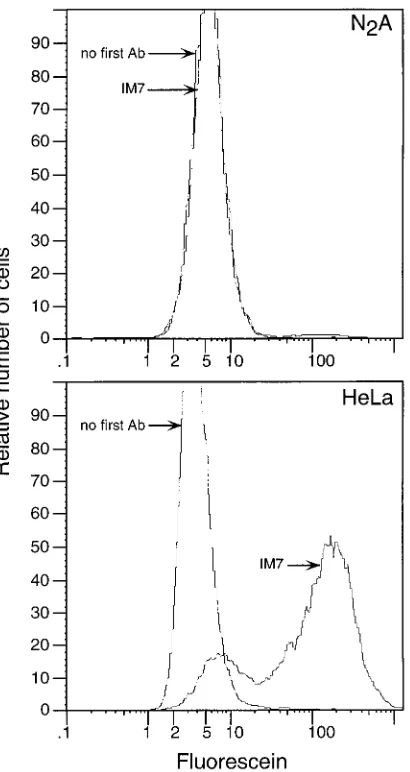
FIG. 4. Flow cytometric analysis of Pgp-1 expression on N2A cells and HeLa cells without first antibody (Ab) and with anti-Pgp-1 MAb IM7. HeLa cells served as a positive control for IM7 reactivity.
[image:5.612.320.552.72.241.2]FIG. 5. One-step growth curve analyses of poliovirus P1/Mahoney, P2/Lan-sing, and P3/Leon at 378C in Pgp-1-negative N2A cells which express Pvr (Pvr1) or lack Pvr (Pvr2). Total virus production at different times postinfection was determined by plaque assay on HeLa cells. Infections were done at high MOI (10 PFU/cell).
TABLE 1. Binding affinity constants of poliovirus for Pvr on CD44-expressing and -nonCD44-expressing SK-N-MC cells
Virus CD44a K
d(pM)
P1/Mahoney 2 44
P1/Mahoney 1 42
P2/Lansing 2 53
P2/Lansing 1 54
P3/Leon 2 61
P3/Leon 1 100
a1,K
ddetermined with SK-N-MC cells expressing CD44;2,Kddetermined
with SK-N-MC cells lacking CD44.
on November 9, 2019 by guest
http://jvi.asm.org/
REFERENCES
1.Bodian, D.1949. Histopathological basis of clinical findings in poliomyelitis. Am. J. Med.6:563–578.
2.Bouchard, M. J., and V. R. Racaniello.Unpublished results.
3.Colston, E., and V. R. Racaniello.1995. Poliovirus variants selected on mutant receptor-expressing cells identify capsid residues that expand recep-tor recognition. J. Virol.69:4823–4829.
4.Colston, E., and V. R. Racaniello.1994. Soluble receptor-resistant poliovirus mutants identify surface and internal capsid residues that control interaction with the cell receptor. EMBO J.13:5855–5862.
5.Dove, A., and V. Racaniello.Unpublished results.
6.Dukes, C. S., Y. Yu, E. D. Rivadeneira, D. L. Sauls, H.-X. Liao, B. F. Haynes, and J. B. Weinberg.1995. Cellular CD44S as a determinant of human immunodeficiency virus type 1 infection and cellular tropism. J. Virol.69:
4000–4005.
7.Fenwick, M. L., and P. D. Cooper.1962. Early interactions between polio-virus and ERK cells. Some observations on the nature and significance of the rejected particles. Virology18:212–223.
8.Freistadt, M. F., G. Kaplan, and V. R. Racaniello.1990. Heterogeneous expression of poliovirus receptor-related proteins in human cells and tissues. Mol. Cell. Biol.10:5700–5706.
9.Harber, J., G. Bernhardt, H. H. Lu, J. Sgro, and E. Wimmer.1995. Canyon rim residues, including antigenic determinants, modulate serotype-specific binding of polioviruses to mutants of the poliovirus receptor. Virology214:
559–570.
10. Haynes, B. F., H.-X. Liao, and K. L. Patton.1991. The transmembrane hyaluronate receptor (CD44): multiple functions, multiple forms. Cancer Cells3:347–350.
11. Herrlick, P., M. Zoller, S. Pals, and P. Helmut.1993. CD44 splice variants: metastases meet lymphocytes. Immunol. Today14:395–399.
12. Hughes, E. M., and J. T. August.1981. Characterization of plasma mem-brane proteins identified by monoclonal antibodies. J. Biol. Chem.256:664– 671.
13. Isacke, C. M., C. A. Sauvage, R. Hyman, J. Lesley, R. Schulte, and I. S. Trowbridge.1986. Identification and characterization of the human Pgp-1 glycoprotein. Immunogenetics23:326–332.
14. Joklik, W. K., and J. E. Darnell.1961. The absorption and early fate of purified poliovirus in HeLa cells. Virology13:439–447.
15. Koike, S., C. Taya, T. Kurata, S. Abe, I. Ise, H. Yonekawa, and A. Nomoto.
1991. Transgenic mice susceptible to poliovirus. Proc. Natl. Acad. Sci. USA
88:951–955.
16. Lin, Y., and V. Racaniello.Unpublished results.
17. Mendelsohn, C., E. Wimmer, and V. R. Racaniello.1989. Cellular receptor for poliovirus: molecular cloning, nucleotide sequence and expression of a new member of the immunoglobulin superfamily. Cell56:855–865. 18. Morrison, M. E., Y.-J. He, M. W. Wien, J. M. Hogle, and V. R. Racaniello.
1994. Homolog-scanning mutagenesis reveals poliovirus receptor residues important for virus binding and replication. J. Virol.68:2578–2588. 19. Picker, L. J., J. de los Toyos, M. J. Telen, B. F. Haynes, and E. C. Butcher.
1989. Monoclonal antibodies against the CD44 [In(Lu)-related p80], and Pgp-1 antigens in man recognize the Hermes class of lymphocyte homing receptors. J. Immunol.142:2046–2051.
20. Racaniello, V. R.1995. Early events in infection: receptor binding and cell entry, p. 73–94.InH. A. Rotbart (ed.), Human enterovirus infections. ASM Press, Washington, D.C.
21. Ren, R., F. C. Costantini, E. J. Gorgacz, J. J. Lee, and V. R. Racaniello.1990. Transgenic mice expressing a human poliovirus receptor: a new model for poliomyelitis. Cell63:353–362.
22. Ren, R., and V. Racaniello.1992. Human poliovirus receptor gene expression and poliovirus tissue tropism in transgenic mice. J. Virol.66:296–304. 23. Rivadeneira, E. D., D. L. Sauls, Y. Yu, B. F. Haynes, and J. B. Weinberg.
1995. Inhibition of HIV-1 infection of mononuclear phagocytes by anti-CD44 antibodies. AIDS Res. Hum. Retroviruses11:541–546.
24. Shepley, M. P.1988. Monoclonal antibody identification of a 100-kDa mem-brane protein in HeLa cells and human spinal cord involved in poliovirus attachment. Ph.D. thesis. Harvard Medical School, Boston, Mass. 25. Shepley, M. P., and V. R. Racaniello.1994. A monoclonal antibody that
blocks poliovirus attachment recognizes the lymphocyte homing receptor CD44. J. Virol.68:1301–1308.
26. Shepley, M. P., B. Sherry, and H. L. Weiner.1988. Monoclonal antibody identification of a 100-kDa membrane protein in HeLa cells and human spinal cord involved in poliovirus attachment. Proc. Natl. Acad. Sci. USA
85:7743–7747.
27. Shtivelman, E., and J. M. Bishop.1991. Expression of CD44 is repressed in neuroblastoma cells. Mol. Cell. Biol.11:5446–5453.
28. Stamenkovic, I., M. Amiot, M. Pesando, and B. Seed.1989. A lymphocyte molecule implicated in lymph node homing is a member of the cartilage link protein family. Cell56:1057–1062.
29. Stryer, L.1988. Biochemistry, 3rd ed. W. H. Freeman and Company, New York, N.Y.
30. Wimmer, E., C. Hellen, and X. Cao.1993. Genetics of poliovirus. Annu. Rev. Genet.27:353–436.