Copyright © 1999, American Society for Microbiology. All Rights Reserved.
Herpes Simplex Virus Type 2 Glycoprotein G-Negative Clinical
Isolates Are Generated by Single Frameshift Mutations
JAN-ÅKE LILJEQVIST,* BO SVENNERHOLM,
ANDTOMAS BERGSTRO
¨ M
Department of Virology, University of Go¨teborg, S-413 46 Go¨teborg, Sweden
Received 20 May 1999/Accepted 20 August 1999
Herpes simplex virus (HSV) codes for several envelope glycoproteins, including glycoprotein G-2 (gG-2) of
HSV type 2 (HSV-2), which are dispensable for replication in cell culture. However, clinical isolates which are
deficient in such proteins occur rarely. We describe here five clinical HSV-2 isolates which were found to be
unreactive to a panel of anti-gG-2 monoclonal antibodies and therefore considered phenotypically gG-2
negative. These isolates were further examined for expression of the secreted amino-terminal and
cell-associated carboxy-terminal portions of gG-2 by immunoblotting and radioimmunoprecipitation. The gG-2
gene was completely inactivated in four isolates, with no expression of the two protein products. For one isolate
a normally produced secreted portion and a truncated carboxy-terminal portion of gG-2 were detected in
virus-infected cell medium. Sequencing of the complete gG-2 gene identified a single insertion or deletion of
guanine or cytosine nucleotides in all five strains, resulting in a premature termination codon. The frameshift
mutations were localized within runs of five or more guanine or cytosine nucleotides and were dispersed
throughout the gene. For the isolate for which a partially inactivated gG-2 gene was detected, the frameshift
mutation was localized upstream of but adjacent to the nucleotides coding for the transmembranous region.
Thus, this study demonstrates the existence of clinical HSV-2 isolates which do not express an envelope
glycoprotein and identifies the underlying molecular mechanism to be a single frameshift mutation.
Glycoprotein G-2 (gG-2) of herpes simplex virus type 2
(HSV-2) is a viral envelope protein with a feature unique
among HSV proteins in the form of cleavage of the gG-2
precursor during processing to a secreted amino-terminal
por-tion (50, 51) and to a cell- and virion-associated, heavily
O
-glycosylated carboxy-terminal portion that constitutes the
ma-ture gG-2 (3, 11, 32, 38, 43, 51). Mama-ture gG-2 has been
identified as a major target for the human antibody response
(1) and, in contrast to other HSV membrane proteins, this
protein has been shown to be an ideal antigen for
dis-criminating serodiagnosis (2, 19, 20, 25, 52) since only
type-specific epitopes have been described (28). Conservation of the
gene coding for the mature portion of gG-2 among clinical
HSV-2 isolates is a factor of essential importance for the
reliability of serological determination of gG-2-specific
anti-bodies from patient sera and also for the correct typing of
HSV-2 isolates by anti-gG-2 monoclonal antibodies (MAbs).
However, eventual differences in the phenotypic expression of
this portion of gG-2 have seldom been described among
clin-ical isolates.
In search of HSV variants, we studied a large number of
clinical isolates by investigating antibody reactivity for the
pur-pose of determining the difference in frequency of expression
of a type-specific glycoprotein C-1 (gC-1) and of a gG-2 MAb
epitope (28, 39). Altogether, 13 MAb escape mutants were
found among the 2,400 HSV-2 isolates tested, whereas none
were detected in an equal number of HSV-1 isolates (29). This
indicated that the variability of the gG-2 epitope was
signifi-cantly higher than that of the gC-1 epitope. Of these 13 HSV-2
isolates, 5 were in addition unreactive with two other
type-specific anti-gG-2 MAbs, which had been previously mapped
to different epitopes (28), and were therefore defined as
phe-notypically gG-2 negative. The function of the two gG-2
pro-tein products is not known. However, as described for other
HSV genes coding for envelope proteins such as glycoprotein
C (gC) (21, 58), gE (30), or gI (23), the gG-2 gene is
dispens-able for virus propagation in cell cultures; i.e., vidispens-able
gG-2-deficient mutant viruses have been constructed in vitro (16). In
contrast, isolation of clinical HSV mutants lacking a
dispens-able gene product occurs rarely (13, 18, 40, 41), indicating that
the viral proteins contribute significant functions in the natural
infection of the host. We identified here five
nonimmunocom-promised patients with recurrent HSV-2 infections caused by
isolates which harbored a completely or partially inactivated
gG-2 gene. Sequencing of the gG-2 gene identified single
frameshift mutations as the molecular mechanism underlying
the lack of expression of the two gG-2 protein products in four
of the isolates. The same mechanism also accounted for a
normally produced secreted portion and a truncated mature
gG-2 in the fifth isolate.
MATERIALS AND METHODS
Cells and virus strains.African green monkey kidney (GMK-AH1) and hu-man epidermoid carcinoma-2 (HEp-2; ATCC CCL 23) cells were cultured in Eagle minimal essential medium supplemented with 2% calf serum and antibi-otics. Rabbit kidney cells (RK13; ATCC CCL 37) were cultured in Eagle minimal essential medium supplemented with 10% fetal calf serum and antibiotics. A local wild-type HSV-2 strain, B4327UR (S. Jeansson, Go¨teborg, Sweden), was used as a control virus. The original specimens of vesicle fluid from five HSV-2-positive patients were passaged once in GMK-AH1 cells for production of viral
stocks and kept at⫺70°C. All experiments, including the gene sequencing, were
performed with these viral stocks.
MAbs.Three type-specific anti-gG-2 MAbs reactive to the carboxy-terminal portion of gG-2, used as reagents in this study, were produced according to standard hybridoma techniques. The MAb epitopes have previously been
localized to the following amino acids:552PPPPEHR558for MAb O1.B9.E5,
557HRGGPEE563for MAb O1.C5.B2, and 579ATGLAFRTP587 for MAb
O3.G11.H7 (28).
EIA on HSV-2-infected cells.In a recent study (29), 13 of 2,400 clinical HSV-2 strains isolated from patients with clinical lesions were unreactive with the anti-gG-2 MAb O1.C5.B2 when infected GMK-AH1 cells were tested by an enzyme immunoassay (EIA). In the current study these mutant strains were tested for reactivity by the same method by using the anti-gG-2 MAbs O1.B9.E5
* Corresponding author. Mailing address: Department of Virology,
University of Go¨teborg, Guldhedsgatan 10 B, S-413 46 Go¨teborg,
Sweden. Phone: 46-31-3424657. Fax: 46-31-3424960. E-mail: jan-ake
9796
on November 9, 2019 by guest
http://jvi.asm.org/
and O3.G11.H7. Briefly, confluent monolayers of GMK-AH1 cells were infected with the respective isolate and when complete cytopathic effect was seen the cells were fixed in 0.25% glutaraldehyde for 30 min, the MAbs were added separately, and the culture was incubated for 1 h at room temperature. Alkaline
phos-phatase-conjugated F(ab⬘)2 goat anti-mouse immunoglobulin G (IgG) at a
1:2,000 dilution (Jackson ImmunoResearch Laboratories, Inc.) was used as
con-jugate, withp-nitrophenyl dissolved in carbonate buffer (pH 9.8) as a substrate.
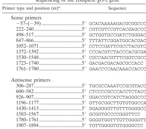
DNA sequencing of the gG-2 gene.Viral DNA was prepared from stock viruses by using the QIAmp Blood Kit (Qiagen) method prior to PCR amplification of
the complete gG-2 gene. Since the gene has an overall G⫹C content of 71.3%
(33), several sets of primers were tested to optimize the amplification and sequencing signals. Nine overlapping oligonucleotide pairs were used as primers (Table 1), and amplified products were separated on a 1% agarose gel prior to extraction of the amplicon bands with the QIAquick Gel Extraction Kit (Qia-gen). PCR cycle sequencing was carried out by using fluorescent labeled stop nucleotides with the dRhodamine Terminator Cycle Sequencing Ready Reaction Kit (Perkin-Elmer Applied Biosystems), and unidirectional extension was per-formed with sense or antisense primers in separate reaction mixtures. After precipitation with ethanol, the labeled samples were analyzed on an automated sequencer (ABI Prism 310 Genetic Analyzer; Perkin-Elmer). The sequences
were compared with theHindIIIlfragment containing the gG-2 gene (US4) for
the HSV-2 reference strain HG52 (33).
Production of hyperimmune sera.Rabbit hyperimmune serum was produced by using a synthetic peptide (247RFRERCLPPQTPAA260) representing part of the secreted portion of gG-2 (51). The peptide was synthesized by using Fmoc (9-fluorenylmethoxy carbonyl) chemistry, purified by high-pressure liquid chro-matography (99% purity), and covalently coupled to bovine serum albumin fraction V (Sigma Chemical Co.) at an approximately 20:1 (peptide-bovine
serum albumin) molar ratio by usingN-succinimidyl
3-(2-pyridyldithio)propi-onate (Pharmacia Biotech) according to conditions given by the manufacturer.
The rabbit was immunized with 200g of the peptide emulsified in 0.5 ml of
Freund complete adjuvant for priming (first injection) and incomplete adjuvant for booster doses (second and third injections) at 3-week intervals. Rabbit hy-perimmune serum directed to the mature gG-2 was prepared by immunization of
a rabbit, as described earlier, with 500g ofHelix pomatialectin-purified gG-2
antigen (37) produced from RK13 cells infected with the B4327UR strain.
Detection of the carboxy-terminal portion of gG-2 by immunoblotting.Cell lysate antigens from strain B4327UR and respective clinical mutant strains were prepared by infecting HEp-2 cells. When complete cytopathic effect was seen, the cells were harvested and lysed in Tris-buffered saline and 1% Nonidet P-40, followed by ultrasonication. The samples were mixed with sample buffer con-taining 2% sodium dodecyl sulfate (SDS) and 5% mercaptoethanol and then subjected to polyacrylamide gel electrophoresis (PAGE) by using a 10% Tris-glycine gel (Novex) and Tris-Tris-glycine-SDS as the running buffer. The proteins were electrotransferred to an Immobilon-P transfer membrane (Millipore Corp.). The gG-2-reactive MAb O1.C5.B2 and a type-common anti-gD MAb
C4.D5 (6), at a final concentration of 16g/ml, were incubated overnight with
strips containing the blotted HSV-2 antigen. Peroxidase-labeled rabbit anti-mouse IgG (Dako) at a 1:100 dilution was used as conjugate with 4-chloro-1-naphthol as the substrate.
Detection of the secreted portion of gG-2 by immunoblotting.GMK-AH1 cells were infected with strain B4327UR and the respective clinical mutant strains. When complete cytopathic effect was seen, the media were harvested and
cen-trifuged at 2,000⫻gfor 10 min before ultracentrifugation at 100,000⫻gfor
1.5 h, followed by centrifugation until dry at 5,000 ⫻g, by using Microsep
microconcentrator tubes with a 10-kDa cutoff (Filtron Skandinavia AB). Proteins
were resuspended in 200l of phosphate-buffered saline, mixed with sample
buffer as described above, separated on a 4 to 12% NuPAGE Bis-Tris gradient
gel (Novex) with 2-(N-morpholino)ethanesulfonic acid–SDS as the running
buffer, followed by immunoblotting to an Immobilon-P transfer membrane. Rabbit hyperimmune serum was added at a 1:20 dilution, and peroxidase-labeled goat anti-rabbit IgG (Dako) at a 1:100 dilution was used as conjugate with 4-chloro-1-naphthol as the substrate.
Amino acid sequencing of the secreted portion of gG-2.The proposed secreted portion of gG-2 detected by immunoblotting was localized from the same Im-mobilon-P transfer membrane stained with Coomassie blue solution. This band was used for amino acid sequencing with an automatic sequencer (Applied Biosystems model 470A). Ten cycles were acquired in which the first eight amino acids were unambiguously determined.
Radioimmunoprecipitation.Confluent GMK-AH1 cells in 50-mm petri dish cultures were infected with the gG-2-negative mutant strains and labeled with 40
l ofD-[6-3H]glucosamine hydrochloride (28 Ci/mmol) (Amersham Life
Sci-ence) at 4 h postinfection. When complete cytopathic effect was seen, the media were harvested as described above except for the concentration step. The rabbit hyperimmune serum was mixed at a 1:100 dilution with the medium, and the
antigen-antibody complexes were precipitated withStaphylococcus aureus(strain
Cowan 1) solution as described previously (38). After SDS-PAGE with a 10% Tris-glycine gel as described above, the gel was soaked in amplifier (Amersham Life Science) for 15 min before it was dried overnight, and subsequent autora-diography was performed with Kodak XRP-1-Omat film.
Type-specific serology. An indirect enzyme-linked immunosorbent assay (ELISA) designed to detect type-specific antibodies against mature gG-2 and gG-1 was performed with sera from patients from whom the gG-2-negative
HSV-2 strains had been isolated.H. pomatialectin-purified gG-2 (100g/ml),
coated at a 1:6,000 dilution in carbonate buffer (pH 9.6) on Maxisorp microtiter plates (Nalge Nunc International), was used as the antigen for the assessment of anti-gG-2 antibodies, with peroxidase-conjugated goat anti-human IgG (Jackson
ImmunoResearch) as the conjugate, at a 1:3,000 dilution, andO
-phenylenedi-amine as the substrate as described previously (28). Similarly, a truncated
re-combinant-produced gG-1, at a concentration of 180g/ml (kindly provided by
SmithKline Beecham Biologicals), was coated in phosphate-buffered saline at a 1:400 dilution. Alkaline phosphatase-conjugated goat anti-human IgG (Jackson ImmunoResearch) was used as conjugate at a 1:3,500 dilution with Sigma 104 phosphatase substrate tablets as the substrate. Sera were obtained from patient
2434 3 months after and from the other patientsⱖ3 years after the gG-2-negative
HSV-2 isolates were collected. Endpoint titers were expressed as the reciprocal of the dilution giving an absorbance value greater than the cutoff. The cutoffs were defined as the mean absorbance values of HSV-1- and HSV-2-negative sera, respectively, plus 0.2 optical density (OD) units.
Nucleotide and protein sequence accession numbers.The gG-2 gene se-quences have been assigned GenBank accession no. AF141854, AF141855, AF141858, AF141856, and AF141857 for strains 2434, 512, 453, VI-147, and VI-4444, respectively. The protein sequence data reported here will appear in the SWISS-PROT Protein Data Bank under accession no. P81780.
RESULTS
gG-negative HSV-2 isolates.
Thirteen clinical HSV-2
iso-lates, which were earlier shown to be unreactive with the
anti-gG-2 MAb O1.C5.B2 in EIA (29), were tested for reactivity
with the two additional anti-gG-MAbs, O1.B9.E5 and
O3.G11.H7. Eight isolates were clearly reactive with these
antibodies and were shown to harbor point mutations within
the anti-gG-2 MAb O1.C5.B2 epitope (unpublished data).
These isolates were therefore excluded from further
charac-terization in the present study. Five isolates showed low
reac-tivity with all three MAbs tested (Table 2) and were considered
gG-2 negative. These strains had been isolated from vesicular
lesions from five different patients with variable duration of the
clinical HSV-2 infection, as well as variable frequency of
re-currences (Table 3). None of the patients were
immunocom-promised, nor were they on any medication.
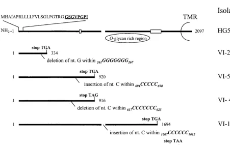
[image:2.612.52.295.88.302.2]Frameshift mutations within the gG-2 gene.
Cyclic gene
sequencing of the complete gG-2 gene was performed by using
TABLE 1. The primer sequences used for amplification and
sequencing of the complete gG-2 gene
Primer type and position (nt)a Sequence
Sense primers
⫺57–(⫺39) ...5⬘
GCACAAAAAGACGCGGCCC
222–240...5⬘
CGTCGTCCGTCACGAGCCC
498–517...5⬘
GCTGGTGCCGATCTGGGACC
847–866...5⬘
TTTATTCGGATGGCACGACC
1052–1071...5⬘
CCTCCGATTCGCCTACGTCC
1372–1392...5⬘
CCCACGTCTACCCACGCGACC
1530–1548...5⬘
CGCCAACGTTTCGGTCGCC
1723–1740...5⬘
GACGACGACAGCGCCACC
1761–1780...5⬘
GAACCCCAACAAACCACCCC
Antisense primers
306–287...5⬘
TGCGCCAAATCCGCGTACC
600–582...5⬘
CTCCCCGCCCACCTCTACC
926–907...5⬘
GGACCGTCATCTAGGGCCCC
1196–1177...5⬘
GTTGCGGCTTGTGTGGCCAT
1430–1413...5⬘
GGAGGGGTTGTTTGGGGCC
1583–1567...5⬘
GCGGTGCCCCGGGTTCC
1780–1761...5⬘
GGGGTGGTTTGTTGGGGTTC
1907–1894...5⬘
TGTTGGGGTGTGGGGCCC
2140–2119...5⬘
TCCCGTCCTTCATCGTTTCTC
aThe nt 2842 to 4938 within the HSV-2HindIIIlfragment, encompassing the
gG-2 gene (US4), for the reference HSV-2 strain HG52 (33) are numbered here as 1 to 2097.
on November 9, 2019 by guest
http://jvi.asm.org/
overlapping sense and antisense primers. Except for a region
encompassing nucleotides (nt) 518 to 614 and 604 to 906,
where only one unidirectional sequence was successfully
achieved, identical sequences were obtained for both the sense
and the antisense primers. nt 590 to 630, in which the
overlap-ping sequencing signals faded, showed a G
⫹
C content of 88%,
which may have contributed to the difficulties resulting in
ter-mination. This region was therefore sequenced in several runs
from at least three different preparations for each isolate, all
showing identical sequences. The isolates harbored a single
frameshift mutation with an insertion or deletion of the
cyto-sine or guanine nucleotides compared to the previously
pub-lished gG-2 gene sequence for reference HSV-2 strain HG52
(33). The mutations of the different strains were localized
within runs of
ⱖ
5 guanine (one isolate) or cytosine (four
iso-lates) nucleotides (Fig. 1). Since unequivocal determination of
the precise site of the mutated base was precluded, only the
stretch of reiterated nucleotides could be localized. The
mu-tations of the different strains were found at different localities
throughout the gene, and the predicted lengths of the
respec-tive truncated transcripts were therefore variable (Fig. 1).
In addition, the isolates showed the following genetic
differ-ences compared with strain HG52 (33): strain VI-2434
har-bored seven single nucleotide substitutions, as well as a
dele-tion of nucleotides
877GTC
879and
1282GCG
1284. Strain VI-512
showed eight single-nucleotide substitutions and a deletion of
nt
1282GCG
1284. Strain VI-453 displayed seven
[image:3.612.52.294.101.200.2]single-nucle-FIG. 1. Schematic representation of the gG-2 gene for HSV-2 reference strain HG52, coding for the secreted gG-2; the proposed cleavage sites (broken line); and the carboxy-terminal high-mannose intermediate which is highlyO-glycosylated, generating the mature gG-2 and the transmembranous region (TMR). nt 2842 to 4938 within the HSV-2HindIIIlfragment of strain HG52 coding for gG-2 (33) are numbered 1 to 2097. Localization of frameshift mutations (deletion or insertion) within runs of reiterated nucleotides and the resulting premature termination codons are depicted for five clinical gG-2-negative HSV-2 isolates. Boxed areas show localization of binding of reagents utilized for detection of respective gG-2 protein products. Deduced signal sequence and sequenced amino-terminal amino acids (boldface and underlined) are aligned for the secreted gG-2.
TABLE 2. Enzyme immunoassay reactivity of three anti-gG-2
MAbs to GMK-AH1 cells infected with five gG-2-negative clinical
HSV-2 isolates
Isolate EIA reactivity (OD values) of MAb a:
O1.B9.E5 O1.C5.B2 O3.G11.H7
B4327UR
b2.58
2.55
2.36
GMK-AH1 cells
c0.14
0.13
0.20
VI-2434
0.18
0.20
0.16
VI-512
0.31
0.31
0.21
VI-453
0.18
0.15
0.16
VI-147
0.28
0.22
0.27
VI-4444
0.16
0.18
0.23
aOD values were calculated as the mean values from duplicate well testing. bA local clinical HSV-2 strain was used as positive control virus.
[image:3.612.78.529.388.673.2]cReactivity with uninfected cells.
TABLE 3. Clinical characterization of five patients harboring
gG-2-negative HSV-2 isolates
Patient no., gender
(age [yrs]) Duration of recurrentHSV-2 infection Site of lesions Frequency ofrecurrences
2434, F (34)
4 mos
Vulva
3/4 mos
512, M (39)
8 yrs
Penis
4/yr
453, M (26)
4 yrs
Penis
1/yr
147, F (35)
4 yrs
Vulva
2/yr
4444, F (51)
3 yrs
Buttock
1/yr
on November 9, 2019 by guest
http://jvi.asm.org/
otide substitutions and deletion of nt
877GTC
879and
1282GCG
1284. Strain VI-147 harbored 11 single-nucleotide
sub-stitutions as well as deletions of nt
1282GCG
1284and
1360AC-GACCCCC
1368, and, finally, strain VI-4444 contained five
sin-gle-nucleotide substitutions and a deletion of nt
1282GCG
1284.
Taken together, the only mutations detected within the gG-2
gene which could explain the lack of reactivity with the three
anti-gG-2 MAbs used in EIA were the single
⫺
1 or
⫹
1
frame-shift mutations.
Expression of the gG-2 protein products by clinical HSV-2
isolates.
The gG-2 precursor protein was reported to be
co-translationally glycosylated, generating a high-mannose
inter-mediate (3, 50, 55) which was cleaved during processing to a
secreted and a terminal portion (50, 51). The
carboxy-terminal high-mannose intermediate was shown to be further
processed by
O
-glycosylation to give the mature gG-2 (3, 11,
32, 38, 43, 51). Accumulation of the carboxy-terminal
high-mannose intermediate was observed in HEp-2 cells (3) and was
therefore detectable by immunoblotting (28). Cell lysates of
HEp-2 cells infected with strain B4327UR and the
gG-2-neg-ative HSV-2 isolates were subjected to SDS-PAGE for
detec-tion by immunoblotting. For strain B4327UR, both the
car-boxy-terminal high-mannose intermediate with an apparent
molecular mass of 77 kDa and the fully glycosylated mature
gG-2 with a molecular mass of approximately 120 kDa were
identified with the anti-gG-2 MAb O1.C5.B2 as described
pre-viously (28) (Fig. 2A). These two gG-2 portions were also
recognized from isolate VI-4444, of which the mature gG-2
was most prominent, showing a slight reduction of the
appar-ent molecular mass (115 kDa). No reactivity with the anti-gG-2
MAb was identified for any of the other four mutant strains.
The anti-gG-2 and anti-gD MAbs were unreactive to
mock-infected cell antigen (data not shown).
To investigate the production of the normally secreted
por-tion of gG-2, virus-infected cell medium was concentrated and
subjected to SDS-PAGE. Rabbit hyperimmune serum
recog-nized a band on immunoblotting that had an apparent
molec-ular mass of 40 kDa for the strains B4327UR and VI-4444 (Fig.
2B), indicating that that portion of gG-2 was expressed by the
isolate VI-4444. No secreted portion of gG-2 could be detected
for the four other mutant strains. The rabbit hyperimmune
serum was obtained after immunization with a peptide
con-taining amino acids localized within the carboxy-terminal end
of the secreted gG-2 (Fig. 1). Since the frameshift mutations
detected for strains VI-2434, VI-512, and VI-453 were found to
be localized upstream of the immunoreactive region, we
can-not exclude the possibility that shorter fragments of the
se-creted gG-2 were expressed. It has been proposed that the
posttranslational cleavage of gG-2 precursor protein occurs
between the amino acids arginine 321 and alanine 322 and
between arginine 342 and leucine 343 (10). Since strain VI-147
harbored a frameshift mutation between these cleavages sites,
the nucleotides coding for residues reactive with rabbit
hyper-immune serum were in frame. Despite this, no secreted gG-2
was detected, suggesting that both cleavage sites may be critical
for correct processing and posttranslational cleavage of the
precursor gG-2.
Since the frameshift mutation detected in strain VI-4444 was
localized upstream of but adjacent to the nucleotides coding
for the transmembranous region (Fig. 1), we investigated
whether a truncated form of the mature gG-2 was secreted into
the medium. When the polyclonal rabbit anti-gG-2 serum was
used for radioimmunoprecipitation of the medium of cells
infected with respective mutant isolates, strain VI-4444
ex-pressed a truncated mature gG-2 with an apparent molecular
mass of 116 kDa (Fig. 3). In agreement with the localization of
the frameshift mutations found for the other gG-2-negative
isolates, no truncated and secreted forms of the mature portion
of gG-2 were detected in any of these strains.
Amino acid sequencing of the secreted portion of gG-2.
The
band of the secreted portion of gG-2 identified in the
immu-noblot was clearly recognized by Coomassie blue staining
so-lution for strain B4327UR (Fig. 2B). This band was subjected
to amino acid sequencing, and the N-terminal amino acids
were as follows: GSGVPGPI. The residues were at positions 23
to 30 after the start codon (Fig. 1) and were identical to the
deduced amino acid sequence of the gG-2 gene for strain
HG52 (33). This confirmed the identification of the secreted
portion of gG-2. Consequently, from the evidence presented
here, the first N-terminally localized stretch of 22 residues of
gG-2 appeared to constitute the signal sequence, which is in
agreement with a previous proposal (33).
[image:4.612.315.545.74.386.2]Seroreactivity in ELISA.
Since the mature gG-2 antigen
usually is used for detection of HSV-2 type-specific antibodies
as a marker of infection (2, 19, 20, 25, 52), it was of interest to
assess whether sera from patients carrying the characterized
FIG. 2. Immunoblot analysis of the gG-2 protein products. (A) HEp-2 cells were infected with a local wild-type HSV-2 strain (B4327UR) and five clinical gG-2-negative HSV-2 isolates. At the point of complete cytopathic effect, cell lysates were subjected to SDS-PAGE and electrotransferred to membranes. The carboxy-terminal high-mannose intermediate (77 kDa) and mature (120 kDa) gG-2 for strain B4327UR, detected by using the type-specific anti-gG-2 MAb O1.C5.B2, are marked with arrows. HSV-2 antigen was visualized by using a type-common anti-gD MAb. (B) Detection of the secreted gG-2 in GMK-AH1 virus-infected cell media by using rabbit hyperimmune serum. The upper band was reactive with rabbit preimmune serum and was considered nonspecific. The lane to the right was from the same membrane, stained with Coomassie blue solution. The arrowed band was subjected to amino acid sequencing. The posi-tions of protein standards are indicated at the left.
on November 9, 2019 by guest
http://jvi.asm.org/
viral isolates contained gG-2 antibodies. In addition, the
sero-reactivity to the HSV-1 type-specific gG-1 antigen was
inves-tigated in parallel. The absorbance values were expressed as
endpoint titers (Table 4) in an indirect ELISA, and, as shown,
three of five patients had detectable serum antibodies against
gG-2.
DISCUSSION
Although point mutations of HSV genes coding for
mem-brane glycoproteins have been described earlier in clinical
HSV isolates (44, 48, 54), the complete inactivation of an HSV
gene coding for a virus envelope protein in clinical HSV
strains, with subsequent lack of protein expression, has to our
knowledge hitherto not been reported. We describe here five
gG-2-negative clinical HSV-2 isolates detected among patients
with recurrent HSV-2 infection, of which four were shown to
have an inactivated gG-2 gene with no expression of the gG-2
protein products. These isolates were found in the search for
clinical HSV variants lacking type-specific epitopes of either
gG-2 or gC-1, two glycoproteins reported to be dispensable for
in vitro infection (9, 16, 17, 21). In contrast, no gC-1-negative
strains were recognized among a large number of clinical
HSV-1 isolates investigated in the same study (29). These
results suggest that HSV-2 strains can reactivate in vivo to
induce clinical lesions in immunocompetent patients despite
the lack of functional gG-2 proteins, i.e., the gG-2 gene may be
classified as nonessential also in vivo at least in some hosts.
An interesting question is whether these patients mostly
reactivate wild-type strains expressing the two gG-2 protein
products and whether the gG-2-negative strains described here
thus could be regarded as single events within each host.
Dur-ing our further studies, two additional isolates were retrieved
from patients 512 and 453, after 2 and 4 years, respectively.
Sequencing of these two isolates identified frameshift
muta-tions within the gG-2 gene identical to those described here
(unpublished observation), suggesting that these
gG-2-nega-tive strains could be repeatedly reactivated in vivo. Moreover,
the finding of identical frameshift mutations of these
addi-tional isolates as described for the original isolates strengthens
the evidence that the detected frameshift mutations were not
merely an artifact of cell culture.
The molecular basis for lack of expression of the mature
gG-2 on virus-infected cells from the five clinical HSV-2
iso-lates investigated was found to be due to frameshift mutations.
In addition, the strains harbored single nucleotide
substitu-tions and delesubstitu-tions of the codons
877GTC
879(two isolates) and
1282GCG
1284(five isolates) compared to the HSV-2 reference
strain HG52 (33). These alterations were also found in clinical
gG-2-positive HSV-2 isolates (unpublished observation) and
were therefore considered as genetic variants present in a
Swedish population. The lack of nt
1360ACGACCCCC
1368de-tected for strain VI-147 was the only alteration which was not
found in gG-2-positive isolates. Since the deletion consisted of
9 nt and consequently the reading frame was retained, it seems
unlikely that this deletion could have contributed to the
inac-tivation of the gene.
The mechanism of silenced expression or truncation of the
coded protein due to frameshift mutations has been described
for different microbiological agents such as yeasts (26, 47),
bacteria (22, 35), and the bacteriophage T4 (42, 49), as well as
for various human viruses. Single neutralization-resistant viral
plaques have been selected after serial cell culture passage of
a respiratory syncytial virus isolate; these were shown to harbor
frameshift mutations within the G gene coding for an envelope
glycoprotein (14). Such mutations have also been identified
within the early region in polyomavirus after selection of
re-vertants by the use of hydroxyurea (57). A spontaneously
orig-inated gC-negative HSV-1 mutant (MP strain) from cell
cul-ture (21) was shown to harbor a frameshift mutation within the
gC-1 gene (12). Moreover, frameshift mutations responsible
for the inactivation of the thymidine kinase gene have been
described for clinical HSV-1 and HSV-2 isolates (15, 46) as
well as for varicella-zoster virus isolates (7, 53) from
immuno-compromised patients. One novel observation in this study was
that clinical HSV-2 strains from immunocompetent patients
could harbor frameshift mutations within the gG-2 gene,
cod-ing for an envelope protein, resultcod-ing in complete inactivation
of the gene. This contrasts with previously described and
char-acterized mutants where prior selection had been exerted via
in vitro cell culture conditions or where isolates were obtained
from patients with severe immune system dysfunctions.
[image:5.612.66.283.73.225.2]The detected frameshift mutations were all due to
single-base insertion or deletion of cytosine or guanine nucleotides,
introducing a premature termination codon within the gG-2
gene. In agreement with other studies, spontaneous frameshift
mutations are especially prone to occur at regions of reiterated
bases (4, 14, 15, 36, 46, 49, 57), and these homopolymer
nu-cleotide stretches are usually found to be mutational hot spots.
The mutations described in the present study were all localized
within either oligo(C) or oligo(G) tracts, a result which may be
due to the fact that the gG-2 gene has an overall high G
⫹
C
FIG. 3. Radioimmunoprecipitation of mature gG-2 in GMK-AH1 virus-in-fected cell media for five clinical gG-2-negative HSV-2 isolates. Proteins were
labeled withD-[6-3H]glucosamine hydrochloride and mixed with rabbit anti-gG-2
polyclonal serum, followed by precipitation withStaphylococcus aureussolution.
Antigens were separated by SDS-PAGE with subsequent autoradiography on
Kodak XRP-1-Omat film. [14C]methylated proteins were used as molecular mass
markers.
TABLE 4. Endpoint titers to the type-specific gG-2 and gG-1
antigens in ELISA of sera from patients harboring gG-2-negative
clinical HSV-2 isolates
Patient no. Endpoint titer
ato:
gG-2 antigenb gG-1 antigenc
2434
400
1,600
512
400
100
453
–
800
147
–
100
4444
200
200
a–, endpoint titer of⬍100.
bH. pomatialectin-purified gG-2.
cRecombinant-produced gG-1.
on November 9, 2019 by guest
http://jvi.asm.org/
[image:5.612.54.293.623.703.2]content (33). The described mutations were preferably
de-tected within runs of cytosine nucleotides (four of five
mu-tants), which also may be due to the nucleotide composition of
the gG-2 gene since the gG-2 gene contains a total of 28
reiterations of
ⱖ
5 cytosine nucleotides compared to two
reit-erations of
ⱖ
5 guanine repeats. The high number of such
repeats may also explain why the frameshift mutations in the
different isolates were found to be dispersed throughout the
gene.
The biological significance of the described gG-2-negative
clinical HSV-2 isolates is currently obscure, and further studies
are hampered because of the hitherto unknown function of the
two gG-2 protein products. The clinical characterization of the
hosts with regard to the site of lesions or the frequency of
recurrences did not reveal any obvious discrepancy compared
to the described natural history of HSV-2 infection (5, 24).
Clinical HSV-1 isolates which harbored a partially inactivated
gC-1 gene and expression of a truncated gC-1 protein found in
the virus-infected cell medium have been described for a
pa-tient with a recurrent eye infection (18). Since this phenotype
was suggested to have maintained gC-1-associated functions
both in vitro and in vivo with regard to cell penetration ability,
as well as with regard to the induction of hemagglutination
inhibition antibodies (34), it is possible that the secreted
ma-ture gG-2 expressed for strain VI-4444 also retained functional
activity.
The detection of antibodies against the mature portion of
gG-2 for patient 4444 suggests that the HSV-2 isolate VI-4444,
which produced a secreted mature gG-2, had induced a B-cell
immune response. Sera from patients 147 and 453 lacked
an-tibodies against gG-2. Since this protein is used as antigen in
type-discriminating serodiagnosis, it is therefore notable that a
few HSV-2-infected patients can lack antibodies due to an
inactivation of the gG-2 gene. Interestingly, sera from patients
2434 and 512 contained antibodies against gG-2, indicating
that these patients also harbored gG-2-positive HSV-2 virus
capable of inducing an antibody response to the mature gG-2.
This observation implied that these patients might have carried
multiple HSV-2 strains, as has been described earlier for
pa-tients infected with HSV-1 (27, 56) or with HSV-2 (8, 31, 45).
However, further studies are needed to clarify the mechanisms
behind this observation, and both reinfection with a
gG-2-positive HSV-2 strain and the selection of different viral clones
from a heterogeneous primary HSV-2 population should be
considered as possible explanations.
In conclusion, this study identifies clinical HSV-2 isolates
which lack the expression of a viral envelope glycoprotein due
to a single frameshift mutation. These strains may prove to be
valuable tools for further study of the function of the secreted
as well as of the mature portion of the gG-2 protein.
ACKNOWLEDGMENTS
We thank Johan Hoebeke for assistance with the amino acid
se-quencing and Ann-Sofi Andersson, Carolina Gustafsson, Maria
Johan-sson, and Anette Roth for skillful technical assistance. We also thank
Nancy Nenonen for critical reading of the manuscript.
This work was supported by grants from the Medical Society of
Go¨teborg, Swedish Medical Research Council (MFR, grant 11225),
the LUA Foundation at Sahlgren’s Hospital, the Central Committee
for Animal Research (CFN, Centrala Fo¨rso¨ksdjursna¨mnden), and the
Swedish Society for Medical Research.
REFERENCES
1.Ashley, R. L., and J. Militoni.1987. Use of densitometric analysis for
inter-preting HSV serologies based on Western blot. J. Virol. Methods18:159–
168.
2.Ashley, R. L., J. Militoni, F. Lee, A. Nahmias, and L. Corey.1988.
Compar-ison of Western blot (immunoblot) and glycoprotein G-specific immunodot enzyme assay for detecting antibodies to herpes simplex virus types 1 and 2
in human sera. J. Clin. Microbiol.26:662–667.
3.Balachandran, N., and L. M. Hutt-Fletcher.1985. Synthesis and processing
of glycoprotein gG of herpes simplex virus type 2. J. Virol.54:825–832.
4.Bebenek, K., J. Abbotts, J. D. Roberts, S. H. Wilson, and T. A. Kunkel.1989. Specificity and mechanism of error-prone replication by human
immunode-ficiency virus-1 reverse transcriptase. J. Biol. Chem.264:16948–16956.
5.Benedetti, J. K., J. Zeh, S. Selke, and L. Corey.1995. Frequency and reac-tivation of nongenital lesions among patients with genital herpes simplex
virus. Am. J. Med.98:237–242.
6.Bergstro¨m, T., E. Sjo¨gren-Jansson, S. Jeansson, and E. Lycke.1992. Map-ping neuroinvasiveness of the herpes simplex virus type 1 encephalitis-in-ducing strain 2762 by the use of monoclonal antibodies. Mol. Cell. Probes
6:41–49.
7.Boivin, G., C. K. Edelman, L. Pedneault, C. L. Talarico, K. K. Biron, and H. H. Balfour, Jr.1994. Phenotypic and genotypic characterization of acy-clovir-resistant varicella-zoster viruses isolated from persons with AIDS. J.
Infect. Dis.170:68–75.
8.Buchman, T. G., B. Roizman, and A. J. Nahmias.1979. Demonstration of exogenous genital reinfection with herpes simplex virus type 2 by restriction
endonuclease fingerprinting of viral DNA. J. Infect. Dis.140:295–304.
9.Cassai, E., R. Manservigi, A. Corallini, and M. Terni.1975–1976. Plaque dissociation of herpes simplex viruses: biochemical and biological characters
of the viral variants. Intervirology6:212–223.
10. Courtney, R. J.Personal communication.
11. Dall’Olio, F., N. Malagolini, G. Campadelli-Fiume, and F. Serafini-Cessi.
1987. Glycosylation pattern of herpes simplex virus type 2 glycoprotein G
from precursor species to the mature form. Arch. Virol.97:237–249.
12. Draper, K. G., R. H. Costa, G. T.-Y. Lee, P. G. Spear, and E. K. Wagner.
1984. Molecular basis of the glycoprotein-C-negative phenotype of herpes
simplex virus type 1 macroplaque strain. J. Virol.51:578–585.
13. Friedman, H. M., J. C. Glorioso, G. H. Cohen, J. C. Hastings, S. L. Harris, and R. J. Eisenberg.1986. Binding of complement component C3b to gly-coprotein gC of herpes simplex virus type 1: mapping of gC-binding sites and demonstration of conserved C3b binding in low-passage clinical isolates.
J. Virol.60:470–475.
14. Garcia-Barreno, B., A. Portela, T. Delgado, J. A. Lopez, and J. A. Melero.
1990. Frame shift mutations as a novel mechanism for the generation of neutralization resistant mutants of human respiratory syncytial virus. EMBO
J.9:4181–4187.
15. Gaudreau, A., E. Hill, H. H. Balfour, Jr., A. Erice, and G. Boivin.1998. Phenotypic and genotypic characterization of acyclovir-resistant herpes
sim-plex viruses from immunocompromised patients. J. Infect. Dis.178:297–303.
16. Harland, J., and M. Brown.1988. Generation of a herpes simplex virus type
2 variant devoid ofXbaI sites: removal of the 0.91 map coordinate site results
in impaired synthesis of glycoprotein G-2. J. Gen. Virol.69:113–124.
17. Heine, J. W., R. W. Honess, E. Cassai, and B. Roizman.1974. Proteins specified by herpes simplex virus. XII. The virion polypeptides of type 1
strains. J. Virol.14:640–651.
18. Hidaka, Y., S. Sakuma, Y. Kumano, H. Minagawa, and R. Mori.1990. Characterization of glycoprotein C-negative mutants of herpes simplex virus
type 1 isolated from a patient with keratitis. Arch. Virol.113:195–207.
19. Ho, D. W. T., P. R. Field, E. Sjo¨gren-Jansson, S. Jeansson, and A. L. Cunningham.1992. Indirect ELISA for the detection of HSV-2 specific IgG
and IgM antibodies with glycoprotein G (gG-2). J. Virol. Methods36:249–
264.
20. Ho, D. W. T., P. R. Field, W. L. Irving, D. R. Packham, and A. L.
Cunning-ham.1993. Detection of immunoglobulin M antibodies to glycoprotein G-2
by Western blot (immunoblot) for diagnosis of initial herpes simplex virus
type 2 genital infections. J. Clin. Microbiol.31:3157–3164.
21. Hoggan, M. D., and B. Roizman.1959. The isolation and properties of a variant of herpes simplex virus producing multinucleated giant cells in
mono-layer cultures in the presence of antibody. Am. J. Hyg.70:208–219.
22. Ito, Y., T. Azuma, S. Ito, H. Suto, H. Miyaji, Y. Yamazaki, Y. Kohli, and M. Kuriyama.1998. Full-length sequence analysis of thevacAgene from
cyto-toxic and noncytocyto-toxicHelicobacter pylori. J. Infect. Dis.178:1391–1398.
23. Johnson, D. C., M. C. Frame, M. W. Ligas, A. M. Cross, and N. D. Stow.
1988. Herpes simplex virus immunoglobulin G Fc receptor activity depends
on a complex of two viral glycoproteins, gE and gI. J. Virol.62:1347–1354.
24. Lafferty, W. E., R. W. Coombs, J. Benedetti, C. Critchlow, and L. Corey.
1987. Recurrences after oral and genital herpes simplex virus infection.
Influence of site of infection and viral type. N. Engl. J. Med.316:1444–1449.
25. Lee, F. K., M. Coleman, L. Pereira, P. D. Bailey, M. Tatsuno, and A. J. Nahmias.1985. Detection of herpes simplex virus type 2-specific antibody
with glycoprotein G. J. Clin. Microbiol.22:641–644.
26. Lemaire, C., S. Robineau, and P. Netter.1998. Molecular and biochemical
analysis ofSaccharomyces cerevisiae cox1mutants. Curr. Genet.34:138–145.
27. Lewis, M. E., W. C. Leung, V. M. Jeffrey, and K. G. Warren.1984. Detection of multiple strains of latent herpes simplex virus type 1 within individual
human hosts. J. Virol.52:300–305.
28. Liljeqvist, J.-Å., E. Trybala, B. Svennerholm, S. Jeansson, E.
on November 9, 2019 by guest
http://jvi.asm.org/
Jansson, and T. Bergstro¨m.1998. Localization of type-specific epitopes of herpes simplex virus type 2 glycoprotein G recognized by human and mouse
antibodies. J. Gen. Virol.79:1215–1224.
29. Liljeqvist, J.-Å., B. Svennerholm, and T. Bergstro¨m.1999. Typing of clinical herpes simplex virus type 1 and 2 isolates with monoclonal antibodies.
J. Clin. Microbiol.37:2717–2718.
30. Longnecker, R., and B. Roizman.1986. Generation of an inverting herpes
simplex virus 1 mutant lacking the L-S junctionasequences, an origin of
DNA synthesis, and several genes including those specifying glycoprotein E
and the␣47 gene. J. Virol.58:583–591.
31. Maitland, N. J., I. W. Smith, J. F. Peutherer, D. H. Robertson, and K. W. Jones.1982. Restriction endonuclease analysis of DNA from genital isolates
of herpes simplex virus type 2. Infect. Immun.38:834–842.
32. Marsden, H. S., A. Buckmaster, J. W. Palfreyman, R. G. Hope, and A. C. Minson.1984. Characterization of the 92,000-dalton glycoprotein induced by
herpes simplex virus type 2. J. Virol.50:547–554.
33. McGeoch, D. J., H. W. M. Moss, D. McNab, and M. C. Frame.1987. DNA
sequence and genetic content of theHindIIIlregion in the short unique
component of the herpes simplex virus type 2 genome: identification of the gene encoding glycoprotein G, and evolutionary comparisons. J. Gen. Virol.
68:19–38.
34. Minagawa, H., Y. Liu, T. Yoshida, Y. Hidaka, Y. Toh, and R. Mori.1997. Pathogenicity of glycoprotein C-deficient herpes simplex virus 1 strain Tn-1
which encodes truncated glycoprotein C. Microbiol. Immunol.41:545–551.
35. Murakami, Y., Y. Nakano, Y. Yamashita, and T. Koga.1997. Identification of a frameshift mutation resulting in premature termination and loss of cell
wall anchoring of the PAc antigen ofStreptococcus mutansGS-5. Infect.
Immun.65:794–797.
36. Okada, Y., G. Streisinger, J. E. Owen, J. Newton, A. Tsugita, and M. Inouye.
1972. Molecular basis of a mutational hot spot in the lysozyme gene of
bacteriophage T4. Nature236:338–341.
37. Olofsson, S., S. Jeansson, and E. Lycke.1981. Unusual lectin-binding
prop-erties of a herpes simplex virus type 1-specific glycoprotein. J. Virol.38:564–
570.
38. Olofsson, S., M. Lundstro¨m, H. Marsden, S. Jeansson, and A. Vahlne.1986. Characterization of a herpes simplex virus type 2-specified glycoprotein with
affinity forN-acetylgalactosamine-specific lectins and its identification as
g92K or gG. J. Gen. Virol.67:737–744.
39. Olofsson, S., A. Bolmstedt, M. Biller, K. Mårdberg, J. Leckner, B. G. Malm-stro¨m, E. Trybala, and T. Bergstro¨m.1999. The role of a single N-linked glycosylation site for a functional epitope of herpes simplex virus type 1
envelope glycoprotein gC. Glycobiology9:73–81.
40. Pereira, L., E. Cassai, R. W. Honess, B. Roizman, M. Terni, and A. Nahmias.
1976. Variability in the structural polypeptides of herpes simplex virus 1 strains: potential application in molecular epidemiology. Infect. Immun.
13:211–220.
41. Pereira, L., D. V. Dondero, D. Gallo, V. Devlin, and J. D. Woodie.1982. Serological analysis of herpes simplex virus types 1 and 2 with monoclonal
antibodies. Infect. Immun.35:363–367.
42. Ripley, L. S., A. Clark, and J. G. deBoer.1986. Spectrum of spontaneous
frameshift mutations. Sequences of bacteriophage T4rIIgene frameshifts. J.
Mol. Biol.191:601–613.
43. Roizman, B., B. Norrild, C. Chan, and L. Pereira.1984. Identification and preliminary mapping with monoclonal antibodies of a herpes simplex virus
type 2 glycoprotein lacking a known type 1 counterpart. Virology133:242–
247.
44. Rozenberg, F., and P. Lebon.1996. Analysis of herpes simplex virus type 1 glycoprotein D nucleotide sequence in human herpes simplex encephalitis.
J. Neurovirol.2:289–295.
45. Sakaoka, H., T. Aomori, T. Gouro, and Y. Kumamoto.1995. Demonstration of either endogenous recurrence or exogenous reinfection by restriction endonuclease cleavage analysis of herpes simplex virus from patients with
recrudescent genital herpes. J. Med. Virol.46:387–396.
46. Sasadeusz, J. J., F. Tufaro, S. Safrin, K. Schubert, M. M. Hubinette, P. K. Cheung, and S. L. Sacks.1997. Homopolymer mutational hot spots mediate
herpes simplex virus resistance to acyclovir. J. Virol.71:3872–3878.
47. Schricker, R., V. Magdolen, G. Strobel, E. Bogengruber, M. Breitenbach, and W. Bandlow.1995. Strain-dependent occurrence of functional GTP:
AMP phosphotransferase (AK3) inSaccharomyces cerevisiae. J. Biol. Chem.
270:31103–31110.
48. Sivadon, V., P. Lebon, and F. Rozenberg.1998. Variations of HSV-1
glyco-protein B in human herpes simplex encephalitis. J. Neurovirol.4:106–114.
49. Streisinger, G., and J. Owen.1985. Mechanisms of spontaneous and induced
frameshift mutation in bacteriophage T4. Genetics109:633–659.
50. Su, H. K., R. Eberle, and R. J. Courtney.1987. Processing of the herpes
simplex virus type 2 glycoprotein gG-2 results in secretion of a 34,000-Mr
cleavage product. J. Virol.61:1735–1737.
51.Su, H. K., J. D. Fetherston, M. E. Smith, and R. J. Courtney.1993. Orien-tation of the cleavage site of the herpes simplex virus glycoprotein G-2.
J. Virol.67:2954–2959.
52. Svennerholm, B., S. Olofsson, S. Jeansson, A. Vahlne, and E. Lycke.1984. Herpes simplex virus type-selective enzyme-linked immunosorbent assay withHelix pomatialectin-purified antigens. J. Clin. Microbiol.19:235–239. 53. Talarico, C. L., W. C. Phelps, and K. K. Biron.1993. Analysis of the
thymidine kinase genes from acyclovir-resistant mutants of varicella-zoster
virus isolated from patients with AIDS. J. Virol.67:1024–1033.
54. Terhune, S. S., K. T. Coleman, R. Sekulovich, R. L. Burke, and P. G. Spear.
1998. Limited variability of glycoprotein gene sequences and neutralizing targets in herpes simplex virus type 2 isolates and stability on passage in cell
culture. J. Infect. Dis.178:8–15.
55. Weldon, S. K., H. K. Su, J. D. Fetherston, and R. J. Courtney.1990. In vitro synthesis and processing of herpes simplex virus type 2 gG-2, using cell-free
transcription and translation systems. J. Virol.64:1357–1359.
56. Whitley, R., A. D. Lakeman, A. Nahmias, and B. Roizman.1982. DNA restriction-enzyme analysis of herpes simplex virus isolates obtained from
patients with encephalitis. N. Engl. J. Med.307:1060–1062.
57. Wilson, J. B., A. Hayday, S. Courtneidge, and M. Fried.1986. A frameshift at a mutational hotspot in the polyoma virus early region generates two new
proteins that define T-antigen functional domains. Cell44:477–487.
58. Zezulak, K. M., and P. G. Spear.1984. Mapping of the structural gene for the herpes simplex virus type 2 counterpart of herpes simplex virus type 1 glycoprotein C and identification of a type 2 mutant which does not express
this glycoprotein. J. Virol.49:741–747.
on November 9, 2019 by guest
http://jvi.asm.org/