THE DISSERTATION ON
MANAGEMENT OF CHOLEDOCHOLITHIASIS WITH SPECIAL
REFERENCE TO USE OF CHOLEDOCHOSCOPY
M.Ch.BRANCH-VI
SURGICAL GASTROENTEROLOGY& PROCTOLOGY
THE TAMILNADU Dr.M.G.R.MEDICAL UNIVERSITY
CHENNAI- TAMIL NADU
CERTIFICATE
Certified that this dissertation entitled “MANAGEMENT OF
CHOLEDOCHOLITHIASIS WITH SPECIAL REFERENCE TO USE OF
CHOLEDOCHOSCOPY” is the bonafide work done by Dr.C.SUGUMAR,
during the period 2004- 2007 ,done under my guidance and supervision and issubmitted in partial fulfilment of the requirement for the M.Ch ( BRANCH –VI )
-Surgical Gastroenterology & proctology of The Tamilnadu Dr.M.G.R Medical
university , August 2007 examination .THE DEAN Prof .SRIKUMARI DAMODARAM
MADRAS MEDICAL COLLEGE M.S; M.Ch (G.E);MAMS;FACS
CHENNAI -3 Department of surgical gastroenterology Madras medical college
ACKNOWLEDGEMENT
I wish to extend my sincere gratitude to professor SRIKUMARI DAMODARAM M.S ;M.Ch ; MAMS;FACS, Head of Department ,Department of Surgical Gastroenterology & Proctology , Madras medical college , Chennai -3, for her expert guidance, support and valuable suggestions and constructive criticism during the conduct of this study .
I thank Prof. MYTHILI BASKARAN, DEAN I/C,Madras Medical College ; Chennai-3 for encouragement to conduct the study.
I also express my special and sincere thanks to Dr. O.L .NAGANATH BABU M.S;M.Ch;FRCS, Dr.Selvaraj M.S, M.Ch and Dr. P.Raghumani M.S for their help and assistance in every stage of my study .
CONTENTS
S NO.
CHAPTER
PAGE NO.
1.
INTRODUCTION
1
2.
AIM OF STUDY
3
3.
MATERIALS AND METHODS
4
4.
RESULTS
6
5.
DISCUSSION
17
6.
CONCLUSION
64
7.
BIBLIOGRAPHY
65
MANAGEMENT OF CHOLEDOCHOLITHIASIS WITH SPECIAL
REFERENCE TO USE OF CHOLEDOCHOSCOPY
INTRODUCTION
Gall stone disease affects people from every society, race, gender and age group. More than 95% of biliary tract disorders are related to gallstone 1. Most bile duct stones are
stones that have passed into bile duct from the gallbladder.
Choledocholithiasis means stones in the bile duct. Stones are non crumbling concretions larger than 2mm in diameter and biliary microlithiasis are particles 2mm or less in diameter although there is no universally accepted definition 2. Sludge is suspension of
cholesterol monohydrate crystals, calcium bilurubinate granules, and or other calcium salts with or without microlithiasis of gall bladder mucus. Sludge is a form of gall stone disease and may predispose to macroscopic stones or directly cause pancreatitis and other morbidity.
Despite good surgical techniques,about 8% to 16% of patients have retained stones in common bile duct after conventional choledocholithotomy 3, 4. Common bile duct stone is
defined as retained if they are discovered within two years of cholecystectomy or recurrence if they are detected more than two years after cholecystectomy.5
To reduce the incidence of retained stones in the common bile duct, operative flexible choledochoscopy was introduced into clinical practice in 1970’s. Moreover surgery has been associated with a discouragingly high incidence of residual stones .Many studies since then have confirmed the value of flexible choledochoscopy as a reliable method of reducing the incidence of retained biliary tract stones.The reported incidence varied between 0% to 7%6.
and requires the expertise of a medical gastroenterologist. In contrast, operative choledochoscopy is a simpler procedure that can easily be learnt and practised by many surgeons at the time of exploration of common bile duct.
AIM OF STUDY
1. To evaluate the role of operative flexible choledochoscopy after choledocholithothomy / transcystic choledochoscopy in reducing the incidence of retained stones in the common bile duct.
2. To study the epidemiology and clinical features of choledocholithiasis. 3. To study the surgical modes of management of common bile duct stone.
4. To study the operative finding in terms of gall bladder / common bile duct stone 5. To study the complications of surgical procedures
MATERIALS AND METHODS
The patients who had their common bile duct explored for proven common bile duct stones during three years period from 2004 to 2007 were reviewed prospectively at Government General Hospital, Chennai in Department of Surgical Gastroenterology.
During this period forty four patients with confirmed common bile duct stone with or without gallstones were chosen.
Initial decision to explore the common bile duct was made by ultrasound in 20patients(45%), ERCP in 8 patients(18%), CT in 10 patients (23%), MRCP in 6 patients (14%) is given in Figure1.
In addition, inclusion criteria and exclusion criteria were used to select the patients for common bile duct exploration using flexible choledochoscopy.
INCLUSION CRITERIA FOR CBD EXPLORATION
1. Cholelithiasis with history of jaundice.
2. Sonographic / ERCP / CT/ MRI evidence of common bile duct stone.
3. Patient with cholelithiasis with raised serum alkaline phosphatase and raised serum gamma glutamyl transferase.
EXCLUSION CRITERIA FOR CBD EXPLORATION
1. Patient with common bile duct stones who refused operative treatment. 2. Patient with common bile duct stones who were medically unfit for operation.
cholecystectomy. Conventional common bile duct exploration was done through the choledochotomy incision or through transcystic route (in dilated cystic duct) using desjardin’s forceps . Then common bile duct was explored either through choledochotomy or through transcystic route using a choledochoscopy. In our study we used flexible video choledochoscopy ( PENTAX FCN -15X ) as diagnostic and therapeutic procedure.
The bile duct stone picked up by the choledochoscopy was removed using dormia basket. Saline irrigation was done to remove biliary sludge.Completion choledochoscopy was done to examine the common bile duct, common hepatic duct, right and left hepatic ducts. After confirming there were no residual stones, patients were subjected to surgical procedure based on common bile duct diameter. Patients with dilated common bile duct (>15mm) underwent choledochoduodenostomy or choledochojejunostomy or primary closure of CBD . Patients with undilated common bile duct (<15mm) underwent closure with ‘T tube’ drainage.
RESULTS
In this prospective analysis of forty four patients, transcystic choledochoscopy was done in 4 patients (9.09%) and transcholedochotomy with choledochoscopy in 40 patients (90.91%). Of these one patient showed retained stone in magnetic resonance cholangio pancreatogram which was done postoperatively after twelve weeks. Figure2 depicts the route of choledochoscopy.
The failure rate of choledochoscopy accounted to 2.27%. The incidence of retained stone was comparatively lower in the study and it was comparable to international standard.
EPIDEMIOLOGY
The patients were compared based on the age, sex, length of hospital stay and history and symptomatology.
Age group
The commonest age group in the study was between 51-60years. Age group ranges from 21 years to 65 years. The male, female ratio according to age wise distribution in given in Figure 3
Sex incidence
Figure1
INVESTIGATION
45%
18% 23%
14%
US ERCP CT MRCP
Figure 2
CHOLEDOCHOSCOPY
9.09%
90.91%
Figure 3
AGE GROUP
Figure 4
40.9% Male
59.1% Female
0 2 4 6 8 10
21-30 31-40 41-50 51-60 61-70
AGE IN YEARS
Male Female
N
O
O
F
P
A
T
IE
N
T
S
SEX INCIDENCE
FEMALE 43%
Length of hospital stay
While comparing the total number of days each patient stayed in the hospital ,. it ranges from 13days to 64days. Average 30.5days / patient.
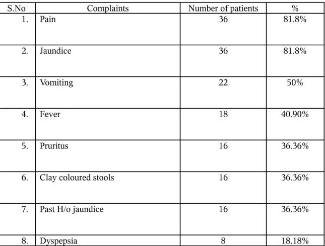
Complaints
The commonest complaint given by the patients was pain (81.8%) and jaundice (81.8%). The other symptoms include vomiting (50%), fever (40.9%) past history of jaundice, clay coloured stools, pruritis (36.36%) dyspepsia (18.18%), loss of appetite (13.63%), loss of weight (9.09%), flatulence (4.5%)
Table 1 depicts the complaints
Table 1
S.No Complaints Number of patients %
1. Pain 36 81.8%
2. Jaundice 36 81.8%
3. Vomiting 22 50%
4. Fever 18 40.90%
5. Pruritus 16 36.36%
6. Clay coloured stools 16 36.36%
7. Past H/o jaundice 16 36.36%
9. Loss of appetite 6 13.63%
10 .
Loss of weight 4 9.09%
11 .
Flatulence 2 4.5%
Co-morbid illness
Out of forty four patients there were about ten patients who had co-morbid illness, such as diabetes mellitus in 4 patients (9%) and hypertension in 6 patients (13.63%)
History of Surgery
There was a past history of surgery in 14 patients (31.8%). The different types of surgery were cholecystectomy, duodenal perforation closure, puerperal sterilization, hysterectomy, truncal vagotomy with pyloroplasty, truncal vagotomy with gastro jejunostomy, cholecystectomy with choledochal cyst excision. The different types of surgery and endoscopic interventional procedures are depicted in Figure5
There was previous history of endoscopic procedure in 8 patients (18.18%), the details of which are given in Table2.
Table 2
S.No Endoscopic Procedure Number of patients
1. ERCP & Sphincterotomy 2
2. ERCP & Stenting 4
CLINICAL FEATURES
The commonest clinical features was jaundice (68.18%) followed by fever (40.9%), pruritus (27.27%), palpable liver (27.27), splenomegaly (4.54%), signs of liver failure (2.27%) and murphy’s sign (2.27%). The symptoms are depicted in Figure 6
On per rectal examination twenty eight patients (63.64%) had yellow coloured stools and 16 patients (36.36%) had clay coloured stools.
Figure 5
PREVIOUSSURGICAL PROCEDURE
Figure 6CLINICAL FEATURES
5 1 2 2 1 1 2 0 1 2 3 4 5 6TYPE OF SURGERY
CC CC WITH CHOLEDOCHAL CYST EXCISION PUEPERAL STERALISATION HYSTERECTOMY TV WITH PYLOROPLASTY
TV WITH GJ
DU PERFORATION CLOSURE
INVESTIGATIONS
Preoperative investigations done were complete blood count, serum liver function tests, urine for bile salt and bile pigment, BT, CT, PT, APTT, INR, Serum Amylase, ultrasound, computerized tomography, MRCP (selected patients) and UGI Scopy. The numbers of patients showing abnormal values are depicted in Table 3
Table 3
S.No Investigation Number of
patients with abnormal value
% of abnormality
1. Complete Hemogram - 0%
2. Serum Bilurubin (>2mg %) 30 68.18%
68.18%
40.90% 22.27%
27.27%
4.54% 2.27%
2.27%
JAUNDICE FEVER
PRURITUS PALPABLE LIVER
SPLENOMEGALY LIVER FAILURE SIGNS
3. SGOT (>40 IU/dl) 10 22.72%
4. SGPT (>4IU/dl) 10 22.72%
5. Serum Alkaline phosphatase (36-141Mu/ml)
22 50%
6. Urine bile salt and pigment positive 30 68.18%
The UGI scopy was done in all patients and was found to be abnormal in 12 of them (27.27%). Findings are given below in Figure7.
Hence endoscopy should be done preoperatively to exclude other causes of mass in the epigastrium.
SURGICAL PROCEDURE AND OPERATIVE FINDINGS
Of the forty four patients, 38 of them (86.36%) underwent cholecystectomy. The remaining 6 patients had already undergone cholecystectomy. Cholecystectomy was done by neck to fundus method in 18 patients and fundus to neck method in 20 patients. After cholecystectomy, choledochoscopy was done through choledochotomy route in 40patients (90.91%) and through transcystic route in 4 patients (9.09%). .
Based on the diameter of the common bile duct, patients were subjected to surgical procedures. Those who had CBD diameter >15mm were treated with choledochoduodenostomy or choledochojejunostomy or primary closure. Those who had CBD diameter <15mm were treated with closure of CBD with T tube. The type of incision is given in the Figure 8
31patients(70.45%), T tube drainage in 6 patients(13.63) and primary closure of CBD in 6 patients (13.63%) and choledochojejunostomy was done in a patient(2.27%) who had excision for type 2 choledochal cyst with retained stone in the left hepatic duct. The type of operative procedure is given in Figure9.
When the operative findings were compared, it was found that out of 38 patients who underwent cholecystectomy, 12patients had cholelithiasis. Out of twelve patients there was single stone in gall bladder in 2 patients (4.54%) and multiple stones in the gall bladder in 10 patients (22.72%).
The common bile duct stone was single in 34 patients (77.27) and multiple in 10 patients (22.72%) . 12 patients (27.27%) showed both common bile duct and gall bladder stones. When the diameter of the common bile duct was analysed, it was found that the diameter ranged from 6mm to 25mm (average 17mm). There were 2 patients (4.55%) who had associated distal CBD stricture. Biopsy of the stricture was done, which confirmed only fibrous tissue.The operative findings are shown below in Figure 10
Figure 7
Figure 8
UGI ENDOSCOPY
7 2
1 2
PANGASTRITIS
DISTAL
ESOPHAGITIS&ANT
RAL GASTRITIS UNHEALTHY MUCOSA IN L.C
INCISION
68.18% 4.54%
27.27%
SUBCOSTAL
MIDLINE
BILATERAL SUBCOSTAL
Figure 9
Figure 9
COMPLICATION
The commonest complication was wound infection in 14 patients (31.81%). The other complications were bile leak in 2 patients (4.55%), retained stone in one patient (2.27%),who had choledochal cyst excision for type 2 choledochal cyst. Figure 11 depicts the complication in detail.
70.45% 13.63%
13.63%
2.27%
SURGICAL PROCEDURE
CHOLEDOCHODUODEN
Figure 10 OPERATIVE FINDINGS Figure 11 COMPLICATION FOLLOW-UP
• Post operative follow up was done for a period of six months.
• ‘T’ tube cholangiogram was done in 6 patients (13.63%) on the 8th-10th post operative day. After confirming that no residual stone in the common bile duct and free flow of
31.81%
4.55%
0% 0% 0%2.27% 0% 5% 10% 15% 20% 25% 30% 35%
TYPE OF COMPLICATION
WOUND INFECTION BILE LEAK DVT PANCREATITIS PNEUMONIA RETAINED STONE 4.54% 22.72% 77.27% 22.72% 27.27% 4.55%
GB SINGLE STONE
dye into the duodenum, the T tube was removed on the 12th-14th post operative day and subsequently discharged.
• Serum liver function test was done on the 14th post operative day, which showed raised serum bilirubin (>2mg %) in eight patients that subsequently normalized on routine check – up.
• Postoperative high frequency ultrasound done on the 14th post operative day was normal in all patients.
• Patients were regularly followed up every fifteen days for six months
DISCUSSION
(A) History
The wish and need to examine the biliary system directly dates from the beginning of surgery on the biliary system.
In 1941, Mac Iver 7 and copperstown described rigid choledochoscopic instrument
equipped with a lens, a distal light source and a rinsing device.
In 1972, Morgenstern and Berci introduced a rigid choledochoscopy using a Hopkin’s system.
In 1970, Olympus introduced the CHF series of choledochoscope with improved fiberoptics and maneuverability.
The flexible choledochoscope was first described by Shore and Lippman in 1965. (8)
The flexible choledochoscopy was gradually improved and modified by Ashby to the present range, the CHF-20 series.
In 1974, Takada et al 9 tried percutaneous transhepatic cholangioscopy through
percutaneous transhepatic biliary drainage tract.
In 1978, Nakajima 9 reported direct endoscopic visualization of biliary and pancreatic
duct system by peroral cholangio pancreatoscopy.
In 1981, Nimura et al 9 reported percutaneous transhepatic cholangioscopy. In 1981,
Ishikawa 9 et al developed percutaneous transhepatic cholecystography.
In 1994, Soda et al 9 reported a new type of small caliber flexible scope (miniscope)
Nevertheless large or impacted stones remained problematic.Technical advances led to techniques that are both safe and effective, including the application of laser in 1981 10 and
choledochoscopic electro hydraulic shockwave lithotripsy (ESWL in 1985) 11.Treatment of
choledocholithiasis via a T tube tract including dissolution therapy of various types and extraction under fluoroscopic control using a steerable catheter as introduced by Burhenne 12.
In recent era, laparoscopic cholecystectomy eliminated the approach route to CBD by which choledochoscopy, can be performed in suspected CBD stone during or after removal of gall bladder. Laparoscopic choledochoscopy has been performed using small diameter fiberoptic instrument 13.
(B) INSTRUMENTS
The choledochoscope commonly used in surgical practice may be rigid or flexible type.In our series we used flexible choledochoscopy 14 (Type PENTAX FCN-15X),
Which is shown in figure(12).
The instrument has been improved considerably in recent years with advances in fiberoptics and miniaturization.
The instrument consists of a small control unit which houses the eye piece and a lever to control angulation of the tip. The working shaft has a length of 40cm with graduated flexibility, being more rigid proximally to facilitate control of the tip. The tip angulation
ACCESSORY EQUIPMENTS
Accessory equipments available for diagnostic and therapeutic procedures include 1. Dormia basket for stone removal
2. Biopsy forceps for choledochoscopic biopsy 3. Guide wire to guide insertion of catheter balloons
4. Electro hydraulic lithotripsy (EHL) shock wave generator and EHL probe 5. Injection needle used for tumour ablation
6. ND Yag laser equipment and a laser fiber probe. 7. Transductal manometry devices.
8. Balloons & inflation devices 9. Irrigation catheter
10. Quadrangular basket forceps 11. Double jointed stone dislodger
(C) APPROACHES TO THE BILE DUCTS
Flexible fiberoptic choledochoscope can reach stones in the bile duct via several different approaches .These can be either surgical or non surgical (minimally invasive) 15.
The minimally invasive route include a percutaneous transhepatic tract after percutaneous transhepatic biliary drainage (PTBD) 16, T tube tract 17, jejunostomy tract 18,
cholecystostomy tract 19, peroral cholangioscopy and pancreatoscopy.
The different types of approaches are
4. Postoperative choledochoscopy 5. T. tube tract choledochoscopy 6. Jejunostomy tract choledochoscopy 7. Cystic duct choledochoscopy
The commonest approach followed is operative choledochoscopy. In operative choledochoscopy, choledochoscope is introduced directly in to the bile duct during laparotomy.It is usually performed as a method of exploration of bile duct in patients with dilated duct due to choledocholithiasis to remove stones or to confirm the absence of residual stones.
FINDINGS AT OPERATIVE MANEUVER
Normal mucous membrane of CBD is pale pink in appearance with a faint yellow tinge. There are usually several longitudional folds present, which are flattened by the presence of the irrigating fluid. Approaching the ampulla, the duct narrows, becomes funnel shaped and curves forward right posteriorly. Distal sphincter area usually appears stellate but may look some what fish mouthed, pin point or patulous in nature. The mucosa of the hepatic ducts is paler than of distal duct.
In cholangitis the appearance range from mucosal congestion and edema to marked ulcerative cholangitis with fibrinous exudates. These inflammatory changes can be marked in papillary region. CBD stone are easily identified as free floating or found impacted in the orifice of ampulla or partially embedded in the ductal wall or in a diverticulum of the distal duct. Multiple stones and biliary mud are frequently found behind the larger calculus.
be flushed into duodenum, in some cases under visual control using warm saline.For larger calculi, the assistant advances the stone basket and with co-ordinated movements, the calculus is entrapped.The chloledochoscopic view bile duct and stone is given in figure(13,14,15,16 & 17)
Figure 11
CHOLEDOCHOSCOPE
Figure 12
Figure 13
Figure 14
CHOLEDOCHOSCOPIC VIEW OF NORMAL INTRAHEPATIC DUCT
Figure 15
Figure 16
Figure 17
RETRIEVAL OF STONE USING DORMIA BASKET
COMPARISON OF CONVENTIONAL EXPLORATION OF CBD WITHOUT
CHOLEDOCHOSCOPY AND WITH CHOLEDOCHOSCOPY
In most series reviewed , stones that are found within the biliary tree after conventional exploration are regarded as retained stones. Retained stones should be expressed as a percentage of the number of stone-positive explorations and should not be related to the total number of common bile duct explorations. The percentage incidence of retained stones is therefore been related to number of stone – positive explorations at which the choledochoscope was used.
The mean incidence of retained stones is 10.6% (median 10.0 percent)
Table 4
RESULTS OF CONVENTIONAL EXPLORATION OF COMMON BILE DULT
S.No Reference Year Stone
positive exploration
No retained stones
% of retained stones
1 .
Isaccs & Davies 1960 NA NA 5
2 .
Way et al 1972 NA NA 10
3 .
Le. Quesne et al 1975 78 1 1.3
4 .
Stubbs & Blumgart 1984 23 3 13
5 .
Gartell & Mc Ginn 1984 32 6 18.7
6 .
Rogers et al 1985 55 7 12.7
7 .
8 .
Neoptolemos et al 1987 59 5 8.5
9 .
Sheridan et al 1987 191 37 19.4
Total 10.6
Table 5
RESULTS OF FLEXIBLE CHOLEDOCHOSCOPY
S.No Reference Year Stone positive
exploration
No. retained
stone
% of retained stone
1 .
Shore and shore 1970 100 4 4.0
2 .
Long land 1975 49 2 4.1
3 .
Stotter et al 1975 20 0 0
4 .
Finnis&Rowntree 1977 70 0 0
5 .
Legrand et al 1978 200 4 2.0
6 .
Yap et al 1980 84 4 4.8
7 .
Bauer et al 1981 36 1 2.8
8 .
Grange&Millard 1981 47 0 0
9 .
Wang 1982 47 1 2.1
1 0
1 1 .
Gartell& McGinn 1984 31 1 3.2
1 2 .
Leggeri at al 1984 128 2 1.6
1 3 .
Ashby 1984 65 1 1.5
1 4 .
May & Corfield 1985 33 3 9.0
1 5 .
Eleftheriadis et al 1985 88 0 0
1 6 .
Ashby 1985 70 1 1.4
1 7 .
Jakimowicz et al 1985 26 0 0
1 8 .
Jakimowicz et al 1986 238 5 2.1
2 0 .
Markowitz et al 1987 102 0 0
2 1 .
Molina et al 1988 36 1 2.8
(D) CHOLEDOCHOLITHIASIS
In era of open cholecystectomy the common bile duct was explored in approximately 15% of all cholecystectomies and stones were removed in approximately 65% of these explorations. The incidence of concomitant choledocholithiasis varied between 8 and 20% (Table 6)21.
Table 6
INCIDENCE OF CHOLEDOCHOLITHIASIS IN PATIENTS WITH GALL STONES
S.No Reference Total
cases of gall stones Explorati on of CBD % Exploratio n Yield stone % Overall incidence of CBD % stone
1 .
Mcsherry & Glenn (1980) 8791 15.5 60 9.5
2 .
Hampson et al (1981) 2889 15 51 8
3 .
Doyle at al (1982) 4000 22 52.5 11.5
4 .
Lygidakis (1983) 3710 11.5 80 9.5
5 .
6 .
Ganey et al (1986) 1024 26 36 9.5
7 .
Denbesten & Berci (1986) 983 24.5 81 20
8 .
R.M.Girard (1986) 10471 11 75 8
Types of choledocholithiasis
The stones in common bile duct may be primary or secondary which vary in epidemiology. Majority of CBD stones are secondary stones which originally form in the gall bladder and later pass down through the cystic duct into CBD. The size of secondary bile duct stone is limited by the diameter of the cystic duct, and thus these stones are significantly smaller than primary stone.
The incidence of primary CBD stone is controversial and varies from 4% to 56% 22
Madden defined primary stones as solitary, ovoid, light brown in color, soft and easily crushable. Saharia et al 23 classified patients as having primary stones if they met all of the
following criteria.
1. Previous cholecystectomy with or without common bile duct exploration 2. Atleast two year asymptomatic period after initial biliary tract surgery. 3. Presence of soft friable, light brown stones or sludge in the common duct 4. Absence of long cystic duct or biliary stricture due to previous surgery.
Pathogenesis of choledocholithiasis
Table 7
CHARACTERISTICS OF CBD STONE
S.No Characteristics Brown pigment
stone Black pigment stone Cholesterol stone
1 .
Color Earthy, muddy, chocolate Black, dark (Yellow core) White, yellow Beige, tan 2 .
Consistency Soft, amorphous Hard Hard
3 .
Surface Dull Smooth, Shiny Smooth, Shiny
4 .
Inner structure Laminated Amorphous Radial, crystalline
5 .
Cholesterol content <20% <20% 70-100%
6 .
Calcium Bilurubinate
10-60% 10-60% <10%
7 .
Chemical content Calcium palmitate Calcium carbonate (or) phosphate Cholesterol 8 .
The secondary CBD stone arise in gall bladder and migrate to CBD and have a spectrum of cholesterol stone and black pigment stone. Black pigment stones occur in condition in which bilurubin excretion is increased as in haemolytic disorders, and in situation associated with profound gall bladder stasis such as prolonged fasting and long term parental nutrition,cirrhosis and ileal disease.
Brown pigment stone are primary CBD stone. .The cause of brown stone is biliary stasis secondary to factors such as sphincter of oddi dysfunction, benign biliary strictures, sclerosing cholangitis and cystic duct dilatation and infestation with Ascaris lumbricoides an Clonorchis.sinensis.
PATHOPHYSIOLOGY OF BILIARY TRACT OBSTRUCTION
Obstruction of biliary tract in one of its many forms is a problem frequently encountered by the general surgeons. Pathophysiological effects of biliary tract obstruction is classified into four sub divisions
1. Physical effect 2. Pathological effect 3. Biochemical effect 4. Other functional effect
1. Physical effect
(a) Secretion ceases when biliary pressure exceeds 300mm of H2O. 24 Cholesterol and
pressure causes bacterial reflux into lymphatic and venous system .
(b) Hepatic blood flow might be reduced in the face of increasing hydrostatic pressure within the liver .
(c) Pain in biliary obstruction is due to distension of gall bladder or bile duct or may be associatied with stretching of the liver capsule
2. Pathological effects
(a) Bile duct and canaliculi
Bile canaliculi become dilated and microvilli distorted and swollen. Bile pigment “thrombi” may be seen in the canaliculi and in the adjacent hepatocytes.Marked inflammatory reaction in the portal tract with a polymorphonuclear leukocyte infiltration (acute cholangiolitis) This cholangiolytic changes are followed by increased fibrogenesis with deposits of reticulin fibers and eventually of collagen bundle..
(b) Fibrotic changes
The reticulin laid down in periportal areas matures to type I collagen, causing scarring fibrosis around the bile ducts, which may further aggravate the cholestasis.
(c) Cholangitis
Acute cholangitis in its most severe form is a life threatening infection originating in the biliary tree. In early stage of obstruction, normal hepatic bile secretion continues and an increase in intra ductal pressure approach the secretory pressure of bile 25-30cm H2O 26
Normally the biliary tree is sterile due to continuous flow of bile. At biliary pressure greater than 15cm H2O there is systemic reflux of radio labeled biliary bacteria 26. The
reticulo-endothelial system within the liver normally kills the vast majority of bacteria that enter the liver, with an estimated 10% entering the systemic circulation. Exact mechanism of bacteremia is unknown.
In presence of gall stones either within the gall bladder or biliary tree, positive culture are seen in 15-50% and 70-90% respectively 27. In complete ductal obstruction secondary to
malignancy25-40% of patients have positive cultures.
The organisms most often isolated are gram negative aerobe, Escherichia coli and Klebsiella pneumonia, the gram positive enterococci and gram negative anaerobe, Bacteriodes fragiles. In 30-50% of patients more than two organisms are present. It was also shown that >10 5 organism are seen in
1. 0% of normal patients
2. 3% of patients with symptomatic gall stones.
3. 36% of patients with common duct stones but without cholangitis 4. 85% of patients with acute cholangitis 28.
(D) Atrophy
Long standing obstruction of biliary tract leads to atrophy of liver
(iii) Biochemical effect:
The following bio-chemical changes are expected to occur in biliary obstruction due to choledocholithiasis
(a) Bilurubin: With complete biliary out flow obstruction serum total bilurubin levels typically increase by 25-43µmol/ L/ day 29 .
(b) Alkaline phosphatase: Elevation of this enzyme is sensitive indicator of biliary tract obstruction.30.Other large molecular size isoenzyme which appears in cholestasis and
(c) Lipids: Cholesterol levels may be elevated in long standing biliary obstruction. .
(e) Carbohydrate metabolism
Abnormal glucose tolerance may be seen in patients with impaired liver function and impaired gluconeogenesis after prolonged bile duct obstruction
(f) Bile salt circulation
Increased serum bile acid level up to sixty times normal due to interruption of entero hepatic circulation of bile salt occurs due to total biliary tract obstruction.
(IV) Other functional effects:
(a) Endotoxemia
In obstructive jaundice endotoxin has been found in the peripheral circulation in up to 50% of patients 31 with bile duct obstruction and play a central role in pathophysiology.
(b) Immunological effect
Extra hepatic biliary obstruction results in cellular immune dysfunction due to depression of T cell function due to direct effect of systemic endotoxemia
HISTORY/ CLINICAL FEATURES
occur when the stones obstruct the common bile duct. The clinical presentation varies depending on the degree and level of obstruction and the presence or absence of biliary infection. Choledocholithiasis occur in any of the following five ways.
1. Without symptoms 2. Biliary colic 3. Jaundice 4. Cholangitis 5. Pancreatitis
Although intermittent right upper quadrant post prandial pain, nausea, vomiting, bloating, fatty food intolerance are often perceived to be symptoms of biliary tract disease, they are not specific.
The classic charcots triad which includes fever with chills, jaundice and pain indicates cholangitis
When the triad is associated with hypotension and mental confusion (Reynolds pentad) 32
Pain is the most frequent presenting symptom.
Fever is an indication of cholangitis and classic charcot’s triad strongly favour the diagnosis. A recent study showed fever in (92%); jaundice in (65%); pain in (42%) & all three symptoms in (19%) of patients
Jaundice occurs when CBD becomes obstructed and conjugated bilurubin enters the blood stream. History of clay coloured stools and tea coloured urine is obtained in approximately 50% of cases.
patient with gall stone develop Pancreatitis.
Pancreatitis pain is different from biliary pain. Pain is located in the epigastric and mid abdominal areas and is sharp, severe, continuous and radiated to back.
History of benign CBD strictures, sclerosing cholangitis, sphincter of oddi dysfunction and cystic dilatation of CBD are important in the diagnosis of biliary stone.
Parasitic infestation with A. lumbricoides or C.Sinensis may result in development of primary CBD stone
Acute cholecystitis should be suspected when there is right upper quadrant pain and tenderness on deep inspiration (Murphy’s sign).
Chronic cholecystitis should be suspected when gall bladder is not palpable and courvoisier’s law is satisfied which states that “when CBD is obstructed by a stone, dilatation of gall bladder is rare; when the duct is obstructed in some other way, the dilatation is common”
INVESTIGATIONS
Laboratory tests are helpful, but the results are not specific for the diagnosis of choledocholithiasis. Patients with cholangitis and pancreatitis have abnormal laboratory test values. Single abnormal laboratory value does not confirm the diagnosis of choledocholithiasis, cholangitis or pancreatitis rather a coherent set of laboratory studies lead to correct diagnosis
1. White blood cell count elevation indicates the presence of infection or inflammation. Elevation above 10,000/mm³ was significant. In our study all cases showed WBC count <10,000/mm³ .
2. Serum bilurubin level elevation indicates obstruction of the CBD. Sr. Bilurubin more than 2mg/dl is considered abnormal. In our study thirty patients (68.18%) had increased values, out of which 4 patients had values greater than 10mg / dl .
3. Liver transaminase levels are elevated in choledocholithiasis. Increased transaminase level was present in ten patients (22.72%) in our study .
4. Serum alkaline phosphatase levels are elevated in patients with obstructive choledocholithiasis.Normal value is 36-141mu / ml. Increased serum alkaline plosphatase level was present in twenty two patients (50%) .
5. Serum gamma glutamyl transpeptidase and 5’ nucleotidase levels are elevated in patients with obstructive choledocholithiasis. Normal serum GGT level is <55u/l. 6. Serum protein levels may be abnormal in severe liver dysfunction associated with
8. Bleeding time, coagulation time may be abnormal in long standing obstructive jaundice . Prothrombin time may be elevated in patients with prolonged CBD obstruction, secondary to depletion of vit-k . Bleeding time was normal in all patients. Coagulation time and prothrombin time was abnormal in one patient who had ascitis with pleural effusion.Activated partial thromboplastin time (APPT). APPT was abnormal in one patient which was normalized after fresh frozen plasma transfusion. 9. Serum Amylase and lipase values are elevated in presence of acute pancreatitIs
complicating choledocholithiasis
10. Serum electrolytes value are important in correcting electrolyte imbalance in patients with obstructive jaundice
IMAGING STUDIES
Imaging studies done in patient with choledocholithiasis are grouped as
1. PREOPERATIVE STUDIES
(a) NON INTERVENTIONAL - Trans abdominal ultrasound
- Endoscopic ultrasound
- Computerized tomography
- Magnetic resonance Cholangio pancreatography - Upper Gastro intestinal Endoscopy
(b) INTERVENTIONAL - Endoscopic Retrograde cholangio pancreatogram
- Percutaneous transhepatic cholangiogram
2. INTRA OPERATIVE STUDIES
• Choledochoscopy
• Cholangiography
3. POST OPERATIVE STUDIES
• T tube cholangiography
• Trans abdominal ultrasound
PREOPERATIVE STUDIES
(1) Trans abdominal ultrasound
Ultrasonogram has been always the first imaging study for jaundiced patients. Dilated bile ducts are seen as tubules lying along side the portal vein branches. This pattern is characteristic and specific .
The level of obstruction can be predicted accurately in 80% of cases. The demonstration of stones in CBD depends on their size and position. Larger stones (>5mm) are readily visualized unless excessive intestinal gas precludes adequate imaging
Ultrasonography is accurate in the diagnosis of all gallbladder stones (97% in elective situation and 80% in presence of acute cholecystitis). But common bile duct stones are missed frequently (sensitivity 15-40%). The detection of CBD stone is impeded by presence of gas in duodenum. CBD dilatation is identified accurately with up to 90% accuracy. Recent advance with the introduction of tissue harmonic imaging (THI) gives better visualization of fluid filled structure and reduced artifacts and enhanced contrast resolution.
In our study, ultrasound was done in all patients. Out of forty four patients, twenty patients (45%) were diagnosed by ultrasound to have choledocholithiasis. Figure.18 shows U/S finding in choledocholithiasis.
(2) Endoscopic ultrasound
Though our institution lacks the specific investigation it needs special mention. Two types of echo endoscope are available for the examination of biliary tree.
The availability of high frequency catheter based ultrasound probes has made it possible to obtain ultrasound images within the biliary tree. Normal bile duct can be imaged as three layered structure.
1. Inner hyperechoic layer represents mucosa with a border echo.
2. Middle hypoechoic layer represents the smooth muscle fiber and fibro elastic tissue.
+--3. Outer hyperechoic layer represents thin and loose connective tissue with a border echo 33
EUS sensitivity and specificity of CBD stone detection are reported in the range of 84% to 100% and 95% to 100% respectively.
(3) Computerized tomography
CT scan is very accurate in detection of biliary tree obstruction and ductal dilatation, both intrahepatic and extrahepatic. The insensitivity of sonography in detecting choledocholithiasis has been well documented with a reported detection rate of 18 to 22%. CT has much higher sensitivity with a reported detection rate up to 76% 34 . Thin collimation
scan (3 to 5mm) should be performed at close intervals through the transition area of the duct in order to detect the calculus. The only reliable indicator of choledocholithiasis is said to be the presence of dense intraluminal calcification or a “target sign” within the duct. CT is of little value when the stones are small and non calcified and bile duct dilatation is minimal or absent. Figure 19 shows CT Findings in choledocholithiasis.
(4) Magnetic resonance cholangio pancreato graphy
“MR Cholangio pancreatography” refers to a range of techniques for imaging the biliary tree and pancreatic duct that all exploit the intrinsically long T2 relaxation value of many fluids, including biliary and pancreatic duct secretions. The techniques may be classified by the MR “refocussing” method used (i.e. gradient echo or spin echo), image data collection (i.e. 2D or 3D), requirement for image processing to suppress background tissues, type of receiver coil and the approach used to minimize motion related artifacts ,those related to respiration.
Early attempts to image the biliary and pancreatic ducts used gradient echo method with body receiver coils. More recenty, spin echo refocussed methods based on RARE (rapid acquisition with relaxation enhancement) 35 and its variants combined with specialized
surface receiver coils have permitted notable improvements in image quality and faster image acquisition. Most techniques have included image post processing, such as MIP (maximum intensity projection) algorithm.
Although the earlier gradient echo technique could detect the presence of biliary obstruction in 100% 36,37 and its level up to 90% 37, they could not be used to determine
accurately the underlying cause of obstruction. Improved results were achieved with RARE based technique.
The review of literature regarding sensitivity (SE), specificity (SP), diagnostic accuracy (DA), positive predicitive value (PPV) and negative predictive value in the diagnosis of choledocholithiasis with MRCP is given in Table 7 38
Table 7
S.No Authors N (%)SE SP (%) DA(%) PP(%) NPV(%)
1 .
Calvo et al (2002) 116 91 84 90 89 88
2 .
Huassein et al (2002) 50 95 96 96 95 96
3 .
Boraschi et al (2002) 95 90 96 94 95 93
4 .
Stiris et al (2000) 50 87.5 94.4 90.0 96.6 81.1
5 .
Lomas et al (1999) 69 100 97 - 82 100
6 .
Pavone et al (1999) 189 90.2 98.5 96.3 95.8 96.4
7 .
-8 .
Lomanto et al (1997) 62 91.6 100 96.8 -
-Stones < 4mm can be imaged routinely even with MIP techniques and the sensitivity may be improved by reviewing the source image, or performing projection RARE sequences.
Figure 18
ULTRASOUND FINDING IN CHOLEDOCHOLITHIASIS
Figure 19
Figure 20
MRCP FINDING IN CHOLEDOCHOLITHIASIS
Figure 22
ERCP FINDING IN CHOLEDOCHOLITHIASIS
(5) Upper gastro intestinal Endoscopy
Preoperative UGI Endoscopy was done in all patients. It was found to be abnormal in twelve patients (27.27%) with the following findings. Pangastritis in (7 patients), distal esophagitis with antral gastritis (2patients); unhealthly mucosa in lesser curvature in (1patient); active duodenal ulcer in (2 patients)
(6) Endoscopic Retro grade Cholangio pancreatography (ERCP)
stenting. Patients who had stents were removed preoperatively.Laparoscopic cholecystectomy followed by ERCP interventional procedure has become the gold standard in the management of choledocholithiasis.
• Small stones can be retrieved with a dormia basket or a fogarty balloon catheter with an intact papilla. In majority of situation, a sphincterotomy is needed before the stones can pass.Stones smaller than 1cm pass spontaneously within 48hours. Stones that are 1 to 2 cm in diameter require extraction with the dormia basket or fogarty catheter in addition to sphincterotomy. Stones larger than 2cm in diameter require further treatment with lithotripsy or chemical dissolution with mono-octanin (Cholesterol stone) and (via) a nasobiliary tube has been considered. If stone extraction is unsuccessful, a biliary drainage procedure, whether internal or external is performed. Figure 22 show ERCP with filling defect in CBD.
(7
) Percutaneous transhepatic cholangiography (PTC)
PTC may be modality of choice in patients in whom ERCP is difficult (e.g) those with previous gastric surgery or distal obstructing CBD stone or lack of an experienced endoscopist; and in patient with extensive intra hepatic stone disease and cholangio hepatitis. A long large bore needle is advanced percutaneously and transhepatically into an intra hepatic duct and cholangiography performed
INTRA OPERATIVE STUDIES
• Choledochoscopy
• Cholangiography
• Intraoperative ultrasound
CHOLANGIOGRAPHY
An area of debate is the use of routine intra operative cholangiography (IOC) during a cholecystectomy. The argument in favour of routine IOC is that it provides accurate information about biliary anatomy and the presence of CBD stones. In operative cholangiography six techniques are used.
1) Cystic duct cholangiography during laparoscopic cholecystectomy 2) Cystic duct cholangiography during open cholecystectomy
3) Cholecysto cholangiography when gall bladder is present
4) Common duct cholangiography when gall bladder is absent or when CBD is dilated 5) Transhepatic cholangiography when the anatomy of extra hepatic bile duct is unclear
or distorted from multiple previous operation on the bile ducts and
6) Post exploration T tube cholangiography performed after exploration of CBD
7) Though cholangiogram is not routinely used in our institution, we performed T tube cholangiogram in 6 patients (13.63%).
T tube cholangiography
likelihood of finding a residual stone on a later post operative T tube cholangiography from 30 to 7% 39. Operative choledochoscopy can reduce the incidence of retained bile duct stones
to 2% or less 40.
Completion T tube cholangiography is used to fill the right upper quadrant with saline solution; saline is then used to irrigate and remove all air from the biliary system. A 20ml syringe is attached directly to T tube. Two cholangiography are obtained one after the instillation of approximately 5ml of contrast medium and the second after the instillation of additional 10ml.
It is important to position the patient in such a way that CBD is to right of and does not overlie the lumbar spine. The injection rate should be slow . The entire biliary system, including the intra hepatic and extra hepatic bile ducts and flow of contrast into the duodenum should be demonstrated.
(2) Intra operative ultrasound
PREOPERATIVE MANAGEMENT OF PATIENTS WITH OBSTRUCTIVE JAUNDICE
Adequate timing of surgical intervention and preparation of patient for surgery are essential in management of patients with obstructive lesion of biliary tract. Adequate preparation entails the correction of metabolic abnormality, improvement of general condition and institution of specific measures designed to minimize the incidence of complication associated with prolonged or severe cholestasis.
DIET:
1. High intake of carbohydrate is essential and amino acid solution containing aromatic amino acid should be used sparingly as they may precipitate encephalopathy.
2. Oral diet is safest and used whenever possible.
3. Elemental diet supplementation in nutritionally compromised patients.
DRUGS
Oral administration of bile salts or lactulose to reduce the intestinal absorption of endotoxin from intestinal microflora and thus minimizing the incidence of renal failure following surgical intervention
ELECTROLYTE IMBALANCE
• Hypokalemia is frequently present and should be corrected
• Isotonic saline should be restricted as the total exchangeable body sodium is elevated.
INFECTION
Infection of biliary tract is much more commonly present in ductal calculus disease. Number of clinical trials have shown that postoperative sepsis in patient having biliary tract surgery is generally reduced by use of short term prophylactic antibiotics, either cephalosporin or aminoglycoside or piperacillin in high risk group.In patients with acute cholangitis with unknown microbiology, antibiotic selection must include the gram negative aerobes and possibly enterococci.
Antibiotics commonly used are
GM Negative aerobe
1. Aminoglycosides such as gentamycin and tobramycin 2. Third generation cephalosporin
3. Ureidopenicillin : Piperacillin (GM positive coverage)
4. Carbapenams :Imipenam and meropenam (GM positive anaerobes)
5. Fluoroquinolones : Travafloxacin (GM negative aerobic and anaerobic coverage)
In pseudomonas
Piperacillin; cefipime (4th generation cephalosporin); carbapenams , travofloxacin
Coagulation disorder
Prolonged prothrombin time resulting from deficiency of vitamin K-dependent factor is most commonly encountered in obstructive jaundice.
deficiency within 1-3days. If not corrected , fresh frozen plasma administered to cover perioperative period .
Severe bleeding in jaundiced patients suspect DIC due to circulating endotoxemia.. For DIC, monitor fibrinogen levels, fibrin degradation products and platelet count. This condition requires control of infection and specific treatment with fresh frozen plasma alone or in combination with heparin .
Renal failure
Renal failure in obstructive jaundice is well known. Adequate hydration and pre-operative induction of a natriuresis / diuresis reduces the incidence of renal failure.
Intra venous fluids (5% dextrose saline) for 12-24hours before surgery This is followed by a osmotic diuretic (mannitol) and a loop diuretic (frusemide) administered intravenously at the time of induction of anaesthesia.
Preoperative administration of oral chenodeoxycholate commencing few days before surgery has shown to reduce the incidence of renal failure
Administration of oral lactulose has shown to reduce the incidence of renal failure in jaundiced patient .
SURGICAL PROCEDURE
cholecystectomy, all the remaining thirty nine patients underwent cholecystectomy. The traditional approach in the treatment of symptomatic gall stones and choledocholithiasis has been open cholecystectomy and CBD exploration. Recently with widespread acceptance of laparascopic cholecystectomy for the treatment of symptomatic gall stones, the management of common bile duct stone has become even more controversial.
MANAGEMENT OPTIONS FOR CHOLEDOCHOLITHIASIS
1. Open cholecystectomy followed by open choledochotomy and stone retrieval 2. Pre-cholecystectomy or post-cholecystectomy endoscopic retrograde
cholangiography and papillotomy with stone removal 3. Laparascopic cholecystectomy with CBD exploration 4. Percutaneous transhepatic stone extraction
5. Open cholecystectomy with open choledochotomy with stone removal followed by choledochoduodenostomy or choledochojejunostomy
6. Open cholecystectomy with choledochotomy with stone removal followed by T tube drainage.
CHOLECYSTECTOMY
The open approach continues to be a safe and effective therapy for the treatment of complicated gall stone disease with choledocholithiasis. Management of patients with CBD stone from both quality and economic perspective includes preoperative ERCP with papillotomy followed by laparoscopic cholecystectomy. Unfortunately many patients are not candidate for this approach due to nonavailability of ERCP.
Cholecystectomy is done in two ways
1. Cholecystectomy from neck toward fundus (Commonly done) 2. Cholecystectomy from fundus toward cystic duct
In our study out of thirty eight patients who underwent cholecystectomy, 18patients had cholecystectomy from neck towards fundus and 20 patients had cholecystectomy from fundus to neck. If the anatomy cannot be clearly identified,the gall bladder should be dissected from fundus downward towards the gall bladder neck, making ductal and vascular anatomy easier to identify.
The extra hepatic bile duct can be examined best from the left side of the operating table by palpating the hepatoduodenal ligament between the left thumb anteriorly and the forefinger in the foramen of winslow. The duodenum must be mobilized (kocher’s maneuver) to allow adequate palpation of the distal CBD and the ampulla.
Choledochotomy
anastomosis. As much as possible, the common duct is left proximally in case the duct is needed for some further procedure [(e.g) repair of a stricture] in this part of the duct. Choledochotomy should be placed distally and close to the duodenum so that the usual distance from this point to the papilla measures ± 7cm Figure 23 shows Choledochotomy
EXPLORATION OF THE DUCT
Exploration should be as atraumatic as possible. Desjardin’s forceps or fogarty balloon is used to explore the CBD. Desjardin’s forceps is used to grasp larger size stones and fogarty balloon is used to remove smaller size stone and sludge. This is followed by introduction of flexible choledochoscopy to examine the CBD, common hepatic duct, the right and left hepatic duct and even further up to second order bile duct. If any stone detected, it is removed by using dormia basket.
Fogarty probe can be used for retrieval of small stone and sludge. The catheter is passed into the duodenum and the balloon inflated and catheter with drawn until it impinges against the papilla. The catheter is held by the syringe on the left hand and the degree of inflation controlled by the thumb and the plunger. With gentle traction superiorly the catheter is gradually pulled up to choledochotomy site, care being taken to prevent any stone slipping in to proximal biliary tree. The catheter is withdrawn and reinserted into each of the main hepatic ducts and the procedure is repeated.
The last step in CBD exploration is to irrigate the duct with saline. Small stones, sludge or debris can be flushed in to duodenum or back into the choledochotomy opening by irrigating the ductal system.
T tube drainage
The standard practice is to use a T tube if the CBD size is less than 15mm and if choledochoscopy showed evidence of spasm or edema of sphincter of oddi.
T tube can become obstructed, particularly if they are tight fitting and can be difficult to extract. This can be avoided by cutting off a strip of the wall of T tube. The modified T tube is held in desjardin’s forceps which conveniently grasps the T junction of the tube allowing it to be slipped into choledochotomy .
The long limb of the tube is placed at the lower end of the opening and repair commenced just above the upper apex of the incision using continuous or interrupted resorbable material. The final stitch should be close to the opening against the T tube.
Choledochoduodenostomy
In our experience we performed 31 patients (70.45%) with Choledochoduodenostomy and is the commonest procedure done in our institution following CBD exploration with flexible choledochoscopy.. It should not be performed with the ducts less than 15mm in diameter and a duct narrower than 12mm is an absolute contraindication.
The circumstances in which this bypass has been effective in association with a dilated duct are
1. Retained or recurrent calculi in the choledochus or hepaticduct. 2. Cholangitis
3. Ampullary stenosis
4. Presence of primary choledochal calculi or bile stasis.
5. Tubular stricture of the transpancreatic portion of the choledochus usually due to chronic pancreatitis.
6. Low iatrogenic stricture.
Contraindication
1. Duodenal ulcer 2. Acute pancreatitis
to the posterior duodenal incision and sutures are tied so that knots are within the lumen. Anterior suture is similarly approximated using interrupted sutures with the knots tied outside.A choledochoduodenostomy is generally preferred because it is simpler and quicker and permits easy endoscopic access if necessary41.
Figure 26, Shows choledochoduodenostomy
Choledochojejunostomy
The technique is usually used to treat when Choledochoduodenostomy is not technically possible. Figure 27 & 28 shows choledochojejunostomy .
• The bile duct is transected, the distal portion is oversewn and an end to side choledochojejunostomy performed
• Biliary intestinal continuity is established either by a loop of jejunum or a roux en y loop. Many surgeons prefer single layer anastomosis usually with 3-0 Dexon or Vicryl
• Choledochojejunostomy with a large CBD (2.5cm) does not need to be stented. If choledochojejunostomy is performed with a normal caliber duct, it may be prudent to stent the anastomosis.
• In our study we performed one case of choledochojejunostomy for a patient who underwent choledochal cyst excision done previously.
Primary closure of the CBD
Routine drainage of CBD following choledochotomy is unnecessary as it prolongs hospital stay and increases post operative morbidity 42,43. The use of T tube is associated with
encrustations leading to difficulty in removal of tube, duct stricture, cellulitis around the T tube, cholangitis, trauma to duct and subsequent biliary leak during tube removal and delayed healing of the CBD wound. The continuous external drainage of bile can sometime lead to nutritional disturbances
Inclusion crteria for primary closure 1. All stones are thoroughly removed.
2. There is no stenosis or obstruction in ampulla of vater and sphincter, which is determined by gentle passage of 12F rubber catheter or 18F metal probe through papilla into the duodenum.
3. Choledochoscopy shows no severe inflammation on the wall of the duct. 4. Absence of pancreatitis.
T tube drainage following choledochotomy is associated with increased bile infection and wound infection. Significant bile leak following T tube removal is said to occur in 1.2 to 30% 44. External loss of bile through the T tube may lead to anorexia and
constipation (Postcholedochostomy acidotic syndrome) 45. The use of T tube following
routine choledochotomy is unnecessary and increase post operative morbidity and mortality.Many papers reported by different authors 46,47,48. support the direct closure of
Figure 23
CHOLEDOCHOTOMY
Figure 24
Figure 25
T TUBE DRAINAGE
Figure 26
Figure 27
CHOLEDOCHOJEJUNOSTOMY
Figure 28
TRANS DUODENAL SPHINCTEROPLASTY
The role of transduodenal sphincteroplasty in the management of CBD stone is controversial. We have not performed this procedure and it needs mention because of its indication in some conditions
It is indicated in
1. Failure of endoscopic sphincterotomy 2. In patients with billroth II gastrectomy
3. In papillitis, where patency of the papillotomy has to be assured long term. 4. In pancreatitis when a drainage procedure of distal duct of wirsung is indicated. 5. Impacted ampullary stone
6. Multiple CBD stone 7. Recurrent CBD stone 8. Papillary stenosis Contraindicated in
1. CBD diameter >2cm
2. Long sphincter stricture >15mm 3. Peri-vaterian diverticulum
4. Duodenal wall and head of pancreas severely inflamed (Barraya et al) 49.
COMPLICATIONS
insufficiency, retained or impacted stone, pneumonia and deep vein thrombosis.
CONCLUSION
It is now well established that choledochoscopy reduces the incidence of retained common bile duct stones in patients with choledocholithiasis. Hence we recommend that operative choledochoscopy should always be done after exploration of the common bile duct stones to reduce the incidence of retained stone.
Surgical exploration of CBD with conventional method without the use of choledochoscopy have shown 8 to 16% of patients with retained stones . With the use of choledochoscopy, the incidence is reduced to 2% as quoted in various studies. In our study percentage of retained stone after flexible choledochoscopy was (2.27%) .
Females were commonly affected by choledocholithiasis. The commonest age group was between 51-60yrs. Commonest complaint was pain or jaundice
BIBLIOGRAPHY
1. KO CW, Lee SP, Epidemiology and Natural History of CBD stones and prediction of disease G.I.Endoscopy 56 (Suppl6): S165 – 169 2002
2. Clinical G I Endoscopy; Gregory G Ginsberg, Michael L, Kochman, Ian Norton, Christopher J Gostout Chapter 46 Pg 671 – 693
3. Havard C, Non – Malignant bile duct obstruction Ann.R.Coll Surg Engl 1990: 26 : 88-93
4. Hicken NF, MCCallister AJ. operative Cholangiography as an aid to reduce the incidence of “OVER LOOKED” common bile duct stones; a study of 1,293 choledocholithotomies surg 1964: 55: 953-8
5. Sabiston textbook of surgery 16th edition page no 1089
6. Rattner D.W; Warshan AL, Impact of choledochoscopy on the management of choledocholithiasis. Experience of 490 CBD exploration at the massachusets general hospital surg 1981: 194: 76-79
7. MCIver MA: An instrument for visualizing the interior of the common duct at operation preliminary note. Surgery 1941; 9: 112-14
8. Shore JM, Lippman HN A Flexible choledochoscope Lancet 1965: i: 1200-1
9. Cholangioscopy; dong – wan seo. Sung koo lee myung – Hwan kim young – 11 min (Lippin cott Williams & Wilkins pg 2
10. Orii k, Nakahara A, Takase Y, Choledocholithotomy by Yag laser with a choledochfiberscope surg 1981: 90: 120-2
11. Gochok, Ikezewa H, yasuo m et al Experience of Choledocho fiberscopic, electro hydraulic shockwave lithotripsy Dig Endos (Jpn) 1985: 26: 183-7
12. Burhenne H.J. Percutaneous extraction of retained biliary tract stone 661 patients AJR Am J Roentgenol 1980; 134: 888-98
13. Kelly WEJ, Sheridan VC, Laparascopic Choledochoscopy with a small caliber endoscope. A safe and effective technique for laparascopic CBD exploration Surgery Endoscopy 1995; 10: 293-6
15. Birkett DH, Williams LF Jr post operative fiberoptic choledochoscopy Ann surg 1981: 194: 630-4
16. Nimura Y, Hayakawa N, Toyodas et al Percutaneous transhepatic cholangioscopy (PTCS) stomach intestine (jpn) 1981 16: 681-9
17. Moss JP, Whelan J Jr. Dedman TC III, Voyles RG, Post operative Choledochoscopy through the T tube tract surg gynecol 1980 151 807-9
18. Hwang MH, KuoRJ, Modified Roux en Y Choledochojejunostomy with post operative Choledocho fiberscopy in the treatment of intrahepatic stones. J.Formos med Assoc 1980: 79: 631-6
19. Siegel JH, Mayer LF, Percutaneous choledochoscopy and cholecystoscopy diagnostic and therapeutic user Endoscopy 1981: 13: 124- 7
20. Br.J.Surg 1990 volume 77 september 975-982. operative choledochoscopy: CBD exploration is incomplete without it. R.W.Motson and L.A.Wetter
21. Blumgart pg 737 surgery of the liver and biliary tract
22. Madden Jl 1973 Common duct stones: their origin and surgical managemnt surgical clinics of north America 53: 1095-1113
23. Saharia PC, Zuidema GD, Cameron Jl 1977, Primary common duct stones.Annals of surgery 1985: 598-602
24. Csendes A, Sepulveda A, Burdiles P, Braghetto I, BastiasJ, Schutte H, Diaz JC, Yarmuch J, Maluenda F 1988 common bile duct pressures in patients with CBD stones with or without acute suppurative cholangitis archives of surgery 123: 697-699
25. Strasburg SM, Dorne BC, Redinger RN, small DN, Egdall RH 1971 Effect of alteration of biliary pressure on bile composition – a method for study: primate biliary physiology V. Gastroenterology 61: 357-362
26. Jacobson K, Ellander J, Rosengren B 1962, Cholangiovenous reflux. Acta chirurgica scandinavica 123:316
27. Lipsett PA, Pitt HA 1990. Acute cholangitis, surgical clinics of north America 70: 1297