Copyright © 2002, American Society for Microbiology. All Rights Reserved.
An Efficient and Versatile Mammalian Viral Vector System for Major
Histocompatibility Complex Class I/Peptide Complexes
Ai Kawana-Tachikawa,
1Mariko Tomizawa,
1Jun-ichi Nunoya,
1Tatsuo Shioda,
2Atsushi Kato,
3Emi E. Nakayama,
2Tetsuya Nakamura,
4Yoshiyuki Nagai,
5and Aikichi Iwamoto
1,4*
Division of Infectious Diseases, Advanced Clinical Research Center,
1and Department of Infectious Disease and Applied
Immunology,
4Institute of Medical Science, University of Tokyo, Minato-ku, Tokyo 108-8639, Department of Viral
Infections, Research Institute of Microbial Diseases, Osaka University, Suita-City, Osaka 565-0871,
2Department
of Viral Diseases and Vaccine Control, National Institute of Infectious Diseases, Musashimurayama
City, Tokyo 208-0011,
3and Toyama Institute of Health, Imizu-gun, Toyama 939-036,
5Japan
Received 3 May 2002/Accepted 28 August 2002
We report a Sendai virus (SeV) vector system for expression of major histocompatibility complex (MHC)
class I/peptide complexes. We cloned the extracellular domain of a human MHC class I heavy chain,
HLA-A*2402, and human

-2 microglobulin (

2m) fused with HLA-A*2402-restricted human immunodeficiency
virus type 1 (HIV-1) cytotoxic T-lymphocyte (CTL) epitopes (e-

2m) in separate SeV vectors. When we
coinfected nonhuman mammalian cells with the SeVs, naturally folded human MHC class I/peptide complexes
were secreted in the culture supernatants. Biotin binding peptide sequences on the C terminus of the heavy
chain were used to tetramerize the complexes. These tetramers made in the SeV system recognized specific
CD8-positive T cells in peripheral blood mononuclear cells of HIV-1-positive patients with a specificity and
sensitivity similar to those of MHC class I tetramers made in an
Escherichia coli
system. Solo infection of
e-

2m/SeV produced soluble e-

2m in the culture supernatant, and cells pulsed with the soluble protein were
recognized by specific CTLs. Furthermore, when cells were infected with e-

2m/SeV, these cells were recognized
by the specific CTLs more efficiently than the protein pulse per se. SeV is nonpathogenic for humans, can
transduce foreign genes into nondividing cells, and may be useful for immunotherapy to enhance
antigen-specific immune responses. Our system can be used not only to detect but also to stimulate antigen-antigen-specific
cellular immune responses.
Class I molecules of the major histocompatibility complex
(MHC)—human leukocyte antigen (HLA) in humans—play
crucial roles in cellular immune responses in humans. An
MHC class I/peptide complex has a heterotrimeric structure
consisting of a polymorphic glycoprotein called the heavy
chain,

-2 microglobulin (

2m), and a short peptide with 8 to
11 amino acids which is a proteolytic fragment of a larger
antigen. A unique conformational structure called an epitope
results from the bumps created by the peptide embedded in a
groove of a heavy chain and a part of the surrounding banks.
Cytotoxic T lymphocytes (CTLs) recognize the epitope
through their T-cell receptors (TCRs) and remove the exotic
cells which present tumors, viral antigens, and so on. When
antigen presentation by MHC class I molecules is insufficient,
the target cells may escape from immune surveillance by CTLs.
Often, invasive tumors partially or totally lose expression of
MHC class I/peptide complexes due to a blockage at any step
required for synthesis or transport to the cell surface (3).
Vi-ruses use different tactics to sneak away from CTLs. Nef in
human immunodeficiency virus type 1 (HIV-1), ICP47 in
her-pes simplex virus type 1, US2, US3, US6, and US11 in
cyto-megalovirus (CMV), and E19 in adenovirus are examples of
viral proteins which disturb the expression of MHC class
I/pep-tide complexes on the surfaces of infected cells (1, 2, 27, 40, 41,
43).
It has been shown that artificially manufactured MHC class
I/peptide complexes are useful for studying epitope-specific
CTL responses or for enhancing these responses in vitro.
Sev-eral methods of manufacturing MHC class I/peptide
com-plexes in vitro have been reported. The first is to make the
three components independently and mix them (11). The
sec-ond is to make two molecules,

2m and a heavy chain fused
with a peptide sequence at the N terminus via a linker
se-quence (15). The third is also to make two molecules, a heavy
chain and

2m fused with a peptide at the N terminus via a
linker sequence (31, 37, 39). Fourth, a fusion protein consisting
of a heavy chain and

2m is mixed with a peptide (22, 36). The
fifth method is to make a single chain construct of the three
components (23).
Escherichia coli
or baculovirus expression
systems have been used to make MHC class I tetramers (4, 39).
In this study, we adopted a Sendai virus (SeV) vector system
for MHC class I/peptide complex expression.
SeV, a member of the family
Paramyxoviridae
, has a
nonseg-mented negative-strand RNA as a genome. It causes severe
respiratory disease in mice but is nonpathogenic for humans
(16, 17, 24). It has been shown that the SeV vector system is
very efficient at production of soluble proteins (44). We would
like to show that this system is also very efficient at producing
heteromeric molecules in the culture supernatant. Moreover,
the same system could be used to express MHC class I/peptide
complexes with intended specificities on the cell surface and to
stimulate antigen-specific CTLs. Thus, the SeV vector system
* Corresponding author. Mailing address: Division of Infectious
Diseases, Advanced Clinical Research Center, Institute of Medical
Science, University of Tokyo, 4-6-1 Shirokanedai, Minato-ku, Tokyo
108-8639, Japan. Phone: 81-3-5449-5359. Fax: 81-3-5449-5427. E-mail:
11982
on November 8, 2019 by guest
http://jvi.asm.org/
appears to be not only an efficient but also a versatile system
for expression of heteromeric cell surface molecules.
MATERIALS AND METHODS
Cell lines and media.Cells of the monkey kidney cell lines LLC-MK2and
CV-1 were cultured in minimal essential medium (MEM) (Sigma, St. Louis, Mo.) supplemented with penicillin and streptomycin, each at 100 U/ml (Invitro-gen, Carlsbad, Calif.), and 10% heat-inactivated fetal calf serum (FCS) (M10). Epstein-Barr virus-transformed B-lymphoblastoid cell lines (B-LCLs) were es-tablished as previously described (26) and maintained in RPMI 1640 (Sigma) supplemented with 100 U of penicillin/ml, 100 U of streptomycin/ml, and 10% heat-inactivated FCS (R10). For CTL culture, R10 supplemented with 10% Lymphocult T (Biotest, Dreieich, Germany) was used.
Construction of recombinant SeV.A genetic construct for a soluble HLA class I heavy chain (A24-BSPhis) was generated by a three-step PCR using a cloned HLA-A*2402 cDNA as a template (35). In A24-BSPhis, the external domain of the HLA-A*2402 molecule (from the␣1 to the␣3 domain of HLA-A*2402) was connected with BirA substrate peptide (BSP), a histidine tag (his), and signals needed for expression in SeV betweenNotI sites (Fig. 1A).
Initially, the HLA-A*2402 cDNA was amplified using the 5⬘primer A24-a (5⬘-T GCGGCCGCCGTACGAGGATGGCCGTCATGGCGCCCCG-3⬘), which hy-bridizes to the 5⬘end of the HLA-A*2402 signal sequence and contains a spacer sequence and aNotI site, and the 3⬘primer A24-d1 (5⬘-GTCCCGCAGCTCCA TCTTCATTGCCTCAAAGATTCCTCCAAGGGATCCCCATCTCAGGGTG AGGGGCTT-3⬘), which hybridizes to the 3⬘end of the HLA-A*2402␣3 domain and encodes BSP (LGGIFEAMKMELRD) (coding sequence underlined) (29). A24-a was used as the 5⬘primer for all three PCR steps. The first PCR product was used as a template for the second PCR and amplified using the 3⬘primer A24-d1his (5⬘-CTACGGCGTACGTCAATGGTGGTGATGGTGGTGGTCC CGCAGCTCCAT-3⬘), which hybridizes to the 3⬘half of BSP and contains a histidine tag (His6), a stop codon, and a spacer sequence. The second PCR
product was used as a template for the third PCR and amplified using the 3⬘ primer A24-d2his (5⬘-TTGCGGCCGCGATGAACTTTCACCCTAAGTTTTT CTTACTACGGCGTACGTCA-3⬘), which hybridizes to the stop codon and the spacer sequence and contains the SeV E and S signal sequences and aNotI site (44).
For epitope-fused2m (e-2m), human2m cDNA was cloned by reverse transcription-PCR from mRNA isolated from peripheral blood mononuclear cells (PBMCs) of a healthy donor. The gene was modified to introduce sequences encoding peptides with a binding motif for HLA-A*2402 and a Gly-Ser linker between the C terminus of the signal sequence and the N terminus of the mature 2m protein (Fig. 1B). Two HLA-A*2402-binding peptides were used as epitopes: one was the peptide from HIV-1 Nef corresponding to amino acids 138 to 147 (Nef138-10; RYPLTFGWCF), and the other was the peptide from HIV-1 Env corresponding to amino acids 584 to 594 (Env584-11; RYLRDQQLLGI) (14). The cloned2m was used as a template for a three-step PCR. Initially,2m was amplified using the 5⬘primer e/b2m-a1 (5⬘-GGAGGTGGCGGGTCCGGAG GTGGTTCTGGTGGAGGTTCGATCCAGCGTACTCCAAAGATT-3⬘), which hybridizes to2m immediately adjacent to the signal sequence and encodes a 13-amino-acid Gly-Ser linker sequence (GGGGSGGGSGGGS), and the 3⬘ primer b2m-d (5⬘-TTGCGGCCGCGATGAACTTTCACCCTAAGTTTTTCTT ACTACGGCGTACGTTACATGTCTCGATCCCACTT-3⬘), which hybridizes to the 3⬘end of2m and encodes a spacer sequence, E and S signals, and aNotI site. b2m-d was used as the 3⬘primer in all three PCR steps. For Nef138-2m, e(nef)-a2 (5⬘-TCTGGCCTGGAGGCTAGATATCCACTGACCTTTGGATG GTGCTTCGGAGGAGGTGGCGGGTCC-3⬘), which hybridizes to the linker sequence, encodes Nef138-10 (underlined), and contains part of the2m signal sequence, was used as the 5⬘ primer for the second PCR. e(env)-a2 (5⬘ -TCTGGCCTGGAGGCTAGATACCTAAGGGATCAACAGCTCCTAGGGAT TGGAGGTGGCGGGTCC-3⬘) was used for Env584-2m (underlined). Then these PCR products were used as a template for the third PCR and amplified using the 5⬘primer e/b2m-a3 (5⬘-TGCGGCCGCCGTACGGCCG AGATGTCTCGCTCCGTGGCCTTAGCTGTGCTCGCGCTACTCTCTCT TTCTGGCCTGGAGGCT-3⬘), which contains a 2m signal sequence, a spacer sequence, and aNotI site. We also made a2m coding fragment by using b2m-a (5⬘-TGCGGCCGCCGTACGGCCGAGATGTCTCGCTCCGT GGCCTTA-3⬘), which hybridizes to the 5⬘end of the2m signal sequence and contains the spacer sequence andNotI site, and b2m-d.
Each PCR fragment was cloned into the pGEM-T vector (Promega Corp., Madison, Wis.), and we confirmed the absence of PCR errors for each fragment by sequence analysis with the BigDye Terminator Cycle Sequencing Ready Reaction kit (Applied Biosystems, Foster City, Calif.). Then each fragment was
introduced into theNotI site of pV(⫺)SeV18⫹b(⫹), which contains a full-length copy of the positive-sense SeV antigenome (15,384 nucleotides) with an addi-tional 18-nucleotide sequence including aNotI site within the N gene and with two point mutations in the P gene which cause a defect in V mRNA without affecting P protein expression (Fig. 1C) (16, 44), and we obtained A24-BSPhis/ pSeV, Nef138-2m/pSeV, Env584-2m/pSeV, and2m/pSeV.
SeV recovery.Viruses were recovered from each plasmid as previously de-scribed (13, 17). Briefly, LLC-MK2cells were infected with a recombinant
vac-cinia virus (VV), vTF7-3, expressing T7 polymerase (9) at a multiplicity of infection (MOI) of 2. Then A24-BSPhis/pSeV, Nef138-2m/pSeV, Env584-2m/
FIG. 1. Construction of SeVs expressing the soluble MHC class I
heavy chain or e-

2m. (A) Structure of A24-BSP
his
. The extracellular
domain of HLA-A*2402 was connected with BSP and the histidine tag
via two amino acids (glycine and serine). (B) Structure of e-

2m. An
HLA-A*2402-restricted epitope and a 13- or 14-amino-acid
glycine-serine linker were inserted between the signal sequence and the coding
sequences of the mature

2m protein. In this study, we used two
HLA-A*2402-restricted HIV-1 CTL epitopes, Nef138-10 and
Env584-11. (C) A24-BSP
his
and e-

2m followed by E and S signals of SeV
were inserted into the
Not
I site in the parental pV(
⫺
)SeV18
⫹b(
⫹
),
which generated a full-length SeV V(
⫺
) antigenome, as described in
Materials and Methods. (D) Expression of A24-BSP
his
and e-

2m in
SeV-infected cells by Western blot analysis. CV-1 cells were infected
with SeVs at an MOI of 3 and were lysed 24 h postinfection, and cell
lysates were separated on an SDS-PAGE gel with a 10 to 20%
gradi-ent, transferred to a polyvinylidene difluoride membrane, and then
detected with an anti-His6
(left) or anti-

2m (right) MAb. In the case
of e-

2m, the culture supernatants which were removed from SeV
particles by centrifugation were also used. (Left) Lane 1, A24-BSP
his
/
SeV; lane 2, wild-type SeV (wt/SeV). (Right) Lanes 1 and 5,
Nef138-
2m/SeV; lanes 2 and 6, Env584-

2m/SeV; lanes 3 and 7,

2m/SeV;
lanes 4 and 8, wt/SeV; lane 9, 20 ng of purified

2m. In lanes 1 to 4, 10
g of cell lysates was added; in lanes 5 to 8, 10
l of the culture
supernatant was added. The numbers to the left of the gels indicate the
sizes (in kilodaltons) of the products.
on November 8, 2019 by guest
http://jvi.asm.org/
pSeV, or2m/pSeV, and plasmids pGEM-N, pGEM-P, and pGEM-L, encoding trans-acting proteins, were transfected simultaneously by using the liposomal transfection reagent DOTAP (N-[1-(2,3-dioleoyloxy)propyl]-N,N,N-trimethyl-ammonium methylsulfate; Roche Diagnostics, Rotkreuz, Switzerland). Cells were maintained in serum-free MEM in the presence of 40g of 1--D
-arabino-furanosylcytosine (araC)/ml and 100g of rifampin/ml to minimize VV cyto-pathogenecity. Forty-eight hours after transfection, the cells were harvested, washed with phosphate-buffered saline (PBS), and inoculated into the allantoic cavities of 10-day-old embryonated chicken eggs. After 3 days of incubation, the allantoic fluid was harvested. When the presence of viruses was confirmed by hemagglutination test, the allantoic fluid, diluted 107- to 108-fold, was used for
the second propagation to remove vTF7-3. We obtained SeV expressing A24-BSPhis (A24-BSPhis/SeV), Nef138-2m (Nef138-2m/SeV), Env584-2m (Env584-2m/SeV), or2m (2m/SeV) at 4.4⫻107, 2.8⫻108, 2.7⫻108, or 3.0
⫻108cell infectious units/ml, respectively.
ELISA for MHC class I//e-2m complexes or e-2m.A sandwich enzyme-linked immunosorbent assay (ELISA) was performed to detect properly folded MHC class I/e-2m complexes. A 1-g/ml concentration of the anti-MHC class I monoclonal antibody (MAb) 3F10 (Ancell, Bayport, Minn.), which recognizes only fully assembled MHC class I/peptide complexes, was used as a capture antibody, and a 125-ng/ml concentration of horseradish peroxidase (HRP)-con-jugated anti-human2m (DAKO A/S, Glostrup, Denmark) was used as a de-tector antibody. For e-2m detection, samples were directly coated, and the bound e-2m was detected with 500 ng of HRP-conjugated anti-human2m/ml. Purified2m (Biogenesis, Poole, England) was used as a standard protein for measurement. 3,3⬘,5,5⬘-Tetramethylbenzidine was used as an HRP substrate in both ELISAs.
Purification of MHC class I//e-2m complexes.MHC class I/e-2m complexes were purified from the culture supernatants of SeV-infected CV-1 cells. The culture supernatants were centrifuged at 40,000⫻gto pellet down the SeV particles and were then purified by affinity chromatography on a Hitrap Chelat-ing HP column (Amersham Pharmacia Biotech, Piscataway, N.J.) in 0.02 M NaHPO4(pH 7.4)–0.5 M NaCl with a 0 to 0.5 M gradient of imidazole.
Preparation of MHC class I//e-2m tetramers.Tetramerization of monomeric MHC class I/e-2m complexes was performed as previously described (4, 7). BSPs of the purified MHC class I/e-2m complexes were biotinylated by using the BirA enzyme (Avidity, Denver, Colo.). One milligram of MHC class I/e-2m complexes was incubated with 10 g of BirA at 25°C for 18 h in a buffer containing 10 mM Tris-Cl (pH 8.0), 50 mM Bicine (pH 8.3), 10 mM ATP, 10 mM magnesium acetate, and 40M biotin. Following the reaction, biotinylated MHC class I/e-2m complexes were purified on a Superdex 200 column (Amersham Pharmacia) in 20 mM Tris-Cl (pH 8.0)–150 mM NaCl. Finally, the buffer was changed to PBS including protease inhibitor cocktail (Roche Diagnostics) and mixed with phycoerythrin (PE)-conjugated streptavidin (Molecular Probe, Eu-gene, Oreg.) at a 1:1 ratio of biotinylated MHC class I/e-2m to biotin binding sites.
MHC class I tetramers made in anE. colisystem were obtained from the National Institute of Allergy and Infectious Diseases (NIAID) MHC Tetramer Core Facility (Emory University Vaccine Center).
Generation of CTL lines and clones.A Nef138-10-specific CTL line was induced from PBMCs of HIV-1-infected individuals carrying HLA-A*2402. A total of 3⫻105PBMCs were cultured in R10 in 96-well round-bottom tissue
culture plates. The next day, 105stimulator cells (autologous phytohemagglutinin
[PHA]-stimulated PBMCs irradiated with 3,300 rads and pulsed with Nef138-10 for 1 h at 10M) were added and cultured for 2 weeks in the presence of 10% Lymphocult-T, which supplied 100 U of interleukin-2 (IL-2)/ml, and 1g of an anti-CD28 MAb (BD Pharmingen, San Diego, Calif.)/ml. The cells were further stimulated with irradiated autologous B-LCLs pulsed with 10M concentrations of peptides for another 7 to 10 days. CMV-specific CTL lines were induced in the same way, by using an HLA-A*2402-restricted peptide from CMV pp65 corre-sponding to amino acids 328 to 337 (CMVpp65/328-9; QYDPVAALF) (19), from PBMCs of HLA-A*2402-positive patients who were also seropositive for both HIV-1 and CMV. For tetramer staining of CTL lines, we used them after the second stimulation. After the fourth stimulation, the cells were cloned by limiting dilution to 0.8 and 8 cells/well in 96-well round-bottom tissue culture plates. Each well contained 105irradiated autologous B-LCLs and 5⫻104
irradiated allogeneic PBMCs in the presence of 10M peptide in the cloning medium (R10 containing 10% lymphocult-T and 5% PHA-blast culture super-natant).
Tetramer staining and fluorescence-activated cell sorter analysis.At first each tetramer was incubated with various dilutions to determine the optimal condi-tions for staining. Cells were stained with PE-labeled tetramers at 37°C for 15 min at the optimal concentration, washed once with 2% FCS–0.1% NaN3in PBS,
and then stained with anti-CD8–allophycocyanin (BD Pharmingen) at 4°C for 20 min. Cells were washed three times and fixed with 1% paraformaldehyde. Stained cells were analyzed by using a FACSCalibur (Becton Dickinson, Moun-tain View, Calif.) with CellQuest software (Becton Dickinson) and Flowjo soft-ware (Tree Star, San Carlos, Calif.).
51Cr release assay.Cytotoxicity was measured by a standard51Cr release assay
as previously described (18). Briefly, HLA-A24 matched allogeneic B-LCLs were labeled with 100Ci of Na251CrO4for 2 h and washed three times with R10.
Labeled target cells (2⫻103) were added to a 96-well round bottom microtiter
plate with a corresponding amount of peptide or the culture supernatant of e-2m/SeV-infected cells. After an hour of incubation, effector cells were added and incubated for 4 h. When SeV-infected LCLs were used as target cells, the cells were infected with SeVs, at an MOI of 10, 20 h before addition of the effector cells. The supernatants were collected and analyzed with a microbeta counter. Spontaneous51Cr release was determined by measuring counts per
minute in the supernatants of wells containing only target cells (cpmspn).
Max-imum release (cpmmax) was determined by measuring the release of51Cr from
target cells in the presence of 2% Triton X-100. Specific lysis was calculated as (cpmexp ⫺cpmspn)/(cpmmax ⫺ cpmspn) ⫻100, where cpmexp represents the
counts per minute in the supernatants of wells containing target and effector cells.
RESULTS
Construction of SeVs expressing soluble MHC class I heavy
chain or e-

2m.
We made SeVs expressing soluble MHC class
I heavy chain, HLA-A*2402, designated A24-BSP
his,
with a
BSP sequence and a histidine tag (Fig. 1A). We also made

2m
connected with an antigenic peptide (epitope) via a 13- or
14-residue linker at the N terminus and named the construct
e-

2m (Fig. 1B). Since it is known that SeV replicates
effi-ciently only when the length of the genome is a multiple of 6
(rule of six) (6), the genome length was adjusted by the linker.
In this study the HLA-A*2402-restricted HIV-1 epitope
Nef138-10 or Env584-11 was used as the antigenic peptide
(14). The constructs carrying A24-BSP
his
or e-

2m were
in-serted into the
Not
I site of pV(
⫺
)SeV18
⫹b(
⫹
), which contains
the V(
⫺
) version of the SeV antigenome (16, 44) (Fig. 1C).
Twenty-four hours after infection, A24-BSP
his
or e-

2m was
expressed in A24-BSP
his
/SeV- or e-

2m/SeV-infected CV-1
cells, respectively (Fig. 1D). Considerable amounts of e-

2m
were secreted from the infected cells into the culture
superna-tants (Fig. 1D, right). Because of the epitope and linker
pep-tides, the mobilities of Nef138-

2m and Env584-

2m on
so-dium dodecyl sulfate-polyacrylamide gel electrophoresis
(SDS-PAGE) gels were slightly slower than that of

2m.
Production of soluble MHC class I/e-

2m complexes by
SeV-infected cells.
It is known that human MHC class I heavy
chain without human

2m fails to be expressed on the cell
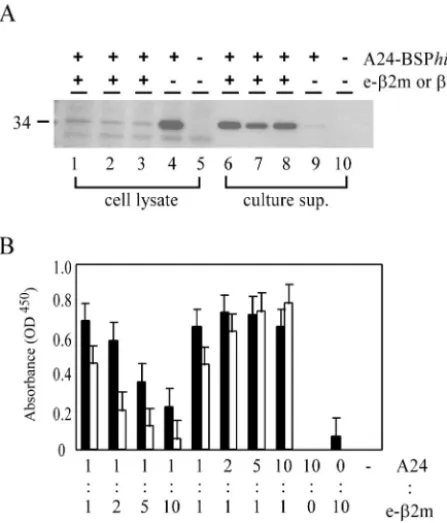
surface because of its conformational instability (42). When
CV-1 cells were infected with A24-BSP
his
/SeV alone,
A24-BSP
his
was detected in the cell lysates but hardly at all in the
culture supernatants (Fig. 2A, lanes 4 and 9). When
Nef138-
2m/SeV, Env584-

2m/SeV, or

2m/SeV was inoculated
to-gether with A24-BSP
his
/SeV, large amounts of A24-BSP
his
were detected in the culture supernatants (Fig. 2A, lanes 6 to
8) while only small amounts of A24-BSP
his
remained in the
cell lysates (Fig. 2A, lanes 1 to 3). Thus, the presence of e-

2m
stabilized A24-BSP
his
by forming stable MHC class I/e-

2m
complexes, which could be secreted from the infected cells very
efficiently.
To confirm that MHC class I/e-

2m was folded properly like
native MHC class I/peptide complexes, culture supernatants of
CV-1 cells coinfected with A24-BSP
his
/SeV and e-

2m/SeV
on November 8, 2019 by guest
http://jvi.asm.org/
were assayed by using a MAb which detects only fully
assem-bled MHC class I/peptide complex (see Materials and
Meth-ods). Cells were coinfected with various ratios of A24-BSP
his
/
SeV to Nef138-

2m/SeV or Env584-

2m/SeV, and culture
supernatants were assayed on day 3. Considerable amounts of
MHC class I/e-

2m complexes were detected in the culture
supernatants. The more the ratio of the MOI of e-

2m/SeV to
that of A24-BSP
his
/SeV was increased, the less A24-BSP
his
/e-
2m complex (A24/e-

2m) was secreted when cells were
in-fected with both Nef138-

2m/SeV and Env584-

2m/SeV (Fig.
2B). In contrast, when the MOI ratio was decreased, A24/
Nef138-

2m secretion was not affected but A24/Env584-

2m
secretion increased. The optimal ratios for A24/e-

2m
com-plex secretion appeared to be dependent on the sequences of
the peptides and partly on the ratio of the two vectors (Fig.
2B). These data also confirmed the proper folding of
A24-BSP
his
.
We purified A24/Nef138-

2m complexes by affinity
chroma-tography using a histidine tag from the culture supernatant of
CV-1 cells coinfected with A24-BSP
his
/SeV and Nef138-

2m/
SeV at an MOI ratio of 1 to 1. About 1 mg of purified A24/
Nef138-

2m complex was obtained from 10
8CV-1 cells.
Staining of specific CD8 T cells by SeV-made tetramers.
Purified A24/Nef138-

2m complexes were biotinylated at the
BSP and multimerized with PE-conjugated streptavidin to
form tetramers as previously described (4, 7). A CTL clone
specific for Nef138-10 was fully stained with the
A24/Nef138-
2m tetramer (Fig. 3A).
We then tested the specificity of A24/Nef138-

2m tetramers
by using CTL lines specific for Nef138-10 or another
HLA-A*2402-restricted epitope derived from CMV. Whereas the
CMV-specific CTL line was not stained with A24/Nef138-

2m
tetramers at all (Fig. 3B), a certain portion of the
Nef138-specific CTL lines was stained with A24/Nef138-

2m tetramers
(Fig. 3C). These data indicated that tetramers formed by
SeV-made MHC class I/e-

2m complexes were fully assembled and
recognized by adequate TCRs.
MHC class I/e-

2m complexes in our system have an
artifi-cial linker peptide near the epitope which may interfere with
recognition by TCRs. In order to test the possibility, we
com-pared the efficiency of our SeV-made tetramers
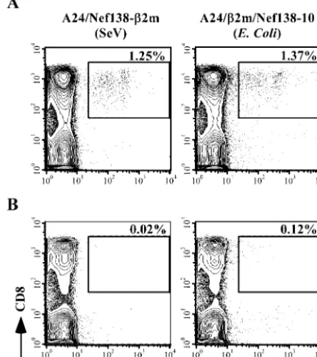
[image:4.603.312.527.72.304.2](A24/Nef138-
2m tetramers) with that of
E. coli
-made tetramers without the
linker peptide (A24/

2m/Nef138-10 tetramers). PBMCs from
HLA-A*2402-positive uninfected or HIV-1-infected
individu-als were examined with both tetramers. A total of 1.25 or
1.37% of CD8 T cells among PBMCs from HIV-1-infected
individuals and 0.02 or 0.12% of CD8 T cells among PBMCs
from non-HIV-1-infected individuals were stained with A24/
Nef138-

2m or A24/

2m/Nef138-10 tetramers, respectively
FIG. 2. Secretion of MHC class I/e-

2m complex from
SeV-coin-fected cells. (A) Detection of A24-BSP
his
in SeV-infected cells and
culture supernatants. CV-1 cells were infected with A24-BSP
his
/SeV
with or without e-

2m/SeV at an MOI of 3. Twenty four hours
postin-fection, cells and culture supernatants were harvested. Cells were
lysed, and culture supernatants were centrifuged at 40,000
⫻
g
. Ten
micrograms of cell lysates (lanes 1 to 5) or 10
l of supernatants (lanes
6 to 10) was separated on an SDS-PAGE gel with a 10 to 20% gradient.
The position of the 34-kDa product is indicated on the left. Lanes 1
and 6, A24BSP
his
/SeV plus Nef138-

2m/SeV; lanes 2 and 7,
A24BSP
his
/SeV plus Env584-

2m/SeV; lanes 3 and 8, A24-BSP
his
/
SeV plus

2m/SeV; lanes 4 and 9, A24-BSP
his
/SeV only; lanes 5 and
10, wild-type SeV. (B) ELISA for MHC class I/e-

2m complex
detec-tion. CV-1 cells were infected with A24-BSP
his
/SeV and either
Nef138-

2m/SeV (solid bars) or Env584-

2m/SeV (open bars) at
var-ious MOI ratios. Culture supernatants were harvested 3 days
postin-fection. After centrifugation at 40,000
⫻
g
, culture supernatants were
assayed in an ELISA specific for fully assembled MHC class I/peptide
complexes (described in Materials and Methods). Averages and
stan-dard deviations for three wells are given. Results for one
representa-tive experiment out of three are shown.
FIG. 3. Staining of CTLs with A24/Nef138-

2m tetramers. (A) A
Nef138-10-specific CTL clone was stained with A24/Nef138-

2m
tet-ramers. (B and C) After a second stimulation with cognate peptides, a
Nef138-10-specific CTL line (B) and a CMVpp65/328-9-specific CTL
line (C) were stained with A24/Nef138-

2m tetramers. A total of 2
⫻
10
5to 5
⫻
10
5cells were stained.
on November 8, 2019 by guest
http://jvi.asm.org/
[image:4.603.50.274.73.334.2](Fig. 4). SeV-made A24/Nef138-

2m tetramers stained
Nef138-10-specific CD8 T cells as sensitively as
E. coli
-made
A24/

2m/Nef138-10 tetramers.
Antigenicity of soluble e-

2m and e-

2m/SeV-infected cells.
Since it has been reported that e-

2m made in
E. coli
can
stimulate CTLs more stably than the peptide itself (38), we
tested this property of SeV-made e-

2m. A CTL killing assay
was performed using HLA-A*2402-positive B-LCLs pulsed
with either Nef138-

2m, Env584-

2m, or the Nef138-10
pep-tide. The Nef138-10 peptide was used at 100 nM, a
concentra-tion at which the killing activity was saturated (data not
shown). CV-1 cells were infected with Nef138-

2m/SeV or
Env584-

2m/SeV, and concentrations of Nef138-

2m and
Env584-

2m in the culture supernatants were about 300 nM.
They were used at a 10-fold dilution, and the final
concentra-tions were about 30 nM. The Nef138-10-specific CTL clone
killed the target cells pulsed with Nef138-

2m as efficiently as
cognate-free peptides, while they did not kill Env584-

2m-pulsed B-LCLs at all (Fig. 5A, left panel). We also examined
culture supernatants of an Nef138-

2m/SeV- or Env584-

2m/
SeV-infected human T-cell line, MT-2. Although the
concen-tration of Nef138-

2m in the MT-2 culture supernatant was
10-fold lower than that for CV-1 (10 to 20 nM) and it was also
used at a 10-fold dilution, the cells were killed equally (Fig. 5A,
right panel). We then tested the possibility of using e-

2m/SeV
as a gene therapy tool. For this purpose, we infected B-LCLs
with Nef138-

2m/SeV or Env584-

2m/SeV and used the
in-fected cells as targets of a CTL assay 20 h after infection. Only
Nef138-

2m/SeV-infected cells, not Env584-

2m/SeV-in-fected cells, were killed specifically by the Nef138-10-specific
CTL clone (Fig. 5B). Although the SeV infectivity of B-LCLs
was lower than 30% (data not shown), 55% of target cells were
killed at a 5:1 effector-to-target-cell ratio, suggesting that
ex-creted Nef138-

2m bound to the MHC class I molecules on
the cell surface. Taken together, these data suggest that
Nef138-

2m can be supplied both endogenously and
exog-enously.
DISCUSSION
We have described a SeV vector system which can be used to
express various forms of MHC class I molecules. First, plenty
of MHC class I tetramers which worked as efficiently and
specifically as those made in
E. coli
could be produced with
ease. Second, soluble e-

2m could stimulate CTLs as
effi-ciently as cognate peptides when applied to target cells
ex-pressing proper MHC class I heavy chains. Third, infection
with the viral vector encoding e-

2m could induce expression
of proper MHC class I molecules with the intended epitope on
the surfaces of target cells expressing proper MHC class I
heavy chains.
[image:5.603.51.273.78.328.2]FIG. 4. Staining of PBMCs from HIV-1-infected or uninfected
in-dividuals with either the A24/Nef138-

2m (SeV-made) tetramer or the
A24/

2m/Nef138-10 (
E. coli
-made) tetramer. A total of 10
6PBMCs
from HIV-1-infected individuals (HLA-A2/24, B35/52) (A) or
non-HIV-1-infected individuals (HLA-A24, B7/35) (B) were stained with
the A24/Nef138-

2m tetramer (left panels) or the A24/

2m/Nef138-10
tetramer (right panels).
FIG. 5. Antigenicity of e-

2m. Standard 4-h
51Cr release assays
were performed using a Nef138-10-specific CTL clone derived from an
HIV-1 infected individual. (A) As target cells, HLA-A24-matched
B-LCLs were pulsed with the culture supernatant of Nef138-

2m/SeV
(solid squares)- or Env584-

2m/SeV (open squares)-infected CV-1
(left) or MT-2 (right) cells. CV-1 or MT2 cells were infected with SeVs
at an MOI of 3, and the culture supernatant was harvested 24 h
postinfection and filtered with a 0.22-
m-pore-size membrane. The
culture supernatants were used at a 10-fold dilution, and Nef138-

2m
and Env584-

2m from CV-1 or MT-2 cells were used at about 30 or 2
nM, respectively. (B) B-LCLs infected with either Nef138-

2m/SeV
(solid diamonds), Env584-

2m/SeV (open diamonds), or wild-type
SeV (crosses) were also used as target cells. B-LCLs were infected with
SeVs at an MOI of 10 and used as target cells 24 h postinfection. As
a control, B-LCLs were pulsed with the Nef138-10 peptide at 100 nM
(solid triangles).
on November 8, 2019 by guest
http://jvi.asm.org/
In the
E. coli
systems which have been used generally to
make tetramers, the heavy chain and

2m must be obtained
and purified separately to high purity and then refolded in the
presence of the peptide in vitro. The yield of purified MHC
class I/peptide complexes is 10 to 15% (11). In our mammalian
system, more than 1 mg of MHC class I/e-

2m complexes was
produced from 10
8CV-1 cells, and almost 100% of purified
MHC class I/peptide complexes could be recovered by affinity
chromatography using a histidine tag. It has been known that

2m and/or peptide absence leads to misfolding and
degrada-tion of the MHC class I heavy chain because of the “quality
control” function in the endoplasmic reticulum (ER) (28).
Actually, when CV-1 cells were infected with A24-BSP
his
/SeV
alone, as shown in Fig. 2A, there was little secretion of
A24-BSP
his
. However, when cells were coinfected with A24-BSP
his
/
SeV and e-

2m/SeV or

2m/SeV, high levels of A24-BSP
his
were secreted. When cells were coinfected with A24-BSP
his
/
SeV and e-

2m/SeV at the most efficient MOI ratio,
A24-BSP
his
scarcely remained in the cells 24 h after infection. It is
inferred that A24-BSP
his
formed stable complexes with e-

2m
immediately after its synthesis and was transported to the cell
surface and secreted. These results also indicated that the
human MHC class I heavy chain, A24-BSP
his
, does not form
stable complexes with the endogenously expressed monkey

2m in CV-1 cells. This makes it possible to obtain MHC class
I/peptide complexes which have a single epitope.
e-

2m display enhanced MHC stabilization and antigenicity
compared with those of free peptides because of the adjuvant
effect of

2m, especially in the case of peptides with lower
affinity with MHC class I (38). Some tetramers are difficult to
make, and one of the factors that determines the success of
tetramer production is the binding affinity of the peptide for
the MHC class I molecule (this information can be found at
the NIAID Tetramer Core Facility website,
http://www.emo-ry.edu/WHSC/TETRAMER/faq.html). Such MHC class
I/peptide complexes, which are impossible to make in the
E.
coli
system, may be produced with this SeV system.
MHC class I molecules which presented glycopeptides were
recognized by
␣
/

T cells (30). SeV-made tetramers from
mammalian cells may turn out to be useful for analysis of
modified peptide antigens.
SeV-made tetramers have artificial linker peptides near the
TCR recognition site and may interfere with recognition of
MHC class I/peptide complexes by some TCRs. When we
compared SeV-made tetramers to
E. coli
-made tetramers
with-out the linker peptides, the same proportion of cells were
stained in PBMCs of HIV-1-infected individuals. This result
suggested that the hindrance of epitope recognition by linker
peptides was minimal, if there was any. This result may also
agree with observations of the crystal structure of soluble
TCRs bound to MHC class I ligands (10, 12), which show that
room exists for linker peptides attached to the C termini of the
antigen peptides (39). Further studies are needed to evaluate
possible interference by the linker peptides with TCR-MHC
class I interaction. Exogenously pulsed and endogenously
ex-pressed Nef138-

2m was recognized by a CTL clone specific
for Nef138-10. Supernatants of Nef138-

2m/SeV-infected
CV-1 or MT-2 cells were used to pulse target cells. The final
concentrations were about 30 or 2 nM, respectively. Cells
pulsed with these culture supernatants were killed by the CTLs
with a sensitivity similar to that of cells pulsed with Nef138-10
peptides at 100 nM, the concentration at which the target cells
could be killed most efficiently (Fig. 5A and data not shown).
This result indicated that e-

2m is more stable than free
pep-tides on MHC class I complexes and is efficiently recognized by
CTLs even at a low concentration. B-LCLs pulsed with
Nef138-

2m derived from MT-2 cells were killed more
effi-ciently than those pulsed with Nef138-

2m derived from CV-1
cells even at a 10-fold-lower concentration. MT-2 is a human
CD4-positive cell line which is transformed with human
T-cell leukemia virus type 1 (HTLV-1). HTLV-1-transformed T
cells are known to produce a wide spectrum of lymphokines,
including gamma interferon and IL-2, which activate T cells (5,
20, 32). Such cytokines secreted by MT-2 cells may be
respon-sible for the killing activity of the CTL clones.
Gene transduction with SeV enabled e-

2m to be expressed
endogenously in human cells. When cells are pulsed with
pep-tides or e-

2m, they are considered to replace the peptides of
MHC class I/peptide complexes on the cell surface by chance.
But when e-

2m is expressed at high levels in a cell, a certain
population of MHC class I heavy chains can associate with
e-

2m in the ER and may present the epitopes on the cell
surface at a high frequency. Fresh e-

2m is also supplied
con-tinuously in the supernatant by the infected cells, so the e-

2m
is likely to associate with heavy chains within the cell (in the
ER) and on the cell surface. In fact, specific lysis was higher
than 50% in spite of a lower percentage of infected cells (less
than 30%) (Fig. 5B and data not shown).
In some studies MHC class I genes have been transduced to
mammalian cells by DNA transfection to present a specific
antigen on the cell surface (15, 23, 36, 37). However, the
efficiency of gene transduction by DNA transfection may be
too low to apply to clinical trials. In a recent report, target cells
which presented peptides covalently linked to

2m delivered
by a retroviral vector were recognized and killed by
appropri-ate CTL clones (31). However, with a retroviral vector,
effi-cient gene transduction into nondividing cells, such as
den-dritic cells (DCs), which are the strongest professional
antigen-presenting cells, is difficult, and expression of the protein is
considered to be low. On the other hand, in this SeV system,
the level of foreign gene expression is very high and the host
range is broad (24). Particularly, expression of genes inserted
into the V(
⫺
) version of the SeV vector is very high (44). The
SeV vector can also transduce foreign genes in nondividing
cells such as neurons (21). In fact, we confirmed that the green
fluorescent protein gene could be transduced into DCs
effi-ciently and expressed at high levels by using an SeV vector
expressing green fluorescent protein (unpublished data).
Fur-thermore, SeV replication is independent of nuclear functions
and does not transform cells by integrating its genetic
infor-mation into the cellular genome.
Recently, there have been many studies which used DCs to
activate immune responses, particularly in cancer therapy.
Pi-lot DC vaccination studies induced specific anticancer
re-sponses, including some clinical responses (8, 25, 33, 34).
e-

2m-pulsed or e-

2m/SeV-infected DCs may be useful for
immunotherapy against cancers and infection with viruses such
as HIV and hepatitis C virus.
on November 8, 2019 by guest
http://jvi.asm.org/
ACKNOWLEDGMENTS
We thank Y. Ishikawa for providing the HLA-A*2402
gene-contain-ing plasmid. We also thank the NIAID Tetramer Facility and the NIH
AIDS Research and Reference Reagent Program for providing the
MHC class I tetramer. We acknowledge Hiroko Tomiyama and
Masa-nori Matsui for helpful advice on CTL culture. We are also grateful to
Hideki Sakahira and Chikaya Moriya for advice on protein
manipula-tions.
This work was partly supported by grants for AIDS research from
the Ministry of Health, Labor and Welfare of Japan; by a Grant-in-Aid
for Scientific Research (A) from the Japan Society for the Promotion
of Science (JSPS); and by the Japan Health Sciences Foundation.
REFERENCES
1. Ahn, K., A. Gruhler, B. Galocha, T. R. Jones, E. J. Wiertz, H. L. Ploegh, P. A. Peterson, Y. Yang, and K. Fruh. 1997. The ER-luminal domain of the HCMV glycoprotein US6 inhibits peptide translocation by TAP. Immunity 6:613–621.
2. Ahn, K., T. H. Meyer, S. Uebel, P. Sempe, H. Djaballah, Y. Yang, P. A. Peterson, K. Fruh, and R. Tampe.1996. Molecular mechanism and species specificity of TAP inhibition by herpes simplex virus ICP47. EMBO J.15: 3247–3255.
3. Algarra, I., T. Cabrera, and F. Garrido.2000. The HLA crossroad in tumor immunology. Hum. Immunol.61:65–73.
4. Altman, J. D., P. A. Moss, P. J. Goulder, D. H. Barouch, M. G. McHeyzer-Williams, J. I. Bell, A. J. McMichael, and M. M. Davis.1996. Phenotypic analysis of antigen-specific T lymphocytes. Science274:94–96.
5. Brown, D. A., F. B. Nelson, E. L. Reinherz, and D. J. Diamond.1991. The human interferon-gamma gene contains an inducible promoter that can be transactivated by tax I and II. Eur. J. Immunol.21:1879–1885.
6. Calain, P., and L. Roux.1993. The rule of six, a basic feature for efficient replication of Sendai virus defective-interfering RNA. J. Virol.67:4822–4830. 7. Crawford, F., H. Kozono, J. White, P. Marrack, and J. Kappler.1998.
Detection of antigen-specific T cells with multivalent soluble class II MHC covalent peptide complexes. Immunity8:675–682.
8. Fong, L., and E. G. Engleman.2000. Dendritic cells in cancer immunother-apy. Annu. Rev. Immunol.18:245–273.
9. Fuerst, T. R., E. G. Niles, F. W. Studier, and B. Moss.1986. Eukaryotic transient-expression system based on recombinant vaccinia virus that syn-thesizes bacteriophage T7 RNA polymerase. Proc. Natl. Acad. Sci. USA 83:8122–8126.
10. Garboczi, D. N., P. Ghosh, U. Utz, Q. R. Fan, W. E. Biddison, and D. C. Wiley.1996. Structure of the complex between human T-cell receptor, viral peptide and HLA-A2. Nature384:134–141.
11. Garboczi, D. N., D. T. Hung, and D. C. Wiley. 1992. HLA-A2-peptide complexes: refolding and crystallization of molecules expressed in Esche-richia coliand complexed with single antigenic peptides. Proc. Natl. Acad. Sci. USA89:3429–3433.
12. Garcia, K. C., M. Degano, L. R. Pease, M. Huang, P. A. Peterson, L. Teyton, and I. A. Wilson.1998. Structural basis of plasticity in T cell receptor recognition of a self peptide-MHC antigen. Science279:1166–1172. 13. Hasan, M. K., A. Kato, T. Shioda, Y. Sakai, D. Yu, and Y. Nagai.1997.
Creation of an infectious recombinant Sendai virus expressing the firefly luciferase gene from the 3⬘proximal first locus. J. Gen. Virol.78:2813–2820. 14. Ikeda-Moore, Y., H. Tomiyama, K. Miwa, S. Oka, A. Iwamoto, Y. Kaneko, and M. Takiguchi.1997. Identification and characterization of multiple HLA-A24-restricted HIV-1 CTL epitopes: strong epitopes are derived from V regions of HIV-1. J. Immunol.159:6242–6252.
15. Kang, X., P. F. Robbins, E. B. Fitzgerald, R. Wang, S. A. Rosenberg, and Y. Kawakami.1997. Induction of melanoma reactive T cells by stimulator cells expressing melanoma epitope-major histocompatibility complex class I fu-sion proteins. Cancer Res.57:202–205.
16. Kato, A., K. Kiyotani, Y. Sakai, T. Yoshida, and Y. Nagai.1997. The paramyxovirus, Sendai virus, V protein encodes a luxury function required for viral pathogenesis. EMBO J.16:578–587.
17. Kato, A., Y. Sakai, T. Shioda, T. Kondo, M. Nakanishi, and Y. Nagai.1996. Initiation of Sendai virus multiplication from transfected cDNA or RNA with negative or positive sense. Genes Cells1:569–579.
18. Kawana, A., H. Tomiyama, M. Takiguchi, T. Shioda, T. Nakamura, and A. Iwamoto.1999. Accumulation of specific amino acid substitutions in HLA-B35-restricted human immunodeficiency virus type 1 cytotoxic T lymphocyte epitopes. AIDS Res. Hum. Retrovir.15:1099–1107.
19. Kuzushima, K., N. Hayashi, H. Kimura, and T. Tsurumi.2001. Efficient identification of HLA-Aⴱ2402-restricted cytomegalovirus-specific CD8⫹ T-cell epitopes by a computer algorithm and an enzyme-linked immunospot assay. Blood98:1872–1881.
20. Lal, R. B., and D. L. Rudolph.1991. Constitutive production of interleukin-6 and tumor necrosis factor-alpha from spontaneously proliferating T cells in patients with human T-cell lymphotropic virus type-I/II. Blood78:571–574.
21. Li, H. O., Y. F. Zhu, M. Asakawa, H. Kuma, T. Hirata, Y. Ueda, Y. S. Lee, M. Fukumura, A. Iida, A. Kato, Y. Nagai, and M. Hasegawa.2000. A cytoplasmic RNA vector derived from nontransmissible Sendai virus with efficient gene transfer and expression. J. Virol.74:6564–6569.
22. Mottez, E., C. Jaulin, F. Godeau, J. Choppin, J. P. Levy, and P. Kourilsky. 1991. A single-chain murine class I major transplantation antigen. Eur. J. Immunol.21:467–471.
23. Mottez, E., P. Langlade-Demoyen, H. Gournier, F. Martinon, J. Maryanski, P. Kourilsky, and J. P. Abastado.1995. Cells expressing a major histocom-patibility complex class I molecule with a single covalently bound peptide are highly immunogenic. J. Exp. Med.181:493–502.
24. Nagai, Y.1999. Paramyxovirus replication and pathogenesis. Reverse genet-ics transforms understanding. Rev. Med. Virol.9:83–99.
25. Nestle, F. O., S. Alijagic, M. Gilliet, Y. Sun, S. Grabbe, R. Dummer, G. Burg, and D. Schadendorf.1998. Vaccination of melanoma patients with peptide-or tumpeptide-or lysate-pulsed dendritic cells. Nat. Med.4:328–332.
26. Nilsson, K., and G. Klein.1982. Phenotypic and cytogenetic characteristics of human B-lymphoid cell lines and their relevance for the etiology of Burkitt’s lymphoma. Adv. Cancer Res.37:319–380.
27. Paabo, S., L. Severinsson, M. Andersson, I. Martens, T. Nilsson, and P. A. Peterson.1989. Adenovirus proteins and MHC expression. Adv. Cancer Res. 52:151–163.
28. Pamer, E., and P. Cresswell.1998. Mechanisms of MHC class I-restricted antigen processing. Annu. Rev. Immunol.16:323–358.
29. Schatz, P. J.1993. Use of peptide libraries to map the substrate specificity of a peptide-modifying enzyme: a 13 residue consensus peptide specifies bioti-nylation inEscherichia coli. Bio/Technology11:1138–1143.
30. Speir, J. A., U. M. Abdel-Motal, M. Jondal, and I. A. Wilson.1999. Crystal structure of an MHC class I presented glycopeptide that generates carbo-hydrate-specific CTL. Immunity10:51–61.
31. Tafuro, S., U. C. Meier, P. R. Dunbar, E. Y. Jones, G. T. Layton, M. G. Hunter, J. I. Bell, and A. J. McMichael.2001. Reconstitution of antigen presentation in HLA class I-negative cancer cells with peptide-2m fusion molecules. Eur. J. Immunol.31:440–449.
32. Tendler, C. L., S. J. Greenberg, W. A. Blattner, A. Manns, E. Murphy, T. Fleisher, B. Hanchard, O. Morgan, J. D. Burton, D. L. Nelson, et al.1990. Transactivation of interleukin 2 and its receptor induces immune activation in human T-cell lymphotropic virus type I-associated myelopathy: pathogenic implications and a rationale for immunotherapy. Proc. Natl. Acad. Sci. USA 87:5218–5222.
33. Thurner, B., I. Haendle, C. Roder, D. Dieckmann, P. Keikavoussi, H. Jonuleit, A. Bender, C. Maczek, D. Schreiner, P. von den Driesch, E. B. Brocker, R. M. Steinman, A. Enk, E. Kampgen, and G. Schuler.1999. Vaccination with mage-3A1 peptide-pulsed mature, monocyte-derived den-dritic cells expands specific cytotoxic T cells and induces regression of some metastases in advanced stage IV melanoma. J. Exp. Med.190:1669–1678. 34. Timmerman, J. M., and R. Levy.1999. Dendritic cell vaccines for cancer
immunotherapy. Annu. Rev. Med.50:507–529.
35. Tokunaga, K., Y. Ishikawa, A. Ogawa, H. Wang, S. Mitsunaga, S. Moriyama, L. Lin, M. Bannai, Y. Watanabe, K. Kashiwase, H. Tanaka, T. Akaza, K. Tadokoro, and T. Juji.1997. Sequence-based association analysis of HLA class I and II alleles in Japanese supports conservation of common haplo-types. Immunogenetics46:199–205.
36. Toshitani, K., V. Braud, M. J. Browning, N. Murray, A. J. McMichael, and W. F. Bodmer.1996. Expression of a single-chain HLA class I molecule in a human cell line: presentation of exogenous peptide and processed antigen to cytotoxic T lymphocytes. Proc. Natl. Acad. Sci. USA93:236–240. 37. Uger, R. A., and B. H. Barber.1998. Creating CTL targets with
epitope-linked2-microglobulin constructs. J. Immunol.160:1598–1605.
38. Uger, R. A., S. M. Chan, and B. H. Barber.1999. Covalent linkage to 2-microglobulin enhances the MHC stability and antigenicity of suboptimal CTL epitopes. J. Immunol.162:6024–6028.
39. White, J., F. Crawford, D. Fremont, P. Marrack, and J. Kappler.1999. Soluble class I MHC with2-microglobulin covalently linked peptides: spe-cific binding to a T cell hybridoma. J. Immunol.162:2671–2676.
40. Wiertz, E. J., T. R. Jones, L. Sun, M. Bogyo, H. J. Geuze, and H. L. Ploegh.1996. The human cytomegalovirus US11 gene product dislocates MHC class I heavy chains from the endoplasmic reticulum to the cytosol. Cell84:769–779. 41. Wiertz, E. J., D. Tortorella, M. Bogyo, J. Yu, W. Mothes, T. R. Jones, T. A.
Rapoport, and H. L. Ploegh.1996. Sec61-mediated transfer of a membrane protein from the endoplasmic reticulum to the proteasome for destruction. Nature384:432–438.
42. Williams, D. B., B. H. Barber, R. A. Flavell, and H. Allen.1989. Role of 2-microglobulin in the intracellular transport and surface expression of murine class I histocompatibility molecules. J. Immunol.142:2796–2806. 43. Yang, O. O., P. T. Nguyen, S. A. Kalams, T. Dorfman, H. G. Gottlinger, S.
Stewart, I. S. Chen, S. Threlkeld, and B. D. Walker.2002. Nef-mediated resistance of human immunodeficiency virus type 1 to antiviral cytotoxic T lymphocytes. J. Virol.76:1626–1631.
44. Yu, D., T. Shioda, A. Kato, M. K. Hasan, Y. Sakai, and Y. Nagai.1997. Sendai virus-based expression of HIV-1 gp120: reinforcement by the V(⫺) version. Genes Cells2:457–466.
on November 8, 2019 by guest
http://jvi.asm.org/