0022-538X193/042221-07$02.00/0
Copyright©1993,AmericanSociety forMicrobiology
RNA-Binding Activity of Hepatitis Delta Antigen
Involves
Two
Arginine-Rich
Motifs and Is Required for Hepatitis
Delta Virus RNA Replication
CHA-ZE LEE,1 JU-HUNG LIN,1MEICHAO,' KEVIN McKNIGHT,2ANDMICHAEL M. C.
LAI`*
HowardHughes Medical Institute and Department of Microbiology, University of Southern CaliforniaSchoolofMedicine, Los Angeles, California 90033-1054,1and Department of Microbiology, Universityof North Carolina, Chapel Hill, North Carolina27599-72902
Received 31July 1992/Accepted 20 January 1993
Hepatitis delta antigen (HIAg) isanRNA-binding protein with binding specificity forhepatitis delta virus (HDV) RNA (J. H. Lin, M. F. Chang, S. C. Baker, S.
Govindarajan,
and M. M. C. Lai, J. Virol. 64:4051-4058, 1990). Byamino acidsequence homology search, we haveidentified within its RNA-bindingdomaintwostretchesofanarginine-rich motif(ARM),whichispresentinmanyprokaryoticand eukaryotic
RNA-binding proteins. The first oneisKERQDHRRRKA and the second isEDEKRERRIAG,and theyare
separated by29aminoacids.Deletion ofeitheroneoftheseARMsequencesresultedinthe total lossofthe in vitroRNA-binding activity ofHDAg. Thus, HDAg is different from otherRNA-binding proteins in that it requirestwoARM-likesequencesforitsRNA-bindingactivity. Replacement of thespacer sequencebetween
the two ARMs withashorterstretch ofsequencealso reduced RNAbindinginvitro.Furthermore, site-specific mutationsof the basic amino acid residues inbothARMs resultedin the totallossorreduction ofRNA-binding
activity.Thebiological significanceof theRNA-bindingactivitywasstudiedby examiningthetrans-activating
activity of the RNA-binding mutants. The plasmids expressing HDAgs with various mutations in the RNA-binding motifs werecotransfected with a replication-defective HDV dimer cDNA constructinto COS cells. Itwasfound that all theHDAgmutantswhich had lostthe in vitroRNA-binding activity alsolost the abilitytocomplementthedefect of HDV RNAreplication. Weconclude that thetrans-activating function of HDAg requiresitsbindingtoHDVRNA.
Hepatitisdelta virus (HDV)isasatellite virus ofhepatitis B virus and isanetiologicalagentof certainsevereforms of
viralhepatitis (32). HDVcontainsasingle-stranded circular
RNA genome of 1.7 kb in length (21, 30, 38), which has extensive intramolecular complementary sequences. Its RNA structure resembles that of viroids, virusoids, and plantsatellite virus RNAs. HDV RNA has an autocatalytic
cleavageandligationactivity (23, 35, 36, 40-42),andappears
to replicate by a rolling-circle mechanism (10, 29). It has been shown that HDV RNA replication requires the pres-ence of a virus-encoded protein, hepatitis delta antigen (HDAg) (22). However,the molecular mechanismbywhich HDAg participates in HDV RNA replication is not clear. HDAgis a phosphoprotein (6) and usuallyconsists oftwo protein speciesof 24 kDa(smallHDAg,195 aminoacids)and 27 kDa (large HDAg, 214amino acids) (3, 39). These two proteins are identical in sequence, but the large HDAg contains 19 additional amino acids atthecarboxylterminus (39).The smallHDAgisrequiredforHDV RNAreplication (22),while thelargeoneinhibits RNAreplication (7, 14)and is required for HDV assembly (5, 33). Both HDAgs are
localized in the nuclei of HDV-infected cells (27, 44). We havepreviouslyshown thatHDAgbindsspecificallytoHDV RNA(26). Thisbinding requires a specific RNAstructure, rather than specific sequences, of HDVRNA (7, 26). The RNA-bindingsitesonHDAghave been mappedwithin the middle one-third region of HDAg (amino acids 79 to 163) (26). Both thelargeand smallHDAgbind to HDV RNA at equal efficiency (20). However, the molecular basis and
* Correspondingauthor.
functional significance of RNA binding by HDAg are not clear.
Two classes ofprokaryotic and eukaryotic RNA-binding proteinshavepreviously beenidentified. One contains RNA recognition motif, which consists of 80 amino acids withtwo
conservedsequences, RNP-1 and RNP-2, of 8 and 6 amino
acids, respectively (1, 31). This class of RNA-binding
pro-teins includes proteins which bind topre-mRNA, mRNA, smallnuclearRNA, andpre-rRNA and are involved in the
regulation of translation, splicing, and a variety of other
activities (1, 31). The other class contains an arginine-rich
motif (ARM) with a core of 4 to 8 amino acids, mostly arginines (25). The ARM sequence itself is sufficient for
specificRNArecognition.Thetatandrevproteinsof human
immunodeficiency virus type 1 (HIV-1), the N protein of bacteriophage lambda,and thecapsid proteinsofsomeRNA viruses belong to this family (25). In this report, we have
demonstrated thatHDAgcontainstwoARM-likesequences,
both of which arerequired for RNAbinding.Alteration of eitherARMsequencediminished theRNA-binding activity. Thus,HDAgrepresentsanewclass ofRNA-binding protein.
Furthermore,theRNA-binding activityofHDAgisrequired for itstrans-activating activity for HDV RNA replication, suggesting that HDAg functions by bindingtoHDV RNA.
MATERIALSANDMETHODS
Computer analysis of HDAg sequence. The HDAg
se-quenceof the Southern Californiaisolate of HDV(30)was
used for sequence analysis using the BESTFIT program from the GeneticsComputer Group (University of Wiscon-sin,Madison). Optimal alignmentofsequenceswasmadeby
2221
on November 9, 2019 by guest
http://jvi.asm.org/
Sallc TTAA
BamHI / EcoRI
i/ BamHI
EcoR
Afill Aflil
Smal
SV40 promoter
AATT
Si nuclease|
EcoRI
EcoRI
BamHI N BamHI
Stul v HDV HDV
Smal
Smali
Smd2
4 SV40 promoter
PECE
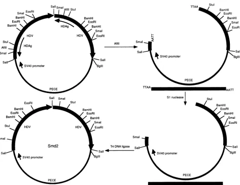
FIG. 1. Schematic diagramofthe construction ofHDVdimermutant Smd2and the relevant restrictionenzymesites. The thickarrow
represents HDV monomer sequencesofgenomicsense.The thinarrowrepresents theHDAg-coding region,which isonthe antigenomic orientation.The thin line representsPECEvectorsequence(13).
usingthe localhomology algorithmof Smith and Waterman (37).
Construction ofplasmids. Allof theconstructs weremade from the cDNA clones of Southern California isolate of HDV RNA(30). HDAg-truncated mutant plasmids L6and L14 were derived from pATH-M (26), which encodes the middle one-third (aminoacids 79 to163) of HDAg fusedto the TrpE protein of Escherichia coli, by the exonuclease digestionmethoddeveloped by Henikoff(17). For construc-tion of plasmids WD and L10, the full-length and the C-terminal two-thirds(aminoacids 108to214)ofthe HDAg-coding region,respectively, were made by polymerase chain reaction to amplify HDAg sequences of the desired regions by using two oligonucleotide primers which contained the appropriateHDVsequencesplusBamHIand HindIII linker sequences. The cDNA products were then inserted into pATH-2 polycloning sites (12). For construction of plasmid D1-2, twocDNAs encoding amino acids 1 to 109 and 136 to 214 of HDAg and containing XbaI site at both ends were made by polymerase chain reaction and inserted into pATH-2. The HDAg-coding sequences inAl, A2, and A2' weremade by polymerase chain reaction using the method described by Higuchi et al. (18) and inserted into pATH-2 polycloningsitesBamHI and HindIII.
The replication-defective HDV dimer construct, Smd2, which wasusedfor thetestingof thetrans-acting
activity
of the HDAg mutants,was constructed by cloning a head-to-taildimer offull-lengthHDV cDNA(30)
intothe Sall siteof the PECE vector, which containsasimian virus 40T-antigen promoter(13). Theresulting plasmidwas digestedintotwofragmentswithAflII;both of thefragmentswereblunt ended with nucleaseS1andreligated(Fig. 1).Asaresult,adeletion of 4nucleotides andaframeshiftwereintroducedatthesite correspondingtoamino acid 130 of the
HDAg-coding
region. Thisplasmidtranscribedareplication-defectiveHDVdimer RNA, which could replicate onlywhen awild-type HDAg wassupplied. PECE-Sm, which expressesawild-type small HDAg,was madeby inserting the EcoRI-BamHI fragment of the HDV cDNAencodingthesmallHDAginto the EcoRI andBglIIsites of thePECEvector.Otherplasmids express-ingvarious HDAg mutants,includingPECE-Al,PECE-A2, PECE-A2', and PECE-D1-2, were made by replacing the StuI-SmaI fragment of PECE-Sm with the corresponding fragments from Al, A2, A2', and D1-2, respectively. The sequences of the resulting plasmids were confirmed by dideoxyribonucleotidechain termination sequencing(34).PreparationofTrpE-HDAg fusion proteins.Bacteriawere grown by previously published procedures (26). Briefly,
.M
on November 9, 2019 by guest
http://jvi.asm.org/
[image:2.612.68.541.77.443.2]culturesofE. coli MC1061 cells transformed withplasmids encoding different TrpE-HDAgmutant fusionproteins were induced with3-indoleacrylic acid(40,ug/ml)for 3 h. Bacteria were lysed by digesting with lysozyme (1 mg/ml) and re-peated freeze-thawing. The lysates were further digested with DNase I. The fusion proteins were collected in the insoluble fraction and resuspended in a buffer containing 0.01 M sodiumphosphate (pH 7.2), 0.1% ,3-mercaptoetha-nol, 1% sodium dodecyl sulfate (SDS), and 6 M urea as previously described(26).
Immunoblot and RNA-protein blot procedures. TrpE-HDAg fusion proteins were separated by SDS-polyacryl-amide gel electrophoresis (PAGE) (7.5% polyacrylamide) and electrotransferred to nitrocellulose membranes. For immunoblot analysis, the membranes were first incubated with5% nonfat milk and then with anti-HDAg antibody(26) followedby125I-labeled proteinAaccordingtothepublished procedures (9). ForRNA-protein blot(Northwestern) anal-ysis, the membranes were incubated in standard binding buffer(SBB;10 mM Tris-HCl
[pH
7.4],1 mMEDTA,50 mM NaCl, 0.02% bovine serum albumin, 0.02% Ficoll, and 0.02%polyvinylpyrrolidone) containing10 ,ug of yeast trans-fer RNA perml and 100 ,ug of cellular RNA from DBT cells per ml for 1 h and then incubated with 32P-labeled HDV genomicRNAin thesamebuffer foranotherhouraccording to thepublishedprocedures(26). Afterwashingthreetimes with SBB at room temperature for 15 min each, the mem-braneswereexposedtoX-rayfilms.RNA mobility shift assay. RNA mobility shift assay was carried out as previously described (26). Briefly, 20 ,ugof partially
purified
TrpE-HDAgproteins was incubated with 10 ng of32P-labeled
HDVgenomic RNA in 10 ,ul ofbuffer containing2.5 mM Tris-HCl(pH7.4),
100 mMNaCl,2 mM MgCl2, 1 mMdithiothreitol,
and20%glycerol
with 40 Uof RNasin(Promega),10 ,ugoftRNA,and 10 ,ug of totalcellular RNAat37°Cfor 30 min.Afterincubation,thesampleswere directly analyzed by electrophoresis under nondenaturing conditions. Electrophoresiswascarried outon a0.6% low-melting agarose gel at 25 V for 15 h at 4°C (26). After electrophoresis, thegelwasdried and visualizedby autora-diography.DNAtransfection. COS 7 cells (16) at 50% confluencyin
6-cm
petri
dishes were incubated with 5 p,g of PECEplasmids containing
replication-defective
HDV dimerDNA, plasmids encoding either the wild-type or mutant small HDAg, and 25 ,ug ofLipofectin
(Bethesda
Research Labo-ratories) for 12 h. The cells were washed with Dulbecco modified Eagle medium(DMEM)
and then incubated with DMEM containing 5% fetal calfserum for another 5days.
Thetotal cellularRNAwasthenextracted by acid
guanidi-nium isothiocyanate
(11)
at 6 daysposttransfection.
The RNA was analyzed by Northern blotanalysis using
32p_ labeledanti-genomic HDV RNAasaprobe(43).RESULTS
RNA-binding motifs ofHDAg.To
identify
the amino acid residuesresponsible
for the RNAbinding
ofHDAg,
wefirst searched for the presence of the knownRNA-binding
motifs inHDAg. Since theRNA-binding
region
is located within the middle one-third domain(amino
acids 79 to163)
of HDAg(26),we examined whether thisregion
containedanRNA recognition motifor an ARM. No RNA
recognition
motifwas found in this
region. However,
two stretches of amino acid sequences have differentdegrees
ofsimilarity
with abatteryof the known ARMs
(25).
Thefirstone wasKERqdhRRRKa
eDeKRERRiag
qTRRRERRcEK
RKKRRgRRRap
sQRRnrRRRNK
BOBRBJRRZZB
1st ARM
of
HDAg
(aa
97-107)
2nd
ARMof
HDAg
(aa
136-146)
N
of
bacteriophage
xTat
of
HIV-1Rev
of
HIV-2
Consensus
FIG. 2. Conserved ARMs in representative RNA-binding pro-teins. Abbreviations:B,basic;0,nonbasicpolar; Z, charged; and J, acidic amino acids; HIV, human immunodeficiency virus. Part of the dataarefrom reference25.
KERQDHRRRKA (from amino acids 97 to 107), and the second was EDEKRERRIAG (from amino acids 136 to 146) (Fig. 2). The presence of two stretches of ARM-like se-quences in the same protein is unique among the known RNA-binding proteins.
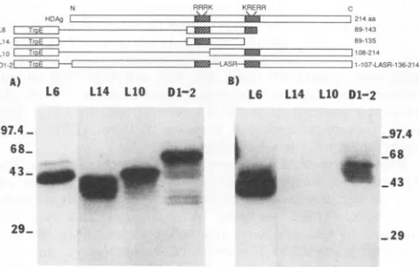
RNA-protein-binding assays of HDAg deletion mutants. To determine whether either of the ARM-like sequences was involved in RNA binding, we made a series of deletion mutants starting from the C-terminal end of the middle domain (M)of HDAg(aminoacid residues 89 to 163), which includes the necessary and sufficient RNA-binding se-quences (26). These mutants were expressed in E.
coli
as TrpE fusionproteins. The partially purified fusion proteins wereseparatedby SDS-PAGE, transferred to nitrocellulose filter membrane, and incubated with32P-labeled
HDV genomic RNAby aNorthwestern procedure, under condi-tions shown to allow specific binding between HDAg and HDV RNA (6, 26). Among these deletion mutants, L6 containsthelongestHDAg sequence(amino acid residues 89 to143)and was found to bind HDV RNA (Fig. 3B). Further truncationatthe C-terminal end resulted in the complete loss of the RNA-binding activity. For examples, mutant L14 (amino acids 89 to 135) and smaller mutants did not bind HDV RNA (Fig. 3B and data not shown) when the sameN RPPR RKRE
HAgi ^~WMO i _ 2 4 aa
T-_TozL -43
A) B)
L6 L14 L10 D1-2
97.4
-68..
43-__M
-97.4
_68
143
29_
_29
FIG. 3. RNA-protein bindingassaysof theHDAgdeletion mu-tants. Thevarious TrpE-HDAgfusionproteinswereseparated by
SDS-PAGE, electrotransferred to nitrocellulose membranes, and
incubated with an anti-HDAg antibodyfor thequantitation of the
fusion proteins by immunoblot analysis (A) or incubated with
32P-labeled HDVgenomicRNA forRNA-bindingassaysby North-western blot analysis (B) as described in Materials and Methods.
Theschematicdiagramof thestructuresof theTrpE-fusion proteins
isshown above. The molecularmass markers (in kilodaltons) are
indicated. The shadedareasrepresenttheARM-like sequences.
L6 L14 110 D1-2
on November 9, 2019 by guest
http://jvi.asm.org/
[image:3.612.326.562.483.634.2].!. _ __ :: :-_: AZ II Y: l=
A? E ; ZZZII_ILZZ±iLIII .1 __ .;-_
P
WD
Al
A2
WD Al A2 A2' B) WD Al A2 A2 97.4 68 43
FIG. 4. RNA-proteinbindingassaysof thesite-specificmutants
of the HDAg ARM sequences. The different TrpE-HDAg fusion proteinswereseparated bySDS-PAGE, electrotransferred to nitro-cellulose membranes, and incubated withanti-HDAg antibodyfor immunoblot analysis (A) or incubated with 32P-labeled HDV genomic RNAfor Northwestern blot analysis (B). The schematic diagram of the structures of the TrpE-fusion proteins is shown above.The molecularmassmarkers(inkilodaltons) areindicated. ThemutatedARMsequencesareidentifiedbylighter shaded boxes, andthesubstituted amino acids areindicated.
amountsof thevariousfusionproteinswereused(Fig.
3A).
Thus, residues 135 to 143 appear to contain sequences necessaryfor HDV RNAbinding.This stretchofsequences corresponds to a major portion of the second ARM-like sequence (Fig. 2). To delineatethe contribution of the first ARM, another mutant,L10,which spansamino acids 108to 214and containsonlythesecond ARM butnotthefirst,was constructed. Thisproteindidnotbind HDV RNA
(Fig. 3B).
Thus, mutants without either the first or second ARM showednobindingtoHDV RNA. This resultindicates that the both ARMs of HDAg arenecessary for RNAbinding. Theimportance of the spacerregionbetween thetwoARMs wasaddressed by another mutant,D1-2,in which the entire spacer sequence(residues108to135)wasreplacedwith four irrelevant amino acids(Leu, Ala, Ser, andArg).Thismutant retained only a small fraction (approximately 5% of the wild-type protein) of the RNA-binding activity (Fig. 3B). Thus, the spacer sequence between thetwo ARMs is also importantforthe RNAbinding.
[image:4.612.349.514.78.264.2]RNA-protein-binding assays of site-specific mutants. The importance of the both ARM-likesequencesinRNAbinding was further assessed by site-specific mutagenesis of these sequences. Three mutants, Al (Arg-104 and Arg-105; changed to Gln and Gly, respectively), A2 (Arg-142 and Arg-143; changed to Ser and Gly, respectively), and A2' (Lys-139, changed to Asn, and Arg-140, changed to Gly) were made on the full-length HDAg-TrpE fusion protein. TheirRNA-binding activities were examined by Northwest-ern blot with
32P-labeled
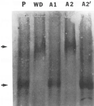
HDV genomic RNA. The same amountsof thefusion proteins were used for each mutant, as evidenced by immunoblot analysis (Fig. 4A). The result showed that the substitutions of Arg in the first ARM(Al) completely eliminated the RNA-binding activity (Fig. 4B). The effects of mutations in the second ARM were more variable. A2', which had substitutions of the first Lys andFIG. 5. RNAmobility shiftanalysis of thearginine mutantsof thetwoARMs.DifferentmutantHDAgproteinsasdescribed in the legendtoFig.4 wereincubated with32P-labeledHDVgenomicRNA andseparated byelectrophoresison0.6%low-melting-pointagarose gels under nondenaturing conditions (26). P, HDVRNA. Arrows indicate the freeRNAandRNA-protein complexes.
Arg, hada 10-fold-lowerbindingactivitythanthewild-type HDAg. In contrast,A2, which had substitutions ofthetwo downstreamarginines, hadafourfold lowerbinding activity (Fig.4B).Thespecificityof thisbindingassayhaspreviously been demonstrated by competition with the excess cold RNA(26).TheRNA-binding activitiesof themutantHDAgs werefurther examined byRNAmobilityshift assays
(Fig.
5). The results showed that the wild-type HDAg and A2 mutantproteinformedan RNA-protein complexwith HDV genomicRNA.Incontrast,onlynegligibleamountsofAlor A2' mutants formed the RNA-protein complex with HDV RNA under the same conditions. Again, thespecificity
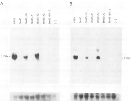
of this assay haspreviouslybeendemonstrated(26). Thus,the two upstream basic amino acids in the second ARM-like sequenceare moreimportantthan the downstreamonesfor the RNAbinding. These studies indicated that both of the ARM-like sequences are necessary for the RNA-binding activity of HDAg.Biological significance of the RNA-binding activity of HDAg. To determine thebiologicalsignificanceof the RNA-binding activityofHDAg,weexaminedwhether the RNA-binding activity ofHDAgwas requiredfor its trans-acting activity for HDV RNA replication (22). We constructed a plasmid expressing a defective HDV dimer which had a frameshift mutation inthe HDAg-coding region. When this plasmidwas transfected into COS 7 cells, very little HDV monomer RNA was detected, indicating that this plasmid couldnotreplicate (Fig. 6, laneSmd2). However,when this construct was cotransfected with a plasmid (PECE-Sm) expressing a wild-type small HDAg, an HDV monomer RNA could be detected (Fig. 6, lane Sm), suggesting that HDAg could activate RNA replication in trans. This result was in agreement with the published finding that HDAg is required for HDV RNA replication (22). When the HDV dimer construct was cotransfected with the different plas-mids expressing an HDAg with various mutations in the RNA-binding motif, onlymutantA2,which retained most of the in vitro RNA-binding activity, could restore the RNA replicationof Smd2. The level of HDV RNA replication was A)
A21
97.4
.... .. ...
43~~~~~~~~~~~
on November 9, 2019 by guest
http://jvi.asm.org/
[image:4.612.58.294.78.262.2]A
LHDAg
SHDAg
S 0
CCS NLS ARM ARM PAS
ti WEM
m
11111111111
rommWE
El
Coiled-coil sequence (CCS)El
Nuclear localization signal (NLS)0
Arginine-rich motif (ARM)In
Packaging-associated sequence (PAS)FIG. 7. Schematicdiagram ofthe functional domains of HDAg.
FIG. 6. Analysis of the trans-activating activity of mutant
HDAgs withadefectiveHDV dimer andmutantHDAg-expressing plasmids. COS 7 cellswerecotransfected with HDV dimer DNA
(Smd2) and various mutantHDAg DNA as described in Results.
Cellular RNAwasextracted 6 days posttransfection, separated in
formaldehyde gels,transferred tonylon membrane, and incubated
with 32P-labeledHDVantigenomic-sense (A)orgenomic-sense (B)
RNA. The membranewasalsoincubatedwith 32P-labeled ribosomal RNA (lower panels) for the quantitation of RNA. The arrow
indicates monomeric HDV genomicRNA. HIb9isanHDVstably
transformed COS 7 cell line which serves as a size marker for
monomerHDV RNA.AllconstructsareinPECEplasmidwith the
simian virus 40earlypromoter(13).
similar when either thewild-type HDAgor A2mutant was
used. Theremainingmutants,Al, A2',andD1-2,which had
no oronlyminimal RNA-binding activities, didnot comple-mentthe defects ofreplicationof Smd2 RNA(Fig. 6A).This resultwasconfirmedbythe relativeamountsofantigenomic RNA synthesized in cells transfected with Smd2 and the different HDAg mutants (Fig. 6B), while the amounts of RNAused for Northern blotanalysiswereshowntobethe
same for all of the samples. We thus conclude that the RNA-binding activity is crucial for thetrans-activating
ac-tivityofHDAg.
DISCUSSION
HDAgis anRNA-binding proteinwithabinding
specific-ityfor HDV RNA(26). The resultspresentedinthis report showed that the RNA-binding properties of HDAg require
twostretches ofthe ARM-likesequenceworkingin concert. Furthermore, a spacer sequence separating the two ARM-like sequences also contributes to the binding activity. Although the ARM-like sequence has previously been de-tected in several prokaryotic and eukaryotic RNA-binding proteins (25),this is the firstproteinshowntorequiretwoof these sequencesfor RNAbinding. Thus, HDAg likely rep-resents a new type of RNA-binding protein which has a
unique specificityfor HDV RNAsequenceorstructure. We havepreviouslyshownthatHDAgbindsspecificallytoHDV RNA butnottoother viralorcellular RNA(26).Thebinding specificity appears not to be dependent on nucleotide
se-quencebutmorelikelyonsecondarystructureof RNA(26). Chao et al. further demonstrated that HDAgbinds only to
the rod structure of HDV RNA (8),which is generated by intramolecular base pairing (4, 38). However, a simple
double-stranded RNA structure derived from an unrelated
virus did not bind to HDAg (26), suggesting that some RNA sequence specificity is also required for the protein-RNA interaction.Furthermore, we have demonstrated that a small RNA fragment (110 nucleotides) containing the ribozyme domain of HDV genomic RNA can also bind to HDAg (unpublished observation). Thisfragment does not form the rod structure but forms a multiple stem-and-loop structure (42). These results suggest that HDAg might recognize a double-stranded RNA structureunique to HDV RNA. Con-ceivablythetwoARM-like sequences may bind to different RNA strandsordifferent regions of such an RNA structure, thusexplainingtherequirement for a spacer sequence. The precise RNA structure of the HDAg-binding sites will re-quire additional studies.
HDV RNAreplication requires a trans-acting function of HDAg(22),althoughtheprecise role of HDAg is not clear. A previous study suggests that HDAg is required for the nuclear transport of HDV RNAbut not directly involved in RNAtranscription perse(28). However, this interpretation cannot explain the failure of large HDAg to trans activate HDV RNAreplication since both large and small HDAgs are transported to the nuclei (44). Conceivably, HDAg may serve as a transcription factor to enhance the efficiency of HDV RNA replication. The result presented in this report showed that theRNA-binding activity of HDAg is required for itstrans-activating activity, suggestingthat HDAg has to bind to HDV RNA to fulfill whatever role HDAg plays in HDV RNAreplication. However, the binding of HDAg to HDV RNAisnotsufficient for HDV RNA replication, since both thelarge and small HDAgs bindtoHDV RNA at equal efficiency (20); yet only the small one can trans-activate RNA replication while the large one inhibits it (7, 14, 22). Conceivably, HDAg binds to RNA, allowing other func-tionaldomains ofHDAgtointeractwith components of the transcriptional machinery. Thisinteraction may be possible only with the small HDAg. This possible mechanism of action of HDAg is similar to that of several other RNA-binding proteins which contain anARM-like sequence and play regulatory roles intranscription. For example, bacterio-phagelambda Nprotein,when itbindstothenutsite, may engage RNApolymeraseandmodifyit in awaytoprevent termination(2, 19). Also,thetatproteinof HIV-1 increases transcriptionprocessivity throughinteraction with theTAR element(24).Both of theseproteins recognize specificRNA sequences through a single ARM
(25).
HDAg may use a similar mechanismtoredirect host cellRNApolymerasefor HDV RNAreplication.
Several functional domains have been identified inHDAg (Fig. 7). The nuclear localization signal
(NLS)
of HDAg directsHDVRNA into thenuclei(44)
sothatRNA replica-tioncanutilize the nuclearmachinery
(28).
Thecoiled-coilor7 :: = Z. 17
on November 9, 2019 by guest
http://jvi.asm.org/
[image:5.612.326.568.72.195.2] [image:5.612.75.298.79.251.2]leucine-zipper
sequencepermits HDAg oligomerization
andis also
required
for RNAreplication (43).
It appears thatHDAg
formsanoligomer
toparticipate
in RNAreplication;
however, oligomerization
ofHDAg
is notrequired
for itsRNA
binding,
since the middle domain ofHDAg,
withoutthecoiled-coil sequence, is sufficient for RNA
binding
(26).
Finally,
the C-terminal 19 amino acids of thelarge
HDAghave an
isoprenylation
site(15)
andprobably
contain thesequences
(packaging-associated sequences) required
forthepackaging
of HDV RNAduring
theassembly
of HDVparticles (5, 33).
Theincorporation
of HDV RNA into virusparticles
mostlikely requires
theRNA-binding activity
ofHDAg
as well.Indeed,
HDV RNA andHDAg
have been shown to form acomplex
inside the virionparticles
(26).
Thus,
HDAg-HDV
RNAbinding
isrequired
notonly
forRNA
replication
but also forassembly
of infectiousvirusparticles.
It should be notedthatsomeof themutantHDAgs
(A2'
andD1-2)
retained a residual amountofRNA-binding
activity
andyetnoHDV RNAreplication
could be detectedwhen
they
werecotransfected with themutantHDVdimer.Thus,
theregion
containing
theRNA-binding
motifs mayhave other activitiesaswell.
Sequence analysis
suggests that there is ahelix-loop-helix
structurebetween the two ARM-like sequences(data
notshown). Conceivably,
the mutations in the ARM sequences may also affect this and otherneighboring
structures. Thispossibility
wouldexplain
thetotal lack of the
trans-activating activity
of mutantD1-2,
which had deleted the spacer sequence and
thus,
thehelix-loop-helix
structure. Theproperties
of these additionalse-quenceswill
require
further studies. ACKNOWLEDGMENTSThis work was
supported
in partby
a Public Health Service Research grant(AI 26741)
from the National Institutes ofHealth. M.C. is a research associate and M.M.C.L is an investigator of HowardHughes
Medical Institute.REFERENCES
1. Bandziulis, R. J., M. S. Swanson, and G. Dreyfuss. 1989.
RNA-binding proteins
asdevelopmental
regulators.Genes Dev.3:431-437.
2. Barik,S.,B.
Ghosh,
W.Whalen,D. Lazinski,and A. Das.1987. Anantiterminationprotein
engagestheelongating
transcription apparatusatapromoter-proximal recognitionsite. Cell50:885-899.
3. Bonino, F., K.H. Heermann,M. Rizzetto, and W. H.Gerlich. 1986.Hepatitisdelta virus:protein compositionof deltaantigen and its
hepatitis
Bvirus-derivedenvelope.J.Virol. 58:945-950. 4. Branch,A.D.,B.J.Benenfeld,B.M.Baroudy,F. V.Wells,J.L.Gerin,and H.D. Robertson.1989. Anultraviolet-sensitiveRNA structuralelement inaviroid-likedomain of thehepatitis delta virus. Science243:649-652.
5.
Chang,
F.L.,P.J.Chen, S.J.Tu,C.J.Wang,and D.S.Chen.1991. The
large
form of hepatitis delta antigen is crucial forassembly
ofhepatitis
deltavirus. Proc. Natl. Acad. Sci. USA88:8490-8494.
6.
Chang,
M.F.,S. C.Baker,L.H.Soe,T.Kamahora,J.G.Keck,S.Makino, S.Govindarajan,and M. M. C. Lai. 1988. Human
hepatitis
delta antigenisanuclearphosphoproteinwithRNA-binding activity.
J.Virol. 62:2403-2410.7. Chao, M.,S.-Y.Hsieh,andJ.Taylor.1990.Role oftwoforms of
hepatitis
delta virus antigen: evidence for a mechanism ofself-limiting
genomereplication. J.Virol. 64:5066-5069.8.
Chao,
M., S. Y. Hsieh, and J.Taylor. 1991. The antigen ofhepatitis
delta virus: examination of in vitro RNA-bindingspecificity.
J. Virol.65:4057-4062.9.
Chao,
Y. C., M. F. Chang, I. Gust, andM. M. C. Lai. 1990.Sequence
conservation and divergenceofhepatitis delta virusRNA.
Virology
178:384-392.10. Chen,P.-J., G. Kalpana,J. Goldberg,W.Mason, B.Werner, J.L. Gerin, andJ. Taylor. 1986. Structure and replication of the genome of thehepatitis delta virus. Proc. Natl. Acad. Sci. USA 83:8774-8778.
11. Chomczynski, P., and N. Sacchi. 1987. Single-step method of RNA isolationby acid guanidinium thiocyanate-phenol-chloro-form extraction. Anal. Biochem. 162:156-159.
12. Dieckmann, C. L., and A. Tzagoloff. 1984. Assembly of the mitochondrial membrane system, ayeast nucleargene
neces-saryfor synthesis of cytochromeb. J. Biol. Chem. 260:1513-1520.
13. Ellis, L.,E.Clauser, D. 0. Morgan, M.Edery,R. A.Roth, and W.J. Rutter. 1986. Replacement of insulin receptor tyrosine residues 1162 and 1163 compromises insulin-stimulatedkinase activityanduptakeof2-deoxyglucose. Cell 45:721-732. 14. Glenn, J. S.,andJ.M. White.1991. trans-dominant inhibitionof
human hepatitis delta virus genome replication. J. Virol. 65: 2357-2361.
15. Glenn, J. S., J. A. Watson, C. M. Havel, andJ. M. White. 1992. Identification ofa prenylationsite in delta virus large antigen. Science256:1331-1333.
16. Gluzman, Y. 1981. SV40-transformed simian cellssupport the replicationofearlySV40mutants. Cell 23:175-182.
17. Henikoff,S. 1984. Unidirectionaldigestionwith exonuclease III
createstargeted breakpoints for DNA sequencing. Gene 28:351-359.
18. Higuchi, R., B. Krummel, and R. K. Saiki. 1988. A general method of in vitropreparation and specific mutagenesis of DNA fragments: study of protein and DNA interactions. Nucleic Acids Res. 16:7351-7367.
19. Horwitz, R. J., J. Li, and J. Greenblatt. 1987. An elongation controlparticle containing the Ngenetranscriptional antitermi-nationprotein of bacteriophage lambda. Cell51:631-641. 20. Hwang, S.,C. Z.Lee,and M. M.C. Lai. 1992. Hepatitis delta
antigen expressed by recombinant baculoviruses: comparisonof biochemicalpropertiesandpost-translational modifications
be-tweenthelargeand small forms.Virology190:413-422. 21. Kos, A.,R.Dikema,A. C.Arnberg,P. H.vanderMerde, and
H. Schellekens. 1986. The HDV possesses a circular RNA. Nature(London)323:558-560.
22. Kuo, M. Y.-P., M. Chao, and J. Taylor. 1989. Initiation of replication of the human hepatitis delta virus genome from cloned DNA: role of deltaantigen.J. Virol. 63:1945-1950. 23. Kuo,M.Y.-P.,L.Sharmeen,G.Dinter-Gottlieb,andJ.Taylor.
1988. Characterizationofself-cleavingRNA sequencesonthe genomeandantigenome of human hepatitis delta virus. J.Virol. 62:4439-4444.
24. Laspia,M.F.,A. P.Rice,andM. B. Mathews. 1989. HIV-1 Tat proteinincreasestranscriptionalinitiation andstabilizes elonga-tion. Cell 59:283-292.
25. Lazinski, D., E. Grzadzielska, and A. Das. 1989. Sequence-specific recognitionofRNAhairpins by bacteriophage antiter-minatorsrequires aconservedarginine-rich motif. Cell 59:207-218.
26. Lin, J. H., M. F. Chang, S. C. Baker, S. Govindarajan, and M. M. C. Lai.1990.Characterization ofhepatitisdelta antigen: specific bindingtohepatitisdelta virus RNA. J. Virol. 64:4051-4058.
27. Macnaughton,T. B., E. J. Gowans, A. R. Jilbert, and C. J. Burrell.1990.Hepatitisdelta virusRNA,protein synthesis and associatedcytotoxicityinastably transfectedcell line. Virology 177:692-698.
28. Macnaughton,T.B.,E.J.Gowans,S. P.McNamara, and C. J. Burrell. 1991.Hepatitisdeltaantigen isnecessaryfor access of hepatitisdelta virus RNAtothe cell transcriptional machinery but isnotpart ofthetranscriptionalcomplex. Virology 184:387-390.
29. Makino, S., M. F. Chang, T. Kamahora, D. M. Vannier, S. Govindarajan,andM. M.C. Lai.1987. Molecularbiology of a humanhepatitisdeltavirusRNA,p.549-564.In W. Robinson, K. Koike, and H. Will(ed.), Hepadnaviruses. Alan R. Liss, Inc.,New York.
30. Makino, S., M. F. Chang, T. Kamahora, D. M. Vannier, S.
on November 9, 2019 by guest
http://jvi.asm.org/
Govindarajan,andM.M. C. Lai. 1987. Molecularcloningand
sequencing of a human hepatitis delta virus RNA. Nature (London) 329:343-346.
31. Mattaj, I. W. 1989.Abindingconsensus: RNAprotein
interac-tionsinsplicing, snRNPs,andsex.Cell57:1-3.
32. Rizzetto, M. 1983. Thedelta agent.Hepatology 3:729-737. 33. Ryu, W. S., M. Bayer, and J. Taylor. 1992. Assembly of
hepatitis delta virus particles. J. Virol.66:2310-2315.
34. Sanger,F., S. Nicklen,and A. R. Coulson. 1977. DNA
sequenc-ing with chain-terminating inhibitors. Proc. Natl. Acad. Sci.
USA 74:5463-5467.
35. Sharmeen, L.,M. Y.-P.Kuo, G.Dinter-Gottlieb,andJ. Taylor. 1988. Antigenomic RNA of human hepatitis delta virus can
undergo self-cleavage. J.Virol. 62:2674-2679.
36. Sharmeen, L.,M. Y.-P.Kuo,andJ.Taylor. 1989.Self-ligating
RNA sequences on the antigenome of human hepatitis delta
virus. J. Virol. 63:1428-1430.
37. Smith, T. F., and M. S. Waterman. 1981. Comparison of
biosequences.Adv.Appl.Math.2:482-489.
38. Wang, K. S., Q. L. Choo, A. J. Weiner, J. H. Ou, R. C.
Najarian, R. M. Thayer, G. T. Mullenbach, K. J. Denniston, J. L. Gerin, and M. Houghton. 1986. Structure, sequence and
expressionof thehepatitisdelta viralgenome.Nature(London)
323:508-514.
39. Weiner, A. J., Q.-L. Choo, K.-S. Wang, S.Govindarajan,A.G.
Redeker,J. L. Gerin,and M.Houghton. 1988.Asingle
antige-nomicopenreadingframe of the hepatitis delta virus encodes
theepitope(s) of both hepatitis delta antigen polypeptides p248 andp278.J. Virol. 62:594-599.
40. Wu, H. N., and M. M. C. Lai. 1990. RNA conformational requirementsofself-cleavageofhepatitisdelta virus RNA. Mol. Cell. Biol. 10:5575-5579.
41. Wu, H. N., Y. J. Lin, F. P. Lin, S. Makino,M. F.Chang,and
M. M. C. Lai. 1989.Human hepatitis delta virus RNA
subfrag-mentscontainanautocleavage activity. Proc. Natl. Acad. Sci.
USA 86:1831-1835.
42. Wu, H. N., Y. J. Wang, C. F. Hung, H. J.Lee, and M. M. C. Lai. 1992. Sequence and structure of the catalytic RNA of hepatitis delta virus genomic RNA. J. Mol. Biol. 223:233-245. 43. Xia, Y. P., and M. M. C. Lai. 1992. Oligomerization of hepatitis
delta antigen is required for both the trans-activating and trans-dominant inhibitory activities of the delta antigen. J. Virol. 66:6641-6648.
44. Xia, Y. P., C. T. Yeh, J. H. Ou, and M. M. C. Lai. 1992. Characterization of nuclear targeting signal of hepatitis delta antigen: nuclear transport as a protein complex. J. Virol.
66:914-921.