THE TAMILNADU Dr. M.G.R. MEDICAL UNIVERSITY
In partial fulfillment for the degree of
MASTER OF DENTAL SURGERY
BRANCH V
ACKNOWLEDGEMENT
I would like to acknowledge and thank my beloved Professor and Head,
Dr. N. R. Krishnaswamy, M.D.S., M. Ortho (RCS, Edin), D.N.B. (Ortho), Diplomate of Indian board of Orthodontics, Department of Orthodontics, Ragas
Dental College and Hospital, Chennai. I consider myself extremely fortunate to
have had the opportunity to train under him. His enthusiasm, integral view on
research, tireless pursuit for perfection and mission for providing ‘high quality
work’, has made a deep impression on me. He has always been a source of
inspiration to strive for the better not only in academics but also in life. His
patience and technical expertise that he has shared throughout the duration of the
course has encouraged me in many ways.
I am privileged to express my extreme gratefulness to my respected guide,
Professor Dr.M.K.Anand, M.D.S., for his guidance which helped me complete this study. His everlasting inspiration, encouragement, constructive criticism and
valuable suggestions conferred upon me have encouraged me.
Dr. Dr.Shakeel Ahmed (Professor), Dr.Rekha Bharadwaj (Reader),
Dr.Shobbana Devi (Reader), Dr.Premalatha (Reader), Dr.Kavitha Iyer
(Reader), Dr.Dhivya Lakshmi (Sr. Lecturer) and Dr.Bharath Ramesh
(Sr.Lecturer) for their support, enthusiasm & professional assistance throughout my
post graduate course.
My deep thanks to my wonderful batch mates, Dr.sheril selvan, Dr.Bajath Balakrishnan, Dr.Lily Arun, Dr.Kowtham Raj, Dr.N.Grace Ruth, Dr.Maryam Baneshi and Dr.Vidhya.R.S , who were cheerfully available at all times to help me. Their support and friendship was constant during these last years,
and I deeply appreciate it.
I also extend my gratitude to my juniors, Dr.Nandhini, Dr. Pradeep kumar, Dr.Sumin.S.G, Dr.Asha George, Dr.Jeevan Jerald, Dr.Divya, Dr.Deepak, Dr.Muthu pradeep, and Dr.Vaishnav for their support.
I would like to thank Mr. Ashok, Mr.Manikandan, Mr.Bhaskar, Sister Lakshmi, Sister Yamini, Sister Kanaka, Mrs. Uma, the Scribbles team for their co-operation and help during my course of study .I would like to mention my
sincere gratitude to C-tars team and Berinnov dental laboratory.
like to express my heartfelt love and gratitude to my mother Mrs.Remani, without whom this venture would have been impossible. I thank the most merciful and
compassionate Almighty God, He guided and helped me throughout my life in
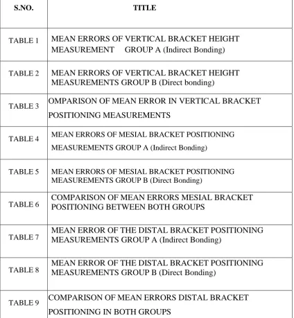
LIST OF TABLES
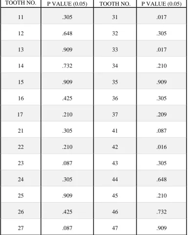
[image:9.595.98.523.285.745.2]S.NO. TITLE
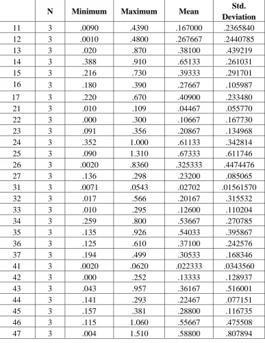
TABLE 1 MEAN ERRORS OF VERTICAL BRACKET HEIGHT
MEASUREMENT GROUP A (Indirect Bonding)
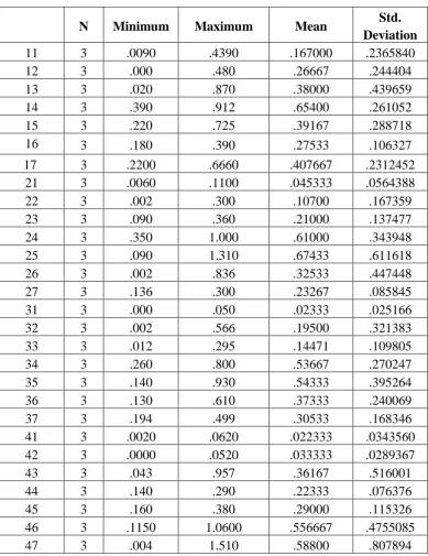
TABLE 2 MEAN ERRORS OF VERTICAL BRACKET HEIGHT MEASUREMENTS GROUP B (Direct bonding)
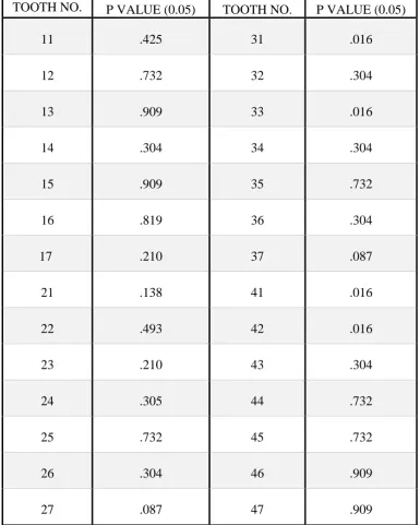
TABLE 3 OMPARISON OF MEAN ERROR IN VERTICAL BRACKET
POSITIONING MEASUREMENTS
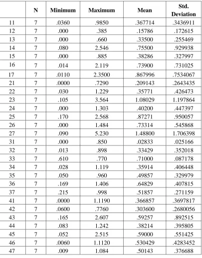
TABLE 4 MEAN ERRORS OF MESIAL BRACKET POSITIONING MEASUREMENTS GROUP A (Indirect Bonding)
TABLE 5 MEAN ERRORS OF MESIAL BRACKET POSITIONING MEASUREMENTS GROUP B (Direct Bonding)
TABLE 6 COMPARISON OF MEAN ERRORS MESIAL BRACKET POSITIONING BETWEEN BOTH GROUPS
TABLE 7 MEAN ERROR OF THE DISTAL BRACKET POSITIONING MEASUREMENTS GROUP A (Indirect Bonding)
TABLE 8 MEAN ERROR OF THE DISTAL BRACKET POSITIONING MEASUREMENTS GROUP B (Direct Bonding)
TABLE 9 COMPARISON OF MEAN ERRORS DISTAL BRACKET
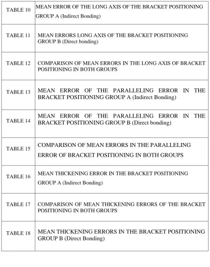
GROUP B (Direct bonding)
TABLE 12 COMPARISON OF MEAN ERRORS IN THE LONG AXIS OF BRACKET POSITIONING IN BOTH GROUPS
TABLE 13 MEAN ERROR OF THE PARALLELING ERROR IN THE
BRACKET POSITIONING GROUP A (Indirect Bonding)
TABLE 14 MEAN ERROR OF THE PARALLELING ERROR IN THE BRACKET POSITIONING GROUP B (Direct bonding)
TABLE 15 COMPARISON OF MEAN ERRORS IN THE PARALLELING
ERROR OF BRACKET POSITIONING IN BOTH GROUPS
TABLE 16 MEAN THICKENING ERROR IN THE BRACKET POSITIONING GROUP A (Indirect Bonding)
TABLE 17 COMPARISON OF MEAN THICKENING ERRORS OF THE BRACKET POSITIONING IN BOTH GROUPS
TABLE 18 MEAN THICKENING ERRORS IN THE BRACKET POSITIONING
[image:10.595.95.524.106.652.2]LIST OF GRAPHS
S.NO. TITLE
GRAPH 1 CLINICAL CROWN HEIGHT
GRAPH 2 MARGINAL RIDGE TO THE CENTRE OF THE BRACKET
GRAPH 3 MARGINAL RIDGE TO FACC
CONTENTS
S .No. TITLE PAGE NO
1. INTRODUCTION 1
2. AIM AND HYPOTHESIS 3
3. OBJECTIVES 4
4. REVIEW OF LITERATURE 5
5. MATERIALS & METHODS 37
6. RESULTS 43
7. DISCUSSION 46
1
INTRODUCTION
Accurate bracket positioning forms the theoretical underpinning for
fixed appliance therapy in orthodontics. Notwithstanding clinician skill along
with distinct angulation and inclination values cut into the bracket, forced and
unforced errors during direct bonding technique make realization of
malocclusion correction a less successful one; not to mention time consuming.
In an attempt to overcome the disadvantages of direct bonding procedure,
many clinicians began to explore the indirect bonding technique first
introduced by Silverman et al69.
The indirect bonding system has many advantages: it makes the patients
comfortable, facilitates easy reproduction of the bracket, provides easy over
correction, allows for the control of in-out movements of the teeth, reduces the
staff required for the treatment, and reduces treatment duration.
In 1982, Aguirre et al1 in their study comparing the two techniques demonstrated that in the upper arch the brackets bonded by the indirect
technique were placed closer to the ideal height and angulation on canines
whereas in the lower arch direct bonding proved to be more accurate with the
mandibular second premolars. Although, neither direct nor indirect bonding
was found to be 100 percent accurate, a bracket failure recording within 3
months of appliance placement showed indirect bonds had a smaller
2
procedure resulted in less chair side time. Koo et al46. in 1999 found that, on average, indirect bonding was more accurate with regard to bracket height,
with no significant difference between direct and indirect bonding for
angulation and mesiodistal position. Similarly, Hodge et al34. stated that the
main advantage of indirect bonding over direct bonding is the reduction in the
envelope of error in bracket position in each of the 3 orientations examined
(vertical, horizontal and angular).
Contrary to the results documented in the above studies, Zachrisson and Brobakken80 in 1978 reported that indirect bonding was associated with
greater failure rate with direct bonding having an advantage over indirect with
regards the closer fit of the bracket base to the tooth surface, removal of
excess flash around bracket base and constant filling out of the bonding
adhesive over the entire contact surface of the bracket.
Orthodontic literature reflects the on- going research with regards direct
and indirect bonding techniques conducted with the assistance of recent
imaging techniques like CBCT, that allow complete visualisation of tissues in
three dimensions including the root of the tooth simply because accurate
3
AIM AND HYPOTHESIS
AIM
a) The primary aim of the present study is to assess the bracket positioning
errors in vertical direction, horizontal direction, bracket angulation and
adhesive thickness.
b) Secondary aim of the present study is to observe and evaluate the
relationship between the total clinical crown height, marginal ridge to
centre of the bracket and marginal ridge to FACC.
HYPOTHESIS
The null hypothesis of the present study is “there is a difference in the
accuracy of bracket positioning between three-dimensional indirect bonding
4
OBJECTIVES
1. To compare the bracket positioning errors between direct and
indirect-direct bonding groups from photos and models.
2. To observe the relationship between the marginal ridges, Centre of the
bracket and FACC point and to fabricate the indirect-direct bonding trays
5
REVIEW OF LITERATURE
Lawrence f. Andrews5 (1972) examined 120 casts of non-orthodontic
patients with normal occlusion found six significant characteristics. He
referred them as ―6 keys to normal occlusion‖ and described their importance.
The 6 keys gave importance to particular molar relation, crown angulation,
crown inclination, absence of rotations, and absence of spaces and flat to
mildly curved occlusal plane. Achieving the final desired occlusion was
considered the purpose of attending to the six keys to normal occlusion
Elliott Silverman69 (1972) Put forward that direct bonding system in orthodontics is a time saved technique which enables the orthodontist to
spend more time on treatment procedures, And the ability to treat borderline
cases as an one traction basis is the another advantage to band less
orthodontics , so that the space occupied by the stainless steel band can be
eliminated.
Elliot Silverman and Morten Cohen67 (1975) introduced a new
technique wherein brackets were fixed on to a cast with sticky glue. When set
a transfer tray or bracket tray was fabricated that held the brackets. The teeth
to be bonded were subjected to pumice prophylaxis and etching then the
transfer tray with a UV light cured composite resin on the bracket bases was
6
Elliot silverman and mortancohen68 (1976) In their second article described a modification of their previous technique. A new material called
―auto tach‖ was introduced instead of the UV light cured composite. Anauto
touch self cured hydrophilic resin cement formulated for maximum
penetration thebrackets were secured on to the cast with nuvatach, which is
sticky glue material and a transfer tray is made after etching and drying the
teeth to be bonded, autotach, which is a 2 paste system, is mixed on a cold slab
and applied to the bracket bases on the transfer tray. Since the setting time for
auto-tach is limited, time should not be wasted after the 2 pastes are mixed to
insert the tray in to the mouth.
Zacharisson and brobakken80 (1978) in this article they compared clinically direct vs. indirect bonding with different bracket types and
adhesives.42 children were included in the study in the maxilla 24 quadrants
were bonded directly and 16 quadrants were bonded indirectly. In the
mandible 28 and 24 were the number of quadrants respectively. Results
showed that failure rates were low for the direct bonded attachments and
overall with direct bonding 6 of 243 and 28 of 201 with indirect bonding was
the number of failures. The difference of 2.5 vs. 13.9% was statistically
significant .The most number of failures was seen in the premolar region in
both mandibular and maxillary arches.
7
horizontal line was established according to methods used by proponents of
the straight-wire appliance. The tangents to the intersection of this plane at the
labial or buccal surfaces of the teeth were measured. These measurements may
be thought of as planes of surface adaptation (PSA). They appeared in a totally
inconsistent and erratic manner and represented such great ranges that it is
possible that present day straight-wire theory has little scientific basis. This
study concluded that achievement of acceptable orthodontic results is possible
with straight-wire therapy, with proper wire bending.
Michael Meyer54 (1978), the introduction of totally preadjusted
appliances has had a significant impact on edgewise orthodontic treatment. It
is important to understand the theoretical aspects of any technological
improvement if it is to be used to advantage. On the other hand, theory cannot
stand alone in a clinical discipline such as orthodontics. With this in mind, this
article contains both theoretical considerations and practical clinical
applications of preadjusted appliances.
Royce G Thomas 66(1979) introduced a new technique that involved the formation of a custom base on the brackets. The technique involved
application of concise or dynabond, which are chemically, cured resins on the
brackets to secure tray was fabricated with vacuum formed tray material. The
clinical procedure involve etching and drying of the teeth followed by
application of concise or dynabond ―sealant; universal resin part (a). On the
8
catalyst resin part (b). The tray is then held in the mouth for one and half
minutes .The author says this technique can provide a simple no rush
atmosphere as no polymerization can occur till the two liquid sealants a and b
come in contact with each other. Another advantage is that the unpredictable
nature of the previously used glue or tape material during bonding of the
brackets on the cast is eliminated
Aguirre, king and Waldron1 (1982) assessed the accuracy of bracket
placement and clinical failure rate comparing the indirect and the direct
bonding technique. The indirect bonding procedure employed was the Thomas
technique. 11 patients were included in the study .2 measurements were made
from photograph of the teeth including vertical and angular measurements.
Maxillary and mandibular arches were divided into hemi arches and one
technique was used on each arch as decided by the flip of a coin. The direct
technique was done with the help of visual inspection and the indirect
technique involved ideally placing the brackets placed in the cast. A camera
with jig attached having a rectangular wire at its end was used were the
rectangular wire engaged the bracket for the photograph a vertical reference
line was marked along the midline marker of the bracket and a horizontal
plane in the rectangular wire. A vertical measurement linear was made along
the vertical plane and the measurement lay between the points at which the
horizontal plane intersected the vertical plane and the incisal and cuspal
9
the angle formed by the mesio-occlusal margin of the vertical and horizontal
planes were intersected as result there was no statistically significant
difference in the vertical placement. Angular bracket placement showed
statistically significant difference in the canines with indirect being more
accurate. Bracket failures recorded three months after bonding showed 4.5 %
for indirect and 5.3% for direct.
Alexander r g et al jco3 (1983) relating efficient non extraction
treatment with importance to bracket torque, recommended -5° torque on the
mandibular incisors to resist anterior flaring of these teeth. The initiation of
torque control with the initial flexible rectangular arch wire in the negatively
torqued brackets will provide the control in positioning the mandibular
anterior teeth-the key to a non-extraction case. Similarly -6° tip on the
mandibular first molars aids distal movement of the molar crowns, which can
create additional arch length. He recommended delaying mandibular arch
bonding to prevent interferences in treatment as maxillary arch is treated
Alexander R G et al jco2 (1983) concerned by the complicated appliance systems, to make treatment more convenient for the patient as well
as for the orthodontist and staff developed an appliance called the vari-simplex
discipline. "Vari" referring to the variety of bracket types used and "simplex"
relating to the kiss principle (keep it simple, sir). The system evolved around
10
angulation, bracket torque, and bracket in-out pertaining to different
malocclusion and treatment modalities.
Richard A. Hocevar33 (1988) compared bond strength and fracture location for brackets bonded with the indirect procedure described by
Thomas‖ versus conventional direct bonding.This in vitro study indicate that
the indirect technique as described results in (1) reduced amount of resin flash,
(2) diminished risk of voids that might weaken the bond and allow plaque
accumulation, (3) minimal adhesive thickness, (4) sufficient bond strength,
and (5) easier cleanup after debonding.
Peter v. Fowler in br j orthod24 (1990) investigated the variability in
the perception of ideal bracket location for the pre-adjusted edgewise
appliance by the same clinician (intra-clinician variability) and between
different clinicians (inter-clinician variability). Probable changes in crown
inclination and root apex positioning, was also evaluated. Both intra- and
inter-clinician variability/or the long axis of the clinical crown (lacc)
angulation were large while very small variations occurred in the location of
the long axis (la) point. Experience and training significantly reduced the
variability in lacc angulation, but had little effect on the la point location.
NIGEL A. Fox25 (1991) examined the shear bond strength of a
conventional composite resin with the bond strength of a fluoride releasing
composite and glass ionomer cement. The results obtained showed that the
11
also examines bond reliability rather than mean bond strength. It is likely that
the two composites will behave in a similar manner in the clinical situation.
Nigel Geoffrey Taylor27 (1992) studied the reliability of bracket positioning, using .022‖ straight wire brackets on anterior teeth of a typodont
study model. In this study Slot angulation showed the largest variability and
vertical bracket placement the least variability and concluded that the bracket
angulation to be the most crtitical dimension for bracket positioning
Nasib balut et al9 (1992) conducted a study to determine the accuracy of bracket placement with the direct bonded technique. Ten orthodontic
faculty members bonded five cases of malocclusion using preadjusted
orthodontic appliance on the models. The 50 sets of models were
photographed were measured to determine vertical and angular discrepancies
in position between adjacent bracket pairs from a constructed reference line.
The upper anterior teeth and the upper and lower canines showed the most
angular discrepancy. The most difficulty in vertical bracket placement was the
upper second premolars, possibly because of the length of their clinical crowns
which are sometimes short. A mean of 0.34 mm for the vertical discrepancies
and a mean of 5.54° for the angular discrepancies are found in placement of
the orthodontic brackets.
Bon Chan Koo46 (1999) compared the accuracy of direct and indirect bonding techniques. In this invitro study nineteen sets of class ii malocclusion
12
nine sets for direct and indirect bonding. Results of the study reveals that both
direct and indirect bonding techniques failed to execute ideal bracket
placement. Overall, indirect bonding showed better bracket placement in
bracket height.
Anoop sondhi72 (1999) has developed a new resin for indirect orthodontic bonding. Also, indirect bonding has the advantages of 1. Precise
bracket placement 2. Optimizing the use of doctor’s time 3. Avoiding band
fitting on posterior teeth 4. Eliminating the need for separators.
Reiner et al 54(1999) major effort is put into the design of brackets to accomplish an optimal completion of orthodontic treatment with respect to 1st,
2nd, and 3rd order corrections. Because such brackets are standardized, the
intra individual variation of the teeth is not taken into consideration. The
influence of vertical bracket displacement on 1st and 3rd order corrections was
studied on the plaster models, including all teeth from central incisors to first
molars, of 28 young persons. The facial contours were evaluated at the mesial,
central, and the distal aspect of the bracket. The contours were calculated with
the formula of a parabola, and the fit was found sufficient. Inter tooth variation
was extreme, the biggest curvature was found among the first mandibular
molars. The variation was also marked between
MCLAUGHLIN R.P and BENNETT J.C in JCO65 (1999) took into consideration several factors such as gingival concerns, incisal and occlusal
13
his concept of straight wire appliance. This led to recommendation of a
theoretical bracket placement chart which eliminated the above mentioned
concerns for achieving better aesthetics and minimizing vertical errors in
bracket positioning leading to unwanted delays resulting due to repositioning
of brackets.
Larry .W. white49 (1999) stated that the hot glue matrix offers a reliable and inexpensive method for transferring accurately placed bracket to
the teeth
R Bibb, P Freeman, R Brown et al11 (2000) The aim of there investigation is to identify the problems that occurred during scanning for the
design and manufacture of facial prostheses. The scanner used in this study
was mainly an optical system of fringe pattern of white light and digital
camera technology to capture approximately the surface of the object. Various
issues were approached by this scanning Methods:1. Preliminary trial of facial
scanning 2. Scanning a surgical subject 3. Prosthesis manufacture.The
accuracy of the scan data was 0.05mm and the accuracy of the model was
+/-0.1mm. At last they say that these techniques may be a valuable aid to shaping
and positioning the prosthesis based on the clinician’s skill and knowledge.
Sean k. Carlson14 (2001) described in detail a 5 step protocol for
assessing and correcting bracket-positioning errors, achieving crown and root
alignment, decreasing treatment time and improving final treatment result.
14
bracket prescription and position, the reset evaluation involving both a clinical
and a radiographic evaluation, scheduling the reset appointment with adequate
time for debonding, rebonding, and rebanding, and finally, secondary
expression and finishing.
Kazuya Watanabe et al and Masatada Koga et al45 in AO (2001)
conducted a study to obtain basic data on bracket designs for the Asian patient
by measuring relative to the occlusal plane by the Andrews’ method using
Setup models of 125 Japanese orthodontic patients. They reached the
following conclusions; No difference was observed in crown angulations.
Crown inclinations of the mandibular central and lateral incisors and canine
were greater in the Class II setup with similar Maxillary molar offset
Yi GK, Dunn Wl28 (2003) the purpose of this study was to compare
the shear bond strength of orthodontic brackets bonded to teeth with either an
indirect bonding technique or a new adhesive resin or a direct bonding
technique and a light-activated adhesive. Fifty-four extracted premolars were
mounted in acrylic blocks and randomly divided into 2 groups. In one group,
orthodontic brackets were bonded to premolars with an indirect bonding
adhesive system; in the other, brackets were bonded with the direct method.
Seventy-two hours later, the brackets were placed in a testing machine and
subjected to a shear force with a crosshead speed of 1 mm/minute. There was
no significant difference between groups. Under the conditions of this study,
15
brackets bonded to tooth enamel, whether they are bonded with the direct or
indirect technique.
P. G. Miles55 (2003) compared and evaluated the clinical failure rates of the chemically-cured composite bonding resins Sondhi Rapid Set (SD) and
Maximum Cure (MC) when used in an indirect bonding technique. Group 1
had the maxillary right and mandibular left quadrants indirectly bonded using
SD adhesive, while the contralateral quadrants were bonded using MC
adhesive. Group 2 had the opposite sides bonded to Group 1. Both
chemically-cured adhesives (SD and MC) examined in this study were suitable for the
indirect bonding of brackets. When comparing the two chemically-cured
adhesives the SD adhesive had a significantly higher number of breakages
than the MC adhesive. When comparing the two chemically-cured adhesives
in each arch, the SD adhesive had a statistically significantly higher number of
breakages than the MC adhesive in the mandibular arch only.
Hodge t m et al34 (2004) performed a prospective, randomized
controlled study using a split mouth design to compare and determine the
accuracy of direct or indirect bracket placement. 26 patients were selected and
labial surfaces of their upper and lower arch teeth were bonded using mbt™
pre-adjusted edgewise appliance randomly with either direct or indirect
method. All brackets were photographed before and after bond-up and
measured from tracings to determine positional differences.There was no
16
of bracket placement. The range of error in the three directions assessed was
greater for direct than indirect placement.
Arndt Klocke44 (2004) this study was to quantify the bond strengths of indirect bonding techniques and adhesives with a custom base of the
bracket. Different types of custom base composites (light-cured, chemically
cured, and thermally cured) were investigated in combination with chemically
cured sealants. In this in vitro investigation, stainless steel brackets were
bonded to 100 permanent bovine incisors using the Thomas technique, the
modified Thomas technique, and light-cured direct bonding for a control
group. The following five groups of 20 teeth each were formed: (1) modified
Thomas technique with thermally cured base composite (Therma Cure) and
chemically cured sealant (Maximum Cure), (2) Thomas technique with
thermally cured base composite (Therma Cure) and chemically cured sealant
(Custom I Q), (3) Thomas technique with light-cured base composite
(Transbond XT) and chemically cured sealant (Sondhi Rapid Set), (4)
modified Thomas technique with chemically cured base adhesive (Phase II)
and chemically cured sealant (Maximum Cure), and (5) control group directly
bonded with light-cured adhesive (Transbond XT).Both the original (group 2)
and the modified (group 1) Thomas technique were able to attain bond
strengths comparable to the light-cured direct bonded control group.
17
composite adhesive each used in combination with a self-etching primer at
different time intervals, and to assess when arch wires could be placed after
bonding and examined the fracture site at debond with the two different
bonding systems.
Eighty freshly extracted human premolars were used. Twenty teeth
were arbitrarily assigned to each of four groups: (1) brackets bonded with
Transbond XT with a Transbond Plus etching primer and debonded within 5
minutes; (2) brackets bonded with Fuji Ortho LC and debonded within 5
minutes; (3) brackets bonded as for group 1 and debonded within 15 minutes;
(4) brackets bonded as for group 2 and debonded within 15 minutes The
present findings designate that brackets bonded with either Transbond XT in
combination with Transbond Plus etching primer and Fuji Ortho LC had
sufficient bond strength at 5 minutes and were even stronger 15 minutes after
initial bonding.
Garino F, Garino GB26 (2005) presenting a computer-aided solution
to orthodontic indirect bonding, allowing accurate positioning of straight-wire
brackets based on a virtual occlusal set-up resulting from the treatment plan.
Computer-aided indirect bonding was applied in two cases: a Class II patient
in the permanent dentition, crowding, rotation of upper and lower bicuspids
and a Class I patient in the permanent dentition with already extracted 1.4, 2.4,
3.4, 4.4, severe crowding, rotation, arch form reduction. Speed brackets were
18
patient corresponds to the final position on the virtual occlusal set-up. In the
second case the resulting occlusion at the end of treatment corresponds to the
final occlusion on the virtual occlusal set-up. The computerized tools
employed in the process enabled precise bracket placement on the stone
model. An algorithm from the virtual set-up is the determinant for the
precision. Computer aided indirect bonding is a method of placing brackets
precisely.
Brandon James Linn50 (2006) in this study they compared the shear bond strength and the sites of bond failure for brackets bonded to teeth, using
two indirect-bonding material protocols and a direct-bonding technique. Sixty
extracted human premolars were collected and arbitrarily divided into three
groups. The direct-bonded group (group 1) used a light-cured adhesive and
primer (Transbond XT). One indirect-bonded group (group 2) consisted of a
chemical-cured primer and light-cured adhesive (Transbond XT), while the
other group (group 3) used a light-cured primer (Orthosolo) and adhesive
(Enlight LV). Forty hours after bonding, the samples were debonded. Results
showed no significant difference in mean bond strength between groups. For
each tooth, an Adhesive Remnant Index (ARI) score was determined. Group 2
was found to have a considerably lower ARI score compared with groups 1
and 3. In addition, Pearson correlation coefficients indicated no strong
19
Cevidanes L.H.S et51 al in AJODO (2006) affirmed that Three-dimensional (3D) imaging techniques can provide better valuable information
to clinicians and researchers including initial diagnosis and superimpositions
and also for assessing growth, treatment changes, and stability. Furthermore,
relationships of soft tissues and the airway can be assessed in 3 dimensions.
Stevens D. R et al74 AJODO (2006) compared standard plaster models with their digital counterparts made with emodel software (version 6.0,
GeoDigm, Chanhassen, Minn) for the analysis of tooth sizes and occlusal
relationships (the Bolton analysis and the peer assessment rating (PAR) index)
and their components using Dental casts from 24 subjects with 8 malocclusion
types. The authors concluded that the preliminary results did not indicate that
digital models would cause an orthodontist to make a different diagnosis of
malocclusion compared with plaster models; digital models are not a
compromised choice for treatment planning or diagnosis
Whetten J. L et al78 in AJODO (2006) to validate digital models as
an alternative for conventional plaster models analyzed ten sets of records of
Class II malocclusion subjects were used for treatment planning by 20
orthodontists on 2 separate occasions. Digital models were used to evaluate
the patients at 1 session and plaster models were used at the other session.
Treatment recommendations were scored and compared for agreement. There
was no statistical difference in intra-rater treatment-planning agreement for
20
traditional plaster models. Digital orthodontic study models are a valid
alternative to traditional plaster study models in treatment planning for Class
II malocclusion patients.
John t. Kalange38 (2007) discussed the pertinent issues relative to the
development and utilization of indirect bonding. Relatively complicated
process, bracket positions are established, archwire geometry is configured,
and custom indirect bonding trays from the setup can be constructed. Ceramic
brackets could not be exposed to such heat. If transparent tray used light cure
resin unit can be used.
Anoop Sondhi73 (2007) gave a new method for effective and efficient
indirect bonding of orthodontic brackets. The custom adhesive bases are easily
formed with transbond xt or apc brackets, and the indirect bonding is
accomplished using a new resin. Bond strength has proven to be excellent, and
the author and others have used this system for the indirect bonding of
complete dental arches, from second molar to second molar, on pediatric,
adult, and orthognathic patients.
Terry a. Guenthner31 (2007) states that (1) Practice efficiency could
be improved, (2) Technique insensitive, (3) Paying attention to the precision
and details Indirect bonding had the advantages include (1) Less chair time,
(2) Just arch wire changes and adjustments, (3) better ease of work, (4) less
21
bonding to be a useful and efficient approach that improves the results of the
treatment.
Masatada koga45 (2007) described quick indirect bonding technique using double silicon bracket transfer tray. This tray is rigid and dimensionally
stable when placed on the teeth and accurately reproduces bracket position for
each tooth. Easy to visualize the bracket position during curing the composite
because of the clear silicone materials. However the disadvantage is high cost
and requires a rapid and efficient application of two silicone materials. He
concluded that the position of the brackets placed on working models are
reproduced on tooth surfaces with a very high level of accuracy. Causes of the
errors may include the use of an excessive amount of soft silicone, inadequate
thickness of the hard silicone, and because of the application of excessive
pressure distortion of tray on the tooth surface occurs.
David armstrong et al7 ejo (2007) conducted an experiment to compare the accuracy of bracket positioning between two methods of bracket
placement locating the bracket at the centre of the clinical crown (cc) and at a
measured distance from the incisal edge (me). Nineteen experienced
orthodontists bonded 20 brackets on one typodont using the cc method and 20
brackets on another typodont using the memethopre-adjusted straight-wire
brackets (victory mbt). The teeth were removed from the typodont and
photographed for imaging analysis. A significant vertical difference between
22
significant differences for mesiodistal errors were noted. It is suggested that
bracket bonding guided by measuring the distance from incisal edge may
result in improved placement for anterior teeth.
Elliot M. Moskowitz et al Sem in Ortho56 (2007) modified the
Thomas technique of indirect bonding of bracket, He used flexible inner tray
made of vinyl polysiloxane impression material and a vacuum formed essix
retainer to cover the inner tray encase the brackets. He claimed that thermal
cured composite provided a virtually unlimited working time for the
placement of orthodontic attachments including high inevitability and highly
reproducibility.
S. Thomas Deahl19 (2007) The purpose of this study was to compare the bond-failure prevalence, numbers of appointments, and treatment times
between direct and indirect bracket bonding for patients treated in private
orthodontic practices. A convenience sample was collected from 11
orthodontic offices; 5orthodontists (772 patients) used a direct bonding
technique, and 6 (596 patients) used indirect technique. In total, they examined
29,963 brackets in 1,368 patients. Bond failures were recorded by tooth
number and by patient during 10 consecutive practice days. This
practice-based study showed no difference in the failure rates, total treatment times and
numbers of appointments between direct and indirect bonding techniques.
23
one by one,while individual pressure on each tooth.He used the Aleenes Tacky
glue then placed bracket on the cast instead of candy.
Thicket et al76 (2007) to show how three pre-adjusted orthodontic bracket systems (Andrews, Roth and MBT) vary drastically in their ability to
control tooth position and appearance. A bracket’s in-built prescriptions of
torque, tip and inout need check both at treatment outset, and as teeth respond
to orthodontic forces. Modifications to bracket position and prescription can
maximize the potential from the pre-adjusted appliance: 1. Bracket inversion;
2. Placement of the contra lateral bracket on the tooth; 3. Use of alternative
bracket systems—Andrews versus Roth versus MBT.
Rangel F.A63 in AJODO (2008) through a technical report offered a viability study of the integration of a digital dental cast into a 3D facial
picture. For the integration, 3 digital data sets were constructed: a digital
dental cast, a digital 3D photograph of the patient with the teeth visible, and a
digital 3D photograph of the patient with the teeth in occlusion. By using an
integrated closest point algorithm, the 3 data sets were matched to place them
in the correct anatomical position. After matching the 3 data sets, a 3D digital
model with the dental cast visible through the transparent picture of the
patient’s face. It is possible to make a data set of a patient’s face with the
dentition positioned into this 3D picture. However, Future research needs to
establish the value of this 3D fused data set in orthodontic diagnosis and
24
Seong-Hun Kim eta al43 in WJO (2008) described and demonstrated a clinical application of a new surgical guide system that used cone-beam
computed tomography (CBCT) images. Acquisition slices for the posterior
maxilla were 0.15 mm in panoramic mode of PSR 9000N model (Asahi
Roentgen, Kyoto, Japan), an implant-positioning program (SimPlant), and
stereolithography to make a surgical guide for accurate placement of
orthodontic mini implants. The surgical guide was easily placed intraorally
and permitted simple and rapid placement of the mini-implant. The site of the
implant placement was accurate, while the vector varied slightly from the
planned vector. Improvements in the CBCT resolution and laser-scanning
technology are likely to eliminate any discrepancies.
Birnie D in Sem Orthod12 (2008) discussed in detail the advantages
of Passive ligation created by the Damon self-ligating bracket system. He
reviewed the various types of bracket designs, the selection of bracket in
relation to various types of malocclusion and the various steps in treatment of
malocclusion with Damon brackets.
Mark Joiner et al37 in AJO (2010) described a modification of Kalange’s technique that was believed improve accuracy and repeatability of
indirect bonding using 2 bow compasses and 3 mechanical pencils. Using the
compasses, two dots are placed on each of the posterior teeth and the dots are
connected. He suggested mandibular canine be placed 1 mm rather than 0.5 to
25
Hend Mohammed El-Zanaty et al23 AJODO (2010) compared the accuracy of dental measurements taken using calipers on plaster dental casts
and those formed from computed tomography scans of the dentition with
a(3DD,Biodent,Cairo,Egypt) dental measurement program. He concluded that
3DD can be an alternative to conventional stone dental models.
Matthew Israel Et Al35 Ao (2011) tested the differences in the accuracy of bracket placement completed by orthoCADiQ indirect bonding
(idb) and that of an in-house fabricated idb system. Forty-six sets of models
were randomly divided into two sample groups. Half of the models had their
bracket positions selected by orthoCAD, while remaining by faculty and
residents in a university orthodontic department. The quality of intra-arch
dental alignment at the end of simulated orthodontic treatment were measured
using the objective grading system (ogs) originally designed by the american
board of orthodontics. No statistical difference was found between total scores
or any of the components evaluated. However, OrthoCADiQ produced a wide
range of ogs scores indicating a lower level of consistency than expected and
was significantly less successful at achieving proper alignment and
buccolingual inclination of the upper first premolars and properly angulating
the lower lateral incisors. OrthoCADiQ did not offer a system that can
position orthodontic brackets better or more reliably than traditional indirect
26
Kyoung-Hui Son71 (2011) this article is to bring in a new virtual orthodontic treatment (VOT) system, which can be used to construct (3D)
virtual models, establish a 3D virtual setup, for placement of the virtual
brackets at the pre-determined position, and fabricate the transfer jig with a
customized bracket base for indirect bonding (IDB) using the
steriolithographic technique. Concluding, The virtual orthodontic treatment
system produced an acceptable treatment result.
Lahcen Ousehal60 (2011) study was conducted in ten models of natural maxillary teeth they compare the accuracy of bracket placement
between two direct bonding instruments: the pole-like Bracket Positioning
Gauge and the star-like Bracket Positioning Gauge. Our results have shown
that: the star-like Bracket Positioning Gauge is more precise in placing
brackets vertically, whereas the pole-like Bracket Positioning Gauge allows a
better angulation of the bracket. Considering each tooth separately, there is no
statistically significant difference between the two positioning gauges, except
that the star-like gauge gives better results in bracket’s height for the second
premolar and the canine, whereas the pole-like gauge allows for a better
positioning and a better vertical angulation of the brackets for the lateral
incisor. For the mesiodistal position there is no statistical significant difference
between these two gauges.
27
enamel etching and adhesive application that helps to reduce plaque
accumulation and demineralization. Thirty patients were bonded with a
split-mouth approach: two randomly selected opposite quadrants were used as the
test sides and the other two as control sides. During the first 6 months, the
plaque presence around the braces was recorded monthly according to a
plaque accumulation index (PAI), as was the presence of demineralization.
PAI values were measured at each of the four bracket sides for every bonded
tooth and they found that during the first 4 months after brackets placement,
this indirect bonding protocol allowed for significant reduction in plaque
accumulation around the braces and reduced onset of white spots during the
orthodontic treatment.
Magdalena Kasparova40 (2013), compared traditional plaster casts,
digital models and 3D printed copies of dental plaster casts based on various
criteria .concluded that it is possible to construct digital data to obtain 3D
printed models and it is accurate.
Jefferson et al36 (2013) this study assessed the time spent for direct (DBB - direct bracket bonding) and indirect (IBB - indirect bracket bonding)
bracket bonding techniques. The time length of laboratorial (IBB) and clinical
steps (DBB and IBB) as well as the prevalence of loose bracket. Seventeen
patients with a mean age of 21 years, requiring orthodontic treatment were
selected for this study. A total of 304 brackets were used. The same bracket
28
statistical analysis by Wilcox on non-par considering the total time length; the
IBB technique was more time-consuming than the DBB. However,
considering only the clinical phase, the IBB took less time than the DBB.
There was no significant difference for the time spent during laboratorial
positioning of the brackets and clinical session for IBB in comparison to the
clinical procedure for DBB metric test at 5% level of significance. The IBB
can be suggested as a valid clinical procedure since the clinical session was
faster and the total time spent for laboratorial positioning of the brackets and
clinical procedure was similar to that of DBB. In addition, both approaches
resulted in similar frequency of loose bracket.
Dale A. Nichols58 (2013) evaluated the placement of orthodontic brackets at different times by various orthodontists Using cone-beam
computed tomography and computer-assisted modeling software. Five
orthodontists with experience in indirect bonding were selected to place
brackets on 10 different casts at 3 time periods (n 5 30 per orthodontist). Each
participant completed an initial indirect bonding setup on each cast; successive
bracket placements were completed twice at monthly intervals for comparison
with the initial setup. The casts were scanned using an iCAT cone-beam
computed tomography scanner and imported into Geomagic Studio software
for superimposition and analysis. The scans for each time period were
superimposed on the initial setup in the imaging software, and differences
29
measurements recorded were the greatest discrepancies between individual
brackets as well as the mean discrepancies and standard deviations between all
brackets on each cast.It can be concluded that when orthodontists select
bracket positions for a patient, they are consistent in selecting the same
positions at future times. When different bracket positions are selected, the
maximum difference at any point will tend to be less than 1.25 mm.
Ana E. Castilla16 (2014) compared bracket transfer accuracy of five
indirect bonding (IDB) techniques. Five IDB techniques were studied: double
polyvinyl siloxane, double vacuum-form, polyvinyl siloxane vacuum-form,
polyvinyl siloxane putty, and single vacuum-form. Brackets were bonded on
25identical stone working models. IDB trays were fabricated over working
models to transfer brackets to another 25 identical stone patient models. In the
working and patient models the mesiodistal, occlusogingival, and faciolingual
positions of each bracket were measured using digital photography and
calipers. Overall differences in bracket position were relatively small, silicone
based trays had consistently high accuracy in transferring brackets, whereas
methods that exclusively used vacuum-formed trays were less consistent.
Anna Menini53 (2014) investigated the effectiveness of indirect bonding technique by evaluating the number of bond failures which occurred
during treatment. Fifty-two patients were selected and divided into two
groups: group A (33 patients) bonded with the direct technique and group B
30
bracket failure were recorded for over 15 months. No statistical significant
differences were found in the total bond failure rate between direct and
indirect techniques, also when comparing the upper and lower arches. The
only significant difference was found comparing the posterior segment of the
lower arches, in which a higher percentage of detachments were recorded in
group B, bonded with the indirect technique. Moreover, no significant
differences between direct and indirect bonding were found when evaluating
crowding level.
Aletta Hazeveld in32 AJODO (2014) investigated the accuracy and
reproducibility of physical dental models reconstructed from digital data by
using 3 rapid prototyping techniques: digital light processing, jetted
photopolymer, and 3-dimensional printing. All models were measured 5 times
with a 2-week interval between measurements. Results displayed high intra
observer agreement. The authors concluded that the dental models
reconstructed by the tested rapid prototyping techniques are considered
clinically acceptable in terms of accuracy and reproducibility and might be
approp riate for selected applications in orthodontics.
Castilla A.E et al16 in AO (2014) compared bracket transfer accuracy of five indirect bonding (IDB) Techniques (double polyvinyl siloxane, double
vacuum-form, polyvinyl siloxane vacuum-form, polyvinyl siloxane putty, and
single vacuum-form. The mesiodistal (M-D), occlusogingival (O-G), and
31
and patient models using digital photography (M-D, O-G) and calipers (F-L).
Between the working and patient models, double-VF had the most teeth with
significant differences and PVS-VF the fewest. When the techniques were
compared, double-VF and single-VF showed significantly less accuracy in the
O-G direction. He concluded that Silicon-based trays had consistently high
accuracy in transferring brackets, whereas methods that exclusively used
vacuum-formed trays were less consistent.
Groth, Neal D. Kravitz, Perry E.Jones Jco30 (2014) stated that SLA printers are generally slower than the others because laser can cure only a
small area at a time. FDM printers extrudes a resin that has been heated just
beyond its melting point, then the heated material becomes hardens once it has
been withdrawn from the liquid resin. And lastly Polyjet, The build platforms
move vertically to accommodate subsequent layers it can be made as thin as
16 microns so that surface quality of PPP models is excellent.
Lincoln Issamu Nojima59 (2015) this article assessed the precision in
bracket placement and efficiency in orthodontic indirect bonding. Accuracy
and reproducibility of indirect technique lead to its efficiency as an
orthodontic bonding method. It provides the advantages related to indirect
bonding to benefit both professionals and patients involved in this process.
The indirect bonding technique should provide high accuracy in bracket
32
Ahmed m. El-timamy22 (2016) introduced a new technique for evaluating the root axes in indirect bonding technique with the help of CBCT
and computer aided devices were used to produce steriolithographic trays for
indirect-direct bonding in a three dimensional way.3D images of brackets with
different manufactures facilitate their implementation of virtual bonding.
CBCT imaging together with 3D printing may help in mounting a new
technique for precise bracket positioning.
Shahnaz mahamood et al52 (2016) 3D printing is a desktop fabrication of making 3D solid objects from a digital file. Charles Hull
(chuk)1984 was the first 3D systems to develop the first working 3D-printer,
later in 1986 he developed an instrument called stereo lithography apparatus.
Notable benefits of 3D printing are 1. Enhanced treatment procedures 2.patient
treatment becomes fast, smooth and with greater precision, 3. Consistent
superior appliances. Various materials are being used for 3D printing such as
ABS plastic, PLA, polyamide, glass filled polyamide, silver, steel, titanium,
wax, polycarbonate and stereo lithography materials .Easy,Automated
model-making with 3D printer dramatically reduces fabrication times and
exponentially increases output, thereby transitioning to a fully digital process,
and no need of storage for bulky physical models.
Syed Abid Altaf Bukhari et al in 13(2017) compared the accuracy and time taken to perform the space analysis (arch length discrepancy and
33
with that of conventional plaster models of 45 patients. He concluded, virtual
models can be used as an alternative to routine plaster models in model
analysis procedures, independent of severity of crowding.
S´ergio Estelita Barros et al10 Ao (2017) evaluated the reliability and
differences between the vertical compensation at the center of clinical crown
and that required to level the marginal ridges. Dental casts from 200 patients
were selected. Result shows The vertical discrepancy between the clinical
crown centers (vdcc) and marginal ridges(vdmr) of adjacent posterior teeth
were statistically significant. Clinically significant differences were observed
between the maxillary second premolar and first molar and between the
mandibular molars. He concluded that vpcc may not be an accurate predictor
of marginal ridge leveling because the vertical compensation is necessary to
level the vpcc that is not similar to level of marginal ridges.
Pitts T. R et al15 in JCO (2017) gave the philosophy behind the protocols for bracket positioning and other biomechanics that contribute to
protecting the smile arc in orthodontic patients. The upper bracket has to be
positioned in such way that the smile arc has to be protected.
Cristian Zaharia et al81 (2017) 3D printing has successfully been used since the 1980s 3D printing technology has been widely used in various
fields of Dentistry for different occasions.3D printed restorations surely are on
top of all. Although some disadvantages are there like cost, available only in
34
and inhalation. By further research can make the 3D printing technology can
be used as widely, although this can act as a substitute for the conventional
work flow.
Jiyeon kim42 (2018) evaluated the effect of cusp height of posterior
teeth (first premolar, second premolar, first molar) on the accuracy of the
computer-aided design and computer-aided manufacturing (cad/cam) indirect
bonding system. Five maxillary arch models were taken without any attrition
divided in to 2 groups. Control group with 0.5mm grinding and 0.5mm of wax
add up in cusp tips. Rapid proto type models were printed for both groups and
transfer jigs were designed using digital model. 3-dimensional program is used
to evaluate the differences between the intended digital bracket position and
actual bracket position after indirect bonding.This study concluded that a
difference in cusp height of maxillary posterior teeth did not produce a
statistically significant difference in the linear and angular dimensions of
bracket placement with the cad/cam indirect bonding system.
Rebong R. E et al64 in AO (2018) evaluated the dimensional accuracy of Polyjet, and stereolithographic (SLA) and fused deposition modeling
(FDM), – produced models, by comparing them to traditional plaster casts.
Plaster models were scanned, saved as stereolithiography files, and printed as
physical models using three different three-dimensional (3D) printers.A digital
caliper was used to obtain measurements on the original plaster models as well
35
reconstructed by FDM technology had the fewest dimensional measurement
differences compared to plaster models.
Oliveira N. S and Pretti H18 in IJOH (2018) evaluated the quality of individual positions of the attachments will make it possible to foresee
positions that may lead to undesired orthodontic movements. The authors
listed imprecision factors associated with traditional bonding methods, as well
as present evidence about the software systems that incorporate digital
technology to vestibular bonding. Although digital systems did not
demonstrate to guarantee accuracy, virtual orthodontics is now an irrevocable
for enabling more efficacy and therapeutic control.
Kalra R.K et al in JCDR39 (2018) compared the accuracy of bracket placement by Direct and Indirect Bonding techniques using digitally captured
and processed images. Ideal bracket positioning of twelve sets were compared
using Digital processing. He concluded that neither technique yielded ideal
bracket placement, however, Indirect bonding was better in terms of accuracy
more often (70% for vertical & 60% for horizontal positioning).
Christian Groth et al29 in Jco (2018), he stated that biggest advantage
of digital printing is their precision and durability. Using CAD/ CAM software
the models can be repaired or altered accurately prior to printing it has
numerous advantages like shorter appointments and patient comfort,
avoidance of impression redundancy ,elimination of shipping expenses, model
36
and maintenance of 3D printer. Then the printed models will be made from the
orthodontic laboratory. As in future 3D printing technology improves the
orthodontist to prescribe, design and manufacture orthodontic products in
offices itself.
37
MATERIALS AND METHODS
The present in-vivo study included 10 patients between the age of 15-30 years from the department of orthodontics who required fixed appliance
therapy. All types of malocclusions including extraction and non-extraction
cases were included. Following criterias were followed for sample selection,
Inclusion criteria
Patients with crowded dentition
Proclined dentition
Non carious teeth
Permanent dentition
Exclusion criteria
Patients having systemic disease
Fractured teeth
Demineralised enamel
Teeth with caries
Fluorosis, amelogenisis imperfect.
This study is a full mouth study .Out of 10 samples patients were
randomly divided in to 2 groups, group A (study group) and group B (control
group).Indirect bonding in group A and conventional direct bonding in
38
mimics software (version20),cone beam computed tomography, biocompatible
transparent 3D printing resin ,models, canon 700D camera. Experienced
orthodontists are selected to place brackets in both groups. .
INDIRECT BONDING METHOD (GROUP A)
Each brackets in the bracket kit (3M Gemini MBT .022 in) is scanned using structured light scanner .The images will be first obtained in
DICOM format and then converted to stereolithographic format.
CBCT took for the patient. Each tooth extracted from the CBCT including the root of the tooth.
Patient’s cast digitally scanned and superimposed on the CBCT images.
On the tooth we took 5 reference planes for bracket positioning, incisal or occlusal, mesial, distal, long axis (vertical), mesiodistal
(horizontal).Then positioned the scanned bracket on each tooth
according to the MBT bracket height chart and positioned accurately
using the mimics software(version20) tools (Geomagic freeform plus software).
After the bracket being placed, a 3D image of U shaped stent will be added to the project.
39
the mimics software the teeth and the bracket will be then subtracted
from the image of the tray to have a negative replica.
The subtracted image of the tray in steriolithographic format will be printed with a 3D printer (SLAprinting technique, machine-formlabs
form2 for rigid splint and polyjet printing technique,machine-stratsays
J750 for soft splint) to obtain a 3D printed bracket positioning tray
with indentation for bracket seating.
The actual brackets will be then transferred to the printed trays and the whole assembly will be ready for direct bonding in patient’s mouth.
Enamel surfaces of the teeth to be bonded indirectly were prepared in a similar manner to those that received direct bonding.
Printed tray with seated bracket was then placed on their perspective arches and light cured (Transbond-XT) for 40 seconds on mesial,
distal, occlusal and gingival surface over the transfer tray.
Then the trays were removed away gently from the teeth to complete the procedure.
DIRECT BONDING TECHNIQUE (GROUP B)
Patients selected for direct bonding was subjected to pumice prophylaxis with a prophy cup.
All the teeth to be bonded were then etched for 30 seconds with 37% orthophosphoric acid and rinsed thoroughly; the teeth were dried with
40
The teeth were then coated with the primer and cured for 20 seconds each.
Light cured resin was applied on the bracket bases and then manually positioned.
Position of the bracket mesio distally was determined visually and height was checked with a MBT bracket positioning gauge.
Transbond-XT was used to bond the bracket to tooth surface.
The bracket was cured for 10seconds on the buccal, 10seconds on the gingival surface, 10seconds on mesial, 10 seconds on the distal
surfaces.
ASSESSMENT
To evaluate the bracket positioning models and photographs of each tooth were taken for both groups and compared the errors.
All photographs were taken on a canon digital camera equipped with 100mm macro lens with 1:1 magnification and ring flash .a jig was
constructed with a rectangular wire (19*25inch) set a distance of
150mm for standardization.
By using the imageJ application all following measurements are taken.
Linear measurements
Vertical height measurements from incisal or cuspal tip
Horizontal measurements (mesial and distal)
41
Marginal ridges to the center of bracket
Marginal ridges to the cusp height
Angular measurements
Angle formed between long axis of the teeth and long axis of
the bracket (axial inclination).
Angle formed between incisal plane and bracket
base(paralleling error).
Angle formed between bracket base and incisal edge
(thickening error).
By using CBCT we measured the approximation of root position and
the bracket position.The measurements taken from the teeth bonded with the 2
techniques were compared statistically to each other. Comparison between the
42
Step1
Digitally scanned steriolithographic file image of the cast
Step 2
Scanned steriolithographic file image of the 3M Gemini MBT bracket (structured light scanning)
Step 3
Root extracted from CBCT Superimposed on the digital model to check the root angulation
3D bracket positioning in mimics software (version20) using Geomagic freeform plus software
Step 4
Three dimensional splint design
Step 6
a) b) c)
a) 3D printed transparent splint with cast (Incisal portion)
b) Transfer tray made up of soft flexible material, Tango+vero clear (gingival portion) c) Bracket transferred for direct bonding
Step 7
Vertical measurements, paralleling error, long axis, mesio distal measurements, marginal ridge to FACC, marginal ridge to centre of brackets and thickening error, has
done in ImageJ application with the help of the plaster model.
CONTROL GROUP (GROUP B) Step1
After bonding photos taken with canon digital camera equipped with 100mm lens and ring flash
Step2
Impression took with alginate, and cast poured
Step3