R E V I E W
Open Access
The digestive tract as the origin of systemic
inflammation
Petrus R. de Jong
1,2*, José M. González-Navajas
3,4and Nicolaas J. G. Jansen
1Abstract
Failure of gut homeostasis is an important factor in the pathogenesis and progression of systemic inflammation,
which can culminate in multiple organ failure and fatality. Pathogenic events in critically ill patients include
mesenteric hypoperfusion, dysregulation of gut motility, and failure of the gut barrier with resultant translocation
of luminal substrates. This is followed by the exacerbation of local and systemic immune responses. All these
events can contribute to pathogenic crosstalk between the gut, circulating cells, and other organs like the liver,
pancreas, and lungs. Here we review recent insights into the identity of the cellular and biochemical players from
the gut that have key roles in the pathogenic turn of events in these organ systems that derange the systemic
inflammatory homeostasis. In particular, we discuss the dangers from within the gastrointestinal tract, including
metabolic products from the liver (bile acids), digestive enzymes produced by the pancreas, and inflammatory
components of the mesenteric lymph.
Keywords:
Acute inflammation, Gastrointestinal failure, Gut-liver crosstalk, Pancreatitis
Abbreviations:
ARDS, Acute respiratory distress syndrome; CCK, Cholecystokinin; DCA, Deoxycholic acid;
FFA, Free fatty acid; G-I, Gastro-intestinal; GPR, G-protein-coupled receptor; I-BABP, Ileal bile acid-binding protein;
ICU, Intensive care unit; IEC, Intestinal epithelial cell; I-FABP, Intestinal fatty acid-binding protein; IL, Interleukin;
I/R, Ischemia/reperfusion; KC, Kupffer cell; L-FABP, Liver fatty acid-binding protein; LPS, Lipopolysaccharide;
MLN, Mesenteric lymph node; MODS, Multiple organ dysfunction syndrome; PLA
2, Phospholipase A2; PRR, Pattern
recognition receptor; SCFA, Short-chain fatty acid; STAT3, Signal transducer and activator of transcription 3;
TLR, Toll-like receptor; TNF, tumor necrosis factor; UDCA, Ursodeoxycholic acid
Background
Gastro-intestinal (G-I) pathology in the critically ill
patient mainly involves the small intestine. The inner
lining of the small intestine is covered by a single
layer of intestinal epithelial cells (IECs) and is organized
as crypts and villi. The crypts are mostly made up of
proliferating cells, whereas the villi are covered by fully
matured epithelial cells to provide 200–300 m
2of
ab-sorptive surface [1]. All IEC types derive from intestinal
stem cells that reside at the crypt bottom [2]. Precursor
cells differentiate into absorptive enterocytes or one of
the secretory cell types. Enterocytes are equipped with
microvilli and express nutrient transporters to maximize
the uptake of solutes, simple carbohydrates, and amino
acids. Secretory cells include mucus-producing Goblet
cells, antimicrobial peptide-producing Paneth cells, and
hormone-producing enteroendocrine cells [3]. Products
released by secretory cell types are vital for the
mainten-ance of the gut barrier and motility. Paneth cells
pro-vide a protective niche for intestinal stem cells and
maintain homeostatic host–microbial interactions [4].
These cellular systems provide fail-safe mechanisms to
ensure continuous turnover of epithelial cells and
maintenance of the intestinal barrier.
The commensal microflora in a healthy gut constitute
approximately 10
3–10
4different bacterial species, mostly
of the
Firmicutes
and
Bacteroidetes
phyla. They are
indis-pensable for the digestion of dietary substrates, exert
pro-proliferative effects on IECs, promote enterocyte
differentiation, prevent colonization of pathogens, and
* Correspondence:[email protected]1
Department of Pediatric Intensive Care, Wilhelmina Children’s Hospital, University Medical Center Utrecht, Utrecht, The Netherlands
2Sanford Burnham Prebys Medical Discovery Institute, 10901 N Torrey Pines
Rd, La Jolla, CA 92037, USA
Full list of author information is available at the end of the article
educate the mucosal and systemic immune system [5].
Furthermore, our microbiota is involved in the generation
of secondary bile acids, which promote the uptake of
dietary lipids and fat-soluble vitamins [6]. The
fermen-tation of complex carbohydrates yields short-chain fatty
acids (SCFAs; e.g., butyrate) that serve as an energy
source for the host and display beneficial effects on
im-mune cells [5], IEC proliferation, differentiation, and gut
barrier function [7]. Importantly, SCFAs also mediate
anti-inflammatory effects on immune cells, which involves
signaling via G-protein-coupled receptor 41 (GPR41) and
GPR43. GPR43 signaling is anti-inflammatory in the gut
[8]. However, GPR43 deficiency results in increased
mortality upon gut barrier loss, most likely due to
sep-tic complications of bacterial translocation associated
with aberrant neutrophil chemotaxis [9]. Thus, the
in-testinal microbiota and its metabolic products are vital
for gut homeostasis.
Systemic stress, such as major trauma, burns, or
sur-gery, can disturb this delicate balance, leading to
epi-thelial denudation of villi, enterocyte dysfunction, gut
barrier loss, and translocation of luminal constituents
to the circulation [10]. This may occur with only mild
systemic inflammation, for example, leakage of
endo-toxins (lipopolysaccharide (LPS)) from the intestinal
lumen to the circulation occurs during open heart
sur-gery [11]. On the other hand, a major shift of intestinal
microbiota to pathogenic species coinciding with reduced
microbial diversity occurs in both systemic inflammatory
response syndrome and neonatal sepsis patients [12]. Both
Gram-negative bacteria (e.g.,
Escherichia coli
,
Klebsiella
,
Enterobacter
spp.) and Gram-positive bacteria (e.g.,
Staphylococcus
,
Enterococcus
,
Streptococcus
spp.) play a
role in bacteremia or sepsis in neonates [13], infants [14],
and adults [15]. Thus, a compromised gut barrier can lead
to bacterial translocation and bacteremia, which could
lead to systemic inflammation and, in susceptible patients,
to sepsis, septic shock, and circulatory collapse, with or
without multiple organ dysfunction syndrome (MODS).
The role of local events in the intestines, the importance
of the gut–liver axis, the contribution of biliary and
pan-creatic enzymes, and, finally, the gut–lung connection are
discussed in the sections below.
Gastrointestinal failure
The aforementioned deleterious events do not always
remain indolent and may lead to the poorly defined
clinical entity of G-I failure. Its symptoms include food
intolerance, G-I hemorrhage, and ileus. In more severe
cases, G-I failure may lead to liver failure, cholecystitis,
and pancreatitis [16, 17]. Postoperative patients frequently
experience intestinal failure of various degrees of severity
[18]. A grading system of acute G-I injury was recently
proposed with increasing severity from grade I (risk of
developing G-I dysfunction or failure), grade II (G-I
dys-function), grade III (G-I failure), to grade IV (G-I failure
with severe impact on distant organ function) [19]. Early
diagnosis of G-I failure is challenging as problems with
enteral feeding, including vomiting, delayed gastric
empty-ing, and diarrhea, can occur in up to 50 % of critically ill
patients [20]. Since enteral nutrition has beneficial effects
on the gut barrier, parenteral feeding may lead to further
deterioration of G-I physiology. Gut dysfunction and G-I
failure are associated with prolonged intensive care unit
(ICU) stay and increased mortality [21, 22]. In fact, the
presence of three or more G-I symptoms (high gastric
residual volume, absent bowel sounds, vomiting, diarrhea,
bowel distension, and G-I bleeding) on the first day of
ICU admission is associated with a threefold increase in
mortality [23]. To aid in the diagnosis of G-I failure,
plasma or urinary levels of intestinal fatty acid-binding
protein (I-FABP), liver fatty acid-binding protein (L-FABP)
and ileal bile acid-binding protein (I-BABP), and/or
citrul-line can be helpful [24, 25]. I-FABP, L-FABP, and I-BABP
are reliable biomarkers of enterocyte damage and/or loss
and their urinary or plasma levels increase during
intes-tinal injury. Plasma levels of citrulline represent enterocyte
mass and/or functionality [26]. In the course of G-I
fail-ure, plasma citrulline levels would therefore be decreased
and are indicative of a loss of the gut barrier [24, 27].
In-deed, low citrulline levels are associated with elevated
serum C-reactive protein (CRP) levels, an increased rate
of nosocomial infections, and higher mortality in critically
ill patients [28]. A recent report showed that I-FABP
can serve as a biomarker for the intensity of intestinal
handling during surgery [29]. These serum markers also
demonstrated that gut barrier loss is prevalent in patients
that underwent non-abdominal surgery [30]. Finally, the
systemic release of I-FABP in the course of surgery
coin-cided with endotoxemia [31], whereas the administration
of endotoxin in healthy subjects induced an increased
intestinal permeability [32]. Together, these data further
support the association between gut barrier dysfunction
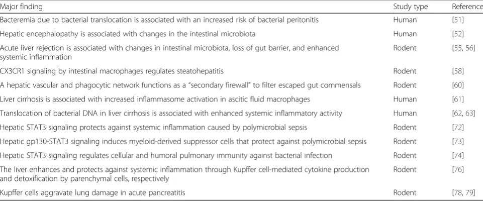
and systemic inflammation, as shown in Fig. 1.
Early event: loss of the gut barrier
The gut barrier must be permissible to allow uptake of
essential nutrients, but also needs to retain harmful
sub-stances that are only micrometers away from the
circula-tion. These include microbial substrates (e.g., LPS), dietary
components (e.g., free fatty acids (FFAs)) and digestive
enzymes produced by the exocrine pancreas. The main
etiological factors that contribute to gut barrier loss
after surgery are splanchnic hypoperfusion (including
ischemia/reperfusion damage) [30], decreased gut
mo-tility, and hypoxia. These processes are under control
of neuronal and endocrine effector arms, in addition to
microcirculation. Hypotension associated with systemic
inflammatory response syndrome usually results in
shunt-ing of blood from the splanchnic vessels to the central
cir-culation. The perfusion of the small intestine operates
within tight margins; the lack of autoregulatory
mecha-nisms results in a hypersensitivity to variations in blood
pressure. The combination of hypoperfusion and
hyp-oxia can further exacerbate the deleterious effects on
intestinal tissue integrity [33]. Hypoperfusion and
is-chemia/reperfusion (I/R) injury are partners in crime
with regard to intestinal pathology [34], even though
the latter displays distinct features. Importantly, short
periods of I/R did not induce intestinal inflammatory
responses in human [35], suggesting that a certain
thresh-old of injury is required for the instigation of G-I failure.
An adequate microcirculation is also required for optimal
blood flow to tissues, particularly in the G-I tract. Various
clinical tools are available to monitor the microcirculation,
including orthogonal polarization spectral and sidestream
darkfield imaging [36]. Finally, the severity and/or
dur-ation of intestinal ischemia are directly correlated with the
degree of gut barrier loss and endothelial dysfunction [37].
The reticulo-endothelial system in the liver provides a
fail-safe mechanism to filter out any toxic translocation
prod-ucts from the portal circulation that leaked from the
intes-tines. However, Kupffer cells (KCs) of the liver—which are
key players in the reticulo-endothelial system—appear to
play a dual role in the regulation of inflammation.
Whereas
KCs
are
an
important
source
of
pro-inflammatory cytokines in systemic inflammation [38],
they may also play an anti-inflammatory role under these
conditions [39, 40]. Finally, a portion of potentially
dele-terious elements can also find a shortcut to the circulation
via the mesenteric lymph.
The gut
–
liver axis in systemic inflammation
[image:3.595.61.539.90.393.2]approximately 70 % of the hepatic blood supply. This
anatomical situation makes the liver a central location
for clearing systemic bacterial infections and for
main-taining immune system homeostasis. Lining the hepatic
sinusoids, KCs are the first macrophage population to
encounter bacteria or microbial products derived from
the intestine. However, the function of KCs is
compro-mised in patients with advanced liver disease and, despite
the threatening consequences of impaired liver function,
systemic infections are the main cause of death in these
patients [41]. Many of these infections are initiated by
translocation of intestinal bacteria and usually result in
bacteremia and, in more severe cases, sepsis [42]. Bacterial
translocation can be demonstrated by analysis of
mesen-teric lymphatics or portal vein blood samples. It is
import-ant to point out here that, in a classic study, portal vein
sampling in trauma patients undergoing laparotomy did
not provide evidence for bacterial translocation by blood
cultures [43]. Subsequent studies with trauma patients
confirmed that blood cultures generally failed to show
bacterial growth [44, 45]. However, more sensitive methods,
such as immunostaining for
E. coli
beta-galactosidase [44]
or electron microscopy [45], provided direct evidence
for bacterial translocation to mesenteric lymph nodes
(MLNs) in most patients. While the presence of
bac-teria in MLNs as a pathological event has been debated
[46, 47], multiple studies have shown that positive
cul-tures from MLN samples obtained from laparotomy
pa-tients occurred in 10–15 % of papa-tients, which correlated
with an increased risk of postoperative sepsis [48, 49] or
postoperative infection [50].
Both human and animal model studies have provided
more mechanistic details on gut–liver crosstalk (Table 1).
[image:4.595.57.538.507.708.2]Bacteremia increases the risk of spontaneous bacterial
peritonitis, which occurs in one out of five hospitalized
cirrhotic patients [51]. At the same time,
microbiota-driven inflammation can also aggravate liver disease. For
example, alterations in colonic microbiota are associated
with endotoxemia and inflammation in patients with
hepatic encephalopathy, a complication of liver cirrhosis
[52, 53]. Gut bacteria dysbiosis may also contribute to
postoperative infections and organ rejection after liver
transplant [54]. In fact, acute liver rejection in rats is
accompanied by alterations in gut microbiota, impaired
integrity of the intestinal barrier, bacterial translocation,
elevation of plasma endotoxin levels, and a systemic
in-flammatory response [55, 56]. Gut microbiota dysbiosis
can also trigger local inflammation in the liver and promote
the progression from moderate liver disease to
steatohepati-tis [57]. A recent study showed that microbiota-dependent
activation of the chemokine receptor CX3CR1 in intestinal
macrophages is crucial for maintaining intestinal
homeo-stasis and barrier integrity and, therefore, for controlling
steatohepatitis progression [58]. Since advanced liver
disease usually leads to circulatory abnormalities (portal
hypertension, splanchnic vasodilation), these events may
constitute a vicious circle. Damage to the intestinal barrier
leads to bacterial translocation and thus liver
inflamma-tion and liver dysfuncinflamma-tion, leading to exacerbainflamma-tion of
circulatory abnormalities and causing further intestinal
injury [59].
Emphasizing the relationship between microbiota and
the immune response in the liver, recent data suggest
that the liver remains sterile when the intestine is healthy.
However, the liver becomes an important
“secondary
fire-wall”
when commensal bacteria penetrate the mesenteric
Table 1
Gut-liver crosstalk in systemic inflammation
Major finding Study type Reference
Bacteremia due to bacterial translocation is associated with an increased risk of bacterial peritonitis Human [51]
Hepatic encephalopathy is associated with changes in the intestinal microbiota Human [52]
Acute liver rejection is associated with changes in intestinal microbiota, loss of gut barrier, and enhanced systemic inflammation
Rodent [55,56]
CX3CR1 signaling by intestinal macrophages regulates steatohepatitis Rodent [58]
A hepatic vascular and phagocytic network functions as a“secondary firewall”to filter escaped gut commensals Rodent [60]
Liver cirrhosis is associated with increased inflammasome activation in ascitic fluid macrophages Human [61]
Translocation of bacterial DNA in liver cirrhosis is associated with enhanced systemic inflammatory activity Human [62,63]
Hepatic STAT3 signaling protects against systemic inflammation caused by polymicrobial sepsis Rodent [72]
Hepatic gp130-STAT3 signaling induces myeloid-derived suppressor cells that protect against polymicrobial sepsis Rodent [73]
Hepatic STAT3 signaling regulates cellular and humoral pulmonary immunity against bacterial infection Rodent [74]
The liver enhances and protects against systemic inflammation through Kupffer cell-mediated cytokine production and detoxification by parenchymal cells, respectively
Rodent [76]
Kupffer cells aggravate lung damage in acute pancreatitis Rodent [78,79]
Summary of key studies that addressed the bidirectional gut–liver crosstalk in the pathogenesis of systemic inflammation. The type of study (animal model or human) is indicated
circulation [60]. Importantly, not only viable bacteria
but also abnormal amounts of microbial products (e.g.,
LPS or bacterial DNA) can breach the intestine and
ar-rive at the liver or the ascitic fluid. These events have
been associated with activation of the inflammasome
complex [61], systemic inflammatory responses [62, 63],
and acute-on-chronic liver failure [64]. The microbial
antigens are recognized by pattern recognition receptors
(PRRs), namely Toll-like receptors (TLRs), which in the
liver are expressed on KCs, hepatic stellate cells,
intrahe-patic lymphocytes, dendritic cells, endothelial cells, and
hepatocytes [65]. TLR-mediated activation of hepatic
stellate cells is associated with the production of
proin-flammatory cytokines [66] and the development of liver
fibrosis [67]. KCs normally produce proinflammatory
cytokines and promote immune cell recruitment after
TLR stimulation; however, continuous stimulation of KCs
with low levels of LPS induces LPS tolerance and release
of anti-inflammatory cytokines such as interleukin (IL)-10
[68]. Furthermore, in the healthy liver LPS is detoxified by
both KCs and hepatocytes and rapidly loses its biological
activity. Thus, it has been speculated that activation of
TLR4 on KCs might be a common event and play a role
in immune homeostasis, whereas activation of other PRRs,
like NOD-like receptors, may be more frequent in
infec-tion [54]. Several studies also pointed to crosstalk between
KCs and migrating neutrophils. Bactericidal neutrophils
migrate to the liver sinusoids during endotoxemia and
sepsis and, along with KCs, form the leading force in the
elimination of bacteria in the liver [69]. In turn,
elimin-ation of neutrophils is necessary to resolve inflammelimin-ation
and previous studies suggested that KCs ingest and
elim-inate neutrophils in the liver sinusoids after microbial
clearance [70]. These interactions may play a critical role
in downregulating pro-inflammatory cytokine and
chemo-kine production by KCs [71].
The liver also maintains immune homeostasis in other
organs, with an important role for hepatocyte signal
trans-ducer and activator of transcription 3 (STAT3) during
sepsis and pneumonia. Hepatic STAT3 activity was
ne-cessary to prevent an excessive systemic inflammatory
response and attenuate lethality after cecal ligation and
puncture-induced sepsis [72]. STAT3-mediated protection
in this model of polymicrobial sepsis was attributed to the
serum amyloid A and CXCL1-dependent mobilization of
myeloid-derived suppressor cells [73]. Similarly, a recent
study showed that pre-existing liver STAT3 activation
modulates host immune responses in a two-hit model of
endotoxemia followed by bacterial lung challenge [74].
Mice with hepatocyte-specific STAT3 deletion showed
re-duced concentrations of acute phase response proteins
serum amyloid A and serum amyloid P, impaired alveolar
macrophage reactive oxygen species generation, higher
lung and blood bacterial loads, and increased mortality in
this model [74]. Thus, physiological amounts of bacterial
ligands arriving at the liver from the gut can contribute to
maintain systemic immune homeostasis through the
in-duction of STAT3 activity. However, gut alterations and
excessive microbiota-dependent liver inflammation may
shift this balance towards a more proinflammatory
phenotype, leading to damage in remote organs such as
brain, lung, pancreas, and heart [75]. A good example
of this crosstalk is the gut–liver–lung axis during acute
pancreatitis, leading to the acute respiratory distress
syndrome (ARDS) and MODS. In this scenario, severe
pancreatitis triggers intestinal barrier dysfunction and
gut inflammation. Translocated microbial products and
inflammatory mediators produced in the gut (e.g.,
tumor necrosis factor (TNF)-
α
, IL-6, and IL-1
β
) then
arrive at the liver via the portal vein and activate KCs,
which produce more pro-inflammatory cytokines that
amplify the inflammatory response [76]. These cytokines
released by the liver are then transported via the systemic
circulation to the
lung,
where
they cause
acute
hemorrhagic necrosis of lung epithelial cells and activation
of pulmonary monocytes and macrophages, ultimately
contributing to ARDS and MODS [77, 78]. Indeed,
inhib-ition of KCs has been shown to reduce
pancreatitis-associated remote organ injury [78, 79]. Together, these
studies suggest that failure of the gut barrier may
consti-tute a fatal event in patients with end-stage liver disease
and that the
“gut–liver inflammation”
axis may play an
important role in the balance between tolerance and
systemic inflammation in critical illness.
Intestinal damage induced by bile acids
on the intestinal epithelial barrier in vitro, this was not
the case for UDCA [82]. Furthermore, exposure of
epi-thelial cells to concentrations of DCA corresponding to
high-fat diets disrupted the epithelial barrier in vitro
and in vivo. In contrast, concentrations of DCA
typic-ally found in low-fat diets did not affect the epithelial
integrity [83]. In this context, the effects of enteral
nutri-tion versus total parenteral nutrinutri-tion on the gut barrier in
critically ill patients are highly relevant. Total parenteral
nutrition increases the gut permeability, which in addition
to the effects of bile acids may involve a variety of
mecha-nisms [84]. Others showed protective effects of lipid-rich
enteral feeding on the gut barrier in experimental shock,
which was reversed by CCK receptor antagonists [85].
These data suggest an intrinsic protective effect of enteral
nutrition on the gut barrier. Thus, even though enteral
feeding stimulates the release of bile acids in the gut
lumen that could potentially damage the epithelial lining,
its advantageous effects on the gut barrier appear to be
dominant.
Intestinal and extra-intestinal injury caused by
pancreatic enzymes
The local and systemic effects of pancreatic enzymes in
the critically ill patient have recently regained attention.
Acinar cells of the exocrine pancreas secrete a variety
of enzymes that, upon activation in the duodenal
lumen, can degrade proteins (trypsinogen,
chymotryp-sinogen, carboxypeptidase, elastases), lipids (pancreatic
lipase, phospholipase), and sugars (pancreatic amylase).
Bicarbonate (HCO3
−) is produced by ductal cells. This
pancreatic juice is secreted in response to CCK.
How-ever, during intestinal ischemia these proteases can
contribute to degradation of the protective mucus layer
and epithelial tight-junctions, leading to an elevated gut
permeability and penetration of serine proteases of the
gut wall [86]. This was first demonstrated by pancreatic
duct ligation, which reduced gut barrier failure in an
experimental model of hemorrhage-associated shock
[87]. Prevention of pancreatic enzyme influx in the
intestinal lumen resulted in reduced degradation of the
mucus layer, less toxicity to endothelial cells, and reduced
activation of circulating neutrophils [87]. The toxic effects
of pancreatic enzymes in vivo, including trypsin,
chymo-trypsin, elastase, amylase, and lipase, are potentiated when
the mucus layer is already compromised [88]. Conversely,
the addition of a mucus layer to IEC monolayers reduced
the disruptive effects of trypsin on the gut barrier [89].
Importantly, the translocation of active proteases to the
circulation is associated with an increased risk of
multi-organ failure [90], most likely caused by local
autodiges-tive processes in the gut that liberate tissue or microbial
factors that turn on systemic inflammatory responses [91].
These data suggest that pharmacological inhibition of
pancreatic enzymes could abrogate these
pathophysio-logical events and ameliorate circulatory derangement in
critical illness. A recent publication demonstrated this
con-cept in three models of experimental shock in
rats—hemor-rhagic shock, peritonitis, and endotoxin shock—with a
focus on serine proteases. All models of shock resulted
in increased protease activity in the gut wall.
Con-versely, intraluminal administration of protease inhibitors
(ANGD, tranexamic acid, and aprotinin) dramatically
re-duced tissue damage in the small intestines as well as
distant organs (heart, lung), which was associated with
significantly improved survival [92]. Notably, direct and
invasive administration of protease inhibitors was required
to yield high intraluminal concentrations in the gut [92],
which limits its direct translation into clinical practice.
Alternatively, continuous delivery of protease inhibitors
via enteral feeding has been shown to be successful in one
case of septic shock [93]. Together, these data suggest that
under ischemic conditions in the gut, pancreatic enzymes
can mediate deleterious local and systemic effects in the
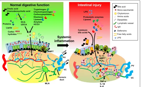
critically ill patient, as summarized in Fig. 2.
The gut
–
lung connection: mesenteric lymph
A direct anatomical link between the gut, systemic
cir-culation, and distant organs is provided by the lymphatic
vessels from the intestines. After feeding, the mesenteric
lymph is enriched for lipids (chylomicrons), fat-soluble
vitamins, and a variety of other lipophilic macromolecules.
These afferent lymphatic vessels drain from the intestinal
villus tips to MLNs and ultimately into the thoracic duct,
heart, and the pulmonary circulation. Within this
net-work, MLNs are the site where luminal antigens are
filtered and taken up by antigen-presenting cells
(mac-rophages, dendritic cells), which can direct adaptive
immune responses. The cellular players of this
special-ized mucosal immune system shuttle between MLNs
and the lamina propria and are programmed for
suc-cessful compartmentalization of the commensal
micro-flora. One of the mechanisms involved in this process
is the deposition of secretory IgA in the mucus layer
[94], an adaption to the unusually large bacterial load
of the G-I tract.
prevented hemorrhagic shock-induced endothelial
hyper-permeability and lung damage [96]. Furthermore,
real-time cross-transfusion of mesenteric lymph from donor
rats with trauma/hemorrhagic shock to naïve recipients
resulted in marked lung injury and local neutrophil
accu-mulation in these recipients [97]. Virtually all studies that
have addressed the components of mesenteric lymph used
animal models (Table 2); there is little information
regard-ing the toxic components of post-shock mesenteric lymph
in human [98]. Bacteria or microbial products do not
play a role in this phenomenon [99]. Rather, various
acute-phase proteins, possibly produced by the
intes-tinal epithelium [100], in addition to pro-inflammatory
lipid mediators [101], and in particular lipase-generated
FFAs [102], are likely causative agents. Their cytotoxicity
appears to be determined by the FFA-to-protein ratio in
mesenteric lymph, as the addition of albumin—a lipid
scavenger—reversed their effects [102]. Serum albumin
precursor was the most (approximately eightfold)
upregu-lated protein in post-shock mesenteric lymph [100], which
could therefore be part of a compensatory mechanism to
inhibit lipid cytotoxicity [103]. On the other hand,
gly-cosylated albumin in post-shock mesenteric lymph has
been associated with intrinsic cytotoxicity [104]. Thus,
albumin can have both protective and deleterious
ef-fects on lung injury.
Another candidate to mediate the toxic effects of
mesenteric lymph is phospholipase A2 (PLA2), an
en-zyme that generates lipid mediators (eicosanoids,
pros-taglandins) [105]. Importantly, high levels of PLA2
are
synthesized and secreted by Paneth cells in the intestine
[106]. Another antimicrobial protein produced by Paneth
cells that was found to be increased in toxic mesenteric
lymph was
α
-defensin 4 [107]. Together, this suggests a
Fig. 2Cellular and molecular players in intestinal injury caused by systemic inflammation. A large variety of pancreatic and hepatobiliary products are involved in the complex digestive function of the small intestine, e.g., proteolytic enzymes, lipases, amylases, bicarbonate, and primary bile acids. The primary bile acids (cholic acid, chenodeoxycholic acid) are converted to secondary bile acids (e.g., lithocholic acid, DCA, UDCA) by the microbiota. The digestive enzymes derived from the host or microbiota ensure the breakdown of macromolecules to soluble nutrients, which is followed by uptake and transport to the portal circulation or intestinal lymph tract. While the intestinal microflora are crucial for these digestive functions in the lumen, their quantity and topography are tightly controlled by the thick mucus layer that contains secretory IgA and antimicrobial peptides (e.g., defensins) produced by mucosal immune cells and the epithelium. In critical illness, circulatory derangement results in gut barrier loss and the translocation of digestive enzymes, cytotoxic bile acids, free fatty acids (FFAs), and microbial substrates to the submucosa, which exacerbates local and systemic inflammatory reactions. The intestinal epithelium is also an important source of pro-inflammatory mediators that are released into the circulation, including IL-17, lipid mediators produced by phospholipase A2 (PLA2), and antimicrobial peptides derived from Paneth cells. These [image:7.595.58.542.88.387.2]potential detrimental role for Paneth cell products in the
course of systemic inflammation and/or shock, which are
distributed via the intestinal lymphatic system to the
cir-culation. Indeed, mice deficient for matrix
metalloprotein-ase 7 (MMP7), the enzyme involved in posttranslational
activation of Paneth cells products, were protected against
systemic LPS-induced lethality [108]. Activation of Paneth
cells and transport of Paneth cell products, including
pro-inflammatory IL-17A, by intestinal macrophages to
the liver have been previously demonstrated in systemic
inflammation [109]. IL-17 is an important activator of
neutrophils and Paneth cells were the main producers
of IL-17 in an experimental model of TNF-
α
-induced
[image:8.595.60.539.99.213.2]intestinal injury and shock [110]. Furthermore, liver I/R
injury and its associated systemic inflammation resulted
in significantly increased IL-17 levels in the portal
ven-ous blood, which was associated with massive Paneth
cell degranulation in the gut and hepatic, intestinal, and
Table 2
Gut
–
lung crosstalk
Major finding Study type Reference
Cross-transfusion of mesenteric lymph from donors after shock/trauma induces acute lung injury in recipients Rodent [97]
Post-shock mesenteric lymph contains increased amounts of bioactive lysophospholipids and PUFAs Rodent [101]
Post-shock mesenteric lymph contains increased amounts of free fatty acids Rodent [102]
The detrimental effects of post-shock mesenteric lymph can be reversed by the addition of albumin Rodent [103]
Inhibition of PLA2inhibits the cytotoxic activity of post-shock mesenteric lymph Rodent [105]
Levels of Paneth cell-derivedα-defensin 4 (an antimicrobial peptide) are increased in post-shock mesenteric lymph Rodent [107]
Endogenous alarmins are abundantly released from the intestines in post-shock mesenteric lymph Rodent [113] Pertinent studies that addressed the interactions between intestinal pathology and lung injury
PLA2phospholipase A2,PUFApoly-unsaturated fatty acids
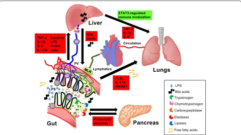
Fig. 3Direct and indirect interactions between the gut and other organs in systemic inflammation. Luminal components of the small intestine can spread to the circulation via the portal vein (PV) and liver. This includes pro-inflammatory constituents such as lipopolysaccharide (LPS), bacterial DNA, whole bacteria, other bacterial products, and free fatty acids (FFAs). Toxic components of the gut lumen, including FFAs, inflammatory products of phospholipase A2 (PLA2), pro-inflammatory cytokines (e.g.,IL-17), and damage-associated molecular substrates such as high mobility group box 1
[image:8.595.58.538.364.633.2]renal injury. Pharmacological or genetic approaches to
abrogate Paneth cell function reversed these effects
[111]. Similarly, genetic knockout for IL-17A also
pre-vented intestinal damage in this I/R model [112]. These
findings further demonstrate a central role for the gut
as a driving force in systemic inflammation (Fig. 2).
Finally, increased levels of alarmins that are generally
released after tissue injury were also found to be elevated
in post-shock mesenteric lymph [113]. These substrates
are endogenous TLR4 ligands and mediate
immunostimu-latory effects via the activation of nuclear factor kappa B
(NF-kB). Mice with genetic mutations in the receptors
or adapter molecules from the TLR4 signaling pathway
were protected against post-shock mesenteric
lymph-mediated lung injury [114]. These findings are consistent
with the
“danger model”
that states that certain
endogen-ous ligands signal the presence of tissue injury to the host
via PRRs [115], including TLR.
Conclusions
In conclusion, the mesenteric lymph is another route for
immunostimulatory proteins to reach the systemic
circu-lation after intestinal injury and post-shock mesenteric
lymph is particularly toxic to the pulmonary
microvascu-lature. Gut-derived toxic factors that leak from MLNs,
including products of pancreatic enzymes, endogenous
danger signals, and Paneth cell products, most likely
partner up to exert these detrimental effects. All the
aforementioned interactions are summarized in Fig. 3.
Finally, basic insights into the intimate relationship
be-tween the G-I tract and the systemic inflammatory system
are expected to lead to more efficacious treatment
modal-ities for critical illness in the future.
Acknowledgments
We thank Asami Takahashi for her assistance in figure preparation.
Funding
PRdJ was supported by a Research Fellowship from the Crohn’s and Colitis Foundation of America (CCFA; RFA2927). JMG-N is supported by grant PI13/00315 from the Institute of Health Carlos III, Government of Spain (co-financed with FEDER funds) and grant UGP-14-123 from the FISABIO Research Foundation.
Authors’contributions
PRdJ, JMG-N, and NJGJ contributed equally to the drafting and final review of the manuscript. All authors read and approved the final manuscript.
Authors’information
PRdJ studied the regulation of systemic inflammation in pediatric patients at the University Medical Center Utrecht (UMCU), The Netherlands. He completed a research fellowship at the University of California, San Diego (UCSD) that addressed regulatory mechanisms of intestinal inflammation, supported by the Crohn’s and Colitis Foundation of America (CCFA). Concurrently he obtained his PhD degree at Utrecht University. He is currently a postdoctoral fellow at the Sanford Burnham Prebys Medical Discovery Institute, La Jolla, CA, USA.
JMG-N received his doctoral degree in Biology from the University of Alicante, Spain. He completed his postdoctoral training in mucosal immunology at UCSD. He is currently a Miguel Servet researcher of the Spanish Institute of Health Carlos III and principal investigator at the
Networked Biomedical Research Center for Hepatic and Digestive Diseases (CIBERehd) in Alicante. His laboratory focuses on the regulation of systemic inflammation, as well as inflammation and inflammation-associated cancer of the gut and liver.
NJGJ is an Associate Professor in Pediatric Intensive Care at the Dept. of Pediatrics, UMC Utrecht, The Netherlands. He currently serves as the Chairman of the Section Infection, Systemic Inflammation and Sepsis of the European Society of Pediatric and Neonatal Intensive Care (ESPNIC), has been the ESPNIC Council Member for the Netherlands, and has served as Chairman of the Dutch Society for Pediatric Intensive Care. His research focuses on regulatory mechanisms in systemic inflammation induced by pediatric cardiac surgery and on brain injury during and after pediatric cardiac surgery. He is principal investigator at the Institute of Child Health at the University Medical Center of Utrecht for Congenital Heart Disease LifeSpan and follow-up program.
Competing interests
The authors declare that they have no competing interests.
Author details
1Department of Pediatric Intensive Care, Wilhelmina Children’s Hospital, University Medical Center Utrecht, Utrecht, The Netherlands.2Sanford Burnham Prebys Medical Discovery Institute, 10901 N Torrey Pines Rd, La Jolla, CA 92037, USA.3Networked Biomedical Research Center for Hepatic and Digestive Diseases (CIBERehd), Hospital General Universitario de Alicante, Alicante, Spain.4Alicante Institute of Health and Biomedical Research (ISABIAL–FISABIO Foundation), Alicante, Spain.
References
1. van der Flier LG, Clevers H. Stem cells, self-renewal, and differentiation in the intestinal epithelium. Annu Rev Physiol. 2009;71:241–60.
2. Sato T, van Es JH, Snippert HJ, Stange DE, Vries RG, van den Born M, Barker N, Shroyer NF, van de Wetering M, Clevers H. Paneth cells constitute the niche for Lgr5 stem cells in intestinal crypts. Nature. 2011;469(7330):415–8.
3. Clevers H, Batlle E. SnapShot: the intestinal crypt. Cell. 2013;152(5):1198. e2. 4. Vaishnava S, Behrendt CL, Ismail AS, Eckmann L, Hooper LV. Paneth cells
directly sense gut commensals and maintain homeostasis at the intestinal host-microbial interface. Proc Natl Acad Sci U S A. 2008;105(52):20858–63. 5. Tremaroli V, Backhed F. Functional interactions between the gut microbiota
and host metabolism. Nature. 2012;489(7415):242–9.
6. Qin J, Li R, Raes J, Arumugam M, Burgdorf KS, Manichanh C, Nielsen T, Pons N, Levenez F, Yamada T, Mende DR, Li J, Xu J, Li S, Li D, Cao J, Wang B, Liang H, Zheng H, Xie Y, Tap J, Lepage P, Bertalan M, Batto JM, Hansen T, Le Paslier D, Linneberg A, Nielsen HB, Pelletier E, Renault P, Sicheritz-Ponten T, Turner K, Zhu H, Yu C, Li S, Jian M, Zhou Y, Li Y, Zhang X, Li S, Qin N, Yang H, Wang J, Brunak S, Dore J, Guarner F, Kristiansen K, Pedersen O, Parkhill J, Weissenbach J, MetaHIT C, Bork P, Ehrlich SD, Wang J. A human gut microbial gene catalogue established by metagenomic sequencing. Nature. 2010;464(7285):59–65.
7. Lewis K, Lutgendorff F, Phan V, Soderholm JD, Sherman PM, McKay DM. Enhanced translocation of bacteria across metabolically stressed epithelia is reduced by butyrate. Inflamm Bowel Dis. 2010;16(7):1138–48.
8. Maslowski KM, Vieira AT, Ng A, Kranich J, Sierro F, Yu D, Schilter HC, Rolph MS, Mackay F, Artis D, Xavier RJ, Teixeira MM, Mackay CR. Regulation of inflammatory responses by gut microbiota and chemoattractant receptor GPR43. Nature. 2009;461(7268):1282–6.
9. Sina C, Gavrilova O, Forster M, Till A, Derer S, Hildebrand F, Raabe B, Chalaris A, Scheller J, Rehmann A, Franke A, Ott S, Hasler R, Nikolaus S, Folsch UR, Rose-John S, Jiang HP, Li J, Schreiber S, Rosenstiel P. G protein-coupled receptor 43 is essential for neutrophil recruitment during intestinal inflammation. J Immunol. 2009;183(11):7514–22.
10. Clark JA, Coopersmith CM. Intestinal crosstalk: a new paradigm for
understanding the gut as the“motor”of critical illness. Shock. 2007;28(4):384–93. 11. Kats S, Schonberger JP, Brands R, Seinen W, van Oeveren W. Endotoxin
release in cardiac surgery with cardiopulmonary bypass: pathophysiology and possible therapeutic strategies. An update. Eur J Cardiothorac Surg. 2011;39(4):451–8.
colonisation in premature neonates predicts neonatal sepsis. Arch Dis Child Fetal Neonatal Ed. 2012;97(6):F456–62.
13. Hsu JF, Chu SM, Lee CW, Yang PH, Lien R, Chiang MC, Fu RH, Huang HR, Tsai MH. Incidence, clinical characteristics and attributable mortality of persistent bloodstream infection in the neonatal intensive care unit. PLoS One. 2015;10(4):e0124567.
14. Pereira CA, Marra AR, Camargo LF, Pignatari AC, Sukiennik T, Behar PR, Medeiros EA, Ribeiro J, Girao E, Correa L, Guerra C, Carneiro I, Brites C, Reis M, de Souza MA, Tranchesi R, Barata CU, Edmond MB, Brazilian SCOPE Study Group. Nosocomial bloodstream infections in Brazilian pediatric patients: microbiology, epidemiology, and clinical features. PLoS One. 2013;8(7):e68144.
15. Phua J, Ngerng W, See K, Tay C, Kiong T, Lim H, Chew M, Yip H, Tan A, Khalizah H, Capistrano R, Lee K, Mukhopadhyay A. Characteristics and outcomes of culture-negative versus culture-positive severe sepsis. Crit Care. 2013;17(5):R202.
16. Ohri SK, Velissaris T. Gastrointestinal dysfunction following cardiac surgery. Perfusion. 2006;21(4):215–23.
17. Reintam A, Parm P, Kitus R, Starkopf J, Kern H. Gastrointestinal failure score in critically ill patients: a prospective observational study. Crit Care. 2008;12(4):R90.
18. Carlson GL, Dark P. Acute intestinal failure. Curr Opin Crit Care. 2010;16(4):347–52.
19. Reintam Blaser A, Malbrain ML, Starkopf J, Fruhwald S, Jakob SM, De Waele J, Braun JP, Poeze M, Spies C. Gastrointestinal function in intensive care patients: terminology, definitions and management. Recommendations of the ESICM Working Group on Abdominal Problems. Intensive Care Med. 2012;38(3):384–94.
20. Mentec H, Dupont H, Bocchetti M, Cani P, Ponche F, Bleichner G. Upper digestive intolerance during enteral nutrition in critically ill patients: frequency, risk factors, and complications. Crit Care Med. 2001;29(10):1955–61. 21. Reintam A, Parm P, Redlich U, Tooding LM, Starkopf J, Kohler F, Spies C,
Kern H. Gastrointestinal failure in intensive care: a retrospective clinical study in three different intensive care units in Germany and Estonia. BMC Gastroenterol. 2006;6:19.
22. Silva MA, Santos Sda G, Tomasi CD, Luz G, Paula MM, Pizzol FD, Ritter C. Enteral nutrition discontinuation and outcomes in general critically ill patients. Clinics (Sao Paulo). 2013;68(2):173–8.
23. Reintam Blaser A, Poeze M, Malbrain ML, Bjorck M, Oudemans-van Straaten HM, Starkopf J, Gastro-Intestinal Failure Trial Group. Gastrointestinal symptoms during the first week of intensive care are associated with poor outcome: a prospective multicentre study. Intensive Care Med. 2013;39(5):899–909. 24. Piton G, Manzon C, Cypriani B, Carbonnel F, Capellier G. Acute intestinal
failure in critically ill patients: is plasma citrulline the right marker? Intensive Care Med. 2011;37(6):911–7.
25. Thuijls G, van Wijck K, Grootjans J, Derikx JP, van Bijnen AA, Heineman E, Dejong CH, Buurman WA, Poeze M. Early diagnosis of intestinal ischemia using urinary and plasma fatty acid binding proteins. Ann Surg. 2011;253(2):303–8. 26. Derikx JP, Blijlevens NM, Donnelly JP, Fujii H, Kanda T, van Bijnen AA,
Heineman E, Buurman WA. Loss of enterocyte mass is accompanied by diminished turnover of enterocytes after myeloablative therapy in haematopoietic stem-cell transplant recipients. Ann Oncol. 2009;20(2):337–42. 27. Typpo KV, Larmonier CB, Deschenes J, Redford D, Kiela PR, Ghishan FK.
Clinical characteristics associated with postoperative intestinal epithelial barrier dysfunction in children with congenital heart disease. Pediatr Crit Care Med. 2015;16(1):37–44.
28. Piton G, Manzon C, Monnet E, Cypriani B, Barbot O, Navellou JC, Carbonnel F, Capellier G. Plasma citrulline kinetics and prognostic value in critically ill patients. Intensive Care Med. 2010;36(4):702–6.
29. van Bree SH, Cailotto C, Di Giovangiulio M, Jansen E, van der Vliet J, Costes L, Depoortere I, Gomez-Pinilla PJ, Matteoli G, Boeckxstaens GE. Systemic inflammation with enhanced brain activation contributes to more severe delay in postoperative ileus. Neurogastroenterol Motil. 2013;25(8):e540–9. 30. Derikx JP, van Waardenburg DA, Thuijls G, Willigers HM, Koenraads M, van
Bijnen AA, Heineman E, Poeze M, Ambergen T, van Ooij A, van Rhijn LW, Buurman WA. New insight in loss of gut barrier during major non-abdominal surgery. PLoS One. 2008;3(12):e3954.
31. Pathan N, Burmester M, Adamovic T, Berk M, Ng KW, Betts H, Macrae D, Waddell S, Paul-Clark M, Nuamah R, Mein C, Levin M, Montana G, Mitchell JA. Intestinal injury and endotoxemia in children undergoing surgery for congenital heart disease. Am J Respir Crit Care Med. 2011;184(11):1261–9.
32. Hietbrink F, Besselink MG, Renooij W, de Smet MB, Draisma A, van der Hoeven H, Pickkers P. Systemic inflammation increases intestinal permeability during experimental human endotoxemia. Shock. 2009;32(4):374–8.
33. Harrois A, Baudry N, Huet O, Kato H, Lohez M, Ziol M, Duranteau J, Vicaut E. Synergistic deleterious effect of hypoxemia and hypovolemia on microcirculation in intestinal villi*. Crit Care Med. 2013;41(11):e376–84. 34. Feinman R, Deitch EA, Watkins AC, Abungu B, Colorado I, Kannan KB, Sheth
SU, Caputo FJ, Lu Q, Ramanathan M, Attan S, Badami CD, Doucet D, Barlos D, Bosch-Marce M, Semenza GL, Xu DZ. HIF-1 mediates pathogenic inflammatory responses to intestinal ischemia-reperfusion injury. Am J Physiol Gastrointest Liver Physiol. 2010;299(4):G833–43.
35. Matthijsen RA, Derikx JP, Kuipers D, van Dam RM, Dejong CH, Buurman WA. Enterocyte shedding and epithelial lining repair following ischemia of the human small intestine attenuate inflammation. PLoS One. 2009;4(9):e7045.
36. Top AP, Tasker RC, Ince C. The microcirculation of the critically ill pediatric patient. Crit Care. 2011;15(2):213.
37. Sun Z, Wang X, Deng X, Lasson A, Wallen R, Hallberg E, Andersson R. The influence of intestinal ischemia and reperfusion on bidirectional intestinal barrier permeability, cellular membrane integrity, proteinase inhibitors, and cell death in rats. Shock. 1998;10(3):203–12.
38. Kim TH, Lee SH, Lee SM. Role of Kupffer cells in pathogenesis of sepsis-induced drug metabolizing dysfunction. FEBS J. 2011;278(13):2307–17. 39. Traeger T, Mikulcak M, Eipel C, Abshagen K, Diedrich S, Heidecke CD, Maier
S, Vollmar B. Kupffer cell depletion reduces hepatic inflammation and apoptosis but decreases survival in abdominal sepsis. Eur J Gastroenterol Hepatol. 2010;22(9):1039–49.
40. Hutchins NA, Chung CS, Borgerding JN, Ayala CA, Ayala A. Kupffer cells protect liver sinusoidal endothelial cells from Fas-dependent apoptosis in sepsis by down-regulating gp130. Am J Pathol. 2013;182(3):742–54. 41. Arvaniti V, D'Amico G, Fede G, Manousou P, Tsochatzis E, Pleguezuelo M,
Burroughs AK. Infections in patients with cirrhosis increase mortality four-fold and should be used in determining prognosis. Gastroenterology. 2010;139(4):1246–56. 1256.e1–5.
42. Gustot T, Durand F, Lebrec D, Vincent JL, Moreau R. Severe sepsis in cirrhosis. Hepatology. 2009;50(6):2022–33.
43. Moore FA, Moore EE, Poggetti R, McAnena OJ, Peterson VM, Abernathy CM, Parsons PE. Gut bacterial translocation via the portal vein: a clinical perspective with major torso trauma. J Trauma. 1991;31(5):629–36. discussion 636–8.
44. Brathwaite CE, Ross SE, Nagele R, Mure AJ, O'Malley KF, Garcia-Perez FA. Bacterial translocation occurs in humans after traumatic injury: evidence using immunofluorescence. J Trauma. 1993;34(4):586–9. discussion 589–90. 45. Reed LL, Martin M, Manglano R, Newson B, Kocka F, Barrett J. Bacterial
translocation following abdominal trauma in humans. Circ Shock. 1994;42(1):1–6.
46. Moore FA. The role of the gastrointestinal tract in postinjury multiple organ failure. Am J Surg. 1999;178(6):449–53.
47. MacFie J. Current status of bacterial translocation as a cause of surgical sepsis. Br Med Bull. 2004;71:1–11.
48. O'Boyle CJ, MacFie J, Mitchell CJ, Johnstone D, Sagar PM, Sedman PC. Microbiology of bacterial translocation in humans. Gut. 1998;42(1):29–35. 49. MacFie J, Reddy BS, Gatt M, Jain PK, Sowdi R, Mitchell CJ. Bacterial translocation
studied in 927 patients over 13 years. Br J Surg. 2006;93(1):87–93.
50. Nieves E, Tobon LF, Rios DI, Isaza A, Ramirez M, Beltran JA, Garzon-Ospina D, Patarroyo MA, Gomez A. Bacterial translocation in abdominal trauma and postoperative infections. J Trauma. 2011;71(5):1258–61.
51. Bajaj JS, O'Leary JG, Reddy KR, Wong F, Olson JC, Subramanian RM, Brown G, Noble NA, Thacker LR, Kamath PS, NACSELD. Second infections independently increase mortality in hospitalized patients with cirrhosis: the North American consortium for the study of end-stage liver disease (NACSELD) experience. Hepatology. 2012;56(6):2328–35.
52. Bajaj JS, Ridlon JM, Hylemon PB, Thacker LR, Heuman DM, Smith S, Sikaroodi M, Gillevet PM. Linkage of gut microbiome with cognition in hepatic encephalopathy. Am J Physiol Gastrointest Liver Physiol. 2012;302(1):G168–75.
53. Gomez-Hurtado I, Such J, Sanz Y, Frances R. Gut microbiota-related complications in cirrhosis. World J Gastroenterol. 2014;20(42):15624–31. 54. Chassaing B, Etienne-Mesmin L, Gewirtz AT. Microbiota-liver axis in hepatic
55. Xie Y, Luo Z, Li Z, Deng M, Liu H, Zhu B, Ruan B, Li L. Structural shifts of fecal microbial communities in rats with acute rejection after liver transplantation. Microb Ecol. 2012;64(2):546–54.
56. Ren Z, Jiang J, Lu H, Chen X, He Y, Zhang H, Xie H, Wang W, Zheng S, Zhou L. Intestinal microbial variation may predict early acute rejection after liver transplantation in rats. Transplantation. 2014;98(8):844–52.
57. Henao-Mejia J, Elinav E, Jin C, Hao L, Mehal WZ, Strowig T, Thaiss CA, Kau AL, Eisenbarth SC, Jurczak MJ, Camporez JP, Shulman GI, Gordon JI, Hoffman HM, Flavell RA. Inflammasome-mediated dysbiosis regulates progression of NAFLD and obesity. Nature. 2012;482(7384):179–85. 58. Schneider KM, Bieghs V, Heymann F, Hu W, Dreymueller D, Liao L,
Frissen M, Ludwig A, Gassler N, Pabst O, Latz E, Sellge G, Penders J, Tacke F, Trautwein C. CX3CR1 is a gatekeeper for intestinal barrier integrity in mice: Limiting steatohepatitis by maintaining intestinal homeostasis. Hepatology. 2015;62(5):1405–16.
59. Prin M, Bakker J, Wagener G. Hepatosplanchnic circulation in cirrhosis and sepsis. World J Gastroenterol. 2015;21(9):2582–92.
60. Balmer ML, Slack E, de Gottardi A, Lawson MA, Hapfelmeier S, Miele L, Grieco A, Van Vlierberghe H, Fahrner R, Patuto N, Bernsmeier C, Ronchi F, Wyss M, Stroka D, Dickgreber N, Heim MH, McCoy KD, Macpherson AJ. The liver may act as a firewall mediating mutualism between the host and its gut commensal microbiota. Sci Transl Med. 2014;6(237):237ra66. 61. Lozano-Ruiz B, Bachiller V, Garcia-Martinez I, Zapater P, Gomez-Hurtado I,
Moratalla A, Gimenez P, Bellot P, Frances R, Such J, Gonzalez-Navajas JM. Absent in melanoma 2 triggers a heightened inflammasome response in ascitic fluid macrophages of patients with cirrhosis. J Hepatol. 2015;62(1):64–71. 62. Frances R, Zapater P, Gonzalez-Navajas JM, Munoz C, Cano R, Moreu R,
Pascual S, Bellot P, Perez-Mateo M, Such J. Bacterial DNA in patients with cirrhosis and noninfected ascites mimics the soluble immune response established in patients with spontaneous bacterial peritonitis. Hepatology. 2008;47(3):978–85.
63. Gonzalez-Navajas JM, Bellot P, Frances R, Zapater P, Munoz C, Garcia-Pagan JC, Pascual S, Perez-Mateo M, Bosch J, Such J. Presence of bacterial-DNA in cirrhosis identifies a subgroup of patients with marked inflammatory response not related to endotoxin. J Hepatol. 2008;48(1):61–7.
64. Zapater P, Frances R, Gonzalez-Navajas JM, de la Hoz MA, Moreu R, Pascual S, Monfort D, Montoliu S, Vila C, Escudero A, Torras X, Cirera I, Llanos L, Guarner-Argente C, Palazon JM, Carnicer F, Bellot P, Guarner C, Planas R, Sola R, Serra MA, Munoz C, Perez-Mateo M, Such J. Serum and ascitic fluid bacterial DNA: a new independent prognostic factor in noninfected patients with cirrhosis. Hepatology. 2008;48(6):1924–31.
65. Nakamoto N, Kanai T. Role of toll-like receptors in immune activation and tolerance in the liver. Front Immunol. 2014;5:221.
66. Paik YH, Schwabe RF, Bataller R, Russo MP, Jobin C, Brenner DA. Toll-like receptor 4 mediates inflammatory signaling by bacterial lipopolysaccharide in human hepatic stellate cells. Hepatology. 2003;37(5):1043–55. 67. Seki E, De Minicis S, Osterreicher CH, Kluwe J, Osawa Y, Brenner DA,
Schwabe RF. TLR4 enhances TGF-beta signaling and hepatic fibrosis. Nat Med. 2007;13(11):1324–32.
68. Knolle P, Schlaak J, Uhrig A, Kempf P, Meyer zum Buschenfelde KH, Gerken G. Human Kupffer cells secrete IL-10 in response to lipopolysaccharide (LPS) challenge. J Hepatol. 1995;22(2):226–9.
69. Gregory SH, Sagnimeni AJ, Wing EJ. Bacteria in the bloodstream are trapped in the liver and killed by immigrating neutrophils. J Immunol. 1996;157(6): 2514–20.
70. Shi J, Gilbert GE, Kokubo Y, Ohashi T. Role of the liver in regulating numbers of circulating neutrophils. Blood. 2001;98(4):1226–30.
71. Holub M, Cheng CW, Mott S, Wintermeyer P, van Rooijen N, Gregory SH. Neutrophils sequestered in the liver suppress the proinflammatory response of Kupffer cells to systemic bacterial infection. J Immunol. 2009;183(5):3309–16. 72. Sakamori R, Takehara T, Ohnishi C, Tatsumi T, Ohkawa K, Takeda K, Akira S,
Hayashi N. Signal transducer and activator of transcription 3 signaling within hepatocytes attenuates systemic inflammatory response and lethality in septic mice. Hepatology. 2007;46(5):1564–73.
73. Sander LE, Sackett SD, Dierssen U, Beraza N, Linke RP, Muller M, Blander JM, Tacke F, Trautwein C. Hepatic acute-phase proteins control innate immune responses during infection by promoting myeloid-derived suppressor cell function. J Exp Med. 2010;207(7):1453–64.
74. Hilliard KL, Allen E, Traber KE, Kim Y, Wasserman GA, Jones MR, Mizgerd JP, Quinton LJ. Activation of hepatic STAT3 maintains pulmonary defense during endotoxemia. Infect Immun. 2015;83(10):4015–27.
75. Gonzalez-Reimers E, Santolaria-Fernandez F, Martin-Gonzalez MC, Fernandez-Rodriguez CM, Quintero-Platt G. Alcoholism: a systemic proinflammatory condition. World J Gastroenterol. 2014;20(40):14660–71. 76. Wang Y, Liu W, Liu X, Sheng M, Pei Y, Lei R, Zhang S, Tao R. Role of liver in
modulating the release of inflammatory cytokines involved in lung and multiple organ dysfunction in severe acute pancreatitis. Cell Biochem Biophys. 2015;71(2):765–76.
77. Closa D, Bardaji M, Hotter G, Prats N, Gelpi E, Fernandez-Cruz L, Rosello-Catafau J. Hepatic involvement in pancreatitis-induced lung damage. Am J Physiol. 1996;270(1 Pt 1):G6–13.
78. Liu HB, Cui NQ, Li DH, Chen C. Role of Kupffer cells in acute hemorrhagic necrotizing pancreatitis-associated lung injury of rats. World J Gastroenterol. 2006;12(3):403–7.
79. Shifrin AL, Chirmule N, Zhang Y, Raper SE. Macrophage ablation attenuates adenoviral vector-induced pancreatitis. Surgery. 2005;137(5):545–51. 80. Martinez-Augustin O, Sanchez de Medina F. Intestinal bile acid physiology
and pathophysiology. World J Gastroenterol. 2008;14(37):5630–40. 81. Mossner J. New advances in cell physiology and pathophysiology of the
exocrine pancreas. Dig Dis. 2010;28(6):722–8.
82. Raimondi F, Santoro P, Barone MV, Pappacoda S, Barretta ML, Nanayakkara M, Apicella C, Capasso L, Paludetto R. Bile acids modulate tight junction structure and barrier function of Caco-2 monolayers via EGFR activation. Am J Physiol Gastrointest Liver Physiol. 2008;294(4):G906–13.
83. Stenman LK, Holma R, Eggert A, Korpela R. A novel mechanism for gut barrier dysfunction by dietary fat: epithelial disruption by hydrophobic bile acids. Am J Physiol Gastrointest Liver Physiol. 2013;304(3):G227–34. 84. Feng Y, Ralls MW, Xiao W, Miyasaka E, Herman RS, Teitelbaum DH. Loss of
enteral nutrition in a mouse model results in intestinal epithelial barrier dysfunction. Ann N Y Acad Sci. 2012;1258:71–7.
85. de Haan JJ, Thuijls G, Lubbers T, Hadfoune M, Reisinger K, Heineman E, Greve JW, Buurman WA. Protection against early intestinal compromise by lipid-rich enteral nutrition through cholecystokinin receptors. Crit Care Med. 2010;38(7):1592–7.
86. Chang M, Kistler EB, Schmid-Schonbein GW. Disruption of the mucosal barrier during gut ischemia allows entry of digestive enzymes into the intestinal wall. Shock. 2012;37(3):297–305.
87. Caputo FJ, Rupani B, Watkins AC, Barlos D, Vega D, Senthil M, Deitch EA. Pancreatic duct ligation abrogates the trauma hemorrhage-induced gut barrier failure and the subsequent production of biologically active intestinal lymph. Shock. 2007;28(4):441–6.
88. Kistler EB, Alsaigh T, Chang M, Schmid-Schonbein GW. Impaired small-bowel barrier integrity in the presence of lumenal pancreatic digestive enzymes leads to circulatory shock. Shock. 2012;38(3):262–7.
89. Chang M, Alsaigh T, Kistler EB, Schmid-Schonbein GW. Breakdown of mucin as barrier to digestive enzymes in the ischemic rat small intestine. PLoS One. 2012;7(6):e40087.
90. Malinoski DJ, Hadjizacharia P, Salim A, Kim H, Dolich MO, Cinat M, Barrios C, Lekawa ME, Hoyt DB. Elevated serum pancreatic enzyme levels after hemorrhagic shock predict organ failure and death. J Trauma. 2009;67(3):445–9.
91. Mitsuoka H, Kistler EB, Schmid-Schonbein GW. Generation of in vivo activating factors in the ischemic intestine by pancreatic enzymes. Proc Natl Acad Sci U S A. 2000;97(4):1772–7.
92. DeLano FA, Hoyt DB, Schmid-Schonbein GW. Pancreatic digestive enzyme blockade in the intestine increases survival after experimental shock. Sci Transl Med. 2013;5(169):169ra11.
93. Lee YT, Wei J, Chuang YC, Chang CY, Chen IC, Weng CF, Schmid-Schonbein GW. Successful treatment with continuous enteral protease inhibitor in a patient with severe septic shock. Transplant Proc. 2012;44(3):817–9. 94. Fanous MY, Phillips AJ, Windsor JA. Mesenteric lymph: the bridge to future
management of critical illness. JOP. 2007;8(4):374–99.
95. Deitch EA. Gut lymph and lymphatics: a source of factors leading to organ injury and dysfunction. Ann N Y Acad Sci. 2010;1207 Suppl 1:E103–11. 96. Deitch EA, Adams C, Lu Q, Xu DZ. A time course study of the
protective effect of mesenteric lymph duct ligation on hemorrhagic shock-induced pulmonary injury and the toxic effects of lymph from shocked rats on endothelial cell monolayer permeability. Surgery. 2001;129(1):39–47.
98. Dzieciatkowska M, Wohlauer MV, Moore EE, Damle S, Peltz E, Campsen J, Kelher M, Silliman C, Banerjee A, Hansen KC. Proteomic analysis of human mesenteric lymph. Shock. 2011;35(4):331–8.
99. Adams Jr CA, Xu DZ, Lu Q, Deitch EA. Factors larger than 100 kd in post-hemorrhagic shock mesenteric lymph are toxic for endothelial cells. Surgery. 2001;129(3):351–63.
100. Fang JF, Shih LY, Yuan KC, Fang KY, Hwang TL, Hsieh SY. Proteomic analysis of post-hemorrhagic shock mesenteric lymph. Shock. 2010;34(3):291–8. 101. Morishita K, Aiboshi J, Kobayashi T, Mikami S, Yokoyama Y, Ogawa K,
Yokota H, Otomo Y. Lipidomics analysis of mesenteric lymph after trauma and hemorrhagic shock. J Trauma Acute Care Surg. 2012;72(6):1541–7. 102. Qin X, Dong W, Sharpe SM, Sheth SU, Palange DC, Rider T, Jandacek R,
Tso P, Deitch EA. Role of lipase-generated free fatty acids in converting mesenteric lymph from a noncytotoxic to a cytotoxic fluid. Am J Physiol Gastrointest Liver Physiol. 2012;303(8):G969–78.
103. Osband AJ, Deitch EA, Hauser CJ, Lu Q, Zaets S, Berezina T, Machiedo GW, Rajwani KK, Xu DZ. Albumin protects against gut-induced lung injury in vitro and in vivo. Ann Surg. 2004;240(2):331–9.
104. Kaiser VL, Sifri ZC, Dikdan GS, Berezina T, Zaets S, Lu Q, Xu DZ, Deitch EA. Trauma-hemorrhagic shock mesenteric lymph from rat contains a modified form of albumin that is implicated in endothelial cell toxicity. Shock. 2005;23(5):417–25.
105. Gonzalez RJ, Moore EE, Ciesla DJ, Biffl WL, Offner PJ, Silliman CC. Phospholipase A(2)–derived neutral lipids from posthemorrhagic shock mesenteric lymph prime the neutrophil oxidative burst. Surgery. 2001;130(2):198–203.
106. Keshav S. Paneth cells: leukocyte-like mediators of innate immunity in the intestine. J Leukoc Biol. 2006;80(3):500–8.
107. Atkins JL, Hammamieh R, Jett M, Gorbunov NV, Asher LV, Kiang JG. Alpha-defensin-like product and asymmetric dimethylarginine increase in mesenteric lymph after hemorrhage in anesthetized rat. Shock. 2008;30(4):411–6.
108. Vandenbroucke RE, Vanlaere I, Van Hauwermeiren F, Van Wonterghem E, Wilson C, Libert C. Pro-inflammatory effects of matrix metalloproteinase 7 in acute inflammation. Mucosal Immunol. 2014;7(3):579–88.
109. Park SW, Kim M, Kim JY, Ham A, Brown KM, Mori-Akiyama Y, Ouellette AJ, D'Agati VD, Lee HT. Paneth cell-mediated multiorgan dysfunction after acute kidney injury. J Immunol. 2012;189(11):5421–33.
110. Takahashi N, Vanlaere I, de Rycke R, Cauwels A, Joosten LA, Lubberts E, van den Berg WB, Libert C. IL-17 produced by Paneth cells drives TNF-induced shock. J Exp Med. 2008;205(8):1755–61.
111. Park SW, Kim M, Brown KM, D'Agati VD, Lee HT. Paneth cell-derived interleukin-17A causes multiorgan dysfunction after hepatic ischemia and reperfusion injury. Hepatology. 2011;53(5):1662–75.
112. Lee HT, Kim M, Kim JY, Brown KM, Ham A, D'Agati VD, Mori-Akiyama Y. Critical role of interleukin-17A in murine intestinal ischemia-reperfusion injury. Am J Physiol Gastrointest Liver Physiol. 2013;304(1):G12–25. 113. Yi J, Slaughter A, Kotter CV, Moore EE, Hauser CJ, Itagaki K, Wohlauer M,
Frank DN, Silliman C, Banerjee A, Peltz E. A“clean case”of systemic injury: mesenteric lymph after hemorrhagic shock elicits a sterile inflammatory response. Shock. 2015;44(4):336–40.
114. Reino DC, Pisarenko V, Palange D, Doucet D, Bonitz RP, Lu Q, Colorado I, Sheth SU, Chandler B, Kannan KB, Ramanathan M, da Xu Z, Deitch EA, Feinman R. Trauma hemorrhagic shock-induced lung injury involves a gut-lymph-induced TLR4 pathway in mice. PLoS One. 2011;6(8):e14829. 115. Matzinger P. The danger model: a renewed sense of self. Science.