0022-538X/06/$08.00⫹0 doi:10.1128/JVI.80.5.2309–2317.2006
Copyright © 2006, American Society for Microbiology. All Rights Reserved.
Structural Characterization of the UL25 DNA-Packaging Protein
from Herpes Simplex Virus Type 1
Brian R. Bowman,
1† Robert L. Welschhans,
1Hariharan Jayaram,
1‡ Nigel D. Stow,
2Valerie G. Preston,
2and Florante A. Quiocho
1*
Verna and Marrs McLean Department of Biochemistry and Molecular Biology, Baylor College of Medicine, Houston, Texas 77030,1and MRC Virology Unit, Institute of Virology, Glasgow G11 5JR, United Kingdom2
Received 25 October 2005/Accepted 8 December 2005
Herpesviruses replicate their double stranded DNA genomes as high-molecular-weight concatemers which are subsequently cleaved into unit-length genomes by a complex mechanism that is tightly coupled to DNA insertion into a preformed capsid structure, the procapsid. The herpes simplex virus type 1 UL25 protein is incorporated into the capsid during DNA packaging, and previous studies of a null mutant have demonstrated that its function is essential at the late stages of the head-filling process, either to allow packaging to proceed to completion or for retention of the viral genome within the capsid. We have expressed and purified an N-terminally truncated form of the 580-residue UL25 protein and have determined the crystallographic structure of the region corresponding to amino acids 134 to 580 at 2.1-Å resolution. This structure, the first for any herpesvirus protein involved in processing and packaging of viral DNA, reveals a novel fold, a distinctive electrostatic distribution, and a unique “flexible” architecture in which numerous flexible loops emanate from a stable core. Evolutionary trace analysis of UL25 and its homologues in other herpesviruses was used to locate potentially important amino acids on the surface of the protein, leading to the identification of four putative docking regions for protein partners.
TheHerpesviridaeconstitute a large family of animal viruses, several members of which are pathogenic to humans, causing such diseases as chicken pox, cold sores, infectious mononu-cleosis, and genital lesions (45). Herpes simplex virus type 1 (HSV-1), the prototypical member of this family, is a large (⬃2,000 Å in diameter) complex virus particle composed of an outer glycoprotein-containing envelope surrounding an amor-phous protein layer or tegument. The tegument layer in turn surrounds an icosahedral capsid (1,250 Å in diameter) which encloses a linear double-stranded DNA (dsDNA) genome of
⬃150 kb (50).
Assembly of the herpesvirus virion is a complicated process that proceeds along an ordered morphogenetic pathway. Ini-tially the capsid shell proteins “polymerize” in a scaffold-me-diated fashion to form an intermediate particle or procapsid (35, 36). The procapsids are closed spherical structures that have a diameter similar to that of the mature capsid but are much more fragile, with open, elongated capsomeres that share very few connections between them (56). During matu-ration, the internal protein scaffold is cleaved by the viral protease and is displaced from inside the capsid as the DNA genome is packaged (10, 15, 28, 44). This process results in the conversion of the spherical procapsid into a more angularized form. Three types of angularized capsids have been identified: A capsids, which are empty with no scaffolding protein or
DNA; B capsids, which contain scaffolding protein but no DNA; and C capsids, which contain the DNA genome but no scaffolding protein (16, 19, 43). C capsids represent the pre-cursors of infectious particles, in contrast to the A and B capsids, which are dead-end products most likely arising from abortive DNA-packaging events and cleavage of the internal scaffold protein in the absence of DNA packaging, respec-tively.
Many aspects of capsid assembly and DNA packaging in the herpesviruses are similar to those in dsDNA bacteriophages such as , T4, and P22 (7, 42). The replicated viral DNA genomes are synthesized as concatemers that are cleaved by the terminase complex at specific DNA sequences to generate unit-length genomes. DNA cleavage appears to be tightly cou-pled to insertion into the capsid, and in the absence of DNA packaging, no unit-length genomes are generated. Analysis of temperature-sensitive and null mutants of HSV-1 has demon-strated that six viral proteins (UL6, UL15, UL17, UL28, UL32, and UL33) are essential for initiation of the cleavage/packag-ing process (7). The terminase is likely to be a complex of UL15 with UL28 and possibly UL33 (2, 5, 12, 60), which actively pumps the DNA into the procapsid through a portal complex (comprised of 12 copies of UL6) located at a unique vertex (11, 37). The portal complex is an integral part of the capsid structure and harbors the binding sites for the terminase complex (58). The functions of UL17 and UL32 are not well defined, although both proteins have been implicated in tar-geting of capsids or capsid proteins to the sites of DNA pack-aging (25, 53).
A seventh gene product essential for HSV-1 DNA packag-ing, UL25, differs from the proteins described above in being required at a later stage of the DNA-packaging process. A null mutant of UL25 (KUL25NS) was isolated and has been
char-* Corresponding author. Mailing address: Verna and Marrs McLean Department of Biochemistry and Molecular Biology, Baylor College of Medicine, Houston, TX 77030. Phone: (713) 798-6565. Fax: (713) 798-8516. E-mail: [email protected].
† Present address: Department of Chemistry and Chemical Biology, Harvard University, Cambridge, MA 02138.
‡ Present address: Howard Hughes Medical Institute and Depart-ment of Biochemistry, Brandeis University, Waltham, MA 02454.
2309
on November 8, 2019 by guest
http://jvi.asm.org/
acterized in two separate studies. McNab et al. (33) reported that A capsids and DNase I-sensitive unit-length genomes ac-cumulated in the nuclei of cells infected with KUL25NS, sug-gesting that, although complete genomes could be packaged, the viral DNA was only transiently associated with the capsid and that UL25 was necessary for its retention. In contrast, Stow (51) reported that DNase-resistant (packaged) DNA was detectable in cells infected with the mutant virus and that although the amount of full-length DNA encapsidated was greatly reduced compared to that of wild-type virus, the mutant was impaired to a much lesser extent for molecules of up to about 100 kb in size. This suggested that UL25 might perform an important function during the final stages of the head-filling process. Despite these differences, the two reports agree that UL25 is essential at a stage subsequent to DNA cleavage and the initiation of DNA packaging. Such a role is consistent with the observation that procapsids contain little to no UL25, while B capsids contain some UL25 but much less than the DNA-containing C capsids (47, 55). It has been postulated that the UL25 protein acquired by the capsid during DNA packaging may function to seal the capsid after the viral DNA has been inserted (33) or to stabilize its structure as the DNA content increases towards that of a full-length genome (51). These hypotheses are also consistent with the observation that the association of other cleavage and packaging proteins with cap-sids is independent of UL25 (61).
There has been some work characterizing the binding part-ners of UL25 (38). In the absence of virus infection UL25 localized in the cytoplasm, but in the presence of the capsid triplex protein VP19C it was transported into the cell nuclei, suggesting that the two proteins interact. Direct interactions with the major capsid protein, VP5, from which the pentons and hexons are assembled, and the second triplex protein, VP23, were also detected. Immunogold labeling studies and capsid stripping experiments with guanidine-HCl suggested that UL25 was associated with the pentons and possibly also the hexons. C capsids are estimated to contain approximately 100 UL25 molecules (38). UL25 is retained in the mature virion, and its location on the outside of the C capsid suggests that it may possibly also play roles in capsid egress from the nucleus or interaction with components of the tegument layer (3, 38).
In order to advance our understanding of the structural basis of UL25 function, we have determined the crystallographic structure of an N-terminal deleted form (⌬1–133) of the 580-residue protein (UL25nt) to 2.1-Å resolution. The structure revealed a novel fold in which the unique flexible architecture appears well suited for participation in the dynamic processes of DNA encapsidation. UL25 and its homologues in other herpesviruses were additionally analyzed using the evolution-ary trace (ET) (20, 27) to locate residues of likely functional importance on the surface of the molecule. This information should provide a useful starting point for future site-directed mutagenesis experiments.
MATERIALS AND METHODS
UL25 expression, purification, and crystallization.HSV-1 nucleotides 48945 to 50555 (29), encoding residues R45 to V580 of UL25, were amplified by PCR and cloned into the pET41 expression vector (the vector contains an
N-terminal glutathioneS-transferase [GST] tag) using ligation independent
cloning (Novagen). It was found that the removal of the N-terminal 44 amino acids increased expression of the UL25 protein severalfold. The truncated
pro-tein was expressed inEscherichia coliBL21(DE3) cells at 16°C for 24 h following
induction with 0.5 mM isopropyl--D-thiogalactopyranoside (IPTG). The cells
were lysed using a Cell Buster (Microfluidics), and the protein was purified to near homogeneity using a glutathione-Sepharose column. Proteolysis to remove the GST tag was performed at 25°C for 24 h with 2 U/ml of enterokinase. Secondary cleavage by the enterokinase at a nonspecific site within the polygly-cine loop between residues 116 to 130 was observed, resulting in a further truncation of the UL25 protein. This secondary cleavage resulted in a nearly 100% conversion from the expected 57-kDa protein to a new, 48-kDa product. To remove minor impurities, the 48-kDa fragment of UL25 was further purified on a Pharmacia S75 gel filtration column equilibrated in 10 mM Tris (pH 8.0), 300 mM NaCl, and 2 mM EDTA. Selected fractions were pooled and concen-trated to 7 to 8 mg/ml. The protein was crystallized at 25°C using the vapor diffusion method, with the drop consisting of a 1:1 ratio of the stock protein solution and a reservoir solution of 15 to 25% polyethylene glycol monomethyl
ether 2000, 200 mM trimethylanimeN-oxide dehydrate, and 100 mM Tris (pH
8.0 to 8.6). Prior to data collection, the crystals were placed in increasing per-centages of glycerol in the crystallization solution to a final concentration of 20% glycerol and then flash frozen in liquid nitrogen.
Structure determination.A two-wavelength data set from a crystal of sel-enomethione-substituted protein was collected on beamline 19ID at the SBC-CAT at the Advanced Photon Source (APS) at the Argonne National Labora-tory. Data were processed with HKL2000 and merged with SCALEPACK (40). The crystals have one molecule in the asymmetric unit and belong to the
or-thogonal space group P212121with unit-cell dimensionsa⫽52.6 Å,b⫽67.6 Å,
c⫽119.6 Å, and␣ ⫽  ⫽ ␥ ⫽90°. The positions of six of the nine
selenome-thionine sites in the molecule were determined using the direct methods program SnBv2.1 (17) with data collected at the selenomethionine peak wavelength. An additional two sites were found from residual maps calculated in SHARP (13). Multiwavelength anomalous dispersion (MAD) phases were calculated to 2.1-Å resolution using SHARP and the peak and inflection point wavelength data sets. The calculated electron density map was solvent flattened with DM (4) and used
to build⬃85% of the total residues using O (23). The model was refined in CNS
(8), interspersed with model building and fitting of water molecules. A total of 410 out of 446 residues (92%) were ordered within the asymmetric unit. Within the molecule, residues 249 to 254, 335 to 345, 417 to 425, 479 to 483, 511 to 513, and 578 to 580 were disordered. The current model, refined at 2.1-Å resolution,
has anRcrystof 22.7 and anRfreeof 25.1. The model has good geometry, with all
but one of the backbone dihedral angles in the most favored or allowed regions.
Sequence alignment and evolutionary trace.The multiple-sequence alignment for the full-length proteins encoded by the human herpesviruses was generated using the CLUSTALW (54) and BOXSHADE programs from the San Diego Supercom-puter Center biology workbench at http://workbench.sdsc.edu/index.html. A multi-ple-sequence alignment for all the UL25nt domains from all known herpesvirus members was generated using CLUSTALW at http://www.ebi.ac.uk/clustalw/ and manually edited to remove residues corresponding to the unresolved portions of the UL25 structure. A probable frameshift error near the C terminus of the gallid herpesvirus 1 sequence was also corrected. This alignment was submitted to the Evolutionary Trace Server (TraceSuite II) (20) at http://www.cryst.bioc.cam
.ac.uk/⬃jiye/evoltrace/evoltrace.html. Secondary-structure predictions were done
using PsiPred (22).
Illustrations.Electrostatic calculations were performed using GRASP. All ribbon diagrams and space-filling models were computed using PyMol (http: //www.pymol.org) as a renderer.
Protein structure accession number.Coordinates have been deposited under PDB accession code 2F5U.
RESULTS AND DISCUSSION
Identification of a stable fragment and structure determi-nation.Full-length UL25 was expressed rather poorly as a GST fusion protein and was difficult to purify in the amounts nec-essary for crystallography. Analysis of the secondary-structure prediction revealed the presence of a long, unstructured N-terminal loop comprising the first 47 residues (Fig. 1). Re-moval of this loop resulted in a construct (R45 to V580; see Materials and Methods) that was expressed robustly and could be purified easily. During purification, removal of the fusion
on November 8, 2019 by guest
http://jvi.asm.org/
tag by protease digestion resulted in a secondary cleavage event at an unidentified site within a predicted glycine-rich loop (Fig. 1) between residues G116 and G130, resulting in a 48-kDa stable fragment, referred to as UL25nt. UL25nt exists as a monomer in solution (as determined by gel filtration) as well as in the crystal (data not shown).
The crystal structure of UL25nt (first ordered N-terminal res-idue A134) was determined by MAD phasing and refined to 2.1 Å resolution (Tables 1 and 2). The structure has a box-like-shaped core with dimensions of roughly 54 by 37 by 40 Å, con-sisting mostly of␣-helices and a few minor-sheets (Fig. 2A). Emanating from the UL25 core are numerous loops, which in some cases stretch quite a distance from the core. Five such loops are quite flexible, as many of the residues that comprise them are disordered in the UL25nt crystals (Fig. 2A).
[image:3.585.46.283.69.649.2]An analysis of the electrostatic surface potential of UL25nt reveals an interesting distribution of surface charges (Fig. 2B and C). One face of the protein contains a large cluster of negatively charged residues, while the face 180° opposite con-tains a large number of positively charged residues. The sig-nificance of this unusual charge distribution is unknown. It has been proposed that UL25 binds DNA (38), and it is possible that the basic patch could mediate this interaction. Addition-ally, these oppositely charged faces may play a role in self-oligomerization or in interactions with other partner proteins during capsid assembly and/or in the subsequent process of tegumentation.
[image:3.585.302.542.86.223.2]FIG. 1. Sequence alignment of UL25 homologues from the eight human herpesviruses. The residues are colored green (completely con-served), yellow (identical residues), and cyan (similar residues). The secondary-structure elements are indicated above the sequence in red (helix), blue (strand), black (loop), and dotted line (disordered). The arrow indicates the position of the first ordered N-terminal residue (A134), and the helix and loop regions predicted for residues 1 to 133 are shown as gray wavy and straight lines, respectively.
TABLE 1. Crystallographic analysis statistics: data collection and phasing
Parameter (unit)
Selenomethionine MAD, APS
beamline 19IDa
Inflection (1) Peak (2)
Wavelength (Å) 0.97948 0.97937
Resolution maximum (Å) 2.1 2.0
Total reflections 182,995 191,657
Unique reflections 48,018 54,508
Completeness (%) 100 (100) 97.9 (81.3)
Redundancy 3.8 (3.5) 3.5 (2.5)
具I典/具典 18.9 (3.5) 17.8 (2.5)
Rsymb 4.8 (27.6) 4.8 (29.6)
Figure of merit 0.64
aValues in parentheses are for outer resolution shell.
bR
[image:3.585.301.541.596.699.2]sym⫽ ⌺hkl兩Ihkl⫺ 具Ihkl典兩/⌺hklIhkl.
TABLE 2. Crystallographic analysis statistics: refinement
Parameter (unit) Value
Resolution (Å)...50–2.1
Rcrysta(%)...22.7
Rfree
a(%)...25.1 AvgBvalues (Å2) ...32.6
Root mean square deviations from ideal
Bond length (Å) ...0.009 Bond angles (°) ...1.2 Dihedral angles (°) ...21.2 Improper torsion angles (°)...0.71
aRfactor⫽ ⌺
hkl储Fo兩⫺兩Fc储/⌺hkl兩Fo兩where兩Fo兩and兩Fc兩are the
ob-served and calculated structure factor amplitudes for reflection hkl, applied to
the work (Rcryst) and test (Rfree) (5% omitted from refinement) sets, respectively.
on November 8, 2019 by guest
http://jvi.asm.org/
The UL25 protein has a novel␣plusprotein-folding motif. A search for overall structural similarities against the DALI (18) database failed to reveal any significant matches. The lack of structural similarity to a known protein makes it difficult to infer structure/function relationships from the structure of UL25 alone. From the previous biochemical studies of this protein it has been suggested that UL25 binds to different partner proteins at particular steps within the capsid assembly
and DNA-packaging process. The molecular architecture of UL25nt (a stable core of compact helices surrounded by long flexible loops) is consistent with the switching between confor-mational states as it interacts with its various protein partners during capsid assembly.
Evolutionary trace and sequence analysis. UL25 is one of the approximately 40 core genes conserved among all members of the alpha-, beta-, and gammaherpesvirus subfamilies (12). An initial alignment of the sequences of the homologous pro-teins encoded by the eight human herpesviruses, representing all three subfamilies, was made to locate regions of highest sequence conservation and likely functional importance.
Although the crystallographic structure does not include the N-terminal 133 residues of UL25, the secondary structure pre-dicted for this region comprises a long alpha helix (residues 48 to 110), possibly involved in oligomerization via a coiled-coil interaction, preceded by a 45-residue unstructured N-terminal loop. The sequence alignment reveals this area, and in partic-ular the N-terminal loop (residues 1 to 45), to be the least conserved portion of the protein (Fig. 1). Moreover, in the human cytomegalovirus (human herpesvirus 5 [HHV-5]) pro-tein, the region between the predicted long alpha helix and the point corresponding to the first ordered amino acid (A134) in the UL25 structure is extended by approximately 50 amino acids. However, deletion mutants of UL25 lacking the N-ter-minal 45 residues complement the growth of the UL25 null mutant poorly, suggesting that this region is nevertheless im-portant for protein function (V. Preston, unpublished results). The greatest sequence similarity lies in the UL25nt portion of the protein. This region of the HSV-1 (HHV-1) protein was, as expected, most closely related to the corresponding proteins of the other human alphaherpesviruses, i.e., HSV-2 (HHV-2) and varicella-zoster virus (HHV-3), which exhibited similari-ties of 93% and 64%, respectively. The proteins encoded by the human beta- and gammaherpesviruses all exhibited sequence similarities to UL25nt in the range of 37 to 39%.
There are 24 residues within the UL25nt region which are completely conserved in the eight human herpesviruses. Map-ping these amino acids onto the known structure of UL25nt revealed that they are mostly hydrophobic core residues
[image:4.585.44.284.65.317.2]lo-FIG. 2. The UL25nt structure. (A) Stereo view of the ribbon rep-resentation of the backbone structure. The secondary-structure ele-ments are colored red (␣-helices), blue (-strands), and green (loops). The dotted black lines (1 to 4) represent the disordered loops, labeled for orientation reference. N and C labels identify the amino- and carboxy-terminal ends, respectively. (B) Electrostatic surface represen-tation of the electronegative face of UL25nt. (C) A 180° rorepresen-tation of the view in panel B, showing the electropositive face of UL25nt.
FIG. 3. Conserved residues of UL25nt. (A) Ribbon diagram of UL25nt, with the conserved residues in Fig. 1 in red. (B) A 180° rotation of the view in panel A.
on November 8, 2019 by guest
http://jvi.asm.org/
[image:4.585.140.451.535.701.2]cated in the interior of the protein (Fig. 1 and 3). These residues are more likely to be critical in folding and maintain-ing the structural integrity of the protein than in playmaintain-ing a key functional role such as participating in protein-protein inter-actions.
In an attempt to identify and localize important functional sites on the surface of UL25nt an evolutionary trace analysis was done on a multiple-sequence alignment of UL25 and ho-mologous protein sequences identified by a PSI-BLAST search. The ET method identifies functionally important resi-dues from phylogenetic tree-based sequence conservation among homologous proteins and maps them onto a represen-tative structure (27). Partitioning of the tree divides the set of proteins into a number of classes, each consisting of related sequences originating from a node within that partition. In general, residues important for protein architecture tend to exhibit the least variation among a family of related proteins, whereas residues important for function are invariant among a particular protein subgroup (i.e., a group of most closely re-lated proteins) but vary between subgroups. Clustering of these class-specific residues on a structure is indicative of a func-tional site, since changes in the amino acid composition cor-relate with evolutionary divergence and functional specificity. This method has been used to detect functional sites in a wide variety of proteins (9, 34, 48).
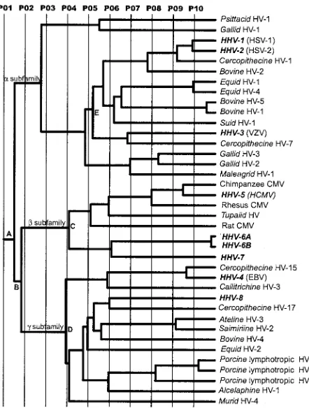
The ET phylogenetic tree for UL25 is shown in Fig. 4, and
the residues identified by the evolutionary trace analysis are shown in Fig. 5. The tree is in very close agreement with results of more detailed investigations of herpesvirus phylogeny based on larger protein sets and the use of alternative approaches for tree construction (30–32). The different partitions (P01 to P10) define the protein classes within the tree. Partitioning of the phylogenetic tree at the root (P01) (Fig. 4) results in a single class of proteins, and the conserved residues correlate with the most fundamental features of the protein. Twelve absolutely conserved residues are identified at this partition (Fig. 5), representing a subset of the residues conserved among the eight human herpesviruses (Fig. 1).
At partition 02 the UL25 homologues are divided into three classes corresponding to the separate subfamilies as a result of branching at nodes A and B. Further partitioning results in the generation of increasing numbers of separate protein classes. For example the gamma herpesviruses resolve into genera
Lymphocryptovirus(gamma-1) andRhadinovirus(gamma-2) at partition 04 (bifurcation at D), and the betaherpesviruses re-solve into the cytomegalo- and roseoloviruses at partition 05 (bifurcation at C).
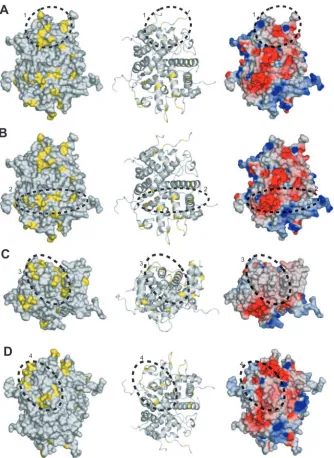
As a consequence of partitioning the phylogenetic tree, the ET analysis reveals conservation patterns that conventional sequence comparisons do not show. At P07, nine protein classes containing more than a single member are defined, which contribute to the positional information shown in Fig. 5. These classes, which are distributed among the three subfamilies, are exemplified by HSV-1, HSV-2, cercopithecine herpesvirus 1, and bovine herpes-virus 2 in the alphaherpesherpes-viruses; human, rhesus, and chimpanzee cytomegaloviruses in the betaherpesviruses; and Epstein-Barr vi-rus (HHV-4) and cercopithecine HV-15 in the gammaherpesvi-ruses. Mapping of the class-specific residues identified at this level to the surface of UL25nt revealed four clusters of particular interest (Fig. 5 and 6). The surface residues and the cluster to which each is classified are summarized in Table 3.
Cluster 1 is located in the apical region of the protein lo-cated on the same face as the view shown in Fig. 2A near disordered loop 2. Cluster 2, located on the same face as cluster 1, traverses laterally across the entire molecule and abuts disordered loop 5. Cluster 3 is made up of loops at the base of UL25nt directly opposite cluster 1. Cluster 4 is located on the face 180° from clusters 1 and 2 and abuts disordered loop 3. The electrostatic surface potentials of the three clusters are shown in Fig. 6. All four potential binding surfaces are rather nondescript, containing both charged and uncharged residues. It is impossible to predict, based solely on electro-statics, either the likely binding partners or the mode of bind-ing. However, it is interesting that three of the four clusters reside on or near areas of the structure where there are dis-ordered loops. It is possible that these flexible areas might become ordered upon binding to another protein partner. The disperse distribution of binding sites on UL25nt suggests that it is capable of binding to several protein partners. The inher-ent elasticity of UL25nt in its apparinher-ent ability to bind multiple protein partners, its unusual charge distribution, and its struc-tural flexibility are consistent with its key function during the dynamic processes associated with DNA encapsidation, capsid stabilization, and DNA retention.
[image:5.585.51.272.69.361.2]Models for UL25 function and biological implications.The HSV-1 UL25 protein has been proposed to be involved in the
FIG. 4. Evolutionary trace of herpesvirus UL25 homologues. P01 to P10 represent the different partitions of the ET phylogenetic tree. The branches defining the three herpesvirus subfamilies are labeled␣, , and␥. Key nodes discussed in the text are identified by A to D. Virus
species names in boldface represent human viruses.
on November 8, 2019 by guest
http://jvi.asm.org/
retention of packaged DNA within the capsid during the late stages of head filling (33, 51). There are phage proteins known which perform a function similar to that of UL25, as many aspects of the DNA-packaging mechanism of herpesviruses are analogous to those of dsDNA bacteriophages such as lambda, T4, and P22.
In many phages, following the packaging of the DNA, the portal is sealed to prevent the escape of the genome in a process known as head completion. Proteins that function to plug the portal during head completion include gp4, gp16, and gp26 of bacteriophage P22 and gp15 and gp16 of SPP1 (39, 41, 52). Mutants defective in any of these proteins exhibit a
phe-notype of unpackaged unit-length genomes similar to that de-scribed for the UL25 deletion mutant KUL25NS by McNab et al. (33), suggesting that UL25 may also be involved in the head completion process for HSV-1. The observations that HSV-1 C capsids contain more UL25 than B capsids and that only ap-proximately half of the UL25 protein in B capsids is associated with pentons (38, 47, 55) are consistent with a role for UL25 in plugging the portal. However, no direct interaction with the UL6 portal protein has been reported, and it has not been determined whether UL25 is present at the unique portal ver-tex of capsids.
[image:6.585.52.536.70.533.2]In the electron cryomicroscopy reconstructions of the entire
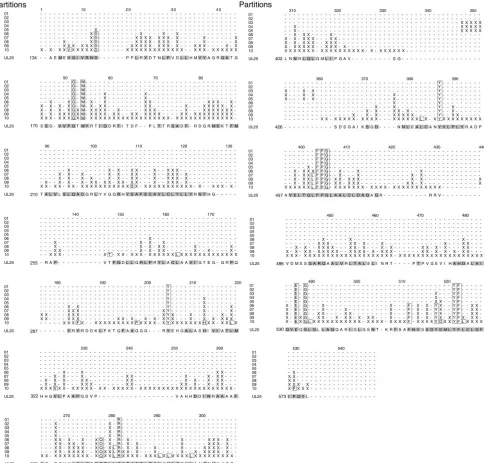
FIG. 5. Evolutionary trace residues for partitions 01 to 10. The trace residues belonging to a given partition occur in the horizontal row corresponding to the partition. The conserved residues are outlined in a box, the class-specific residues are demarcated by an X, and the dashes reflect gaps in the alignment.
on November 8, 2019 by guest
http://jvi.asm.org/
capsid (62) the location of UL25 was not identified, suggesting that it does not conform to the icosahedral symmetry of the capsid and therefore its density is averaged out. However, as mentioned above, UL25 appears to associate with pentons (38), and such an interaction could be envisaged to have a role in either DNA retention or capsid stabilization. The penton channel is rather large, with a minimum diameter of⬃40 Å, and is electropositive in character (6), suggesting that escape of packaged DNA is more likely through one or more of the penton vertices than through the hexon channels, which are quite narrow (⬃20 Å in diameter) and are very electronega-tive. The dimensions of UL25 certainly allow for it or an
oligomer to fit within the penton channel and act to potentially seal the DNA in the capsid.
The gpD protein of bacteriophage lambda and gpShp of bacteriophage 21 function as head stabilization proteins (57, 59). DNA packaging in bacteriophage lambda is accompanied by expansion of the prohead to an icosahedral shape and the binding of gpD. Interestingly, gpD is necessary for the pack-aging of full-length genomes, whereas gpD deletion mutants with genomes⬍82% of wild-type length are viable (49). This deficiency at the late stages of DNA packaging is similar to the phenotype of KUL25NS described by Stow (51), suggesting that rather than physically blocking the penton or portal
chan-FIG. 6. Evolutionary trace analysis. (A) The left and middle panels are a space-filling model and a ribbon trace, respectively, of UL25nt, with the class-specific exterior residues identified by the evolutionary trace shown in yellow. Cluster 1 is circled and identified with a 1. This cluster is also depicted in the electrostatic surface potential representation of the UL25nt structure (right). All three views are the same. (B, C, and D) Corresponding depictions of clusters 2, 3, and 4, respectively.
on November 8, 2019 by guest
http://jvi.asm.org/
[image:7.585.126.460.66.524.2]nel, UL25 might act to stabilize the capsid against the increas-ing internal pressures generated durincreas-ing the packagincreas-ing process. As is the case with lambda gpD, new UL25 binding sites might be revealed as the shape of the HSV-1 capsid is reconfigured during DNA entry. In addition to hexons and pentons, these sites could involve two other reported partners of UL25, the capsid triplex proteins VP19C and VP23 (38). Binding sites for the gpSoc and gpHoc proteins of bacteriophage T4 are simi-larly exposed on the phage head following the structural tran-sitions associated with DNA packaging (26). Although neither protein is required for DNA encapsidation, gpSoc does have a role in stabilizing the capsid against extremes of pH and tem-perature (21, 46). Like UL25, lambda gpD is characterized by a surface of irregular secondary structure with the majority of residues in coils and loops (59).
A possible parallel to UL25 is seen in the outer scaffold protein D from FX174, in which a rigid core structure is sur-rounded by flexible regions that are capable of existing in different conformations depending on their local environment (14). It is possible that UL25 could act in a scaffold-like man-ner, but being added rather than removed from the capsid during the maturation process. This could facilitate subsequent stages of capsid maturation such as nuclear egress or acquisi-tion of the tegument layer. Although the structures of phage proteins involved in DNA retention or head stabilization have been determined (39, 59) no significant similarity to UL25 in their folding pattern is apparent.
In addition to its role in DNA packaging, UL25 may also perform other important functions. It is likely that UL25 in-corporated into C capsids establishes interactions that are im-portant for capsid egress from the nucleus and the acquisition of the tegument layer by the virion. Studies on a UL25 ts
mutant also indicate an essential early function for the virion-contained protein (1; V. Preston, unpublished results), and in this context it is interesting to note that UL25 has been impli-cated in the association of incoming capsids with microtubules (24). Although our structure does not include the N-terminal 133 amino acids, the predicted coiled coil (amino acids 49 to 110) is likely to contribute to oligomerization or other protein-protein interactions, and genetic studies on N-terminally trun-cated proteins confirm that residues in this region are impor-tant for virus replication (V. Preston, unpublished results). It remains to be determined whether DNA packaging and/or some other function is impaired in these truncated proteins.
Conclusion. We have presented the first crystallographic structure of a herpesvirus protein involved in DNA encapsida-tion. The structure revealed a novel fold with a unique “flex-ible” architecture, suggesting its adaptability in changing con-formational states depending on its local environment. The
electrostatic calculation reveals a distinct division of surface charges in which one face is highly electronegative, while the opposite face is electropositive. Evolutionary trace analysis incorporating UL25 homologues from the three herpesvirus subfamilies identified four clusters of residues that potentially form binding sites for other interacting proteins and mapped them to the structure. The distribution of binding sites, in combination with its “flexible” architecture and distinctive electrostatic pattern, suggests an inherent elasticity for UL25, which is a necessary characteristic of a protein involved in the dynamic processes of capsid assembly and DNA encapsidation. Although the structure determination cannot presently dis-criminate between possible models for UL25 function, it pro-vides a basis for future studies on the role of this protein in DNA packaging and virion morphogenesis.
ACKNOWLEDGMENTS
We thank W. Chiu and C. M. Moure for technical assistance and useful discussions and the staff at the APS SBC-CAT for help with data collection.
This research was supported in part by grants from the NIH (R01-AI38469) to Wah Chiu and the Welch Foundation (Q0581) to F.A.Q. Initial extensive work was funded by a grant from the Howard Hughes Medical Institute to F.A.Q.
REFERENCES
1.Addison, C., F. J. Rixon, J. W. Palfreyman, M. O’Hara, and V. G. Preston.
1984. Characterisation of a herpes simplex virus type 1 mutant which has a temperature-sensitive defect in penetration of cells and assembly of capsids.
Virology138:246–259.
2.Adelman, K., B. Salmon, and J. D. Baines.2001. Herpes simplex virus DNA packaging sequences adopt novel structures that are specifically recognized by a component of the cleavage and packaging machinery. Proc. Natl. Acad.
Sci. USA98:3086–3091.
3.Ali, M. A., B. Forghani, and E. M. Cantin.1996. Characterization of an essential HSV-1 protein encoded by the UL25 gene reported to be involved
in virus penetration and capsid assembly. Virology216:278–283.
4.Bailey, S.1994. The CCP4 suite—programs for protein crystallography. Acta
Crystallogr. D50:760–763.
5.Beard, P. M., N. S. Taus, and J. D. Baines.2002. DNA cleavage and packaging proteins encoded by genes U(L)28, U(L)15, and U(L)33 of herpes
simplex virus type 1 form a complex in infected cells. J. Virol.76:4785–4791.
6.Bowman, B. R., M. L. Baker, F. J. Rixon, W. Chiu, and F. A. Quiocho.2003.
Structure of the herpesvirus major capsid protein. EMBO J.22:757–765.
7.Brown, J. C., M. A. McVoy, and F. L. Homa.2002. Packaging DNA into
herpesvirus capsids, p. 111–153.InA. Holzenburg and E. Bogner (ed.),
Struc-ture-function relationships of human pathogenic viruses. Kluwer Academic/ Plenum Publishers, New York, N.Y.
8.Brunger, A. T., P. D. Adams, G. M. Clore, W. L. DeLano, P. Gros, R. W. Grosse-Kunstleve, J. S. Jiang, J. Kuszewski, M. Nilges, N. S. Pannu, R. J. Read, L. M. Rice, T. Simonson, and G. L. Warren.1998. Crystallography and NMR system: a new software suite for macromolecular structure
determi-nation. Acta. Crystallogr. D54:905–921.
9.Chakravarty, S., A. M. Hutson, M. K. Estes, and B. V. Prasad.2005. Evo-lutionary trace residues in noroviruses: importance in receptor binding,
an-tigenicity, virion assembly, and strain diversity. J. Virol.79:554–568.
10.Church, G. A., and D. W. Wilson.1997. Study of herpes simplex virus
maturation during a synchronous wave of assembly. J. Virol.71:3603–3612.
11.Dasgupta, A., and D. W. Wilson.1999. ATP depletion blocks herpes simplex
virus DNA packaging and capsid maturation. J. Virol.73:2006–2015.
12.Davison, A. J.2002. Evolution of the herpesviruses. Vet. Microbiol.86:
69–88.
13.delaFortelle, E., and G. Bricogne.1997. Maximum-likelihood heavy-atom parameter refinement for multiple isomorphous replacement and
multi-wavelength anomalous diffraction methods. Methods Enzymol.276:472–494.
14.Dokland, T., R. McKenna, L. L. Ilag, B. R. Bowman, N. L. Incardona, B. A. Fane, and M. G. Rossmann.1997. Structure of a viral procapsid with
mo-lecular scaffolding. Nature389:308–313.
15.Gao, M., L. Matusick-Kumar, W. Hurlburt, S. F. DiTusa, W. W. Newcomb, J. C. Brown, P. J. McCann III, I. Deckman, and R. J. Colonno.1994. The protease of herpes simplex virus type 1 is essential for functional capsid
formation and viral growth. J. Virol.68:3702–3712.
16.Gibson, W., and B. Roizman.1972. Proteins specified by herpes simplex virus. 8. Characterization and composition of multiple capsid forms of
sub-types 1 and 2. J. Virol.10:1044–1052.
TABLE 3. Evolutionary trace residues within the identified four clusters
Cluster Trace residuesa
1...G225, R288, R305, P327, G331, H348, R362, G363, N365 2...R148, D150, N152, D156, L214, N508
3...G169, S170, G172, R180, G202, R203, K206, P413 4...R390, N396, Y398, D400, L402, R552
aThe listed residues are from partition 07. Only the external residues are
listed.
on November 8, 2019 by guest
http://jvi.asm.org/
17.Hauptman, H. A.1997. Shake-and-bake: an algorithm for automatic solution
ab initio of crystal structures. Methods Enzymol.277:3–13.
18.Holm, L., and C. Sander.1995. Dali: a network tool for protein structure
comparison. Trends Biochem. Sci.20:478–480.
19.Homa, F. L., and J. C. Brown.1997. Capsid assembly and DNA packaging in
herpes simplex virus. Rev. Med. Virol.7:107–122.
20.Innis, C. A., J. Shi, and T. L. Blundell.2000. Evolutionary trace analysis of TGF-beta and related growth factors: implications for site-directed
mu-tagenesis. Protein Eng.13:839–847.
21.Ishii, T., and M. Yanagida.1977. The two dispensable structural proteins (soc and hoc) of the T4 phage capsid; their purification and properties, isolation and characterization of the defective mutants, and their binding
with the defective heads in vitro. J. Mol. Biol.109:487–514.
22.Jones, D. T.1999. Protein secondary structure prediction based on
position-specific scoring matrices. J. Mol. Biol.292:195–202.
23.Jones, T. A., J. Y. Zou, S. W. Cowan, and Kjeldgaard.1991. Improved methods for building protein models in electron density maps and the
loca-tion of errors in these models. Acta Crystallogr. A47:110–119.
24.Kaelin, K., S. Dezelee, M. J. Masse, F. Bras, and A. Flamand.2000. The UL25 protein of pseudorabies virus associates with capsids and localizes to
the nucleus and to microtubules. J. Virol.74:474–482.
25.Lamberti, C., and S. K. Weller. 1998. The herpes simplex virus type 1 cleavage/packaging protein, UL32, is involved in efficient localization of
capsids to replication compartments. J. Virol.72:2463–2473.
26.Leiman, P. G., S. Kanamaru, V. V. Mesyanzhinov, F. Arisaka, and M. G. Rossmann.2003. Structure and morphogenesis of bacteriophage T4. Cell
Mol. Life Sci.60:2356–2370.
27.Lichtarge, O., and M. E. Sowa.2002. Evolutionary predictions of binding
surfaces and interactions. Curr. Opin. Struct. Biol.12:21–27.
28.McClelland, D. A., J. D. Aitken, D. Bhella, D. McNab, J. Mitchell, S. M. Kelly, N. C. Price, and F. J. Rixon.2002. pH reduction as a trigger for
dissociation of herpes simplex virus type 1 scaffolds. J. Virol.76:7407–7417.
29.McGeoch, D. J., M. A. Dalrymple, A. J. Davison, A. Dolan, M. C. Frame, D. McNab, L. J. Perry, J. E. Scott, and P. Taylor.1988. The complete DNA sequence of the long unique region in the genome of herpes simplex virus
type 1. J. Gen. Virol.69:1531–1574.
30.McGeoch, D. J., A. Dolan, and A. C. Ralph.2000. Toward a comprehensive
phylogeny for mammalian and avian herpesviruses. J. Virol.74:10401–10406.
31.McGeoch, D. J., and D. Gatherer.2005. Integrating reptilian herpesviruses
into the familyHerpesviridae. J. Virol.79:725–731.
32.McGeoch, D. J., D. Gatherer, and A. Dolan.2005. On phylogenetic relation-ships among major lineages of the Gammaherpesvirinae. J. Gen. Virol.
86:307–316.
33.McNab, A. R., P. Desai, S. Person, L. L. Roof, D. R. Thomsen, W. W. Newcomb, J. C. Brown, and F. L. Homa.1998. The product of the herpes simplex virus type 1 UL25 gene is required for encapsidation but not for
cleavage of replicated viral DNA. J. Virol.72:1060–1070.
34.Mihalek, I., I. Res, H. Yao, and O. Lichtarge.2003. Combining inference from evolution and geometric probability in protein structure evaluation. J.
Mol. Biol.331:263–279.
35.Newcomb, W. W., F. L. Homa, D. R. Thomsen, F. P. Booy, B. L. Trus, A. C. Steven, J. V. Spencer, and J. C. Brown.1996. Assembly of the herpes simplex virus capsid: characterization of intermediates observed during cell-free
cap-sid formation. J. Mol. Biol.263:432–446.
36.Newcomb, W. W., F. L. Homa, D. R. Thomsen, B. L. Trus, N. Cheng, A. Steven, F. Booy, and J. C. Brown.1999. Assembly of the herpes simplex virus procapsid from purified components and identification of small complexes
containing the major capsid and scaffolding proteins. J. Virol.73:4239–4250.
37.Newcomb, W. W., R. M. Juhas, D. R. Thomsen, F. L. Homa, A. D. Burch, S. K. Weller, and J. C. Brown.2001. The UL6 gene product forms the portal
for entry of DNA into the herpes simplex virus capsid. J. Virol.75:10923–
10932.
38.Ogasawara, M., T. Suzutani, I. Yoshida, and M. Azuma.2001. Role of the UL25 gene product in packaging DNA into the herpes simplex virus capsid: location of UL25 product in the capsid and demonstration that it binds
DNA. J. Virol.75:1427–1436.
39.Orlova, E. V., B. Gowen, A. Droge, A. Stiege, F. Weise, R. Lurz, M. van Heel, and P. Tavares.2003. Structure of a viral DNA gatekeeper at 10 A resolution
by cryo-electron microscopy. EMBO J.22:1255–1262.
40.Otwinowski, Z., and W. Minor.1997. Processing of X-ray diffraction data
collected in oscillation mode. Macromol. Crystallogr. A276:307–326.
41.Poteete, A. R., and J. King.1977. Functions of two new genes in Salmonella
phage P22 assembly. Virology76:725–739.
42.Prevelige, P. E., Jr., and J. King.1993. Assembly of bacteriophage P22: a
model for ds-DNA virus assembly. Prog. Med. Virol.40:206–221.
43.Rixon, F. J.1993. Structure and assembly of herpesviruses. Semin. Virol.
4:135–144.
44.Rixon, F. J., and D. McNab.1999. Packaging-competent capsids of a herpes simplex virus temperature-sensitive mutant have properties similar to those
of in vitro-assembled procapsids. J. Virol.73:5714–5721.
45.Roizman, B. N., and A. E. Sears.1996. Herpes simplex viruses and their
replication, p. 2231–2295.InB. N. Fields, D. M. Knipe, P. M. Howley, R. M.
Chanock, J. L. Melnick, T. P. Monath, B. Roizman, and S. E. Strauss (ed.), Field’s virology. Lippincott-Raven, Philadelphia, Pa.
46.Ross, P. D., L. W. Black, M. E. Bisher, and A. C. Steven.1985. Assembly-dependent conformational changes in a viral capsid protein. Calorimetric comparison of successive conformational states of the gp23 surface lattice of
bacteriophage T4. J. Mol. Biol.183:353–364.
47.Sheaffer, A. K., W. W. Newcomb, M. Gao, D. Yu, S. K. Weller, J. C. Brown, and D. J. Tenney.2001. Herpes simplex virus DNA cleavage and packaging
proteins associate with the procapsid prior to its maturation. J. Virol.75:
687–698.
48.Sowa, M. E., W. He, K. C. Slep, M. A. Kercher, O. Lichtarge, and T. G. Wensel. 2001. Prediction and confirmation of a site critical for effector
regulation of RGS domain activity. Nat. Struct. Biol.8:234–237.
49.Sternberg, N., and R. Weisberg.1977. Packaging of coliphage lambda DNA.
II. The role of the gene D protein. J. Mol. Biol.117:733–759.
50.Steven, A. C., and P. G. Spear. 1997. Herpesvirus capsid assembly and
envelopment, p. 312–351.InW. Chiu, R. M. Burnett, and R. L. Garcea (ed.),
Structural biology of viruses. Oxford University Press, New York, N.Y. 51.Stow, N. D.2001. Packaging of genomic and amplicon DNA by the herpes
simplex virus type 1 UL25-null mutant KUL25NS. J. Virol.75:10755–10765.
52.Strauss, H., and J. King.1984. Steps in the stabilization of newly packaged
DNA during phage P22 morphogenesis. J. Mol. Biol.172:523–543.
53.Taus, N. S., B. Salmon, and J. D. Baines.1998. The herpes simplex virus 1 UL 17 gene is required for localization of capsids and major and minor capsid proteins to intranuclear sites where viral DNA is cleaved and
pack-aged. Virology252:115–125.
54.Thompson, J. D., D. G. Higgins, and T. J. Gibson.1994. CLUSTAL W: improving the sensitivity of progressive multiple sequence alignment through sequence weighting, position-specific gap penalties and weight matrix choice.
Nucleic Acids Res.22:4673–4680.
55.Thurlow, J. K., F. J. Rixon, M. Murphy, P. Targett-Adams, M. Hughes, and V. G. Preston.2005. The herpes simplex virus type 1 DNA packaging protein UL17 is a virion protein that is present in both the capsid and the tegument
compartments. J. Virol.79:150–158.
56.Trus, B. L., F. P. Booy, W. W. Newcomb, J. C. Brown, F. L. Homa, D. R. Thomsen, and A. C. Steven. 1996. The herpes simplex virus procapsid: structure, conformational changes upon maturation, and roles of the triplex
proteins VP19c and VP23 in assembly. J. Mol. Biol.263:447–462.
57.Wendt, J. L., and M. Feiss.2004. A fragile lattice: replacing bacteriophage lambda’s head stability gene D with the shp gene of phage 21 generates the
Mg2⫹-dependent virus, lambda shp. Virology326:41–46.
58.White, C. A., N. D. Stow, A. H. Patel, M. Hughes, and V. G. Preston.2003. Herpes simplex virus type 1 portal protein UL6 interacts with the putative
terminase subunits UL15 and UL28. J. Virol.77:6351–6358.
59.Yang, F., P. Forrer, Z. Dauter, J. F. Conway, N. Cheng, M. E. Cerritelli, A. C. Steven, A. Pluckthun, and A. Wlodawer.2000. Novel fold and capsid-binding properties of the lambda-phage display platform protein gpD. Nat. Struct.
Biol.7:230–237.
60.Yu, D., and S. K. Weller.1998. Genetic analysis of the UL 15 gene locus for
the putative terminase of herpes simplex virus type 1. Virology243:32–44.
61.Yu, D., and S. K. Weller.1998. Herpes simplex virus type 1 cleavage and packaging proteins UL15 and UL28 are associated with B but not C capsids
during packaging. J. Virol.72:7428–7439.
62.Zhou, Z. H., M. Dougherty, J. Jakana, J. He, F. J. Rixon, and W. Chiu.2000.
Seeing the herpesvirus capsid at 8.5 A. Science288:877–880.