0022-538X/95/$04.0010
Copyrightq1995, American Society for Microbiology
Hepatitis C Virus-Encoded Nonstructural Protein NS4A
Has Versatile Functions in Viral Protein Processing
YASUNORI TANJI, MAKOTO HIJIKATA, SHINYA SATOH, TAKASI KANEKO,
ANDKUNITADA SHIMOTOHNO*
Virology Division, National Cancer Center Research Institute, Chuo-ku, Tokyo 104, Japan
Received 16 September 1994/Accepted 30 November 1994
A transient protein expression system in COS-1 cells was used to study the role of hepatitis C virus (HCV)-encoded NS4A protein on HCV nonstructural polyprotein processing. By analyzing the protein
expres-sion and processing of a deletion mutant polypeptide, NSD4A, which encodes the entire putative HCV
nonstructural polyprotein except the region encoding NS4A, the versatile functions of NS4A were revealed.
Most of the NS3 processed from NSD4A was localized in the cytosol fraction and was degraded promptly.
Coproduction of NS4A stabilizes NS3 and assists in its localization in the membrane. NS4A was found to be
indispensable for cleavage at the 4B/5A site but not essential for cleavage at the 5A/5B site in NSD4A. The
functioning of NS4A as a cofactor for cleavage at the 4B/5A site was also observed when 30 amino acids around this site was used as a substrate and a serine proteinase domain of 167 amino acids, from Gly-1049 to Ser-1215, was used as an enzyme protein, suggesting that possible domains for the interaction of NS4A were in those regions of the enzyme protein (NS3) and/or the substrate protein. Two proteins, p58 and p56, were produced
from NS5A. For the production of p58, equal or excess molar amounts of NS4A relative to NSD4A were
required. Deletion analysis of NS4A revealed a minimum functional domain of NS4A of 10 amino acids, from Gly-1678 to Ile-1687.
Hepatitis C virus (HCV), a member of the Flaviviridae fam-ily, has a positive-stranded RNA of about 9.5 kb (5, 13, 19). The precursor polyprotein translated from the longest open reading frame of the genome undergoes proteolytic processing conducted by both host and viral proteinases (2, 4, 6, 9–12, 15, 16, 22). For HCV type 1b (HCV-1b), the major genotype in Japan (13), the order of the individual proteins on the viral
genome is NH2-C (p22)-E1 (gp35)-E2 (gp70)-NS2 (p21)-NS3
(p70)-NS4A (p4)-NS4B (p27)-NS5A (p58/p56)-NS5B (p66)-COOH, where C, E, and NS are the putative core, envelope, and nonstructural proteins, respectively (11). A viral serine proteinase, Cpro-2, located in the N-terminal part of NS3 is employed for processing the region downstream of NS3 by two different modes of cleavage, intramolecular (cis) and intermo-lecular (trans) proteolytic cleavages. The NS3/NS4A (abbrevi-ated 3/4A) junction is processed only in a cis cleavage manner, whereas NS4A/NS4B (4A/4B), NS4B/NS5A (4B/5A), and NS5A/NS5B (5A/5B) junctions are cleaved in a trans cleavage manner (20, 22). The Cpro-2 domain which is necessary and sufficient for the cleavage at the 5A/5B site maps from Gly-1049 to Ser-1215 of the HCV precursor polyprotein (20).
In vitro transcription and translation analysis of the HCV nonstructural polyprotein showed that NS4A helps to anchor NS3 to the surface of the microsomal membrane by mutual association of these two proteins (12). Furthermore, the NS3-NS4A complex associates with NS4B, NS5A, and NS5B to form a complex structure which may be involved in virus rep-lication. These observations may imply that NS4A plays an important role in complex formation. Recently, it was reported that NS4A is necessary for cleavage at the 4B/5A site and that it enhances cleavage at NS3-dependent cleavage sites (3, 7).
For this report, we analyzed the processing of a mutated
NSD4A polypeptide which encodes the entire putative HCV
nonstructural polyprotein except the region of NS4A. Our experiments revealed versatile functions of NS4A. NS4A acts not only in the cleavage at the 4B/5A site but also in the stabilization and localization of NS3. A stoichiometrical assay and deletion analysis were also carried out to reveal the func-tions of NS4A.
MATERIALS AND METHODS
Construction of expression plasmids.The construction of plasmids pCMV/ N729-3010 and pCMV/N1049-1215 has been described previously (11, 20). Com-binations of positive- and a negative-stranded primers used for PCR (described below) are indicated in parentheses after the PCR products. The oligonucleo-tides used in the constructions are summarized in Table 1. To obtain plasmid pCMV/NSD4A, which encodes the entire HCV nonstructural polyprotein except NS4A, the BamHI-PstI fragment of pCMV/N729-3010 was inserted into the
BamHI-PstI site of pUC18 and used as a template for making PCR product 1 (F1
and M13 reverse primer) and PCR product 2 (M13 universal primer and R1). Both PCR products were ligated after phosphorylation, digested with BamHI and PstI, and then inserted into the BamHI-PstI site of pUC18. The resultant plasmid was pUC18/NSD4A, which carries the BamHI-PstI fragment of pCMV/ N729-3010 except for the region encoding NS4A. pCMV/NSD4A was obtained by replacing the PstI fragment of pCMV/N729-3010 with the
BamHI-PstI fragment of pUC18/NSD4A. PCR product 3 (F2 and R2) was digested with
PstI-EcoRI and inserted into the PstI-EcoRI site of pKS(1)/CMV (11) to obtain pCMV/N1658-1711, which encodes NS4A. To obtain plasmids that produce proteins with 30 amino acids (aa) around the 4B/5A or 5A/5B cleavage sites fused in frame to their C termini by an Escherichia coli dihydrofolate reductase (DHFR), PCR products 4 (F3 and R3) and 5 (F4 and R4) were digested with PstI and HindIII and were replaced with the PstI-HindIII fragment of pCMV/N729-1233D (20). The resultant plasmids were pCMV/N1953-1982D and pCMV/ N2400-2429D, respectively. Deletion mutants of NS4A were constructed by replacing the PstI-EcoRI fragments of PCR products 6 (F5 and R2), 7 (F6 and R2), 8 (F7 and R2), 9 (F2 and R5), 10 (F2 and R6), and 11 (F2 and R7) with the
PstI-EcoRI fragment of pCMV/N1658-1711. The resultant plasmids were
pCMV/N1668-1711, pCMV/N1678-1711, pCMV/N1688-1711, pCMV/N1658-1697, pCMV/N1658-1687, and pCMV/N1658-1677, respectively.
Expression of HCV polyproteins in COS-1 cells.DNA transfections were performed as described previously (10). Lysates of COS-1 cells transfected with the series of pCMV-derived plasmids were fractionated by sodium dodecyl sul-fate-polyacrylamide gel electrophoresis (SDS-PAGE) and subjected to immuno-blot analysis as described previously (10). The antibodies used in this experiment were anti-NS3 antibody (a-NS3), anti-NS4A antibody (a-NS4A), anti-NS4B
* Corresponding author. Mailing address: Virology Division, Na-tional Cancer Center Research Institute, 5-1-1, Tsukiji, Chuo-ku, To-kyo 104, Japan. Phone: 81-3-3542-2511, ext. 4700. Fax: 81-3-3543-2181. Electronic mail address: [email protected].
1575
on November 9, 2019 by guest
http://jvi.asm.org/
antibody (a-NS4B), anti-NS5A antibody (a-NS5A), anti-NS5B antibody (a -NS5B), and anti-DHFR antibody (a-DHFR) (10, 11, 15).a-NS4A anda-NS5A were generous gifts from A. Takamizawa (Osaka University, Osaka, Japan).
Pulse-chase analysis.After being incubated for 1 day in a 35-mm-diameter dish, COS-1 cells transfected with the series of pCMV-derived plasmids were used for pulse-chase analysis. Cells were incubated in 0.5 ml of methionine-free Eagle’s minimum essential medium (Flow Laboratories) with 5% dialyzed fetal calf serum for 1 h, and then they were labeled for 15 min in the same medium supplemented with 400 mCi of [35S]methionine (ICN) per ml. After pulse-labeling, the cells were either lysed immediately in 100ml of Laemmli’s sample buffer without dye or lysed following a chase for various times in Dulbecco’s modified Eagle’s medium containing methionine and cysteine (75mg/ml each).
Immunoprecipitation of metabolically labeled cell extract with [35
S]methi-onine.Cell lysates metabolically labeled with [35S]methionine were boiled and diluted 10-fold with extraction buffer (50 mM Tris hydrochloride [pH 7.5], 150 mM NaCl, 5 mM EDTA, 0.5% Nonidet P-40, 1 mM phenylmethylsulfonyl flu-oride). One milliliter of the diluted lysates was preadsorbed with 50ml of protein G-Sepharose suspension (Pharmacia) and incubated with a-NS3, a-NS4A,
a-NS4B,a-NS5A, anda-NS5B for 1 h. The immunocomplex was recovered by the addition of 30ml of protein G-Sepharose and was washed three times with 50 mM Tris hydrochloride (pH 7.4)–500 mM NaCl–5 mM EDTA–1% Nonidet P-40–5% sucrose and once with 10 mM Tris hydrochloride (pH 7.4)–50 mM NaCl–1 mM EDTA. The immunoprecipitates were analyzed by SDS–8% PAGE or tricine–SDS–16% PAGE (18). The gels were dried and exposed to imaging plates (Fuji Photo Film Co., Ltd).
Subcellular localization of viral proteins.pCMV-derived plasmids were trans-fected into COS-1 cells as described above. After being incubated for 1 day in a 35-mm-diameter dish, the cells were incubated in 1 ml of methionine-free Eagle’s minimum essential medium with 5% fetal calf serum supplemented with 200mCi of [35
S]methionine per ml for 4 h. Cells were collected with a rubber scraper and homogenized in 200ml of MSB medium (20 mM Tris hydrochloride [pH 8], 25 mM NaCl, 0.5 mM CaCl2, 1 mM phenylmethylsulfonyl fluoride, 1 mM dithio-threitol). The homogenate was overlaid on 60% (lower layer)240% (upper layer) sucrose solutions in MSB medium and centrifuged (200,0003g, 60 min).
After centrifugation, the portion on the 40% sucrose layer was defined as the cytosol fraction and the portion on the 60% sucrose layer was defined as the membrane fraction. Both fractions were diluted 10-fold with extraction buffer and immunoprecipitated as described above.
Nucleotide sequence accession number.The sequence of the cDNA of pCMV/ N729-3010 has been deposited in the DDBJ, EMBL, and GenBank DNA data-bases under accession number D16435.
RESULTS
Effects of coproduced NS4A on the expression of HCV
polyproteins encoded in pCMV/NSD4A. To understand the
function of NS4A in HCV nonstructural polyprotein
process-ing, pCMV/NSD4A, which encodes the entire putative HCV
nonstructural polyprotein except the NS4A region, was ex-pressed in COS-1 cells, and the protein products produced from this mutated polypeptide were analyzed with region-spe-cific antisera. By comparing the products produced from
NSD4A with those of the nonstructural region encoded in
pCMV/N729-3010 (Fig. 1A), which produces all of the HCV
nonstructural proteins, the function of NS4A in HCV non-structural polyprotein processing was analyzed. The produc-tion of NS3, NS4A, NS4B, NS5A, and NS5B from N729-3010
(abbreviated as NS in Fig. 2) was detected bya-NS3,a-NS4A,
a-NS4B, a-NS5A, anda-NS5B, respectively (Fig. 2, lanes 2).
On the other hand, protein production from the NSD4A
polypeptide was different from that from N729-3010 (Fig. 2A through D, lanes 3). The level of NS3 production was quite low
R3 59-CCGAAGCTTGTCCCAAACATCCCTTAG-39 1982–1977
R4 59-CCGAAGCTTCAAGGCACCTGTCCATGT-39 2429–2424
R5 59-ATGAATTCTTAGTCGGGAACAACAGC-39 1697–1693
R6 59-CAGAATTCTTAAATGATCCTACCCAC-39 1687–1683
R7 59-GCGAATTCTTAGGTTGTCAGGCAATA-39 1677–1673
a
Underlined nucleotides indicate sequences complementary to the genome of HCV. b
[image:2.612.59.555.81.231.2]Corresponding amino acids in the HCV genome (13).
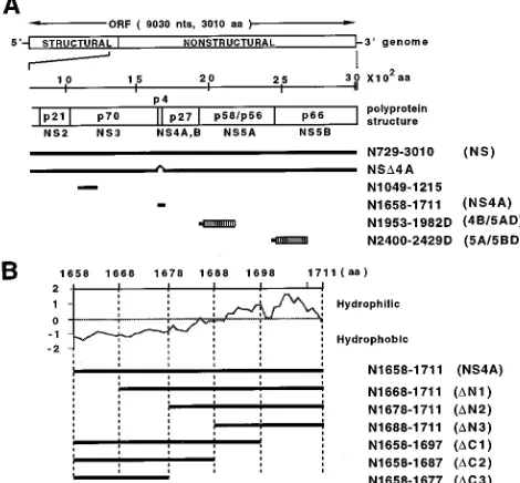
FIG. 1. Schematic representations of HCV polyprotein fragments produced by the expression constructs. (A) The genomic and polyprotein structures of the nonstructural region from NS2 to NS5B are shown enlarged below the HCV open reading frame (ORF). The scale indicates the amino acid position. The regions of polypeptides in the HCV precursor polyprotein are shown by thick bars. The designations of HCV polypeptide regions synthesized in COS-1 cells are shown on the right. Numbers indicate amino acid positions from the N terminus to the C terminus of the HCV precursor polyprotein. Abbreviations for the HCV polypeptides are indicated in parentheses. Hatched boxes indicate E.
coli DHFR fused in frame at the C-terminal end of the HCV polypeptide
(abbreviated as D in the peptide designations). nts, nucleotides. (B) HCV NS4A hydrophobicity profile as estimated by using the DNASIS program with a search length of 7 aa (Hitachi Software Engineering Co., Ltd.). A series of peptides with deletions from the N and C termini of NS4A is shown below the hydrophobicity profile.
on November 9, 2019 by guest
http://jvi.asm.org/
[image:2.612.316.551.382.600.2]in NSD4A. No NS4B or NS5A could be detected bya-NS4B or
a-NS5A, respectively, while an 85-kDa polyprotein (p85) was
detected bya-NS5A. Since the size of this polypeptide, 85 kDa,
was almost identical to the sum of the molecular masses of NS4B and NS5A, this polypeptide was likely to be a processing intermediate composed of these two viral proteins. The failure
ofa-NS4B to detect p85 was possibly due to the low efficiency
of blotting of the high-molecular-weight protein and the low titer of this antibody in this experiment. However, p85 was
detected by a-NS4B in the pulse-chase experiment as
de-scribed below. The production of NS5B as detected bya-NS5B
indicates that cleavage at the 5A/5B site normally occurs in this mutant polypeptide. By coproduction with NS4A, the
produc-tion of NS3, NS4B, and NS5A from NSD4A was restored (Fig.
2A, C, and D, lanes 4). The restoration of NS3 in the presence of NS4A seemed not to be due to efficient cleavage at the artificial NS3-NS4B site. Cleavage at this site occurs efficiently in the absence of NS4A, because the intermediate product, p85(NS4B-NS5A), accumulates in the absence of NS4A (Fig. 2D, lane 3). Instead, the restoration of NS3 by coproduction with NS4A seemed to be caused by the stabilization of NS3. The production of p58(NS5A) relative to p56(NS5A)
gener-ated by coproduction of NSD4A and NS4A was reduced
com-pared with that for N729-3010 (Fig. 2D, lanes 2 and 4).
NS4A is necessary for the stabilization of NS3.Since
cleav-age at NS2/3 is not affected by viral nonstructural proteins encoded in the region downstream of NS3 (11), and the viral proteins p85(NS4B-NS5A) and NS5B, encoded in the region
downstream of NS3, are produced from NSD4A, the greatly
reduced production of NS3 from this mutated polypeptide was thought to be due to the instability of NS3 in cells producing the mutated polypeptide. To clarify this possibility, pulse-chase
analysis using a transient expression system in COS-1 cells was
conducted. Plasmid pCMV/NSD4A with or without pCMV/
N1658-1711, which encodes NS4A, was used for transfection.
Transfectants were pulse-labeled for 15 min with [35
S]methi-onine and chased for various times after the addition of excess amounts of unlabeled methionine. The cell lysates were ana-lyzed at chase time points at 0, 20, 60, 180, 360, and 1,440 (1 day) min for the production of viral proteins by
immunopre-cipitation with a-NS3, a-NS4A, a-NS4B, a-NS5A, and
a-NS5B.
Previously, we showed that the processing of NS3 from the precursor polyprotein of N729-3010 was almost completed within the 15-min pulse period and that NS3 was stably present for more than 1 day of the chase period (21). Contrary to this
observation, the levels of NS3 in NSD4A-producing cell lysates
were low at the start of the chase time, while a protein of
approximately 150 kDa, immunoprecipitated by a-NS3 and
[image:3.612.59.294.69.282.2]likely to be the processing intermediate of NS3-NS4B-NS5A, was detected in significant amounts until 60 min of chase (Fig. 3A, lanes 2 through 5). The amount of NS3 reached a maxi-mum at a chase time of 180 min and then started to decline.
FIG. 2. Detection of processed products of NSD4A and the effect of NS4A on NSD4A processing. Lysates of COS-1 cells transfected with pKS(1)/CMV (10
mg), an original vector plasmid (Vec.) without an HCV insert (lanes 1); pCMV/ N729-3010 (5mg) plus pKS(1)/CMV (5mg) (lanes 2); pCMV/NSD4A (5mg) plus pKS(1)/CMV (5mg) (lanes 3); or pCMV/NSD4A (5mg) plus pCMV/ N1658-1711 (5mg) (lanes 4) were separated by SDS–8% PAGE (A, D, and E), SDS–10% PAGE (C), or tricine–SDS–16% PAGE (B) and analyzed by immu-noblotting witha-NS3 (A),a-NS4A (B),a-NS4B (C),a-NS5A (D), ora-NS5B (E). Molecular mass markers (in kilodaltons) are shown on the left. The posi-tions of processing products NS3, NS4A, NS4B, NS5A, and NS5B and the precursor polyprotein NS4B-NS5A are indicated on the right with arrows. NS, N729-3010.
FIG. 3. Pulse-chase analysis of viral precursor polyprotein processing. Cells were transfected with pCMV/NSD4A (5 mg) plus pKS(1)/CMV (5 mg) or pCMV/NSD4A (5mg) plus pCMV/1658-1711 (5mg) and were pulse-labeled for 15 min and then chased as indicated. The lysate at each time point was immu-noprecipitated witha-NS3 (A),a-NS4A (B),a-NS4B (C),a-NS5A (D), or
a-NS5B (E). Immunoprecipitation was performed with lysates transfected with pKS(1)/CMV (lanes 1), pCMV/NSD4A (lanes 2 through 7), or pCMV/NSD4A plus pCMV/N1658-1711 (lanes 8 through 13). Gel conditions were as follows: SDS–8% PAGE for panels A, D, and E; SDS–10% PAGE for panel C; and tricine–SDS–16% PAGE for panel B. Molecular mass markers (in kilodaltons) are shown on the left. The positions of processing products NS3, NS4A, NS4B, NS5A, and NS5B and precursor polyproteins are shown on the right with arrows. Vec., vector.
on November 9, 2019 by guest
http://jvi.asm.org/
After 1 day (1,440 min) of chase, no detectable NS3 was present, suggesting that NS3 does not exist stably in the cell without NS4A. On the other hand, when NS4A was
copro-duced with NSD4A, the NS3 level increased until 180 min of
chase, and the relative amount of NS3 remained almost con-stant thereafter, indicating that NS3 was stable in the cell (Fig. 3A, lanes 11 through 13). Coproduced NS4A was also stably present in the cell (Fig. 3B, lanes 8 through 13). A precursor polyprotein with a molecular mass of 85 kDa was detected by
both a-NS4B and a-NS5A antibodies in lysates transfected
with pCMV/NSD4A, but NS4B and NS5A were not detected
throughout the chase period (Fig. 3C and D, lanes 2 through 7). On the other hand, NS4B and NS5A were produced when
NS4A was coproduced with NSD4A (Fig. 3C, lanes 8 through
13, and 4D, lane 8). The production of NS5B from NSD4A was
seen at the start of the chase, and the level of the protein was not affected by coproduction of NS4A (Fig. 3E, lanes 2 through 13).
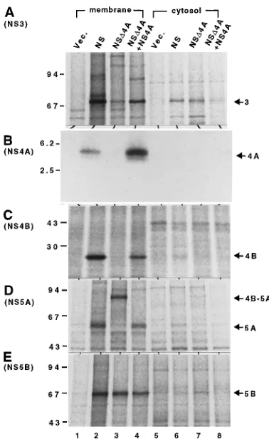
Subcellular localization of HCV proteins.It is reported that
NS4A is likely to associate with NS3 and that it is responsible for the membrane association of NS3 (12). Therefore, the prompt degradation of NS3 in the absence of NS4A was likely due to the loss of association of NS3 with the membrane. Association with the membrane may protect against
proteo-result indicates that NS4A, which is known to associate with membrane, affects the localization of NS3, possibly through mutual interaction.
Besides NS3, the majorities of NS4A, NS4B, NS5A, NS5B, and related precursor polyproteins were detected in the mem-brane fraction both with and without NS4A (Fig. 4B through E).
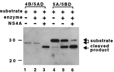
Role of NS4A in HCV nonstructural protein processing.To
investigate the role of NS4A in HCV nonstructural protein processing, a viral serine proteinase (Cpro-2)-dependent trans cleavage assay was performed. The function of NS4A in the processing of HCV nonstructural proteins was assayed by cleavage of DHFR fusion peptides containing 30 amino acid residues around the Cpro-2 cleavage site with a protein, N1049-1215, which has a Cpro-2 activity. The 30 aa used for this assay consisted of 20 aa located upstream of each cleavage site and 10 aa located downstream of each cleavage site. E. coli DHFR was fused in frame at the C-terminal end of each 30-aa segment to facilitate the detection of the C-terminal cleavage
product bya-DHFR (11). Cleavage was detected in the
sub-strate proteins of 4B/5AD and 5A/5BD. Both enzyme and NS4A proteins were required for the cleavage at 4B/5A (Fig. 5, lanes 1 through 3). The fact that NS4A was necessary for cleavage at the 4B/5A site implies that 30 aa around the 4B/5A site are sufficient for cleavage by N1049-1215 plus NS4A. On the other hand, approximately half of the 5A/5BD was pro-cessed by enzyme protein only, while coproduction of NS4A led to an increase in cleavage efficiency (Fig. 5, lanes 4 through 6). The faint band seen in the same position as the cleaved product in the absence of Cpro-2 may be a degraded product (Fig. 5, lane 4).
Stoichiometrical assay of NS4A function.To investigate the
stoichiometrical relationship between NS4A and the
enzyme-substrate protein of NSD4A, we examined the cleavage
effi-ciency at the 4B/5A site by using different amounts of pCMV/
NSD4A and pCMV/N1658-1711 plasmids for transfection. The
efficiencies of 4B/5A cleavage were evaluated by detecting
cleaved NS5A products witha-NS5A. As shown in Fig. 6, the
production of NS5A was reduced to an extent corresponding to the decrease in the amount of NS4A-encoding plasmid.
However, production of p56(NS5A) was detected bya-NS5A
even when the amount of NS4A-encoding plasmid was far less
than that of the NSD4A-encoding plasmid. A proportional
relationship between the production of protein and the amount of the plasmid was found if the amount of plasmid
used was less than 5mg per well in this assay system (data not
shown). Therefore, it was obvious that the presence of the smallest amount of NS4A relative to NS3 was sufficient to
cleave at the 4B/5A site of NSD4A. For instance, even if the
ratio of pCMV/NSD4A to pCMV/N1658-1711 was 99mg to 1
mg (i.e., the molar ratio of nonstructural polyprotein to NS4A
was 38:1), more than half of the substrate protein NSD4A was
cleaved at the 4B/5A site to produce p56(NS5A) (Fig. 6, lane 8).
[image:4.612.80.269.69.377.2]The production of p58(NS5A) was strongly influenced by the amount of NS4A-encoding plasmid. When excess amounts
FIG. 4. Localization of processing products of NSD4A and the effect of NS4A on their subcellular localization. Lysates of COS-1 cells transfected with pKS(1)/CMV (10mg) (lanes 1 and 5), pCMV/N729-3010 (5mg) plus pKS(1)/ CMV (5mg) (lanes 2 and 6), pCMV/NSD4A (5mg) plus pKS(1)/CMV (5mg) (lanes 3 and 7), or pCMV/NSD4A (5mg) plus pCMV/N1658-1711 (5mg) (lanes 4 and 8) were separated into membrane (lanes 1 through 4) and cytosol (lanes 5 through 8) fractions as indicated in the text; fractionated by SDS–8% PAGE (A, D, and E), SDS–10% PAGE (C), or tricine–SDS–16% PAGE (B) after immu-noprecipitation witha-NS3 (A),a-NS4A (B),a-NS4B (C),a-NS5A (D), or
a-NS5B (E). Vec., vector. NS, N729-3010.
on November 9, 2019 by guest
http://jvi.asm.org/
of NS4A relative to NSD4A were expressed in the cell, the relative production of p58(NS5A) and p56(NS5A) was almost the same as that in lysates of N729-3010-producing cells (Fig. 6, lanes 1 through 3). On the other hand, when the smaller
amount of NS4A relative to NSD4A was expressed in the cell,
the relative production of p58(NS5A) was reduced (Fig. 6,
lanes 5 through 8). When 0.2 or 0.1mg of plasmid encoding
NS4A was used for transfection, no p58(NS5A) production was detected, while the uncleaved p85 product was detected (Fig. 6, lanes 7 and 8). These results suggest that NS4A also controls the production of p58(NS5A). Equal or excess amounts of NS4A relative to the enzyme-substrate protein
(NSD4A) were required for the production of p58(NS5A).
Deletion mapping of NS4A. As shown in Fig. 1B, NS4A
consists of two distinct parts, an N-terminal hydrophobic half and a C-terminal hydrophilic half. The roles of these two re-gions in NS4A functioning are not known. An assay of NS4A-dependent cleavage at the 4B/5A site was used to determine the region of the NS4A protein essential for this function. A series of deletion mutants (Fig. 1B) derived from N1658-1711
(NS4A) was used for the assay. The activity of each deletion mutant was determined by detecting NS5A production from
NSD4A. When DN1, DN2, DC1, and DC2 were coproduced
with NSD4A, different levels of p56(NS5A) were detected by
a-NS5A (Fig. 7, lanes 3, 4, 6, and 7). The production of NS5A
was greatly reduced whenDN1 orDC2 was used as the source
of NS4A. However, there was no NS5A production whenDN3
orDC3 was coproduced. These data indicate that the
C-termi-nal part of NS4A downstream of aa 1688 and the N-termiC-termi-nal part upstream of aa 1677 are not essential for NS4A activity, although a significant effect of these regions on cleavage at the 4B/5A site was observed. The minimum domain for NS4A activity appears to be from aa 1678 to aa 1687 of the HCV precursor polyprotein, although whether or not the expected minimum domain of N1678-1687 itself is functional remains to be determined.
DISCUSSION
The transient expression system in COS-1 cells and pulse-chase analysis showed the production of NS3 and NS5B from
NSD4A, indicating that cleavage at the NS2/3 and 5A/5B sites
proceeds without the presence of NS4A. However, the rate of NS3 production in this construct was delayed compared with that in N729-3010 (21). During 180 min of chase, a putative
precursor protein was detected bya-NS3, suggesting that
pro-cessing to NS3 was not completed by the end of the 180-min chase. Since NS4A was excluded from the mutated polypeptide
NSD4A, NS4B was followed directly by NS3. Therefore, the
amino acid sequence around the C-terminal end of NS3 was changed from DLEVVT/STW to DLEVVT/ASH (/ indicates the cleavage site). It is possible that the delayed processing of
NS3 in NSD4A compared with the result for N729-3010 was
caused by the artificial amino acid sequence between NS3 and NS4B. However, this change of amino acid sequence at the cleavage site seems not to affect the further processing of nonstructural precursor polyprotein. On the other hand, the
production of NS5B in lysates transfected with pCMV/NSD4A
was detected at the end of the pulse period, and no precursor
protein reactive with a-NS5B was detected. Therefore,
effi-cient cleavage proceeds at the 5A/5B site of the NSD4A
polyprotein in the absence of NS4A.
The amount of NS3 produced from NSD4A in the absence
[image:5.612.85.271.69.187.2]of NS4A reached a maximum after 180 min of chase, and then
FIG. 5. Role of NS4A in HCV nonstructural protein processing. Lysates of COS-1 cells transfected with the plasmids indicated above the gel were fraction-ated by SDS–10% PAGE and analyzed by immunoblotting with a-DHFR. N1049-1215, a serine proteinase encoded by pCMV/N1049-1215, was used as the enzyme protein. Substrate proteins were 4B/5AD (lanes 1 through 3) and 5A/ 5BD (lanes 4 through 6). The amounts of the plasmids used were as follows: substrate-encoded plasmid (3.3mg) plus pKS(1)/CMV (6.7mg) (lanes 1 and 4); substrate-encoded plasmid (3.3mg) plus enzyme-encoded plasmid (3.3mg) plus pKS(1)/CMV (3.3mg) (lanes 2 and 5); and substrate-, enzyme-, and NS4A-encoded plasmids (3.3mg each) (lanes 3 and 6). Molecular mass markers (in kilodaltons) are shown on the left. The positions of the substrate protein and cleaved products are indicated on the right with arrows.
FIG. 6. Stoichiometrical assay of NS4A function in 4B/5A cleavage. Lysates of COS-1 cells transfected with 10mg of a plasmid encoding NS (lane 1) or with different relative amounts of plasmids encoding NSD4A and NS4A (lanes 2 through 9) were fractionated by SDS–8% PAGE. The amounts of plasmids used and the molar ratios of pCMV/N1658-1711 to pCMV/NSD4A are indicated at the top. Molecular mass markers (in kilodaltons) are shown on the left. The positions of the processing products are indicated on the right with arrows.
FIG. 7. Effects of NS4A products containing deletions on cleavage at the 4B/5A site. Lysates of COS-1 cells transfected with a plasmid encoding NSD4A (5mg) plus pKS(1)/CMV (5mg) (lane 1) or a plasmid encoding NS4A or one of its deletion mutants (5 mg each) (lanes 2 through 8) were fractionated by SDS–8% PAGE and then analyzed by immunoblotting witha-NS5A.
on November 9, 2019 by guest
http://jvi.asm.org/
tural proteins revealed that without the coexistence of NS4A,
more than 50% of the NS3 produced from NSD4A was
local-ized in the cytosol fraction, while when NS3 was coproduced with NS4A, most of the NS3 was found in the membrane fraction. Previously we showed that a possible association be-tween NS4A and NS3 is important for the membrane anchor-ing of NS3 (12). In view of the previous data and of the evidence presented here, one aspect of NS4A functioning may be to anchor NS3 to the membrane of the endoplasmic retic-ulum.
Since NS5B was produced efficiently from NSD4A, it is
be-lieved that NS4A is not essential for cleavage at the 5A/5B site as reported by other groups (3, 7). However, it is noteworthy that the cleavage at the 5A/5B site of the 5A/5BD protein was enhanced when NS4A was present (Fig. 5, lanes 5 and 6). In this assay system, the serine proteinase domain was supplied in
trans to the substrate, which contained only 30 amino acid
residues surrounding the cleavage site; thus, the efficiency of cleavage might have been lowered. The molecular mechanism for the enhancement of 5A/5B cleavage by Cpro-2 with NS4A is not clear. However, stabilization of Cpro-2 and membrane association of Cpro-2 caused by NS4A, which may facilitate close association with substrates, may be considered to en-hance the cleavage function.
The fact that the presence of NS4A is indispensable for processing at the 4B/5A site was confirmed in this study as it has been by others (3, 7). The possible function of NS4A in stabilizing NS3 may not be sufficient for this cleavage because NS3 could be detected in the absence of NS4A by the time that cleavage at the 4B/5A site was completed in the presence of NS4A (Fig. 3A and D). Thus, it is likely that an additional function of NS4A, which cooperates with the function of NS3, involves cleavage at the 4B/5A site. For this function, 10 amino acid residues harboring the central region of NS4A (aa 1678 to 1687) were shown to be essential, although a great reduction in activity by this limited domain was evident (Fig. 7). The serine proteinase domain, aa 1049 to 1215, was sufficient to cleave at the 4B/5A site of a substrate containing only 30 amino acid residues surrounding the 4B/5A cleavage site in the presence of NS4A (Fig. 5). This result is in contrast to the results of Bartenschlager and coworkers (3), in which the N-terminal truncation of NS3 abolished NS4A-dependent 4B/5A cleavage. The domain of Cpro-2 (N1049-1215) used in the present study contains a further deletion of 15 aa from the N terminus of their construct. The reason for the conflicting results is not known; however, they may have been caused by the use of HCV clones with different sequences or by differences in the assay systems.
Proteins with different molecular weights, p56(NS5A) and p58(NS5A), were produced from NS5A. The biochemical na-ture of these proteins remains to be clarified. The production of p58(NS5A) from mutated HCV nonstructural polyproteins
in which amino acids at the P1 and P19positions in the 3/4A or
4A/4B sites are replaced with asparagine is drastically reduced, leading to impaired cleavage at these sites (21). These results
in the production p58(NS5A) differs from that in the cleavage at the 4B/5A site.
In this paper we describe the versatile functions of NS4A of HCV. The function of NS4A seems to differ from that of the NS2B product of flaviviruses, which is required for NS3-de-pendent proteolytic cleavage of the flavivirus precursor polyprotein (1, 4, 8, 14, 17). The complicated function of NS4A seems to be involved not only in the strict regulation of the proteolytic processing of the precursor polyprotein but also in the function of the processed protein, a function that may be crucial for viral proliferation.
ACKNOWLEDGMENTS
This work was supported in part by a grant-in-aid from the Ministry of Health and Welfare for a Comprehensive 10-Year Strategy for Cancer Control and a grant-in-aid for scientific research from the Ministry of Education, Science and Culture, Japan.
ADDENDUM IN PROOF
We observed that NS5A products are phosphorylated and the larger product is the hyperphosphorylated form of the smaller product. Production of the hyperphosphorylated form depends on the presence of NS4A (T. Kaneko, Y. Tanji, S. Satoh, M. Hijikata, S. Asabe, K. Kimura, and K. Shimotohno, Biochem. Biophys. Res. Commun. 205:320–326, 1994).
REFERENCES
1. Arias, C. F., F. Preugschat, and J. H. Strauss. 1993. Dengue 2 virus NS2B and NS3 form a stable complex that can cleave NS3 within the helicase domain. Virology 193:888–899.
2. Bartenschlager, R., L. Ahlborn-Laake, J. Mous, and H. Jacobsen. 1993. Nonstructural protein 3 of the hepatitis C virus encodes a serine-type pro-teinase required for cleavage at the NS3/4 and NS4/5 junctions. J. Virol.
67:3835–3844.
3. Bartenschlager, R., L. Ahlborn-Laake, J. Mous, and H. Jacobsen. 1994. Kinetic and structural analyses of hepatitis C virus polyprotein processing. J. Virol. 68:5045–5055.
4. Chambers, T. J., A. Grakoui, and C. M. Rice. 1991. Processing of the yellow fever virus nonstructural polyprotein: a catalytically active NS3 proteinase domain and NS2B are required for cleavages at dibasic sites. J. Virol. 65: 6042–6050.
5. Choo, Q. L., K. H. Richman, J. H. Han, K. Berger, C. Lee, C. Dong, C.
Gallegos, D. Coit, S. R. Medina, P. J. Barr, A. J. Weiner, D. W. Bradley, G. Kuo, and M. Houghton.1991. Genetic organization and diversity of the hepatitis C virus. Proc. Natl. Acad. Sci. USA 88:2451–2455.
6. Eckart, M. R., M. Selby, F. Masiarz, C. Lee, K. Berger, K. Crawford, C. Kuo,
M. Houghton, and Q.-L. Choo.1993. The hepatitis C virus encodes a serine protease involved in processing of the putative nonstructural proteins from the viral polyprotein precursor. Biochem. Biophys. Res. Commun. 192:399– 406.
7. Failla, C., L. Tomei, and R. D. Francesco. 1994. Both NS3 and NS4A are required for proteolytic processing of hepatitis C virus nonstructural pro-teins. J. Virol. 68:3753–3760.
8. Falgout, B., M. Pethel, Y.-M. Zhang, and C.-J. Lai. 1991. Both nonstructural proteins NS2B and NS3 are required for the proteolytic processing of den-gue virus nonstructural proteins. J. Virol. 65:2467–2475.
9. Grakoui, A., D. W. McCourt, C. Wychowski, S. M. Feinstone, and C. M. Rice. 1993. Characterization of the hepatitis C virus-encoded serine proteinase: determination of proteinase-dependent polyprotein cleavage sites. J. Virol.
67:2832–2843.
on November 9, 2019 by guest
http://jvi.asm.org/
10. Hijikata, M., N. Kato, Y. Ootsuyama, M. Nakagawa, and K. Shimotohno. 1991. Gene mapping of the putative structural region of the hepatitis C virus genome by in vitro processing analysis. Proc. Natl. Acad. Sci. USA 88:5547– 5551.
11. Hijikata, M., H. Mizushima, T. Akagi, S. Mori, N. Kakiuchi, N. Kato, T.
Tanaka, K. Kimura, and K. Shimotohno. 1993. Two distinct proteinase activities required for the processing of a putative nonstructural precursor protein of hepatitis C virus. J. Virol. 67:4665–4675.
12. Hijikata, M., H. Mizushima, Y. Tanji, Y. Komoda, Y. Hirowatari, T. Akagi,
N. Kato, K. Kimura, and K. Shimotohno.1993. Proteolytic processing and membrane association of putative nonstructural proteins of hepatitis C virus. Proc. Natl. Acad. Sci. USA 90:10773–10777.
13. Kato, N., M. Hijikata, Y. Ootsuyama, M. Nakagawa, S. Ohkoshi, T.
Sug-imura, and K. Shimotohno.1990. Molecular cloning of the human hepatitis C virus genome from Japanese patients with non-A, non-B hepatitis. Proc. Natl. Acad. Sci. USA 87:9524–9528.
14. Lin, C., S. M. Amberg, T. J. Chambers, and C. M. Rice. 1993. Cleavage at a novel site in the NS4A region by the yellow fever virus NS2B-3 proteinase is a prerequisite for processing at the downstream 4A/4B signalase site. J. Virol. 67:2327–2335.
15. Manabe, S., I. Fuke, O. Tanishita, C. Kaji, Y. Gomi, S. Yoshida, C. Mori, A.
Takamizawa, I. Yosida, and H. Okayama.1994. Production of nonstructural proteins of hepatitis C virus requires a putative viral protease encoded by
NS3. Virology 198:636–644.
16. Mizushima, H., M. Hijikata, Y. Tanji, K. Kimura, and K. Shimotohno. 1994. Analysis of N-terminal processing of hepatitis C virus nonstructural protein 2. J. Virol. 68:2731–2734.
17. Preugschat, F., C.-W. Yao, and J. H. Strauss. 1990. In vitro processing of dengue virus type 2 nonstructural proteins NS2A, NS2B, and NS3. J. Virol.
64:4364–4374.
18. Schagger, H., and J. G. von Heijine. 1987. Tricine-sodium dodecyl sulfate-polyacrylamide gel electrophoresis for the separation of proteins in the range from 1 to 100 kDa. Anal. Biochem. 166:368–379.
19. Takamizawa, A., C. Mori, I. Fuke, S. Manabe, S. Murakami, J. Fujita, E.
Onishi, T. Andoh, I. Yoshida, and H. Okayama.1991. Structure and orga-nization of the hepatitis C virus genome isolated from human carriers. J. Virol. 65:1105–1113.
20. Tanji, Y., M. Hijikata, Y. Hirowatari, and K. Shimotohno. 1994. Identifica-tion of the domain required for trans-cleavage activity of hepatitis C viral serine proteinase. Gene 145:215–219.
21. Tanji, Y., M. Hijikata, Y. Hirowatari, and K. Shimotohno. 1994. Hepatitis C virus polyprotein processing: kinetics and mutagenic analysis of serine pro-teinase-dependent cleavage. J. Virol. 68:8418–8422.
22. Tomei, L., C. Failla, E. Santolini, R. De Francesco, and N. La Monica. 1993. NS3 is a serine protease required for processing of hepatitis C virus polypro-tein. J. Virol. 67:4017–4026.