Copyright © 1998, American Society for Microbiology. All Rights Reserved.
Exclusive and Persistent Use of the Entry Coreceptor CXCR4
by Human Immunodeficiency Virus Type 1 from a Subject
Homozygous for CCR5
D
32
NELSON L. MICHAEL,
1JULIE A. E. NELSON,
2VINEET N. KEWALRAMANI,
3GEORGE CHANG,
4STEPHEN J. O’BRIEN,
5JOHN R. MASCOLA,
1,6BARBARA VOLSKY,
3MARK LOUDER,
4GILBERT C. WHITE II,
7DAN R. LITTMAN,
3,8RONALD SWANSTROM,
2,9AND
THOMAS R. O’BRIEN
10*
Division of Retrovirology, Walter Reed Army Institute of Research,
1The Henry M. Jackson Foundation for the Advancement of
Military Medicine,
4and Laboratory of Genomic Diversity
5and Viral Epidemiology Branch,
10National Cancer Institute,
Rockville, and Department of Infectious Diseases, Naval Medical Research Institute, Bethesda,
6Maryland; Lineberger
Comprehensive Cancer Center,
2Division of Hematology and Oncology,
7and Department of Biochemistry and
Biophysics,
9University of North Carolina, Chapel Hill, North Carolina; and Skirball Institute
3and
Howard Hughes Medical Institute,
8New York University Medical Center, New York, New York
Received 19 November 1997/Accepted 24 March 1998
Individuals who are homozygous for the 32-bp deletion in the gene coding for the chemokine receptor and
major human immunodeficiency virus type 1 (HIV-1) coreceptor CCR5 (CCR5
2
/
2
) lack functional cell
surface CCR5 molecules and are relatively resistant to HIV-1 infection. HIV-1 infection in CCR5
2
/
2
indi-viduals, although rare, has been increasingly documented. We now report that the viral quasispecies from one
such individual throughout disease is homogenous, T cell line tropic, and phenotypically syncytium inducing
(SI); exclusively uses CXCR4; and replicates well in CCR5
2
/
2
primary T cells. The recently discovered
coreceptors BOB and Bonzo are not used. Although early and persistent SI variants have been described in
longitudinal studies, this is the first demonstration of exclusive and persistent CXCR4 usage. With the caveat
that the earliest viruses available from this subject were from approximately 4 years following primary
infection, these data suggest that HIV-1 infection can be mediated and persistently maintained by viruses
which exclusively utilize CXCR4. The lack of evolution toward the available minor coreceptors in this subject
underscores the dominant biological roles of the major coreceptors CCR5 and CXCR4. This and two similar
subjects (R. Biti, R. Ffrench, J. Young, B. Bennetts, G. Stewart, and T. Liang, Nat. Med. 3:252–253, 1997; I.
Theodoreu, L. Meyer, M. Magierowska, C. Katlama, and C. Rouzioux, Lancet 349:1219–1220, 1997) showed
relatively rapid CD4
1T-cell declines despite average or low initial viral RNA load. Since viruses which use
CXCR4 exclusively cannot infect macrophages, these data have implications for the relative infection of the
T-cell compartment versus the macrophage compartment in vivo and for the development of CCR5-based
therapeutics.
Cellular entry of human immunodeficiency virus type 1
(HIV-1) requires binding both to CD4 (14, 33, 40) and to one
of the seven transmembrane G-protein-coupled chemokine
re-ceptors recently discovered to act as corere-ceptors (2, 6, 8, 11, 16,
19, 20, 24, 46). Viruses able to infect cultured T-cell lines (T
tropic) are syncytium inducing (SI), are frequently found in
late-stage HIV disease, and utilize the chemokine receptor
CXCR4; macrophage-tropic (M-tropic) viruses are non-SI
(NSI) in T-cell lines, are found throughout disease, and utilize
CCR5 (2, 6, 8, 11, 16, 19, 20, 24, 46). Two other chemokine
receptors, CCR2B and CCR3, function as minor HIV-1 entry
coreceptors (11, 19, 48), with CCR3 likely playing a role in
central nervous system HIV-1 infection (27). Recently, two
seven-transmembrane receptors with extensive sequence
ho-mology to CCR5 and CXCR4—Bonzo (3, 17) and BOB (17,
22)—have been shown to mediate entry of simian
immunode-ficiency virus (SIV), as well as some M-tropic HIV-1 and
HIV-2 strains. Another seven-transmembrane receptor,
GPR1, mediates the entry of SIV but not HIV-1 (22). The CC
chemokines RANTES, MIP-1
a
, and MIP-1
b
are natural
li-gands for CCR5 (49, 51), and the CXC chemokine
stromal-cell-derived factor 1 (SDF-1) is the only known natural ligand
for CXCR4 (8, 46, 49, 51). Ligand binding to both receptors is
associated with G-protein-coupled signal transduction and
leu-kocyte chemoattraction (8, 46, 49, 51), as well as partial
viral-entry antagonism (2, 8, 11–13, 16, 20, 29, 46, 58). Viral viral-entry
and signal transduction are separable in vitro functions for
CCR5 (4, 23, 26), but the two may be biologically related to
viral pathogenesis in vivo.
Most viral isolates recovered during primary and early
chronic infection are NSI regardless of the route of infection
(56, 59). Evolution of coreceptor use from CCR5 to CXCR4 is
coincident with viral phenotypic switch from NSI to SI and
progression to AIDS in approximately half of all
HIV-1-sero-positive subjects (31, 32, 34, 43, 54). A 32-bp inactivating
de-letion in CCR5 (CCR5
D
32) common to northern European
populations (41) has been associated with delayed disease
pro-gression in heterozygotes (15, 18, 21, 28, 43, 50, 60) and
espe-cially in those harboring NSI virus (18, 43). Subjects
homozy-gous for CCR5
D
32 (CCR5
2
/
2
) are at a sharply reduced risk
for virus transmission (15, 21, 28, 38, 43, 52, 60). However,
reports of HIV-1 infected CCR5
2
/
2
individuals, by our group
* Corresponding author. Mailing address: Viral Epidemiology
Branch, National Cancer Institute, National Institutes of Health, EPN
434, 6130 Executive Blvd., Rockville, MD 20852. Phone: (301)
496-8115. Fax: (301) 402-0817. E-mail: [email protected].
6040
on November 9, 2019 by guest
http://jvi.asm.org/
and others, demonstrate that this risk reduction is finite (5, 7,
47, 55). We now report the viral phenotype, replication
kinet-ics, macrophage tropism, and coreceptor usage of viruses
de-rived early and late in disease from an HIV-1-infected CCR5
2
/
2
subject.
MATERIALS AND METHODS
Case history and specimen availability.Details of the clinical history have been reported previously (47). Briefly, patient UNC116 was born in 1969 with severe hemophilia A and received over 500,000 U of non-heat-treated factor VIII concentrates between 1978 and 1984. He had no other HIV-1 transmission risk factors. UNC116 was enrolled in the Multicenter Hemophilia Cohort Study (MHCS) with estimated HIV-1 infection in January 1982 and serologic diagnosis in 1985. His time to a CD41T-cell count of less than 200 cells/ml (4.4 years) and progression to AIDS (13.7 years) were at the 7th and 42nd percentiles, respec-tively, for the MHCS. The serum HIV-1 RNA concentration of 3.7 log10
cop-ies/ml in 1986 was median for the MHCS. We previously showed that multiple peripheral blood mononuclear cell (PBMC) DNA samples from UNC116 were
CCR52/2(47). The subject’s parents were CCR52/2and CCR51/2with confirmation of lineage by microsatellite DNA typing. Serum and plasma sam-ples were available throughout this subject’s course, but PBMC samsam-ples were available only at later time points.
HIV-1 molecular clones and isolates. The following infectious molecular clones and viral isolates were obtained through the AIDS Research and Refer-ence Program, Division of AIDS, National Institute of Allergy and Infectious Diseases, National Institutes of Health: pYU-2 from Beatrice Hahn and George Shaw; pNL4-3 from Malcolm Martin; HIV-1ADA-Mfrom Howard Gendelman;
HIV-1BaLfrom Suzanne Gartner, Mikulas Popovic and Robert Gallo;
HIV-1SF162from Jay Levy; and HIV-189.6from Ronald Collman. HIV-1 and HIV-2
infectious molecular clones LAI and ROD, respectively, were obtained from Keith Peden. A molecular clone of JR-CSF was a gift of Irvin S. Y. Chen, and subtype E primary isolates CM235 and CM240 were collected by the Walter Reed Army Institute of Research. pNL4-3 is an infectious molecular clone of an SI, T-tropic virus (1). pYU-2 is an NSI, M-tropic infectious molecular clone (37) which uses the coreceptors CCR5 and CCR3 (11, 37). Both clones belong to genetic subtype B. HIV-1ADA-M, HIV-1BaL, HIV-1JR-CSF, HIV-1CM235,
HIV-1CM240, and HIV-1SF162are NSI viruses which exclusively use CCR5 (17; also,
data not shown), while HIV-189.6is an SI, dual-tropic strain which uses CCR5,
CXCR4, CCR3, and CCR2B (19). Plasmids were propagated in Escherichia coli DH5a and purified for transfection and PCR experiments by using Qiagen columns (Qiagen, Inc., Chatsworth, Calif.).
Amplification and DNA sequencing of env V1 through V3 cDNA.Separate 100-ml aliquots of serum drawn from the subject in December 1985 and June 1986 were mixed with 900ml of RNAzol (Tel-Test, Inc., Friendswood, Tex.) prior to the addition of 10mg of an E. coli rRNA carrier (Boehringer Mannheim, Indianapolis, Ind.) and RNA purification as recommended by the manufacturer. cDNA fragments representing HIV-1 env domains V1 through V3 were subse-quently amplified, cloned, and sequenced as described previously (42). Control amplifications in the absence of template were uniformly negative.
Viral RNA was isolated from a second 100-ml aliquot of serum drawn in June 1986 by using a Qiagen viral RNA isolation kit. The RNA was eluted with 50ml of H2O. Reverse transcriptase PCR (RT-PCR) was performed with primers
V3L2 (59-ACTCAACTGCTGTTAAATGG-39) and V3R1 (59-CACTTCTCCA ATTGTCCCTCA-39) to produce an approximately 650-bp product spanning V3 to V5. The reaction conditions were as follows. Ten-microliter RT reaction mixtures consisted of 5ml of viral RNA, 13PCR buffer (50 mM KCl, 10 mM Tris [pH 8.3]), 5 mM MgCl2, 1 mM each deoxynucleoside triphosphate (United States
Biochemicals, Inc.), 7.5 pmol of primer V3R1, 10 U of RNase inhibitor (Perkin-Elmer), and 25 U of Moloney murine leukemia virus (MLV) RT (Perkin-Elmer). Reverse transcription was done at 42°C for 30 min, followed by 10 min at 99°C to inactivate the RT. Forty microliters of a PCR master mix (13PCR buffer, 2.5 mM MgCl2, 7.5 pmol of primer V3L2, 1.25 U of Perkin-Elmer AmpliTaq
poly-merase) was then added to the RT reaction mixture. PCR was carried out in a Stratagene Gradient-40 Robocycler with a program of 96°C for 3 min, followed by 40 cycles of 96°C for 45 s, 52°C for 45 s, and 72°C for 1 min. The RT-PCR product was cloned into the pT7Blue vector (Novagen) and propagated in DH5a. Clones were screened by amplifying the 141-bp V3 fragment by colony PCR (36) with primers V3L4 and V3R5 and analyzing the colony PCR products by the V3 heteroduplex tracking assay (V3-HTA) as described elsewhere (45). The clones were sequenced with an ABI PRISM dye terminator kit (Perkin-Elmer). cDNA amplicons representing regions V1 through V3 and V3 through V5 were obtained in two separate laboratories. These sequences were both unique to the laboratories where they were generated and highly related to each other, mitigating the possibility of contamination.
V3-HTA.RT-PCR of the V3 region was performed as described elsewhere (45) to generate a 141-bp product. The reaction conditions for this RT-PCR were the same as above, except that primers V3L4 and V3R5 were used. Cycling conditions were as described above with an annealing temperature of 51°C and an extension time of 45 s. The PCR product described above was analyzed by V3-HTA (45) to determine whether the plasma sample contained SI-like variants
and whether it was homogeneous. Heteroduplexes were formed between the RT-PCR product and a35S-labeled probe which contains a V3 sequence nearly
identical to the subtype B consensus from the JR-FL molecular clone (35). The heteroduplexes were separated on a 12% polyacrylamide gel as described else-where (45).
Construction of isogenic recombinant provirus.HXB2 proviral clones chi-meric in the V3 region of env were generated from a cloned serum cDNA sequence as previously described (39) except that the V3 fragments were gen-erated by PCR and not by overlapping oligonucleotide synthesis (Fig. 1a). HIV clones chimeric in domains V3 through V5 were generated as follows. A recom-binant env subclone (pKMJ) was constructed for convenient replacement of a 560-bp BglII-BglII fragment spanning V3 to V5 in JR-FL. The pKMJ vector was constructed by subcloning the 3.1-kb EcoRI-XhoI fragment of the pNL4-3 pro-viral clone (1) into pET21a (Novagen), in which the BglII site had been destroyed by Klenow fill-in. The 2.9-kb SacI-XhoI fragment was then replaced with the same region from the pUC112-1 subclone of JR-FL. This subclone has only two
BglII sites which flank V3 to V5, which allows direct exchange of V3 to V5
sequences. Two of the 650-bp clones (1-7 and 5-3) generated from the June 1986 sample were chosen for insertion into a recombinant infectious clone. The V3 to V5 sequences were reamplified from clones 1-7 and 5-3 with primers 59BGL (59-ATTAGATCTGAAAATTTCACGGAC-39) and 39BGL (59-TCCTCCTCC AGGTCTGAAGATCTC-39). Reaction mixtures contained 13PCR buffer, 2.5 mM MgCl2, 200mM each deoxynucleoside triphosphate, 5 pmol of each primer,
1 ng of plasmid, and 2.5 U of Gibco BRL Taq polymerase, in a final volume of 50ml. Cycling conditions were as described above. The products were ligated into the pT7Blue vector, and clones with inserts were sequenced by using the ABI PRISM dye terminator kit. The BglII insert from one clone of each sequence (1-7 and 5-3) was ligated into the pKMJ vector to replace the JR-FL V3 to V5 sequence. Control recombinant clones were constructed by inserting the
BglII-BglII fragments from the pYU-2 and pNL4-3 molecular clones into pKMJ. The EcoRI-XhoI fragment from each subclone was then ligated into pNL4-3 to make
infectious proviral clones (Fig. 1b). Recombinant molecular-clone DNA was prepared from E. coli STBL2 (Gibco BRL) by using a Qiagen Plasmid Maxi kit. The inserts of all isogenic recombinant proviral constructions were confirmed by DNA sequence analysis.
Preparation of viral stocks from molecular clones. Twenty-microgram amounts of proviral clones were transfected into 1.5 3107COS-1 cells by
electroporation (0.22 kV, 960mF) and subsequently maintained in a humidified, 5% CO2environment at 37°C for 48 h in Dulbecco modified Eagle medium
FIG. 1. Construction of isogenic recombinant proviruses. (a) Exogenous V3 region env sequences were inserted into a portion of the HIV-1HXB2env gene
contained within the SalI-BamHI fragment and then inserted into the HIV-1HXB2proviral background. (b) Exogenous sequences V3 through V5 contained by BglII sites were inserted into the HIV-1JR-FLbackground bounded by SacI and XhoI sites and then inserted into the HIV-1NL4-3proviral background.
on November 9, 2019 by guest
http://jvi.asm.org/
(DMEM) supplemented with 10% fetal calf serum (FCS) (PAA Laboratories, Inc., Newport Beach, Calif.), 100 U of penicillin per ml, 100mg of streptomycin per ml, and 2 mML-glutamine. Supernatants were saved, and 3 ml of fresh medium was added to each well for an additional 24 h. Supernatant fluids were then cleared by centrifugation at 3003g, filtered through 0.22-mm syringe filters (Millipore, Bedford, Mass.), and monitored for p24 antigen production by HIV-1 core antigen enzyme-linked immunosorbent assay (Coulter, Hialeah, Fla.). Ap-proximately 23106phytohemagglutinin (PHA)-stimulated seronegative donor
PBMC were exposed to 1 ml of the p24 antigen-positive COS-1 cell supernatant. After overnight infection, cells were washed and maintained in culture medium (RPMI 1640 supplemented with 15% FCS, 100 U of penicillin per ml, 100mg of streptomycin per ml, and 2 mML-glutamine) containing 20 U of human recom-binant interleukin 2 (IL-2). Virus replication was monitored by p24 antigen production, and culture supernatant harvested at peak p24 antigen production was used to measure the 50% tissue culture infective dose (TCID50). Stocks for
HIV-1 isolates LAI and JR-CSF, HIV-2 isolate ROD, and MLV-pseudotyped HIV-1 were prepared as described elsewhere (17).
Twenty-microgram samples of proviral DNA of the June 1986 chimeric clones (1-7 and 5-3) and the YU-2 and NL4-3 control chimeric clones were used in calcium phosphate transfection of HeLa cells as previously described (10). Su-pernatants were harvested, and virus production was quantitated by RT assay (9). Virus stocks were produced by infection of uninfected PHA-stimulated PBMC. Recovery of plasma virus.Frozen plasma from June 1994 was thawed at 37°C, and 100ml was added to triplicate wells of a 96-well microtiter plate. An aliquot of 53104 PHA-stimulated PBMC was added to each well and incubated
overnight. Cells were washed and maintained in IL-2-supplemented culture medium. One-half of the culture supernatant was harvested and replaced every 2 to 3 days for 20 days. Culture supernatant from one p24 antigen-positive well was used to prepare an expanded virus stock.
PBMC replication kinetics.In triplicate wells of a 96-well microtiter plate, 1.53105PHA-stimulated PBMC per well were infected overnight with;100
TCID50of each virus stock. Cells were washed and maintained in
IL-2-supple-mented culture medium. One-half of the culture supernatant was harvested and replaced every other day for 20 days. Culture supernatant from triplicate wells was pooled, and p24 antigen was measured in batch at the end of the time course. Macrophage replication kinetics.Macrophages were prepared from PBMC of HIV-1-seronegative donors by overnight adherence to plastic flasks precoated with heat-inactivated normal human serum. Nonadherent cells were removed by vigorous washing with culture medium, and macrophages were detached with cold Ca21- and Mg21-free phosphate-buffered saline (PBS) as described else-where (25). Macrophages were then seeded at 105cells per well in flat-bottom
96-well microtiter plates and maintained in culture medium supplemented with 10% normal human serum for 7 days prior to infection. To evaluate virus growth kinetics, cells in triplicate wells were exposed overnight to;400 TCID50of virus
stock and washed thoroughly, and culture supernatant p24 antigen was moni-tored every 3 to 4 days for 30 days. M-tropic (ADA-M, SF162, and 89.6) and T-tropic (MN) viruses were propagated in PHA-stimulated PBMC for use as positive and negative macrophage infectivity controls, respectively.
MT-2 phenotyping assay.The ability of the PBMC-derived viruses to induce syncytia in MT-2 cells was evaluated by using the AIDS Clinical Trials Group consensus protocol (30). MT-2 cells were exposed to 200 TCID50of each virus in
duplicate wells. Viruses were judged to be of the SI phenotype if three or more syncytia per low-magnification (1003) field were observed during a 21-day ob-servation period.
Coreceptor usage in GHOST cells.Virus coreceptor preferences were assayed by single-round infection using a novel indicator cell panel (32a). Human osteo-sarcoma (HOS) cells were stably transduced with human CD4 encoded on the MLV vector pMV7neo. HOS.T4 cells were next stably cotransfected with pCMV-hph and a reporter construct encoding humanized green fluorescent protein (GFP) under the control of the HIV-2 long terminal repeat (LTR). GFP-HOS.T4 (GHOST) clones were screened for their ability to induce GFP after transduction with the HIV-1 tat gene encoded on an MLV vector. One clone which demonstrated a high level of Tat-dependent activated transcription of GFP was chosen and subsequently stably transduced with the MLV vector pBABE-puro (44), encoding various chemokine receptor or HIV/SIV coreceptor genes. GHOST cells were maintained in DMEM supplemented with 10% FCS, 500mg of G418 per ml, and 100 mg of hygromycin per ml. GHOST cells expressing coreceptor genes were additionally supplemented with 1mg of puro-mycin per ml. In addition to the parental line which served as a background control, 10 other GHOST cell lines expressing human CCR1, CCR2B, CCR3, CCR4, CCR5, CXCR4, GPR1, BOB/GPR15, or Bonzo/STRL33 were used in analyses.
To determine virus coreceptor usage, 23104cells were plated per well of
12-well dishes the night prior to infection. The next day, equivalent amounts of virus stocks were applied in the presence of 10mg of Polybrene per ml onto cells for 2 h, after which virus-containing medium was replaced with growth medium. Two days postinfection, cells were detached from plates with PBS–0.02% EDTA, fixed in 2% paraformaldehyde–PBS for 1 h, washed twice with PBS, and resus-pended in PBS–2% FCS–0.2% sodium azide. Cells were then analyzed for humanized-GFP expression on a Becton Dickinson FACScan using CellQuest software. For each infection, the percentage of FL1-positive cells in mock-infected GHOST cells was subtracted from that of virally mock-infected GHOST cells.
The percentage of FL1-positive virally infected parental cells was then subtracted from that of all other GHOST cell infections to control for low-level, endogenous CXCR4 expression. Finally, these corrected percentages of FL1-positive cells were normalized for relative cell line GFP production reflected by parallel infections with amphotropic-MLV envelope (MLV-E)-pseudotyped HIV-1 (which enters GHOST cells independently of CD4 or coreceptor utilization) by multiplying them by the ratio of the mean GHOST MLV-E percent FL1 posi-tives to coreceptor-specific MLV-E percent FL1 posiposi-tives.
Coreceptor usage in HOS.CD4.coreceptor cells (p24 assay).Virus coreceptor entry was evaluated by using HOS cells transduced with CD4 alone or in com-bination with CCR1, CCR2B, CCR3, CCR4, CCR5, or CXCR4 (generously provided by N. Landau, Aaron Diamond AIDS Research Center). HOS cells (maintained in DMEM supplemented with 10% FCS, 100 U of penicillin per ml, 100mg of streptomycin per ml, 2 mML-glutamine, and 1mg of puromycin per ml) were seeded at 53103per well in flat-bottom 96-well microtiter plates. In
triplicate wells, HOS cells were exposed overnight to 500 to 1,000 TCID50of
virus, and gently washed, and p24 antigen in the culture supernatant was mea-sured on days 4 and 7. p24 production in HOS.CD4 wells was subtracted from that in HOS.CD4.coreceptor wells to account for low-level CXCR4 expression in these cells.
Nucleotide sequence accession numbers.Nucleotide sequences V1 to V3 and V3 to V5 derived from patient UNC116 have been assigned GenBank accession no. AF034375 to AF034385.
RESULTS
HIV-1 V3 sequences from patient UNC116 are homogenous
and are SI by both genotype and phenotype.
We previously
showed that HIV-1 env sequences amplified from clinical
sam-ples obtained from patient UNC116 between 1985 and 1992
were of the SI genotype based on the presence of positively
charged amino acids in the deduced amino acid sequence of
the V3 loop region (47) and contained minimal sequence
di-versity (47; also, data not shown). The presence of the SI
genotype and homogeneity were confirmed at the viral cDNA
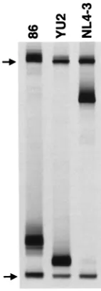
population level by the newly described V3-HTA (45) (Fig. 2).
V3 sequences were amplified by RT-PCR from viral RNA
isolated from a June 1986 serum sample from subject UNC116.
Heteroduplexes were formed between these RT-PCR products
and a radioactively labeled probe containing the near
consen-FIG. 2. Analysis by V3-HTA. V3 loop RT-PCR products from the June 1986 serum sample from patient UNC116 (lane 86) and control YU-2 and NL4-3 recombinant proviral clones were annealed with the JR-FL V3 probe that was
35S labeled on one strand. The heteroduplexes were resolved on a native
poly-acrylamide gel and visualized by autoradiography. Only the bottom half of the autoradiograph is shown. The positions of the single-stranded probe (upper arrow) and double-stranded probe homoduplexes (lower arrow) are indicated. Heteroduplexes in each lane resolved between the single-stranded probe and probe homoduplexes.
on November 9, 2019 by guest
http://jvi.asm.org/
[image:3.612.390.461.69.273.2]sus V3 sequence for subtype B viruses. After resolution of the
heteroduplexes, a single dominant band was seen on the
au-toradiograph, indicating limited V3 quasispecies diversity in
the 1986 serum sample. The intermediate mobility of the
het-eroduplexes from UNC116, compared with the faster NSI
con-trol YU-2 and the slower SI concon-trol NL4-3 (whose envelope
sequences are derived from HIV-1
LAI[1]), indicated that the
sequences had clustering of mutations that has been associated
with the SI genotype (45). Nucleotide sequencing of molecular
clones containing env cDNA sequences V1 to V3 and V3 to V5
from the December 1985 and June 1986 samples revealed a
range of interclone divergence of 2.1 to 5.7%, which rose to
only 7.5 to 9.9% by inclusion of cDNA sequences derived from
a June 1994 plasma sample; all clones were of the SI genotype
and were highly associated phylogenetically (data not shown;
the sequences have been deposited in GenBank).
The earliest available specimens for UNC116 consisted of
small volumes of serum that had been stored under nonideal
conditions for successful viral culture. Rather than attempt
viral culture of this serum, we constructed isogenic
recombi-nant proviruses containing exogenous sequences of either V3
or V3 through V5 (Fig. 1) derived from UNC116 env cDNAs
as described in Materials and Methods. The predominant V3
sequence in the December 1985 serum sample was put into the
HXB2 proviral clone to create the 12/85 chimera (Fig. 1a). The
complete HXB2 proviral clone was used as a control for this
recombinant. The 6/86 chimeras were made by putting two
different V3 to V5 sequences (1-7 and 5-3) derived from the
June 1986 serum sample into a JR-FL env gene within an
NL4-3 proviral clone (Fig. 1b). Each of these V3 to V5
se-quences had the same V3 sequence as that used in the 12/85
recombinant virus. Control recombinant viruses were
con-structed by placing the V3 to V5 sequences from the YU-2
(NSI control) and NL4-3 (SI control) proviral clones into the
same NL4-3/JR-FL recombinant provirus. A viral isolate from
late in disease was obtained from a June 1994 plasma specimen
with high viral load.
The three early (12/85 and 6/86) viral chimeras, the late
(6/94) plasma viral isolate, and the control viruses were tested
for biological phenotype in the MT-2 cell assay. All of the
viruses except the YU-2 recombinant control virus were
phe-notypically SI (Table 1 and data not shown).
The viral quasispecies displays exclusive and persistent
CXCR4 coreceptor utilization.
Equivalent amounts of the viral
stocks were used to infect GHOST cells to determine relative
coreceptor utilization in a single-step infection assay. This
HOS cell line is stably transduced with human CD4, GFP
under the control of the Tat-sensitive HIV-2 promoter, and
specific human coreceptor cDNAs. Infection of these GHOST
cells by HIV-1 results in the production of GFP that can be
detected by flow cytometry less than 48 h postinfection.
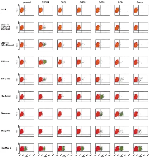
Whereas mock infection of these cells results in virtually no
signal above background (red in Fig. 3), infection with CD4/
coreceptor-independent
amphotropic-MLV-E-pseudotyped
HIV-1 virions results in 5 to 32% of these cells becoming
positive for GFP production (green in Fig. 3). Inspection of the
fluorescence-activated cell sorter profiles in Fig. 3 and the
relative coreceptor utilization in Table 1 for the 12/85 viral
chimera and 6/94 plasma isolate show CXCR4 use to the
ex-clusion of the other known entry coreceptors (CCR2B, CCR3,
CCR5, BOB, and Bonzo). CCR1, CCR4, and GPR1 were also
tested and were not used by these stocks (data not shown). The
SI control virus HIV-1
LAIexclusively used CXCR4, whereas
the NSI control virus HIV-1
JR-CSFexclusively used CCR5.
HIV-2
RODused primarily CXCR4 but also CCR3, BOB, and
Bonzo; SIV
mac1A11used CCR5 and BOB; and SIV
agmTYOused CCR5, BOB, and Bonzo. Exclusive CXCR4 usage was
confirmed for the 12/85 and 6/86 viral chimeras and 6/94
plasma isolate in a HOS.CD4.coreceptor assay using p24
an-tigen readout, in which CXCR4, CCR1, CCR2B, CCR3,
CCR4, and CCR5 were tested (data not shown). Lower viral
titers for the 6/86 viral chimeras compared with the 12/85
chimeras or 6/94 plasma isolate obviated coreceptor typing
with GHOST cells, whose readout is 2 days.
HOS.CD4.core-ceptor typing with p24 antigen readout at 7 days afforded
greater sensitivity for viruses with delayed replication kinetics.
The viral quasispecies replicates in CCR5
1
/
1
and CCR5
2
/
2
PBMC but not in macrophages.
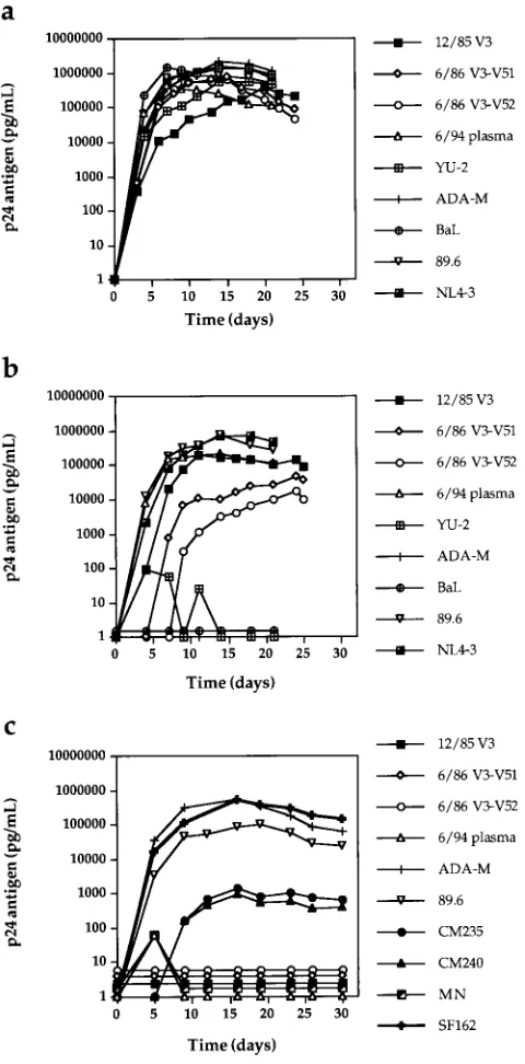
The early viral chimeras
and late plasma isolate from UNC116 displayed typical
repli-cation kinetics in CCR5
1
/
1
PBMC compared with virus
de-rived from the M-tropic molecular clone pYU-2, virus dede-rived
from the T-tropic molecular clone pNL4-3, the dual-tropic
isolate 89.6, and the NSI isolates ADA-M and BaL (Fig. 4a).
The 12/85 chimera and the 89.6 and NL4-3 viruses also
repli-cated well in seronegative CCR5
2
/
2
PBMC, while YU-2,
ADA-M, and BaL did not (Fig. 4b). The replication of the 6/86
V3-V5 viral chimeras was delayed in these cells. This
observa-tion could reflect stochastic selecobserva-tion of relatively less fit
mem-bers of the quasispecies or the reduced activity of a chimeric
protein or involve subtle host cell differences between the
CCR5
1
/
1
and
2
/
2
PBMC.
To rule out the use of as yet undiscovered coreceptors on
primary macrophages, the replication kinetics of the UNC116
and control viruses were studied in CCR5
1
/
1
primary
mac-rophages (Fig. 4c). All of the UNC116 viruses and the SI
control HIV-1
MNfailed to replicate in macrophages, but the
NSI primary isolates CM235, CM240, SF162, and ADA-M, as
well as the dual-tropic 89.6, replicated to various degrees in
primary macrophages. Thus, if the UNC116 viruses are
capa-ble of using as yet undiscovered HIV-1 entry coreceptors, these
putative receptors are not expressed in primary macrophages
under culture conditions.
DISCUSSION
[image:4.612.50.289.91.191.2]Given the importance of CCR5 coreceptor utilization in
transmission and early HIV-1 disease, the virology of
HIV-1-infected CCR5
2
/
2
individuals provides an important insight
into HIV-1 pathogenesis. We now report that the viral
quasi-species from one such individual is persistently homogenous,
SI, and T tropic and exclusively uses the HIV-1 entry
corecep-tor CXCR4 during the observation period. Although early and
TABLE 1. Biological phenotype and coreceptor usage of
experimental and control viral stocks
Virus (phenotypea) % GHOST cell infection b
CXCR4 CCR2B CCR3 CCR5 BOB Bonzo
12/85 V3 chimera
c(SI)
100.0
0
0
0
0
0
6/94 plasma isolate (SI)
100.0
0
0
0
0
0
HIV-1
LAI(SI)
100.0
0
0
0
0
0
HIV-2
ROD(SI)
85.5
0
2.7
0
6.8
5.0
HIV-1
JRCSF(NSI)
0
0
0
100
0
0
SIV
mac1A110
0
0
71.2 28.8
0
SIV
agmTYO0
0
0
75.7
6.1 18.2
aDetermined by the ACTG consensus MT-2 cell assay.
bNormalized to MLV-E-pseudotyped HIV-1 infection as described in Mate-rials and Methods.
cThe 6/86 V3-V5 chimera stock was not tested in the GHOST cell assay. This SI stock was tested in HOS.CD4.coreceptor cells with p24 readout and found to exclusively use CXCR4 (data not shown).
on November 9, 2019 by guest
http://jvi.asm.org/
persistent SI variants (32, 34) or evolution to exclusive CXCR4
usage (53) has been previously found in longitudinal cohorts,
UNC116 represents the first demonstration of exclusive and
persistent CXCR4 usage in an HIV-1-infected individual,
al-beit with the important caveat that the earliest viral sampling in
this study occurred approximately 4 years postinfection.
[image:5.612.50.549.67.601.2]As most other primary viruses reported to date can use
CCR5 for cell entry, we speculate that patient UNC116 was
FIG. 3. HIV-1 entry coreceptor typing of viral stocks in GHOST cells. HOS cells nonpermissive for HIV-1 infections were transduced with human CD4 and the GFP under the control of the HIV-2 LTR to generate parental GHOST cells. These cells were then individually transduced with the HIV-1 entry coreceptor cDNAs indicated at the top. HIV-1 entry and subsequent tat gene expression transactivates the HIV-2 LTR and upregulates the expression of GFP. Following infection of parental and coreceptor-transduced cells with equivalent amounts of specific viral stocks (indicated at the left), the cells were fixed and analyzed by flow cytometry. For each fluorescence-activated cell sorter profile, linear forward side scatter is plotted against log fluorescence intensity for GFP in the FL1 window. Basal-level production of GFP is red; transactivated GFP expression is green.
on November 9, 2019 by guest
http://jvi.asm.org/
initially exposed to a swarm containing viruses capable of using
both CCR5 and CXCR4 but only viruses with CXCR4 entry
capacity were propagated in the context of the host
environ-ment. The quasispecies did not expand to use “minor”
core-ceptors such as CCR2B, CCR3, BOB, Bonzo, or GPR1, a
finding consistent with the dominant roles of CCR5 and
CXCR4 as HIV-1 entry coreceptors. As virus from UNC116
was first sampled roughly 4 years postinfection, it is not clear
when selection for exclusive CXCR4 use occurred, but our
data suggest that virus with CXCR4-restricted coreceptor
uti-lization was present during most, if not all, of the period of
chronic HIV infection. As these observations are based on a
single patient, it will be critical to learn if they apply to other
CCR5
2
/
2
seropositive individuals. Within this context, it may
be notable that virus from a laboratory worker initially infected
with the CXCR4-exclusive, T-tropic strain HIV-1
IIIBwas
sub-sequently recovered in both autologous macrophage culture
and donor PBMC. The PBMC-derived virus was capable of
passage into the human T-cell line H9 (57), indicative of the
utilization of both CCR5 and CXCR4. Together, these reports
suggest that HIV-1 quasispecies may evolve to fit the CCR5
environment of the host.
UNC116 V3 sequences embedded in the env background of
the exclusive CXCR4-using virus HIV-1
HXB2and sequences
V3 to V5 embedded in the env background of the exclusive
CCR5-using virus HIV-1
JR-FLresulted in viral chimeras
re-stricted to CXCR4. Although we cannot formally exclude the
possibility that sequences outside the V3 region contributed to
coreceptor selectivity, this finding confirms previous data
showing the strong influence of the V3 domain upon CCR5
and CXCR4 usage (13).
SI variant, CXCR4-using HIV-1 quasispecies are associated
with increased in vivo virulence in cohort studies in which the
presence of CCR5
2
/
2
infected individuals is either absent or
undocumented but is presumably very rare (31, 32, 34, 43, 54).
The increased pathogenicity of such strains appears to be
in-dependent of the viral load (31). In agreement with those
observations, UNC116 and two similar patients with estimable
dates of infection (7, 55) showed relatively rapid CD4
1T-cell
decline despite average to low early viral RNA load. Because
case reports of HIV-1-infected patients with the CCR5
2
/
2
genotype are few and potentially biased toward those with a
better clinical course, available data are insufficient to predict
the prognosis of such persons. However, these reports provide
no evidence that, once infected with HIV-1, CCR5
2
/
2
pa-tients have a better prognosis than other persons.
We speculate that HIV-1 infection in CCR5
2
/
2
individuals
involves the persistent and dominant (if not exclusive) use of
the coreceptor CXCR4 with cell-free infection largely confined
to the T-cell (not macrophage) compartment. Infection of
macrophages in such individuals may occur through cell-cell
fusion events, although this could limit the ability of
macro-phages to serve as a reservoir of viral replication. Limited data
suggest that HIV-1 quasispecies may evolve in response to the
host’s CCR5 environment and that HIV-1-infected CCR5
2
/
2
patients have no better prognosis than other infected persons.
These points should be carefully considered in the design of
therapeutic modalities such as blockade of CCR5 by small
molecules or bone marrow transplantation with CCR5
2
/
2
stem cells.
ACKNOWLEDGMENTS
We thank Deborah Birx, Suzanne Gartner, James Goedert, Mika
Popovic, Merlin Robb, and Steven Wolinsky for helpful discussions.
We also thank Irvin Chen for providing the HIV-1
JR-FLsubclone
[image:6.612.51.291.69.555.2]pUC112-1 and Malcolm Martin for providing the pNL4-3 clone.
This work was supported in part by National Cancer Institute
con-tract NO1-CP-85649 with Research Triangle Institute, U.S. Army
Medical Research and Materiel Command contract
DAMD17-94-J-4430, and cooperative agreement DAMD17-93-V-3004 between the
U.S. Army Medical Research and Materiel Command and the Henry
M. Jackson Foundation for the Advancement of Military Medicine.
J.A.E.N. was supported by postdoctoral training grant T32-CA-09156
from the National Cancer Institute. V.N.K. was supported by a
post-doctoral fellowship from the Damon Runyon Walter Winchell
Foun-dation. D.R.L. is supported by grants from the NIH and is an
inves-tigator of the Howard Hughes Medical Institute.
FIG. 4. Replication kinetics of viral stocks in CCR51/1(a) and CCR52/2
(b) PBMC and CCR51/1macrophages (c). One hundred and 500 TCID50of PBMC-derived viral stocks were used to infect PHA- and IL-2-stimulated PBMC and monocyte-derived macrophages, respectively. Virus replication was moni-tored by supernatant p24 antigen production.
on November 9, 2019 by guest
http://jvi.asm.org/
REFERENCES
1. Adachi, A., H. E. Gendelman, S. Koenig, T. Folks, R. Willey, A. Rabson, and M. A. Martin.1986. Production of acquired immunodeficiency syndrome-associated retrovirus in human and nonhuman cells transfected with an infectious molecular clone. J. Virol. 59:284–291.
2. Alkhatib, G., C. Combadiere, C. C. Broder, Y. Feng, P. E. Kennedy, P. M. Murphy, and E. A. Berger.1996. CC CKR5: a RANTES, MIP-1a, MIP-1b
receptor as a fusion cofactor for macrophage-tropic HIV-1. Science 272: 1955–1958.
3. Alkhatib, G., F. Liao, E. A. Berger, J. M. Farber, and K. W. C. Peden. 1997. A new SIV co-receptor, STRL33. Nature 388:238.
4. Atchison, R. E., J. Gosling, F. S. Monteclaro, C. Franci, L. Digilio, I. F. Charo, and M. A. Goldsmith.1996. Multiple extracellular elements of CCR5 and HIV-1 entry: dissociation from response to chemokines. Science 274: 1924–1926.
5. Balotta, C., P. Bagnarelli, M. Violin, A. L. Ridolfo, D. Zhou, A. Berlusconi, S. Corvasce, M. Corbellino, M. Clementi, M. Clerici, M. Moroni, and M. Galli.1997. HomozygousD32 deletion of the CCR-5 chemokine receptor gene in an HIV-1-infected patient. AIDS 11:F67–F71.
6. Berson, J. F., D. Long, G. J. Doranz, J. Rucker, F. R. Jirik, and R. W. Doms. 1996. A seven-transmembrane domain receptor involved in fusion and entry of T-cell-tropic human immunodeficiency virus type 1 strains. J. Virol. 70: 6288–6295.
7. Biti, R., R. Ffrench, J. Young, B. Bennetts, G. Stewart, and T. Liang. 1997. HIV-1 infection in an individual homozygous for the CCR5 deletion allele. Nat. Med. 3:252–253. (Letter.)
8. Bleul, C. C., M. Farzan, H. Choe, C. Parolin, I. Clark-Lewis, J. Sodroski, and T. A. Springer.1996. The lymphocyte chemoattractant SDF-1 is a ligand for LESTR/fusin and blocks HIV-1 entry. Nature 382:829–832.
9. Buckheit, R. W., and R. Swanstrom. 1991. Characterization of an HIV-1 isolate displaying an apparent absence of virion-associated reverse transcrip-tase activity. AIDS Res. Hum. Retroviruses 7:295–302.
10. Chen, C., and H. Okayama. 1987. High-efficiency transformation of mam-malian cells by plasmid DNA. Mol. Cell. Biol. 7:2745–2752.
11. Choe, H., M. Farzan, Y. Sun, N. Sullivan, B. Rollins, P. D. Ponath, L. Wu, C. R. Mackay, G. LaRosa, W. Newman, N. Gerard, C. Gerard, and J. Sodroski. 1996. Theb-chemokine receptors CCR3 and CCR5 facilitate infection by primary HIV-1 isolates. Cell 85:1135–1148.
12. Cocchi, F., A. L. De Vico, A. Garzino-Demo, S. K. Arya, R. C. Gallo, and P. Lusso.1995. Identification of RANTES, MIP-1 alpha, and MIP-1 beta as the major HIV-suppressive factors produced by CD81T cells. Science 270: 1811–1815.
13. Cocchi, F., A. L. De Vico, A. Garzino-Demo, A. Cara, R. C. Gallo, and P. Lusso.1996. The V3 domain of the HIV-1 gp120 envelope glycoprotein is critical for chemokine-mediated blockade of infection. Nat. Med. 2:1244– 1247.
14. Dalgleish, A. G., P. C. Beverley, P. R. Clapham, D. H. Crawford, M. F. Greaves, and R. A. Weiss. 1984. The CD4 (T4) antigen is an essential component of the receptor for the AIDS retrovirus. Nature 312:763–767. 15. Dean, M., M. Carrington, C. Winkler, G. A. Huttley, M. W. Smith, R.
Allikmets, J. J. Goedert, S. P. Buchbinder, E. Vittinghoff, E. Gomperts, S. Donfield, D. Vlahov, R. Kaslow, A. Saah, C. Rinaldo, R. Detels, Hemophilia Growth and Development Study, Multicenter AIDS Cohort Study, Multi-center Hemophilia Cohort Study, San Francisco City Cohort, ALIVE Study, and S. J. O’Brien.1996. Genetic restriction of HIV-1 infection and progres-sion to AIDS by a deletion allele of the CKR5 structural gene. Science 273:1856–1862.
16. Deng, H., R. Liu, W. Ellmeier, S. Choe, D. Unutmaz, M. Burkhart, P. Di Marzio, S. Marmon, R. E. Sutton, C. M. Hill, C. B. Davis, S. C. Peiper, T. J. Schall, D. R. Littman, and N. R. Landau.1996. Identification of a major co-receptor for primary isolates of HIV-1. Nature 381:661–666.
17. Deng, H., D. Unutmaz, V. N. KewalRamani, and D. R. Littman. 1997. Expression cloning of new receptors used by simian and human immunode-ficiency viruses. Nature 388:296–300.
18. de Roda Husman, A.-M., M. Koot, M. Cornelissen, I. P. M. Keet, M. Brou-wer, S. Broersen, M. Bakker, M. T. L. Roos, M. Prins, F. de Wolf, R. A. Coutinho, F. Miedema, J. Goudsmit, and H. Schuitemaker.1997. The asso-ciation between CCR5 genotype and the clinical course of HIV-1 infection. Ann. Intern. Med. 127:882–890.
19. Doranz, B. J., J. Rucker, Y. Yi, R. J. Smyth, M. Samson, S. C. Peiper, M. Parmentier, R. G. Collman, and R. W. Doms.1996. A dual-tropic primary HIV-1 isolate that uses fusin and the beta-chemokine receptors CKR-5, CKR-3, and CKR-2b as fusion cofactors. Cell 85:1149–1158.
20. Dragic, T., V. Litwin, G. P. Allaway, S. R. Martin, Y. Huang, K. A. Na-gashima, C. Cayanan, P. J. Maddon, R. A. Koup, J. P. Moore, and W. A. Paxton.1996. HIV-1 entry into CD41cells is mediated by the chemokine receptor CC-CKR-5. Nature 381:667–673.
21. Eugen-Olsen, J., A. K. N. Iversen, P. Garred, U. Koppelhus, C. Pedersen, T. L. Benfield, A. M. Sorensen, T. Katzenstein, E. Dickmeiss, J. Gerstoft, P. Skinhoj, A. Svejgaard, J. O. Nielsen, and B. Hoffman.1997. Heterozygosity for a deletion in the CKR-5 gene leads to prolonged AIDS-free survival and
slower CD4 T-cell decline in a cohort of HIV-seropositive individuals. AIDS 11:305–310.
22. Farzan, M., H. Choe, K. Martin, L. Marcon, W. Hofmann, G. Karlsson, Y. Sun, P. Barrett, N. Marchand, N. Sullivan, N. Gerard, C. Gerard, and J. Sodroski.1997. Two orphan seven-transmembrane segment receptors which are expressed in CD4-positive cells support simian immunodeficiency virus infection. J. Exp. Med. 186:405–411.
23. Farzan, M., H. Choe, K. A. Martin, Y. Sun, M. Sidelko, C. R. Mackay, N. P. Gerard, J. Sodroski, and C. Gerard.1997. HIV-1 entry and macrophage inflammatory protein-1b-mediated signaling are independent functions of the chemokine receptor CCR5. J. Biol. Chem. 272:6854–6857.
24. Feng, Y., C. Broder, P. E. Kennedy, and E. A. Berger. 1996. HIV-1 entry cofactor: functional cDNA cloning of a seven-transmembrane G protein-coupled receptor. Science 272:872–877.
25. Gartner, S., P. Markovits, D. M. Markovitz, M. H. Kaplan, R. C. Gallo, and M. Popovic.1986. The role of mononuclear phagocytes in HTLV-III/LAV infection. Science 233:215–219.
26. Gosling, J., F. S. Monteclaro, R. E. Atchinson, H. Arai, C.-L. Tsou, M. A. Goldsmith, and I. F. Charo.1997. Molecular uncoupling of C-C chemokine receptor 5-induced chemotaxis and signal transduction from HIV-1 corecep-tor activity. Proc. Natl. Acad. Sci. USA 94:5061–5066.
27. He, J., Y. Chen, M. Farzan, H. Choe, A. Ohagen, S. Gartner, J. Busciglio, X. Yang, W. Hofmann, W. Newman, C. R. Mackay, J. Sodroski, and D. Gabuzda.1997. CCR3 and CCR5 are co-receptors for HIV-1 infection of microglia. Nature 385:645–649.
28. Huang, Y., W. A. Paxton, S. M. Wolinsky, A. U. Neumann, L. Zhang, T. He, S. Kang, D. Ceradini, Z. Jin, K. Yazdanbakhsh, K. Kunstman, D. Erickson, E. Dragon, N. R. Landau, J. Phair, D. D. Ho, and R. A. Koup.1996. The role of a mutant CCR5 allele in HIV-1 transmission and disease progression. Nat. Med. 2:1240–1243.
29. Jansson, M., M. Popovic, A. Karlsson, F. Cocchi, P. Rossi, J. Albert, and H. Wigzell.1996. Sensitivity to inhibition by beta-chemokines correlates with biological phenotypes of primary HIV-1 isolates. Proc. Natl. Acad. Sci. USA 93:15382–15387.
30. Japour, A. J., S. A. Fiscus, J.-M. Arduino, D. L. Mayers, P. S. Reichelderfer, and D. R. Kuritzkes.1994. Standardized microtiter assay for determination of syncytium-inducing phenotypes of clinical human immunodeficiency virus type 1 isolates. J. Clin. Microbiol. 32:2291–2294.
31. Jurriaans, S., B. Van Gemen, G. J. Weverling, D. Van Strijp, P. Nara, R. Coutinho, M. Koot, H. Schuitemaker, and J. Goudsmit.1994. The natural history of HIV-1 infection: virus load and virus phenotype independent determinants of clinical course? Virology 204:223–233.
32. Karlsson, A., K. Parsmyr, K. Aperia, E. Sandstrom, E. M. Fenyo, and J. Albert.1994. MT-2 cell tropism of human immunodeficiency virus type 1 isolates as a marker for response to treatment and development of drug resistance. J. Infect. Dis. 170:1367–1375.
32a.Kewal Ramani, V. N., B. Volsky, and D. R. Littman. Unpublished data. 33. Klatzmann, D., E. Champagne, S. Chamaret, J. Gruest, D. Guetard, T.
Hercend, J. C. Gluckman, and L. Montagnier.1984. T-lymphocyte T4 mol-ecule behaves as the receptor for human retrovirus LAV. Nature 312:767– 768.
34. Koot, M., I. P. M. Keet, A. H. V. Vos, R. E. Y. de Goede, M. T. L. Roos, R. A. Coutinho, F. Miedema, P. T. A. Schellekens, and M. Tersmette.1993. Prog-nostic value of HIV-1 syncytium-inducing phenotype for rate of CD41cell depletion and progression to AIDS. Ann. Intern. Med. 118:681–688. 35. Koyanagi, Y., S. Miles, R. T. Mitsuyasu, J. E. Merrill, H. V. Vinters, and I. S.
Chen.1987. Dual infection of the central nervous system by AIDS viruses with distinct cellular tropisms. Science 236:819–822.
36. Lee, A. B., and T. A. Cooper. 1995. Improved direct PCR screen for bacterial colonies: wooden toothpicks inhibit PCR amplification. BioTechniques 18: 225–226.
37. Li, Y., H. Hui, C. J. Burgess, R. W. Price, P. M. Sharp, B. H. Hahn, and G. M. Shaw.1992. Complete nucleotide sequence, genome organization, and biological properties of human immunodeficiency virus type 1 in vivo: evi-dence for limited defectiveness and complementation. J. Virol. 66:6587– 6600.
38. Liu, R., W. A. Paxton, S. Choe, D. Ceradini, S. R. Martin, R. Horuk, M. E. MacDonald, H. Stuhlmann, R. A. Koup, and N. R. Landau.1996. Homozy-gous defect in HIV-1 coreceptor accounts for resistance of some multiply-exposed individuals to HIV-1 infection. Cell 86:367–377.
39. Lusso, P., F. Cocchi, C. Balotta, P. D. Markham, A. Louie, P. Farci, R. Pal, R. C. Gallo, and M. S. Reitz, Jr.1995. Growth of macrophage-tropic and primary human immunodeficiency virus type 1 (HIV-1) isolates in a unique CD41T-cell clone (PM1): failure to downregulate CD4 and to interfere with cell-line-tropic HIV-1. J. Virol. 69:3712–3720.
40. Maddon, P. J., A. G. Dalgleish, J. S. McDougal, P. R. Clapham, R. A. Weiss, and R. Axel.1986. The T4 gene encodes the AIDS virus receptor and is expressed in the immune system and the brain. Cell 47:333–348. 41. Martinson, J. J., N. H. Chapman, D. C. Rees, Y.-T. Liu, and J. B. Clegg.
1997. Global distribution of the CCR5 gene 32-basepair deletion. Nat. Genet. 16:100–103.
42. Michael, N. L., G. Chang, P. K. Ehrenberg, M. T. Vahey, and R. R. Redfield.
on November 9, 2019 by guest
http://jvi.asm.org/
1993. HIV-1 proviral genotypes from the peripheral blood mononuclear cells of an infected patient are differentially represented in expressed sequences. J. Acquired Immune Defic. Syndr. 6:1073–1085.
43. Michael, N. L., G. Chang, L. G. Louie, J. R. Mascola, D. Dondero, D. L. Birx, and H. W. Sheppard.1997. The role of viral phenotype and CCR-5 gene defects in HIV-1 transmission and disease progression. Nat. Med. 3:338–340. 44. Morgenstern, J. P., and H. Land. 1990. Advanced mammalian gene transfer: high titre retroviral vectors with multiple drug selection markers and a complementary helper-free packaging cell line. Nucleic Acids Res. 18:3587– 3596.
45. Nelson, J. A. E., S. A. Fiscus, and R. Swanstrom. 1997. Evolutionary variants of the human immunodeficiency virus type 1 V3 region characterized by using a heteroduplex tracking assay. J. Virol. 71:8750–8758.
46. Oberlin, E., A. Amara, F. Bachelerie, C. Bessia, J.-L. Virelizier, F. Arenzana-Seisdedos, O. Schwartz, J.-M. Heard, I. Clark-Lewis, D. F. Legier, M. Loetscher, M. Baggiolini, and B. Moser.1996. The CXC chemokine SDF-1 is the ligand for LESTER/fusin and prevents infection by T-cell-line-adapted HIV-1. Nature 382:833–835.
47. O’Brien, T. R., C. Winkler, M. Dean, J. A. Nelson, M. Carrington, N. L. Michael, and G. C. White II.1997. HIV-1 infection in a man homozygous for CCR5 delta 32. Lancet 349:1219. (Letter.)
48. Rana, S., G. Besson, D. G. Cook, J. Rucker, R. J. Smyth, Y. Yi, J. D. Turner, H.-H. Guo, J.-G. Du, S. C. Peiper, E. Lavi, M. Samson, F. Libert, C. Lies-nard, G. Vassart, R. W. Doms, M. Parmentier, and R. G. Collman.1997. Role of CCR5 in infection of primary macrophages and lymphocytes by macrophage-tropic strains of human immunodeficiency virus: resistance to patient-derived and prototype isolates resulting from theDccr5 mutation.
J. Virol. 71:3219–3227.
49. Raport, C. J., J. Gosling, V. L. Schweickart, P. W. Gray, and I. F. Charo. 1996. Molecular cloning and functional characterization of a novel human CC chemokine receptor (CCR5) for RANTES, MIP-1b, and MIP-1a. J. Biol. Chem. 271:17161–17166.
50. Rappaport, J., Y. Y. Cho, H. Hendel, E. J. Schwartz, F. Schachter, and J. F. Zagury.1997. 32 bp CCR-5 gene deletion and resistance to fast progression in HIV-1 infected heterozygotes. Lancet 349:922–923. (Letter.)
51. Sampson, M., O. Labbe, C. Mollereau, G. Vassart, and M. Parmentier. 1996. Molecular cloning and functional expression of a new human CC-chemokine receptor gene. Biochemistry 35:3362–3367.
52. Sampson, M., F. Libert, B. L. Doranz, J. Rucker, C. Liesnard, C.-M. Farber, S. Saragosti, C. Lapoumeroulie, J. Cognaux, C. Forceille, G. Muyldermans,
C. Verhofstede, G. Burtonboy, M. George, T. Imai, S. Rana, Y. Yi, R. J. Smyth, R. G. Collman, R. W. Doms, G. Vassart, and M. Parmentier.1996. Resistance to HIV-1 infection in Caucasian individuals bearing mutant al-leles of the CCR-5 chemokine receptor gene. Nature 382:722–725. 53. Scarlatti, G., E. Tresoldi, A. Bjorndal, R. Fredriksson, C. Colognesi, H. K.
Deng, M. S. Malnatt, A. Plebani, A. G. Siccardi, D. L. Littman, E. M. Fenyo, and P. Lusso. 1997. In vivo evolution of HIV-1 co-receptor usage and sensitivity to chemokine-mediated suppression. Nat. Med. 3:1259–1265. 54. Sheppard, H. W., W. Lang, M. S. Ascher, E. Vittinghoff, and W. Winkelstein.
1993. The characterization of non-progressors: long-term HIV-1 infection with stable CD41T-cell levels. AIDS 7:1159–1166.
55. Theodorou, I., L. Meyer, M. Magierowska, C. Katlama, and C. Rouzioux. 1997. HIV-1 infection in an individual homozygous for CCR5 delta 32: Seroco study group. Lancet 349:1219–1220. (Letter.)
56. van’t Wout, A. B., N. A. Kootstra, G. A. Mulder-Kampinga, N. Albrecht-van Lent, H. J. Scherpbier, J. Veenstra, K. Boer, R. A. Coutinho, F. Miedema, and H. Schuitemaker.1994. Macrophage-tropic variants initiate human im-munodeficiency virus type 1 infection after sexual, parenteral, and vertical transmission. J. Clin. Invest. 94:2060–2067.
57. Weiss, S. H., J. J. Goedert, S. Gartner, M. Popovic, D. Waters, P. Markham, F. di Marzo Veronese, M. H. Gail, W. E. Barkley, J. Gibbons, F. A. Gill, M. Leuther, F. M. Verpmese, G. M. Shaw, R. C. Gallo, and W. A. Blattner.1988. Risk of human immunodeficiency virus (HIV-1) infection among laboratory workers. Science 239:68–71.
58. Wu, L., W. A. Paxton, N. Kassam, N. Ruffing, J. B. Rottman, N. Sullivan, H. Choe, J. Sodroski, W. Newman, R. A. Koup, and C. R. Mackay.1997. CCR levels and expression pattern correlate with infectibility by macrophage-tropic HIV-1, in vitro. J. Exp. Med. 185:1681–1691.
59. Zhu, T., H. Mo, N. Wang, D. S. Nam, Y. Cao, R. A. Koup, and D. D. Ho. 1993. Genotypic and phenotypic characterization of HIV-1 in patients with pri-mary infection. Science 261:1179–1181.
60. Zimmerman, P. A., A. Buckler-White, G. Alkhatib, T. Spalding, J. Kubofcik, C. Combadiere, D. Weissman, O. Cohen, A. Rubbert, G. Lam, M. Vac-carezza, P. E. Kennedy, V. Kumaraswami, J. V. Giorgi, R. Detels, J. Hunter, M. Chopek, E. A. Berger, A. S. Fauci, T. B. Nutman, and P. M. Murphy. 1997. Inherited resistance to HIV-1 conferred by an inactivating mutation in CC chemokine receptor 5: studies in populations with contrasting clinical phenotypes, defined racial background, and quantified risk. Mol. Med. 3:23– 36.
on November 9, 2019 by guest
http://jvi.asm.org/