JOURNAL OFVIROLOGY, Nov. 2008, p. 11054–11065 Vol. 82, No. 22 0022-538X/08/$08.00⫹0 doi:10.1128/JVI.01341-08
Copyright © 2008, American Society for Microbiology. All Rights Reserved.
Importance of the V1/V2 Loop Region of Simian-Human
Immunodeficiency Virus Envelope Glycoprotein gp120
in Determining the Strain Specificity of the
Neutralizing Antibody Response
䌤
Melissa E. Laird,
1Tatsuhiko Igarashi,
2Malcolm A. Martin,
3and Ronald C. Desrosiers
1*
New England Primate Research Center, Department of Microbiology and Molecular Genetics, Harvard Medical School,
Southborough, Massachusetts 01772-91021; Laboratory of Primate Models, Experimental Research Center for
Infectious Diseases, Institute for Virus Research, Kyoto University, Kyoto 606-8057, Japan2; and
Laboratory of Molecular Microbiology, National Institute of Allergy and Infectious Diseases,
National Institutes of Health, Bethesda, Maryland 208923
Received 26 June 2008/Accepted 28 August 2008
Plasma samples from individuals infected with human immunodeficiency virus type 1 (HIV-1) are known to be highly strain specific in their ability to neutralize HIV-1 infectivity. Such plasma samples exhibit significant neutralizing activity against autologous HIV-1 isolates but typically exhibit little or no activity against heter-ologous strains, although some cross-neutralizing activity can develop late in infection. Monkeys infected with the simian-human immunodeficiency virus (SHIV) clone DH12 generated antibodies that neutralized SHIV DH12, but not SHIV KB9. Conversely, antibodies from monkeys infected with the SHIV clone KB9 neutralized SHIV KB9, but not SHIV DH12. To investigate the role of the variable loops of the HIV-1 envelope glycoprotein gp120 in determining this strain specificity, variable loops 1 and 2 (V1/V2), V3, or V4 were exchanged individually or in combination between SHIV DH12 and SHIV KB9. Despite the fact that both parental viruses exhibited significant infectivity and good replication in the cell lines examined, 3 of the 10 variable-loop chimeras exhibited such poor infectivity that they could not be used further for neutralization assays. These results indicate that a variable loop that is functional in the context of one particular envelope background will not necessarily function within another. The remaining seven replication-competent chimeras allowed unam-biguous assignment of the sequences principally responsible for the strain specificity of the neutralizing activity present in SHIV-positive plasma. Exchange of the V1/V2 loop sequences conferred a dominant loss of sensitivity to neutralization by autologous plasma and a gain of sensitivity to neutralization by heterologous plasma. Substitution of V3 or V4 had little or no effect on the sensitivity to neutralization. These data demonstrate that the V1/V2 region of HIV-1 gp120 is principally responsible for the strain specificity of the neutralizing antibody response in monkeys infected with these prototypic SHIVs.
In order to achieve complete and durable protection against human immunodeficiency virus (HIV), it has been suggested that a vaccine may have to elicit a potent, broadly neutralizing antibody response as well as a strong cell-mediated immune response (93). Multiple studies have shown that passive trans-fer of HIV-specific antibodies is able to provide sterile protec-tion against HIV type 1 (HIV-1) or simian-HIV (SHIV) chal-lenge, whether administered intravenously or mucosally, in various animal models of infection (3, 32, 57, 58, 70, 73, 85). Moreover, several unusual, well-characterized monoclonal an-tibodies that are broadly acting, relatively potent, and capable of neutralizing viruses in a cross-clade manner have been iso-lated from HIV-infected individuals (7, 9, 13, 34, 78, 88, 91). These studies demonstrate that neutralizing antibodies can, at least in theory, provide protection against HIV-1 infection. However, envelope-based immunogens developed to date have not been capable of eliciting a robust antibody response with
the broad target specificity and high plasma concentrations that are likely to be necessary for protection (5, 12, 20, 22, 24–26, 31, 56, 59, 62, 92).
HIV-1 infection in humans typically elicits high levels of antibodies directed against the viral surface glycoprotein gp120. It has been estimated that as much as 5% of all immu-noglobulin in some HIV-infected individuals may be directed to the virus-encoded surface glycoprotein (6). However, the abilities of these antibodies to neutralize HIV-1 infectivity are extremely limited in both potency and breadth. The neutraliz-ing antibodies that are elicited upon HIV-1 infection tend to be highly strain specific; they neutralize autologous virus most effectively but exhibit little or no neutralizing activity against heterologous HIV-1 strains (10, 11, 61, 74, 81). Due to rapid sequence evolution within the envelope protein in response to immune pressure and selection, the original antibody popula-tion loses potency over the course of infecpopula-tion as persistently replicating viruses acquire resistance to antibody-mediated de-tection. There is a responsive shift in the production of neu-tralizing antibodies over time, apparently evolving new target specificities to counter newly emerged viral variants (1, 29, 33, 81, 95).
The HIV-1 envelope glycoprotein is the primary target for
* Corresponding author. Mailing address: New England Primate Re-search Center, One Pine Hill Drive, Southborough, MA 01772-9102. Phone: (508) 624-8040. Fax: (508) 624-8190. E-mail: ronald_desrosiers @hms.harvard.edu.
䌤Published ahead of print on 3 September 2008.
11054
on November 8, 2019 by guest
http://jvi.asm.org/
Downloaded from
on November 8, 2019 by guest
http://jvi.asm.org/
Downloaded from
on November 8, 2019 by guest
http://jvi.asm.org/
antibody-mediated neutralization (2, 4). In order to circum-vent antibody recognition, HIV-1 has evolved a number of shielding strategies to occlude conserved epitopes and limit the accessibility of glycoprotein spikes on the surfaces of virions to antibody binding. The mature envelope spike exists as a trimer of gp120-gp41 noncovalently associated heterodimers. Each trimer is tightly packed in a closed conformation such that epitopes that otherwise might serve as targets for antibody binding and neutralization reside in the core of the protein and are not readily exposed (45, 66, 67, 77). Additionally, gp120 contains five distinct regions that are characterized by exten-sive sequence variation. Four of these hypervariable regions are thought to form loops through intrachain disulfide bonds that are exposed on the outer surface of the envelope protein, resulting in the occlusion of conserved core envelope epitopes from antibody recognition prior to viral entry (38, 40, 50, 54, 96).
A number of studies have been performed to explore the roles of the variable loops as immunological decoys or in shielding conserved elements within the core of the envelope. Surprisingly, the deletion of the entire variable loop 1 and 2 (V1/V2) complex, as many as 100 amino acids, can still yield replication-competent HIV-1 or simian immunodeficiency vi-rus (SIV) (44, 97). These V1/V2 deletion mutants are substan-tially more sensitive to neutralization by a panel of monoclonal antibodies targeting multiple epitopes on gp120 (14, 44, 46, 52, 83, 84, 87, 97). Several highly strain-specific neutralizing anti-bodies targeting the V1 and/or V2 loop have been identified, although exact epitopes have not yet been defined (18, 27, 28, 30, 89). A considerable body of evidence suggests that the majority of anti-V3 antibodies are also strain specific and can efficiently neutralize tissue culture-adapted virus strains but not primary isolates (65). In SIV infection, the V4 loop con-tains a conformational, neutralization-sensitive epitope in which sequence changes emerge to escape antibody detection (43, 90).
The present study examines the role of variable-loop se-quences in the strain specificity of antibody-mediated neu-tralization. To investigate if certain variable loops might be responsible for the strain-specific neutralizing activity char-acteristic of HIV-1-positive plasma, we constructed a panel of chimeric viruses between SHIV DH12 and SHIV KB9, exchanging each variable loop between both SHIV enve-lopes individually and in combination. We then examined the effects of these variable-loop exchanges on virus infec-tivity and sensiinfec-tivity to highly strain-specific plasma samples collected from SHIV DH12- and SHIV KB9-infected mon-keys. Seven of the 10 SHIV-derived variable-loop chimeras were replication competent and capable of inducing robust secreted alkaline phosphatase (SEAP) production in the C8166-45 LTR-SEAP cell line. We demonstrate that the exchange of the V1/V2 loop complex between SHIV DH12 and SHIV KB9 is consistently associated with a dramatic loss of sensitivity to autologous plasma and a gain of sensi-tivity to heterologous plasma. Our results demonstrate that the V1/V2 loop is the principal determinant of the strain specificity of the neutralizing-antibody response in SHIV-infected monkeys.
MATERIALS AND METHODS
PCR mutagenesis and plasmid construction.Mutations withinenvwere gen-erated by a two-step PCR method that combined the technique of splicing by overlap extension PCR (69) to generate a primary product with site-directed PCR mutagenesis to incorporate the mutations into the hemigenomic plasmids.
For SHIV KB9, initial mutagenesis was performed on the 3⬘half of the genome,
which was obtained from the NIH AIDS Research and Reference Reagent Program (Division of AIDS, National Institute of Allergy and Infectious Dis-eases, NIH) (47). The full-length proviral plasmid DNA of SHIV DH12 was kindly provided by T. Igarashi and M. A. Martin (86). To facilitate optimal mutagenesis efficiency for the SHIV DH12 envelope sequence, the envelope gene was subcloned from the full-length genome.
For the splicing by overlap extension step of our mutagenesis protocol, two overlapping PCR products were generated, with the desired substitution incor-porated in the region of overlap. Specifically, the amplified and incorincor-porated fragment was the complete variable-loop sequence under investigation. Each
primer included, in its 5⬘portion, nucleotides complementary to the backbone
envelope sequence and, in its 3⬘half, nucleotides complementary to the
variable-loop sequence of interest. These primers were combined with the plasmid DNA containing the variable-loop sequence. The result was a pool of PCR products containing the amplified variable-loop sequence flanked by single-stranded over-hangs of sequence complementary to the reciprocal envelope sequence. These purified variable-loop amplicons were used in lieu of primers for site-directed mutagenesis (Stratagene, La Jolla, CA) reactions using the envelope subclones as template DNA.
The following paired primers were used to generate the amplicons necessary for creating the variable-loop chimeric envelopes (the first capitalized letter indicates the SHIV envelope background, and the SHIV strain that is the source of the variable loop exchanged is indicated by the lowercase abbreviation prior to the variable-loop identification; within the primer sequence, the loop sequences
that are being introduced are underlined): K.dV1V2F, 5⬘CC CCA CTC TGT
GTT ACT TTA AAT TGC ACT GAT TTG AAG AAT GG 3⬘; K.dV1V2R, 5⬘
GGC CTG TGT AAT GAC TGA GGT GTT ACA ACT TAT CAA CCT ATA
GC 3⬘; K.dV3F, 5⬘CAG CTA AAT GAA TCT GTA GTA ATT AAT TGT
ACA AGA CCC AAC AAC AAT ACA AGA AAA GGG 3⬘; K.dV3R, 5⬘CCA
TTT TGC TCT ACT AAT GTT ACA ATG TGC TTG TCT TAT ATC TCC 3⬘;
K.dV4F, 5⬘GGA GGG GAA TTT TTC TAC TGT AAT ACA AAA AAA CTG
3⬘; K.dV4R, 5⬘CC TAC TTT CTG CCA CAT ATT TAT AAT TTG TTT TAT
TCT GCA TGG GAG TGT GAT AGT GTC ATT TCC 3⬘; D.kV1V2F, 5⬘CC
CCA CTC TGT GTT ACT TTA CAT TGC ACT AAT TTG AAT ATC AC 3⬘;
D.kV1V2R, 5⬘GG ACA GGC CTG TGT AAG GGT TGA GGT GTT ACA
ACT TAT TAA CCT ATA CTT AG 3⬘; D.kV3F, 5⬘CAG CTG AAT GAA ACT
GTA GAA ATT AAT TGT ACA AGA CCC AAC AAC AAT ACA AGA GAA
AGG 3⬘; D.kV3R, 5⬘G CCA TTT TAC TTT ACT AAT GTT ACA ATG TGC
TTG TCT TAT ATC TCC 3⬘; D.kV4F, 5⬘GT GGA GGG GAA TTT TTC TAC
TGT AAT ACA GCA CAA CTG TTT AAT AGT ACT TGG 3⬘; D.kV4R, 5⬘C
CTG CCA CAT GTT TAT AAT TTG TTT TAT TCT GCA TTG GAG TGT
GAT TAT GTC ATT TCC 3⬘(Sigma Genosys, The Woodlands, TX).
The variable-loop-exchanged envelopes within the envelope subclones were sequenced to verify accuracy. In the constructs that contained variable loops exchanged in combination, this process was repeated sequentially. Sequencing of these combinatorial constructs was performed subsequent to each mutagenesis step. Full-length proviral DNA clones of each mutant were constructed.
DNA sequencing.Cloned plasmids containing selected mutations were se-quenced with a CEQ8000 genetic-analysis system, using a dye terminator cycle-sequencing chemistry kit as specified by the manufacturer (Beckman-Coulter, Fullerton, CA).
Virus stocks and cell culture.To generate virus stocks, 5g of the appropriate
full-length proviral DNA plasmid was transfected into 1.5⫻106HEK 293T cells
using the calcium phosphate method (Promega, Madison, WI). The medium was changed 24 h posttransfection. Supernatants were harvested on day 3 posttrans-fection. Virus concentrations in the supernatant were quantified by determining the concentration of p27 capsid protein using an antigen capture assay, according to the manufacturer’s instructions (Advanced Bioscience Laboratories, Inc., Kensington, MD). HEK 293T, MT4, C8166, and C8166-45 LTR-SEAP cells were cultured and maintained as described previously (10, 60).
Infectivity assay.Viral infectivity was quantified using the C8166-45 LTR-SEAP indicator cell line (60). C8166-45 LTR-LTR-SEAP cells contain a Tat-inducible SEAP reporter construct allowing SIV infection of the cells to be measured by SEAP concentrations in the cell-free supernatant. Nine serial twofold dilu-tions were made, starting from 10 ng of p27 capsid equivalents of HEK 293T cell-produced stocks. C8166-45 LTR-SEAP cells (8,000) were added to each
on November 8, 2019 by guest
http://jvi.asm.org/
well. The cells were incubated at 37°C in a humidified CO2incubator. Three days
postinfection, SEAP activity was measured in the cell-free supernatant (Applied Biosystems, Foster City, CA).
SHIV-positive plasmas.Five plasma samples taken at selected time points postinfection from rhesus macaques infected with SHIV DH12 were obtained from Malcolm Martin (National Institute of Allergy and Infectious Diseases, NIH, Bethesda, MD). Four plasma samples taken at selected time points postin-fection from rhesus macaques infected with SHIV KB9 were available from a previous study performed at the New England Primate Research Center.
Neutralization. The neutralization sensitivity of each virus to both SHIV-positive monkey plasmas was assayed using the SEAP reporter cell assay as described previously (60). Virus equivalent to 2.5 ng of p27 capsid protein was determined to be the lowest level of virus input sufficient to give a reliable SEAP signal within the linear range of the assay for all of the virus strains used. SEAP activity was quantified on the earliest day postinfection when levels were suffi-ciently high over background to give consistent measurements (at least 10-fold), ranging from 3 days for SHIV DH12, SHIV KB9, SHIV KDV3, and SHIV KDV4 to 5 days for SHIV DKV12, SHIV DKV4, SHIV DKV1234, and SHIV KDV1234 and 7 days for SHIV DKV124.
To perform neutralization assays, 96-well plates were organized as follows. Twenty-five microliters of medium (RPMI 1640 supplemented with 10% fetal
bovine serum, penicillin/streptomycin,L-glutamine, and HEPES buffer [R10])
was added to each well in the first three columns. To the wells in the remaining
columns (columns 4 to 12), 25-l aliquots of successive twofold dilutions of test
plasma or antibody, resuspended in medium, were added. All plasmas were heat inactivated at 56°C for 30 min before use in neutralization assays. For monkey
plasma, 25l of a 1:10 dilution of plasma was mixed with 75l of the test virus.
This constituted a 1:40 dilution in the representative graph. Virus equivalent to
2.5 ng of p27 capsid protein in a total volume of 75l of R10 was then added to
each well in columns 3 to 12. Virus-free medium was added to each well in columns 1 and 2 (mock infection). The plate was incubated for 1 h at 37°C. Following this incubation, 8,000 C8166-45 LTR-SEAP cells resuspended in 100
l of R10 were added to each well. The plate was placed in a humidified chamber
within a CO2incubator at 37°C for 3 to 7 days. SEAP activity was measured.
Neutralizing activities for all antibodies and plasma samples were measured in triplicate using a Victor V multilabel counter (Perkin-Elmer, Norwalk, CT) and reported as the average percent SEAP activity.
RESULTS
Stringent strain-specific neutralization of SHIV DH12 and SHIV KB9. The choice of SHIV DH12 and SHIV KB9 as representative viruses in this study was based on several fac-tors: (i) both SHIVs are well-characterized, genetically modi-fiable molecular clones; (ii) positive plasmas from monkeys infected with the matched molecular clones over several time points postinfection were available; and (iii) positive plasmas from infected animals displayed extremely stringent strain-specific neutralizing activity.
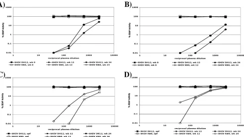
To assess the specificity of the neutralizing activity present in monkeys infected with SHIV DH12 or SHIV KB9, neutraliza-tion assays were performed using plasma samples from animals RHTPP and Rh418, collected 0, 12, and 24 weeks postinfection with SHIV DH12 and plasma samples from animals 477-99 and 481-99, collected 0, 12, and 24 weeks postinfection, with SHIV KB9 (Fig. 1). Assays performed with plasma collected prior to infection (week zero) demonstrated no preexisting nonspecific neutralizing activity for either SHIV DH12 or SHIV KB9. Highly strain-specific neutralizing activity was
ob-FIG. 1. Strain-specific neutralizing activity of positive plasmas when assayed with SHIV DH12 and SHIV KB9. (A) Neutralization of SHIV DH12 and SHIV KB9 with positive plasma from a SHIV DH12-infected monkey, RHTPP, collected at 0, 12, and 24 weeks (wk) postinfection. (B) Neutralization of SHIV DH12 and SHIV KB9 with positive plasma from a SHIV DH12-infected monkey, Rh418, collected at 0, 12, and 24 weeks postinfection. (C) Neutralization of SHIV DH12 and SHIV KB9 with positive plasma from a SHIV KB9-infected monkey, 477-99 collected 12 and 24 weeks postinfection, as well as with plasma from a SHIV-negative, specific-pathogen-free (SPF) monkey. (D) Neutralization of SHIV DH12 and SHIV KB9 with positive plasma from a SHIV KB9-infected monkey, 481-99, collected 12 and 24 weeks postinfection, as well as with plasma from an SPF monkey.
11056 LAIRD ET AL. J. VIROL.
on November 8, 2019 by guest
http://jvi.asm.org/
[image:3.585.48.540.67.345.2]served in plasma samples collected from all four experimen-tally infected monkeys at 12 and 24 weeks postinfection. Plasma from animals RHTPP and Rh418, infected with SHIV DH12, showed extensive neutralization of SHIV DH12 yet had no effect against the heterologous SHIV KB9 (Fig. 1A and B, respectively, and Table 1). Conversely, plasma from monkeys 477-99 and 481-99, infected with SHIV KB9, strongly neutral-ized SHIV KB9, but not SHIV DH12 (Fig. 1C and D, respec-tively, and Table 2). Most or all of the neutralizing activity was accounted for by purified immunoglobulin fractions, indicating that the neutralization was antibody mediated (data not shown).
Further studies were directed at determining whether the variable loops in gp120 were principally responsible for the strain specificity of the neutralizing antibody response and, if so, which variable loop or loops were primarily involved.
Construction of recombinant variable-loop chimeric SHIVs.
SHIV DH12 and SHIV KB9 were originally constructed using SIVmac239 as the backbone and differ genetically in theenv, tat, rev, vpu, andvpr genes, which are derived from multiple different HIV-1 species (47, 79, 86). Gp120 envelope
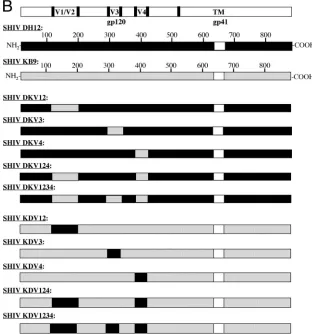
[image:4.585.45.542.81.233.2]se-quences outside of the variable loops share an 82% amino acid identity between these two SHIV clones. Within the variable loops, however, homology is dramatically reduced, maintaining only 58% amino acid identity (Fig. 2A). To investigate the extent to which the variable loops of gp120 contribute to the strain restriction of the neutralizing antibody response, each variable-loop sequence was exchanged alone or in combination with other loop sequences between SHIV DH12 and SHIV KB9 (Fig. 2B). The nomenclature of each SHIV-derived vari-able-loop chimera reflects this construction. For each variable-loop chimeric virus, the backbone SHIV envelope sequence is identified by the first abbreviated letter (D for SHIV DH12 and K for SHIV KB9), the SHIV clone from which the ex-changed variable loop was derived is indicated by the second letter, and the exchanged variable loop is identified by the numerical designation of the loop sequence. For example, SHIV DKV12 was constructed in the SHIV DH12 background to contain the V1/V2 loop derived from SHIV KB9. Virus stocks were generated by HEK 293T cell transfection, and equivalent p27 quantities were used for all comparative exper-iments.
TABLE 1. Neutralization of SHIV-DH12 chimeras with positive plasmas
Animal Infecting SHIV Time point of
sera (wk) KB9 wt
a,b
Neutralization activity for SHIV-DH12 and DH12-derived chimeric virusb
DH12
wt DKV12 DKV3 DKV4 DKV124 DKV1234
RHTPP DH12 10 ⫺ 9,000 ⫺ ND 12,000 ⫺ ⫺
12 ⫺ 7,000 ⫺ ND 15,000 ⫺ ⫺
24 ⫺ 2,000 ⫺ ND 7,500 ⫺ ⫺
Rh418 DH12 12 ⫺ 20,000 ⫺ ND 20,000 ⫺ 40
24 ⫺ 10,000 ⫺ ND 10,000 ⫺ ⫺
477-99 KB9 12 2,500 ⫺ 15,000 ND 40 20,000 8,000
24 5,000 ⫺ 8,000 ND ⫺ 9,000 7,000
481-99 KB9 12 1,000 ⫺ 130 ND ⫺ 300 420
24 1,100 ⫺ 650 ND 40 1,100 800
a
wt, wild type.
b
The numbers indicate the reciprocals of the dilutions of plasma required to reduce the infectivity of the indicated viruses by 50%.⫺, 50% neutralization was not
achieved, even at the lowest dilution (highest concentration) of plasma tested. ND, not determined due to inadequate infectivity of designated viruses.
TABLE 2. Neutralization of SHIV-KB9 chimeras with positive plasmas
Animal Infecting SHIV Time point of
sera (wk) DH12 wt
a,b
Neutralization activity for SHIV-KB9 and KB9-derived chimeric virusb
KB9
wt KDV12 KDV3 KDV4 KDV124 KDV1234
RHTPP DH12 10 9,000 ⫺ ND ⫺ ⫺ ND 3,000
12 7,000 ⫺ ND ⫺ ⫺ ND 5,000
24 2,000 ⫺ ND ⫺ ⫺ ND 2,000
Rh418 DH12 12 20,000 ⫺ ND ⫺ ⫺ ND 10,500
24 10,000 ⫺ ND ⫺ ⫺ ND 5,300
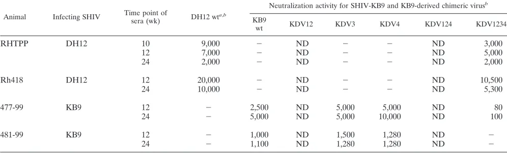
477-99 KB9 12 ⫺ 2,500 ND 5,000 5,000 ND 80
24 ⫺ 5,000 ND 5,000 10,000 ND 100
481-99 KB9 12 ⫺ 1,000 ND 1,500 1,280 ND ⫺
24 ⫺ 1,100 ND 1,280 1,280 ND ⫺
awt, wild type.
bThe numbers indicate the reciprocals of the dilutions of plasma required to reduce the infectivity of the indicated viruses by 50%.⫺, 50% neutralization was not
achieved, even at the lowest dilution (highest concentration) of plasma tested. ND, not determined due to inadequate infectivity of designated viruses.
on November 8, 2019 by guest
http://jvi.asm.org/
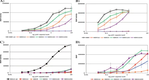
[image:4.585.43.541.549.700.2]Infectivities of the SHIV-derived variable-loop chimeras.At 3 days postinfection, three of the five SHIV KB9-derived vari-able-loop chimeras, SHIV KDV3, SHIV KDV4, and SHIV KDV1234, demonstrated substantial infectivity that was only slightly less than that of parental SHIV KB9 (Fig. 3A). Al-though quantifying SEAP at day 3 postinfection best approxi-mated a single round of infection, additional assays were per-formed at day 5 postinfection to assess whether SHIV KDV12 and SHIV KDV124 required further rounds of replication to produce quantifiable SEAP, reflective of impaired infectivity of these particular variable-loop chimeras (Fig. 3B). However,
at all time points examined, SHIV KDV12 and SHIV KDV124 produced little or no SEAP activity and were not used further for neutralization assays.
[image:5.585.133.445.218.552.2]The infectivities of the SHIV DH12-derived variable-loop chimeras displayed a somewhat different pattern than those of the reciprocal SHIV KB9-derived chimeras in that all of the SHIV DH12-derived chimeras were substantially less infec-tious than the parental SHIV DH12 when assayed for SEAP production 3 days postinfection (Fig. 3C). At day 5 postinfec-tion, four of the five SHIV DH12-derived chimeras (SHIV DKV12, SHIV DKV4, SHIV DKV124, and SHIV DKV1234)
FIG. 2. Variable-loop exchanges made between SHIV DH12 and SHIV KB9. (A) Comparative amino acid alignment of variable-loop sequences between SHIV DH12 and SHIV KB9. (B) Schematic representation of a variable-loop exchange(s) within the SHIV DH12 and SHIV KB9 gp120 envelope glycoproteins.
11058 LAIRD ET AL. J. VIROL.
on November 8, 2019 by guest
http://jvi.asm.org/
demonstrated substantial infectivity (Fig. 3D). SHIV DKV3 displayed low and inconsistent SEAP production at all time points postinfection examined and was also not included in further neutralization assays.
All SHIV-derived variable-loop chimeras were also assessed for infectivity in the CD4⫹CCR5⫹HeLa-derived TZM-bl cell line (23, 76, 94), the human B-cell/T-cell hybrid line LTR-SEAP-CEMx174 (60), and a newly derived LTR-SEAP-MT4 cell line. SHIV DKV3, SHIV KDV12, and SHIV KDV124 also demonstrated little or no infectivity in these cells, not allowing further assays in any of the cell lines examined (data not shown). Viral growth curves were performed in both C8166-45 and MT4 cells for all parental SHIVs and SHIV-derived chi-meras. Replication of the SHIV-derived variable-loop chime-ras in these two cell lines largely recapitulated and was consis-tent with the infectivity measurements described above (data not shown).
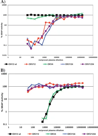
Comparative neutralization of the SHIV DH12-derived vari-able-loop chimeras by DH12-positive and KB9-positive mon-key plasma.We next measured the neutralization sensitivities of the parental SHIV DH12 and the infectious SHIV DH12-derived variable-loop chimeras to plasma samples taken from monkeys infected with either SHIV DH12 or SHIV KB9 at multiple time points postinfection. Figure 4 shows two represen-tative neutralization curves demonstrating the neutralization sensitivities of parental SHIV DH12 and the SHIV DH12-derived variable-loop chimeras when assayed with SHIV
[image:6.585.43.542.71.338.2]KB9-positive plasma from animal 477-99 (Fig. 4A) and SHIV DH12-positive plasma from animal Rh418 (Fig. 4B), both col-lected 12 weeks postinfection. Reflective of the neutralization sensitivities shown in Fig. 1, parental SHIV DH12 displayed complete resistance to neutralization by SHIV KB9-positive plasma and was extremely sensitive to neutralization by the matched SHIV DH12-positive plasma. Fifty percent neutral-ization of SHIV DH12 by the positive plasma from animal Rh418 was achieved at a dilution of 1:20,000. Moreover, the neutralization phenotype of SHIV DKV4 closely mirrored that of the parental SHIV DH12. SHIV DKV4 was also highly resistant to heterologous SHIV KB9-positive plasma and ex-tremely sensitive to autologous SHIV DH12-positive plasma, achieving 50% neutralization at the same dilution, 1:20,000. However, when the SHIV KB9 V1/V2 loop was introduced into the SHIV DH12 envelope background, the neutralization pattern was reversed. The SHIV DKV12 variable-loop chi-mera demonstrated extreme sensitivity to neutralization by the SHIV KB9-positive plasma from animal 477-99, achieving 50% neutralization at a dilution of 1:15,000. Additionally, SHIV DKV12 had lost sensitivity to the SHIV DH12-positive plasma collected from animal Rh418; no neutralization was observed even at the lowest dilution of plasma tested, 1:40. This gain of sensitivity to heterologous plasma and loss of neutralization by autologous plasma was dominant, as the two SHIV DH12-derived chimeras that contained the SHIV KB9 V1/V2 loop complex in combination with other variable loops, SHIV
FIG. 3. Comparative infectivities of parental and SHIV-derived variable-loop chimeric viruses. Virus stocks were obtained from transfection of HEK 293T cells, normalized by the amount of p27, and used to infect C8166-45 LTR-SEAP cells. (A) SEAP activity was measured at 3 days postinfection with SHIV KB9 and SHIV KB9-derived variable-loop chimeras. (B) SEAP activity was measured at 5 days postinfection with SHIV KB9-derived chimeras. (C) SEAP activity was measured at 3 days postinfection with SHIV DH12 and SHIV DH12-derived variable-loop chimeras. (D) SEAP activity was measured at 5 days postinfection with SHIV DH12-derived chimeras. SEAP production of some viruses at high p27 concentrations was not included in this analysis because limited additional rounds of wild-type (wt) infection had lysed the majority of target
SEAP-producing cells by day 5.
on November 8, 2019 by guest
http://jvi.asm.org/
DKV124 and SHIV DKV1234, both maintained complete re-sistance to SHIV DH12-positive plasma and substantial sensi-tivity to neutralization by SHIV KB9-positive plasma.
To ensure that the distinctive neutralization sensitivities of the SHIV DH12-derived variable-loop chimeras were not spe-cific to a particular plasma sample or time point postinfection, we performed more extensive analyses using several additional plasmas from independent experiments to extend the results (Table 1). All plasma samples examined confirmed the neu-tralization phenotypes demonstrated by Fig. 4. Specifically, four plasma samples collected from two monkeys infected with SHIV KB9 (477-99 and 481-99) at 12 and 24 weeks postinfec-tion were able to effectively neutralize all of the SHIV DH12-derived variable-loop chimeras that contained the SHIV KB9
V1/V2 region, to which the parental SHIV DH12 and SHIV DKV4 remained completely resistant. Moreover, these V1/V2 loop chimeras acquired total resistance to neutralization by five SHIV DH12-positive plasmas, taken from two infected animals (RHTPP and RH418) at 10, 12, and 24 weeks postin-fection, all of which efficiently neutralized the parental SHIV DH12 and SHIV DKV4.
[image:7.585.134.448.71.509.2]Comparative neutralization of the SHIV KB9-derived vari-able-loop chimeras by DH12-positive and KB9-positive mon-key plasmas.One of the benefits of the design of these exper-iments was our ability to measure both gain and loss of function in both directions with the two sets of reciprocal chimeras and the two sets of plasma specific for one SHIV clone or the other. We thus performed neutralization assays
FIG. 4. Representative neutralization curves of SHIV DH12 and SHIV DH12-derived loop chimeras with plasma from SHIV-infected monkeys. (A) Neutralization of SHIV DH12 and SHIV DH12-derived loop chimeras by plasma from a SHIV DH12-infected monkey, Rh418, collected 13 weeks postinfection. (B) Neutralization of SHIV DH12 and SHIV DH12-derived loop chimeras by plasma from a SHIV KB9-infected monkey, 477-99, collected 12 weeks postinfection. wt, wild type.
11060 LAIRD ET AL. J. VIROL.
on November 8, 2019 by guest
http://jvi.asm.org/
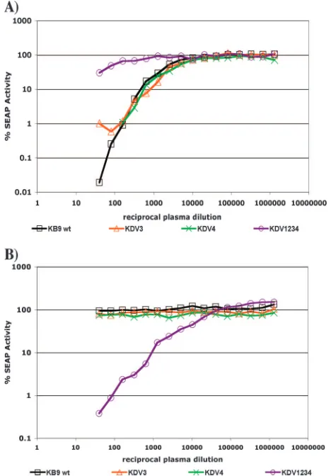
with the reciprocal SHIV KB9-derived variable-loop chimeras, using SHIV KB9-positive plasma from animal 477-99 collected 12 weeks postinfection (Fig. 5A) and SHIV DH12-positive plasma from animal Rh418 collected 12 weeks postinfection (Fig. 5B). As previously observed (Fig. 1), the parental SHIV KB9 was sensitive to neutralization by the SHIV KB9-positive plasma from animal 477-99, with 50% neutralization occurring at a dilution of 1:2,500. Furthermore, SHIV KB9 was com-pletely resistant to neutralization by the SHIV DH12-positive plasma from animal Rh418. Both SHIV KDV3 and SHIV KDV4 demonstrated neutralization phenotypes similar to that of the parental SHIV KB9. Fifty percent neutralization of SHIV DKV3 and SHIV DKV4 was achieved by the autolo-gous, SHIV KB9-positive plasma at a dilution of 1:5,000, and both chimeras were completely resistant to the heterologous SHIV DH12-positive plasma at all dilutions tested. The ex-change of the DH12 V1/V2 loop complex in SHIV KDV1234, however, conveyed the same reversal of neutralization sensi-tivity observed with the reciprocal exchange of the V1/V2 loop sequences described above. SHIV KDV1234 demonstrated al-most complete resistance to SHIV KB9-positive plasma, only
reaching 50% neutralization at the very low plasma dilution of 1:80, and acquired specific sensitivity to SHIV DH12-positive plasma, achieving 50% neutralization at a dilution of 1:10,500. We again performed more extensive analyses using several SHIV-positive plasmas to assess the neutralization sensitivities of the SHIV KB9-derived variable-loop chimeras across inde-pendent experiments and multiple time points postinfection (Table 2). Similar to the previous experiment, the trends ob-served (Fig. 5) were consistent with the results obob-served using plasmas from four animals at several time points and from independent experiments. In all SHIV KB9-positive plasmas examined, parental SHIV KB9, SHIV KDV3, and SHIV KDV4 demonstrated similar, high-titer neutralization sensitiv-ities, whereas SHIV KDV1234 showed little to no sensitivity, even at the lowest dilutions of plasma tested. Furthermore, when screened for sensitivity to SHIV DH12-positive plasma, parental SHIV KB9, SHIV KDV3, and SHIV KDV4 were completely resistant; however, SHIV KDV1234 was extremely sensitive to all SHIV DH12-positive heterologous plasma sam-ples, achieving 50% neutralization at high dilutions of plasma, varying between 1:3,000 and 1:10,500 (Table 2).
DISCUSSION
Studies that use SHIVs benefit from the combination of an established animal model for HIV infection and access to an extensive array of HIV-1 reagents, including stored plasma samples and genetically modifiable molecular clones. Using two well-characterized SHIV clones and plasma collected from animals infected with the virus matched to each clone, we designed a straightforward panel of variable-loop chimeras in each SHIV background to evaluate the role of each loop se-quence in determining the strain specificity of antibody-medi-ated neutralization. The beauty of these experiments was that, by using two sets of reciprocal chimeric constructs and matched SHIV-positive plasma samples, we were able to mea-sure both gain and loss of sensitivity to strain-specific neutral-ization based on the presence or absence of a particular vari-able-loop sequence. All SHIV-derived varivari-able-loop chimeras that included an exchanged V1/V2 variable loop lost sensitivity to positive plasma samples from monkeys infected with the autologous virus and acquired specific sensitivity to positive plasma from monkeys infected with the SHIV strain containing that V1/V2 sequence originally. The plasma dilutions at which the V1/V2 variable-loop chimeras were 50% neutralized were quite similar to the dilutions needed to neutralize the viruses from which the V1/V2 sequences were derived. The presence of these exchanged V1/V2 loop complexes was the dominant factor in establishing a neutralization phenotype for each vari-able-loop chimera. These results demonstrate unambiguously that the V1/V2 variable-loop complex is principally responsible for the strain-specific neutralizing activity observed in plasma from monkeys infected with these prototypic SHIVs.
[image:8.585.46.280.70.406.2]The V3 loop of gp120 is often viewed as a major target of anti-HIV antibody responses and an important immunogen to be considered in vaccine design (8, 37, 42, 53, 64, 71, 72, 82). Arguments for the importance of V3 include the targeting of V3 by antibodies from the majority of HIV-1-infected individ-uals (15, 92), the ability of some anti-V3 monoclonal antibod-ies to potently neutralize HIV-1 infectivity (19, 35, 36, 48), and
FIG. 5. Representative neutralization curves of SHIV KB9 and SHIV KB9-derived loop chimeras with plasma from SHIV-infected monkeys. (A) Neutralization of SHIV KB9 and SHIV KB9-derived loop chimeras by plasma from a SHIV DH12-infected monkey, Rh418, collected 13 weeks postinfection. (B) Neutralization of SHIV KB9 and SHIV KB9-derived loop chimeras by plasma from a SHIV KB9-in-fected monkey, 477-99, collected 12 weeks postinfection. wt, wild type.
on November 8, 2019 by guest
http://jvi.asm.org/
V3’s structural conservation (21, 41, 51, 98). Even though much effort has gone into defining the nature and character-istics of anti-V3 antibody responses, others have pointed out that most anti-V3 antibodies are actually quite limited in their abilities to neutralize primary isolates of HIV-1 (7, 39, 55, 63). Our results indicate that HIV-1 V3 is not a target of the neutralizing-antibody response to any appreciable extent in monkeys infected with the prototypic SHIV strains DH12 and KB9. It is, of course, possible that V3 may be an important target for neutralization in the context of R5-only HIV-1 in-fection or at later time points in the course of monkey inin-fection with these same SHIV strains (49). However, the results re-ported here provide strong evidence for the predominance of the V1/V2 loop complex in determining the strain-specific neu-tralizing-antibody response that characterizes both HIV-1 and SHIV infections.
Using very different approaches and reagent sets, Pinter et al. and Ching et al. have also recently concluded that the V1/V2 region is the dominant determinant of HIV-1 neutral-ization sensitivity (16, 75). In contrast to their studies, our studies employed plasma matched to the cloned virus with which the monkeys were infected, did not use viruses that were globally sensitive to antibody-mediated neutralization, and em-ployed variable-loop swaps in both directions. Nonetheless, all three studies similarly found a dominant role for V1/V2 in determining sensitivity to antibody-mediated neutralization. It will be important in the future to perform analogous experi-ments with CCR5-using clade B HIV-1 isolates and matched plasma collected from HIV-1-infected individuals.
Others have found more complex determinants for the strain specificity of the neutralizing-antibody response (17, 68). The study by Moore et al. published in 2008 (68) is the only study in addition to our own that performed reciprocal exchanges of variable loops in both directions. Although Moore et al. found a substantial role for V1/V2 in determining the strain specific-ity of the neutralizing-antibody response to clade C HIV-1 infection, the C3-V4 region also contributed importantly (68). It is possible that clade, tropism, and individual-to-individual variation could contribute to the degree of dominance of the V1/V2 region.
There are multiple mechanisms by which the V1/V2 loop complex may be acting in order to dramatically alter the neu-tralization sensitivities of the SHIV-derived V1/V2 variable-loop chimeras. Most directly, the V1/V2 variable-loop sequence may contain the epitope targets of neutralizing antibodies in the plasma from monkeys infected with either SHIV DH12 or SHIV KB9. In this scenario, exchanging the V1/V2 loop com-plex between the two SHIVs will concomitantly switch the targets for antibody recognition and neutralization. Alterna-tively, the V1/V2 variable-loop complex might shield particular epitopes from antibody recognition while allowing others to be bound and neutralized by circulating antibody. The conforma-tional change in envelope following a V1/V2 loop exchange may shift this shielding to occlude previously exposed epitopes and expose previously shielded epitopes to antibody recogni-tion, leading to reciprocal gains and losses of neutralization sensitivity. Lastly, the V1/V2 loop complex might be critically involved in the formation of complex, conformational neutral-izing determinants. Consequently, the V1/V2 loop exchange would disrupt such epitopes and render the resultant virus
refractory to neutralization by autologous SHIV-positive plasma while creating a conformational structure that could be recognized and neutralized by heterologous SHIV-positive plasma. Extensive epitope mapping will be necessary to discern which of these potential mechanisms is principally responsible for the V1/V2-dependent determination of the strain-specific neutralizing activity described by this study.
Despite robust replication and infectivity of both parental viruses in C8166-45 LTR-SEAP cells, three of the SHIV-de-rived variable-loop chimeras (SHIV DKV3, SHIV KDV12, and SHIV KDV124) exhibited poor infectivity and were not included in further studies. Our inability to obtain infectious recombinant viruses with these chimeras strongly suggests that although one variable loop is able to function within the con-text of its parental envelope spike, this same loop was non-functional when introduced into a different envelope context. The reduced infectivity resulting from the variable-loop ex-change does not appear to be an inherent characteristic of the amino acid sequence, as infectivity was not impaired in the reciprocal exchange. These differences are most likely based upon inherent differences in the abilities of the envelope com-plexes to tolerate a heterologous variable-loop exchange. gp120 is thought to be stabilized in its tight, compact confor-mation within the envelope spike by intratrimeric interactions between monomers and by an extensive glycosylation network, the pattern of which differs between envelope species (80, 95). Mismatched variable-loop sequences and/or differential N-linked glycosylation patterns of the SHIV-derived variable-loop chimeras may destabilize the trimeric envelope frame-work such that specific loop exchanges (DKV3, KDV12, and KDV124) result in viruses with severely reduced infectivity.
One of the most daunting challenges for HIV-1 vaccine strategies aimed at eliciting a protective neutralizing antibody response is overcoming the enormous sequence variability that is a hallmark of the envelope protein. Thus far, such attempts have demonstrated little success, as the neutralizing activities elicited by the particular envelope immunogens tested have characteristically displayed low potency and/or high strain-spe-cific neutralizing activity. An ideal immunogen would elicit potent neutralizing antibodies that were capable of neutraliz-ing a broad range of diverse primary HIV-1 isolates and would avoid inducing antibodies that were weak and strain specific. There are at least two distinct approaches to achieve this. The first is to design an envelope-based immunogen that will elicit antibodies focused on conserved elements within gp120 that are able to access these epitopes in the context of the mature trimer spike on the surface of the virion. An alternative ap-proach is to include a mixture of envelope sequences in an immunogen pool that can cover as broad a range of sequence variation as possible, thus inducing antibodies capable of neu-tralizing a large spectrum of primary isolates. This approach seems daunting if one is trying to adequately represent the sequence diversity of the entire envelope protein. However, if the sequence variation within the V1/V2 variable loop is the principal determinant of antibody-mediated neutralization, as indicated by the present study, this would considerably limit the range of sequences that would need to be included in such an envelope immunogen pool.
11062 LAIRD ET AL. J. VIROL.
on November 8, 2019 by guest
http://jvi.asm.org/
ACKNOWLEDGMENTS
This work was supported by PHS grants AI 025328 (R.C.D.) and RR00168 (NEPRC) and by funding from the International AIDS Vaccine Initiative. Additionally, this work was supported by the Intra-mural Research Program of the National Institute of Allergy and Infectious Diseases, National Institutes of Health (M.A.M.).
We thank the NEPRC DNA-sequencing core for technical support and the NEPRC Division of Primate Resources for experimental mon-key infection and blood sampling. We also thank Thomas Postler, Elizabeth MacKenzie, and Jacqueline Bixby for technical and editing assistance.
REFERENCES
1.Albert, J., B. Abrahamsson, K. Nagy, E. Aurelius, H. Gaines, G. Nystrom, and E. M. Fenyo.1990. Rapid development of isolate-specific neutralizing antibodies after primary HIV-1 infection and consequent emergence of virus
variants which resist neutralization by autologous sera. AIDS4:107–112.
2.Allan, J. S., J. E. Coligan, F. Barin, M. F. McLane, J. G. Sodroski, C. A. Rosen, W. A. Haseltine, T. H. Lee, and M. Essex.1985. Major glycoprotein antigens that induce antibodies in AIDS patients are encoded by HTLV-III.
Science228:1091–1094.
3.Baba, T. W., V. Liska, R. Hofmann-Lehmann, J. Vlasak, W. Xu, S. Ayehunie, L. A. Cavacini, M. R. Posner, H. Katinger, G. Stiegler, B. J. Bernacky, T. A. Rizvi, R. Schmidt, L. R. Hill, M. E. Keeling, Y. Lu, J. E. Wright, T. C. Chou, and R. M. Ruprecht.2000. Human neutralizing monoclonal antibodies of the IgG1 subtype protect against mucosal simian-human immunodeficiency virus
infection. Nat. Med.6:200–206.
4.Barin, F., M. F. McLane, J. S. Allan, T. H. Lee, J. E. Groopman, and M. Essex.1985. Virus envelope protein of HTLV-III represents major target
antigen for antibodies in AIDS patients. Science228:1094–1096.
5.Beddows, S., N. Schulke, M. Kirschner, K. Barnes, M. Franti, E. Michael, T. Ketas, R. W. Sanders, P. J. Maddon, W. C. Olson, and J. P. Moore.2005. Evaluating the immunogenicity of a disulfide-stabilized, cleaved, trimeric form of the envelope glycoprotein complex of human immunodeficiency
virus type 1. J. Virol.79:8812–8827.
6.Binley, J. M., P. J. Klasse, Y. Cao, I. Jones, M. Markowitz, D. D. Ho, and J. P. Moore.1997. Differential regulation of the antibody responses to Gag
and Env proteins of human immunodeficiency virus type 1. J. Virol.71:2799–
2809.
7.Binley, J. M., T. Wrin, B. Korber, M. B. Zwick, M. Wang, C. Chappey, G. Stiegler, R. Kunert, S. Zolla-Pazner, H. Katinger, C. J. Petropoulos, and D. R. Burton.2004. Comprehensive cross-clade neutralization analysis of a panel of anti-human immunodeficiency virus type 1 monoclonal antibodies.
J. Virol.78:13232–13252.
8.Bolognesi, D. P.1989. Prospects for prevention of and early intervention
against HIV. JAMA261:3007–3013.
9.Buchacher, A., R. Predl, K. Strutzenberger, W. Steinfellner, A. Trkola, M. Purtscher, G. Gruber, C. Tauer, F. Steindl, A. Jungbauer, et al. 1994. Generation of human monoclonal antibodies against HIV-1 proteins; elec-trofusion and Epstein-Barr virus transformation for peripheral blood
lymphocyte immortalization. AIDS Res. Hum. Retrovir.10:359–369.
10.Burns, D. P., C. Collignon, and R. C. Desrosiers.1993. Simian immunode-ficiency virus mutants resistant to serum neutralization arise during
persis-tent infection of rhesus monkeys. J. Virol.67:4104–4113.
11.Burton, D. R.1997. A vaccine for HIV type 1: the antibody perspective. Proc.
Natl. Acad. Sci. USA94:10018–10023.
12.Burton, D. R.2002. Antibodies, viruses and vaccines. Nat. Rev. Immunol.
2:706–713.
13.Burton, D. R., J. Pyati, R. Koduri, S. J. Sharp, G. B. Thornton, P. W. Parren, L. S. Sawyer, R. M. Hendry, N. Dunlop, P. L. Nara, et al.1994. Efficient neutralization of primary isolates of HIV-1 by a recombinant human
mono-clonal antibody. Science266:1024–1027.
14.Cao, J., N. Sullivan, E. Desjardin, C. Parolin, J. Robinson, R. Wyatt, and J. Sodroski.1997. Replication and neutralization of human immunodeficiency virus type 1 lacking the V1 and V2 variable loops of the gp120 envelope
glycoprotein. J. Virol.71:9808–9812.
15.Carrow, E. W., L. K. Vujcic, W. L. Glass, K. B. Seamon, S. C. Rastogi, R. M. Hendry, R. Boulos, N. Nzila, and G. V. Quinnan, Jr.1991. High prevalence of antibodies to the gp120 V3 region principal neutralizing determinant of HIV-1MN in sera from Africa and the Americas. AIDS Res. Hum. Retrovir.
7:831–838.
16.Ching, L. K., G. Vlachogiannis, K. A. Bosch, and L. Stamatatos.2008. The first hypervariable region of the gp120 Env glycoprotein defines the neutral-ization susceptibility of heterologous human immunodeficiency virus type 1 isolates to neutralizing antibodies elicited by the SF162gp140 immunogen.
J. Virol.82:949–956.
17.Cho, M. W., M. K. Lee, C. H. Chen, T. Matthews, and M. A. Martin.2000. Identification of gp120 regions targeted by a highly potent neutralizing an-tiserum elicited in a chimpanzee inoculated with a primary human
immu-nodeficiency virus type 1 isolate. J. Virol.74:9749–9754.
18.Cole, K. S., M. Alvarez, D. H. Elliott, H. Lam, E. Martin, T. Chau, K. Micken, J. L. Rowles, J. E. Clements, M. Murphey-Corb, R. C. Montelaro, and J. E. Robinson.2001. Characterization of neutralization epitopes of simian immunodeficiency virus (SIV) recognized by rhesus monoclonal an-tibodies derived from monkeys infected with an attenuated SIV strain.
Vi-rology290:59–73.
19.Conley, A. J., M. K. Gorny, J. A. Kessler II, L. J. Boots, M. Ossorio-Castro, S. Koenig, D. W. Lineberger, E. A. Emini, C. Williams, and S. Zolla-Pazner.
1994. Neutralization of primary human immunodeficiency virus type 1 iso-lates by the broadly reactive anti-V3 monoclonal antibody, 447-52D. J. Virol.
68:6994–7000.
20.Connor, R. I., B. T. Korber, B. S. Graham, B. H. Hahn, D. D. Ho, B. D. Walker, A. U. Neumann, S. H. Vermund, J. Mestecky, S. Jackson, E. Fen-amore, Y. Cao, F. Gao, S. Kalams, K. J. Kunstman, D. McDonald, N. McWilliams, A. Trkola, J. P. Moore, and S. M. Wolinsky.1998. Immuno-logical and viroImmuno-logical analyses of persons infected by human immunodefi-ciency virus type 1 while participating in trials of recombinant gp120 subunit
vaccines. J. Virol.72:1552–1576.
21.Cormier, E. G., and T. Dragic.2002. The crown and stem of the V3 loop play distinct roles in human immunodeficiency virus type 1 envelope glycoprotein
interactions with the CCR5 coreceptor. J. Virol.76:8953–8957.
22.Derby, N. R., Z. Kraft, E. Kan, E. T. Crooks, S. W. Barnett, I. K. Srivastava, J. M. Binley, and L. Stamatatos.2006. Antibody responses elicited in ma-caques immunized with human immunodeficiency virus type 1 (HIV-1) SF162-derived gp140 envelope immunogens: comparison with those elicited during homologous simian/human immunodeficiency virus SHIVSF162P4
and heterologous HIV-1 infection. J. Virol.80:8745–8762.
23.Derdeyn, C. A., J. M. Decker, J. N. Sfakianos, X. Wu, W. A. O’Brien, L. Ratner, J. C. Kappes, G. M. Shaw, and E. Hunter.2000. Sensitivity of human immunodeficiency virus type 1 to the fusion inhibitor T-20 is modulated by
coreceptor specificity defined by the V3 loop of gp120. J. Virol.74:8358–
8367.
24.Donners, H., T. Vermoesen, B. Willems, D. Davis, and G. van der Groen.
2003. The first generation of candidate HIV-1 vaccines can induce antibodies able to neutralize primary isolates in assays with extended incubation phases.
Vaccine22:104–111.
25.Doria-Rose, N. A., G. H. Learn, A. G. Rodrigo, D. C. Nickle, F. Li, M. Mahalanabis, M. T. Hensel, S. McLaughlin, P. F. Edmonson, D. Montefiori, S. W. Barnett, N. L. Haigwood, and J. I. Mullins.2005. Human immunode-ficiency virus type 1 subtype B ancestral envelope protein is functional and elicits neutralizing antibodies in rabbits similar to those elicited by a
circu-lating subtype B envelope. J. Virol.79:11214–11224.
26.Earl, P. L., W. Sugiura, D. C. Montefiori, C. C. Broder, S. A. Lee, C. Wild, J. Lifson, and B. Moss.2001. Immunogenicity and protective efficacy of
oligomeric human immunodeficiency virus type 1 gp140. J. Virol.75:645–
653.
27.Edinger, A. L., M. Ahuja, T. Sung, K. C. Baxter, B. Haggarty, R. W. Doms, and J. A. Hoxie.2000. Characterization and epitope mapping of neutralizing monoclonal antibodies produced by immunization with oligomeric simian
immunodeficiency virus envelope protein. J. Virol.74:7922–7935.
28.Etemad-Moghadam, B., Y. Sun, E. K. Nicholson, G. B. Karlsson, D. Schenten, and J. Sodroski.1999. Determinants of neutralization resistance in the envelope glycoproteins of a simian-human immunodeficiency virus
passaged in vivo. J. Virol.73:8873–8879.
29.Frost, S. D., T. Wrin, D. M. Smith, S. L. Kosakovsky Pond, Y. Liu, E. Paxinos, C. Chappey, J. Galovich, J. Beauchaine, C. J. Petropoulos, S. J. Little, and D. D. Richman.2005. Neutralizing antibody responses drive the evolution of human immunodeficiency virus type 1 envelope during recent
HIV infection. Proc. Natl. Acad. Sci. USA102:18514–18519.
30.Fung, M. S., C. R. Sun, W. L. Gordon, R. S. Liou, T. W. Chang, W. N. Sun, E. S. Daar, and D. D. Ho.1992. Identification and characterization of a neutralization site within the second variable region of human
immunode-ficiency virus type 1 gp120. J. Virol.66:848–856.
31.Gao, F., E. A. Weaver, Z. Lu, Y. Li, H. X. Liao, B. Ma, S. M. Alam, R. M. Scearce, L. L. Sutherland, J. S. Yu, J. M. Decker, G. M. Shaw, D. C. Montefiori, B. T. Korber, B. H. Hahn, and B. F. Haynes.2005. Antigenicity and immunogenicity of a synthetic human immunodeficiency virus type 1
group m consensus envelope glycoprotein. J. Virol.79:1154–1163.
32.Gauduin, M. C., P. W. Parren, R. Weir, C. F. Barbas, D. R. Burton, and R. A. Koup.1997. Passive immunization with a human monoclonal antibody pro-tects hu-PBL-SCID mice against challenge by primary isolates of HIV-1.
Nat. Med.3:1389–1393.
33.Geffin, R., C. Hutto, C. Andrew, and G. B. Scott.2003. A longitudinal assessment of autologous neutralizing antibodies in children perinatally
in-fected with human immunodeficiency virus type 1. Virology310:207–215.
34.Gorny, M. K., A. J. Conley, S. Karwowska, A. Buchbinder, J. Y. Xu, E. A. Emini, S. Koenig, and S. Zolla-Pazner.1992. Neutralization of diverse hu-man immunodeficiency virus type 1 variants by an anti-V3 huhu-man
monoclo-nal antibody. J. Virol.66:7538–7542.
35.Gorny, M. K., K. Revesz, C. Williams, B. Volsky, M. K. Louder, C. A. Anyangwe, C. Krachmarov, S. C. Kayman, A. Pinter, A. Nadas, P. N. Nyambi, J. R. Mascola, and S. Zolla-Pazner.2004. The v3 loop is accessible
on November 8, 2019 by guest
http://jvi.asm.org/
on the surface of most human immunodeficiency virus type 1 primary isolates
and serves as a neutralization epitope. J. Virol.78:2394–2404.
36.Gorny, M. K., C. Williams, B. Volsky, K. Revesz, S. Cohen, V. R. Polonis, W. J. Honnen, S. C. Kayman, C. Krachmarov, A. Pinter, and S. Zolla-Pazner.2002. Human monoclonal antibodies specific for conformation-sen-sitive epitopes of V3 neutralize human immunodeficiency virus type 1
pri-mary isolates from various clades. J. Virol.76:9035–9045.
37.Goudsmit, J., C. Debouck, R. H. Meloen, L. Smit, M. Bakker, D. M. Asher, A. V. Wolff, C. J. Gibbs, Jr., and D. C. Gajdusek.1988. Human immunode-ficiency virus type 1 neutralization epitope with conserved architecture elicits early type-specific antibodies in experimentally infected chimpanzees. Proc.
Natl. Acad. Sci. USA85:4478–4482.
38.Gregory, T., J. Hoxie, C. Watanabe, and M. Spellman.1991. Structure and function in recombinant HIV-1 gp120 and speculation about the disulfide bonding in the gp120 homologs of HIV-2 and SIV. Adv. Exp. Med. Biol.
303:1–14.
39.Hartley, O., P. J. Klasse, Q. J. Sattentau, and J. P. Moore.2005. V3: HIV’s
switch-hitter. AIDS Res. Hum. Retrovir.21:171–189.
40.Hoxie, J. A.1991. Hypothetical assignment of intrachain disulfide bonds for
HIV-2 and SIV envelope glycoproteins. AIDS Res. Hum. Retrovir.7:495–
499.
41.Huang, C. C., M. Tang, M. Y. Zhang, S. Majeed, E. Montabana, R. L. Stanfield, D. S. Dimitrov, B. Korber, J. Sodroski, I. A. Wilson, R. Wyatt, and P. D. Kwong.2005. Structure of a V3-containing HIV-1 gp120 core. Science
310:1025–1028.
42.Javaherian, K., A. J. Langlois, G. J. LaRosa, A. T. Profy, D. P. Bolognesi, W. C. Herlihy, S. D. Putney, and T. J. Matthews.1990. Broadly neutralizing antibodies elicited by the hypervariable neutralizing determinant of HIV-1.
Science250:1590–1593.
43.Javaherian, K., A. J. Langlois, S. Schmidt, M. Kaufmann, N. Cates, J. P. Langedijk, R. H. Meloen, R. C. Desrosiers, D. P. Burns, D. P. Bolognesi, et
al.1992. The principal neutralization determinant of simian
immunodefi-ciency virus differs from that of human immunodefiimmunodefi-ciency virus type 1. Proc.
Natl. Acad. Sci. USA89:1418–1422.
44.Johnson, W. E., J. Morgan, J. Reitter, B. A. Puffer, S. Czajak, R. W. Doms, and R. C. Desrosiers.2002. A replication-competent, neutralization-sensitive variant of simian immunodeficiency virus lacking 100 amino acids of
enve-lope. J. Virol.76:2075–2086.
45.Johnson, W. E., H. Sanford, L. Schwall, D. R. Burton, P. W. Parren, J. E. Robinson, and R. C. Desrosiers.2003. Assorted mutations in the envelope gene of simian immunodeficiency virus lead to loss of neutralization resis-tance against antibodies representing a broad spectrum of specificities. J.
Vi-rol.77:9993–10003.
46.Kang, S. M., F. S. Quan, C. Huang, L. Guo, L. Ye, C. Yang, and R. W. Compans.2005. Modified HIV envelope proteins with enhanced binding to
neutralizing monoclonal antibodies. Virology331:20–32.
47.Karlsson, G. B., M. Halloran, J. Li, I. W. Park, R. Gomila, K. A. Reimann, M. K. Axthelm, S. A. Iliff, N. L. Letvin, and J. Sodroski.1997. Character-ization of molecularly cloned simian-human immunodeficiency viruses
caus-ing rapid CD4⫹lymphocyte depletion in rhesus monkeys. J. Virol.71:4218–
4225.
48.Krachmarov, C. P., S. C. Kayman, W. J. Honnen, O. Trochev, and A. Pinter.
2001. V3-specific polyclonal antibodies affinity purified from sera of infected humans effectively neutralize primary isolates of human immunodeficiency
virus type 1. AIDS Res. Hum. Retrovir.17:1737–1748.
49.Kraft, Z., N. R. Derby, R. A. McCaffrey, R. Niec, W. M. Blay, N. L. Haigwood, E. Moysi, C. J. Saunders, T. Wrin, C. J. Petropoulos, M. J. McElrath, and L. Stamatatos.2007. Macaques infected with a CCR5-tropic simian/human immunodeficiency virus (SHIV) develop broadly reactive anti-HIV
neutral-izing antibodies. J. Virol.81:6402–6411.
50.Kwong, P. D., R. Wyatt, J. Robinson, R. W. Sweet, J. Sodroski, and W. A. Hendrickson.1998. Structure of an HIV gp120 envelope glycoprotein in complex with the CD4 receptor and a neutralizing human antibody. Nature
393:648–659.
51.Kwong, P. D., R. Wyatt, Q. J. Sattentau, J. Sodroski, and W. A. Hendrickson.
2000. Oligomeric modeling and electrostatic analysis of the gp120 envelope
glycoprotein of human immunodeficiency virus. J. Virol.74:1961–1972.
52.Laakso, M. M., F. H. Lee, B. Haggarty, C. Agrawal, K. M. Nolan, M. Biscone, J. Romano, A. P. Jordan, G. J. Leslie, E. G. Meissner, L. Su, J. A. Hoxie, and R. W. Doms.2007. V3 loop truncations in HIV-1 envelope impart resistance to coreceptor inhibitors and enhanced sensitivity to neutralizing antibodies.
PLoS Pathog.3:e117.
53.LaRosa, G. J., J. P. Davide, K. Weinhold, J. A. Waterbury, A. T. Profy, J. A. Lewis, A. J. Langlois, G. R. Dreesman, R. N. Boswell, P. Shadduck, et al.
1990. Conserved sequence and structural elements in the HIV-1 principal
neutralizing determinant. Science249:932–935.
54.Leonard, C. K., M. W. Spellman, L. Riddle, R. J. Harris, J. N. Thomas, and T. J. Gregory.1990. Assignment of intrachain disulfide bonds and charac-terization of potential glycosylation sites of the type 1 recombinant human immunodeficiency virus envelope glycoprotein (gp120) expressed in Chinese
hamster ovary cells. J. Biol. Chem.265:10373–10382.
55.Li, M., F. Gao, J. R. Mascola, L. Stamatatos, V. R. Polonis, M. Koutsoukos,
G. Voss, P. Goepfert, P. Gilbert, K. M. Greene, M. Bilska, D. L. Kothe, J. F. Salazar-Gonzalez, X. Wei, J. M. Decker, B. H. Hahn, and D. C. Montefiori.
2005. Human immunodeficiency virus type 1envclones from acute and early
subtype B infections for standardized assessments of vaccine-elicited
neu-tralizing antibodies. J. Virol.79:10108–10125.
56.Li, Y., K. Svehla, N. L. Mathy, G. Voss, J. R. Mascola, and R. Wyatt.2006. Characterization of antibody responses elicited by human immunodeficiency virus type 1 primary isolate trimeric and monomeric envelope glycoproteins
in selected adjuvants. J. Virol.80:1414–1426.
57.Mascola, J. R., M. G. Lewis, G. Stiegler, D. Harris, T. C. VanCott, D. Hayes, M. K. Louder, C. R. Brown, C. V. Sapan, S. S. Frankel, Y. Lu, M. L. Robb, H. Katinger, and D. L. Birx.1999. Protection of macaques against patho-genic simian/human immunodeficiency virus 89.6PD by passive transfer of
neutralizing antibodies. J. Virol.73:4009–4018.
58.Mascola, J. R., G. Stiegler, T. C. VanCott, H. Katinger, C. B. Carpenter, C. E. Hanson, H. Beary, D. Hayes, S. S. Frankel, D. L. Birx, and M. G. Lewis.
2000. Protection of macaques against vaginal transmission of a pathogenic HIV-1/SIV chimeric virus by passive infusion of neutralizing antibodies. Nat.
Med.6:207–210.
59.McMichael, A. J., and T. Hanke.2003. HIV vaccines 1983–2003. Nat. Med.
9:874–880.
60.Means, R. E., T. Greenough, and R. C. Desrosiers.1997. Neutralization sensitivity of cell culture-passaged simian immunodeficiency virus. J. Virol.
71:7895–7902.
61.Moore, J. P., Y. Cao, D. D. Ho, and R. A. Koup.1994. Development of the anti-gp120 antibody response during seroconversion to human
immunode-ficiency virus type 1. J. Virol.68:5142–5155.
62.Moore, J. P., Y. Cao, J. Leu, L. Qin, B. Korber, and D. D. Ho.1996. Inter-and intraclade neutralization of human immunodeficiency virus type 1: ge-netic clades do not correspond to neutralization serotypes but partially
correspond to gp120 antigenic serotypes. J. Virol.70:427–444.
63.Moore, J. P., Y. Cao, L. Qing, Q. J. Sattentau, J. Pyati, R. Koduri, J. Robinson, C. F. Barbas III, D. R. Burton, and D. D. Ho.1995. Primary isolates of human immunodeficiency virus type 1 are relatively resistant to neutralization by monoclonal antibodies to gp120, and their neutralization is
not predicted by studies with monomeric gp120. J. Virol.69:101–109.
64.Moore, J. P., and P. L. Nara.1991. The role of the V3 loop of gp120 in HIV
infection. AIDS5(Suppl. 2):S21–S33.
65.Moore, J. P., P. W. Parren, and D. R. Burton.2001. Genetic subtypes, humoral immunity, and human immunodeficiency virus type 1 vaccine
de-velopment. J. Virol.75:5721–5729.
66.Moore, J. P., Q. J. Sattentau, R. Wyatt, and J. Sodroski.1994. Probing the structure of the human immunodeficiency virus surface glycoprotein gp120
with a panel of monoclonal antibodies. J. Virol.68:469–484.
67.Moore, J. P., and J. Sodroski.1996. Antibody cross-competition analysis of the human immunodeficiency virus type 1 gp120 exterior envelope
glyco-protein. J. Virol.70:1863–1872.
68.Moore, P. L., E. S. Gray, I. A. Choge, N. Ranchobe, K. Mlisana, S. S. Abdool Karim, C. Williamson, and L. Morris.2008. The c3-v4 region is a major target of autologous neutralizing antibodies in human immunodeficiency
virus type 1 subtype C infection. J. Virol.82:1860–1869.
69.Morrison, H. G., and R. C. Desrosiers.1993. A PCR-based strategy for
extensive mutagenesis of a target DNA sequence. BioTechniques14:454–
457.
70.Nishimura, Y., T. Igarashi, N. L. Haigwood, R. Sadjadpour, O. K. Donau, C. Buckler, R. J. Plishka, A. Buckler-White, and M. A. Martin.2003. Transfer of neutralizing IgG to macaques 6 h but not 24 h after SHIV infection confers sterilizing protection: implications for HIV-1 vaccine development.
Proc. Natl. Acad. Sci. USA100:15131–15136.
71.Palker, T. J., M. E. Clark, A. J. Langlois, T. J. Matthews, K. J. Weinhold, R. R. Randall, D. P. Bolognesi, and B. F. Haynes.1988. Type-specific
neu-tralization of the human immunodeficiency virus with antibodies to env
-encoded synthetic peptides. Proc. Natl. Acad. Sci. USA85:1932–1936.
72.Palker, T. J., T. J. Matthews, A. Langlois, M. E. Tanner, M. E. Martin, R. M. Scearce, J. E. Kim, J. A. Berzofsky, D. P. Bolognesi, and B. F. Haynes.1989. Polyvalent human immunodeficiency virus synthetic immunogen comprised of envelope gp120 T helper cell sites and B cell neutralization epitopes.
J. Immunol.142:3612–3619.
73.Parren, P. W., P. A. Marx, A. J. Hessell, A. Luckay, J. Harouse, C. Cheng-Mayer, J. P. Moore, and D. R. Burton.2001. Antibody protects macaques against vaginal challenge with a pathogenic R5 simian/human immunodefi-ciency virus at serum levels giving complete neutralization in vitro. J. Virol.
75:8340–8347.
74.Parren, P. W., J. P. Moore, D. R. Burton, and Q. J. Sattentau.1999. The neutralizing antibody response to HIV-1: viral evasion and escape from
humoral immunity. AIDS13(Suppl. A):S137–S162.
75.Pinter, A., W. J. Honnen, Y. He, M. K. Gorny, S. Zolla-Pazner, and S. C. Kayman.2004. The V1/V2 domain of gp120 is a global regulator of the sensitivity of primary human immunodeficiency virus type 1 isolates to
neu-tralization by antibodies commonly induced upon infection. J. Virol.78:
5205–5215.
76.Platt, E. J., K. Wehrly, S. E. Kuhmann, B. Chesebro, and D. Kabat.1998.
11064 LAIRD ET AL. J. VIROL.
on November 8, 2019 by guest
http://jvi.asm.org/
Effects of CCR5 and CD4 cell surface concentrations on infections by mac-rophagetropic isolates of human immunodeficiency virus type 1. J. Virol.
72:2855–2864.
77.Poignard, P., E. O. Saphire, P. W. Parren, and D. R. Burton.2001. gp120:
biologic aspects of structural features. Annu. Rev. Immunol.19:253–274.
78.Purtscher, M., A. Trkola, G. Gruber, A. Buchacher, R. Predl, F. Steindl, C. Tauer, R. Berger, N. Barrett, A. Jungbauer, et al.1994. A broadly neutral-izing human monoclonal antibody against gp41 of human immunodeficiency
virus type 1. AIDS Res. Hum. Retrovir.10:1651–1658.
79.Reimann, K. A., J. T. Li, G. Voss, C. Lekutis, K. Tenner-Racz, P. Racz, W. Lin, D. C. Montefiori, D. E. Lee-Parritz, Y. Lu, R. G. Collman, J. Sodroski, and N. L. Letvin.1996. Anenvgene derived from a primary human immu-nodeficiency virus type 1 isolate confers high in vivo replicative capacity to a chimeric simian/human immunodeficiency virus in rhesus monkeys. J. Virol.
70:3198–3206.
80.Reitter, J. N., R. E. Means, and R. C. Desrosiers.1998. A role for
carbohy-drates in immune evasion in AIDS. Nat. Med.4:679–684.
81.Richman, D. D., T. Wrin, S. J. Little, and C. J. Petropoulos.2003. Rapid evolution of the neutralizing antibody response to HIV type 1 infection.
Proc. Natl. Acad. Sci. USA100:4144–4149.
82.Rusche, J. R., K. Javaherian, C. McDanal, J. Petro, D. L. Lynn, R. Grimaila, A. Langlois, R. C. Gallo, L. O. Arthur, P. J. Fischinger, et al.1988. Anti-bodies that inhibit fusion of human immunodeficiency virus-infected cells bind a 24-amino acid sequence of the viral envelope, gp120. Proc. Natl.
Acad. Sci. USA85:3198–3202.
83.Sanders, R. W., L. Schiffner, A. Master, F. Kajumo, Y. Guo, T. Dragic, J. P. Moore, and J. M. Binley.2000. Variable-loop-deleted variants of the human immunodeficiency virus type 1 envelope glycoprotein can be stabilized by an intermolecular disulfide bond between the gp120 and gp41 subunits. J. Virol.
74:5091–5100.
84.Saunders, C. J., R. A. McCaffrey, I. Zharkikh, Z. Kraft, S. E. Malenbaum, B. Burke, C. Cheng-Mayer, and L. Stamatatos.2005. The V1, V2, and V3 regions of the human immunodeficiency virus type 1 envelope differentially
affect the viral phenotype in an isolate-dependent manner. J. Virol.79:9069–
9080.
85.Shibata, R., T. Igarashi, N. Haigwood, A. Buckler-White, R. Ogert, W. Ross, R. Willey, M. W. Cho, and M. A. Martin.1999. Neutralizing antibody di-rected against the 1 envelope glycoprotein can completely block
HIV-1/SIV chimeric virus infections of macaque monkeys. Nat. Med.5:204–210.
86.Shibata, R., F. Maldarelli, C. Siemon, T. Matano, M. Parta, G. Miller, T. Fredrickson, and M. A. Martin.1997. Infection and pathogenicity of chi-meric simian-human immunodeficiency viruses in macaques: determinants
of high virus loads and CD4 cell killing. J. Infect. Dis.176:362–373.
87.Stamatatos, L., and C. Cheng-Mayer.1998. An envelope modification that renders a primary, neutralization-resistant clade B human immunodeficiency
virus type 1 isolate highly susceptible to neutralization by sera from other
clades. J. Virol.72:7840–7845.
88.Stiegler, G., R. Kunert, M. Purtscher, S. Wolbank, R. Voglauer, F. Steindl, and H. Katinger.2001. A potent cross-clade neutralizing human monoclonal antibody against a novel epitope on gp41 of human immunodeficiency virus
type 1. AIDS Res. Hum. Retrovir.17:1757–1765.
89.Sullivan, N., M. Thali, C. Furman, D. D. Ho, and J. Sodroski.1993. Effect of amino acid changes in the V1/V2 region of the human immunodeficiency virus type 1 gp120 glycoprotein on subunit association, syncytium formation,
and recognition by a neutralizing antibody. J. Virol.67:3674–3679.
90.Torres, J. V., A. Malley, B. Banapour, D. E. Anderson, M. K. Axthelm, M. B. Gardner, and E. Benjamini. 1993. An epitope on the surface envelope glycoprotein (gp130) of simian immunodeficiency virus (SIVmac) involved in
viral neutralization and T cell activation. AIDS Res. Hum. Retrovir.9:423–
430.
91.Trkola, A., M. Purtscher, T. Muster, C. Ballaun, A. Buchacher, N. Sullivan, K. Srinivasan, J. Sodroski, J. P. Moore, and H. Katinger.1996. Human monoclonal antibody 2G12 defines a distinctive neutralization epitope on the
gp120 glycoprotein of human immunodeficiency virus type 1. J. Virol.70:
1100–1108.
92.VanCott, T. C., J. R. Mascola, R. W. Kaminski, V. Kalyanaraman, P. L. Hallberg, P. R. Burnett, J. T. Ulrich, D. J. Rechtman, and D. L. Birx.1997. Antibodies with specificity to native gp120 and neutralization activity against primary human immunodeficiency virus type 1 isolates elicited by
immuni-zation with oligomeric gp160. J. Virol.71:4319–4330.
93.Walker, B. D., and D. R. Burton.2008. Toward an AIDS vaccine. Science
320:760–764.
94.Wei, X., J. M. Decker, H. Liu, Z. Zhang, R. B. Arani, J. M. Kilby, M. S. Saag, X. Wu, G. M. Shaw, and J. C. Kappes.2002. Emergence of resistant human immunodeficiency virus type 1 in patients receiving fusion inhibitor (T-20)
monotherapy. Antimicrob. Agents Chemother.46:1896–1905.
95.Wei, X., J. M. Decker, S. Wang, H. Hui, J. C. Kappes, X. Wu, J. F. Salazar-Gonzalez, M. G. Salazar, J. M. Kilby, M. S. Saag, N. L. Komarova, M. A. Nowak, B. H. Hahn, P. D. Kwong, and G. M. Shaw.2003. Antibody
neutral-ization and escape by HIV-1. Nature422:307–312.
96.Wyatt, R., P. D. Kwong, E. Desjardins, R. W. Sweet, J. Robinson, W. A. Hendrickson, and J. G. Sodroski.1998. The antigenic structure of the HIV
gp120 envelope glycoprotein. Nature393:705–711.
97.Wyatt, R., N. Sullivan, M. Thali, H. Repke, D. Ho, J. Robinson, M. Posner, and J. Sodroski.1993. Functional and immunologic characterization of hu-man immunodeficiency virus type 1 envelope glycoproteins containing
dele-tions of the major variable regions. J. Virol.67:4557–4565.
98.Zolla-Pazner, S.2004. Identifying epitopes of HIV-1 that induce protective
antibodies. Nat. Rev. Immunol.4:199–210.