DNA Virus Replication Compartments
Melanie Schmid,a* Thomas Speiseder,aThomas Dobner,aRamon A. Gonzalezb
Heinrich Pette Institute, Leibniz Institute for Experimental Virology, Hamburg, Germanya
; Facultad de Ciencias, Universidad Autónoma del Estado de Morelos, Cuernavaca, Morelos, Mexicob
Viruses employ a variety of strategies to usurp and control cellular activities through the orchestrated recruitment of macromolecules to specific cytoplasmic or nuclear compartments. Formation of such specialized virus-induced cellular microenvironments, which have been termed viroplasms, virus factories, or virus replication centers, complexes, or compartments, depends on molecular interac-tions between viral and cellular factors that participate in viral genome expression and replication and are in some cases associated with sites of virion assembly. These virus-induced compartments function not only to recruit and concentrate factors required for es-sential steps of the viral replication cycle but also to control the cellular mechanisms of antiviral defense. In this review, we summarize characteristic features of viral replication compartments from different virus families and discuss similarities in the viral and cellular activities that are associated with their assembly and the functions they facilitate for viral replication.
V
iral genomes are replicated, expressed, and assembled in associ-ation with intracellular structures that are formed or reorganized by viral and cellular macromolecules. These complex molecular as-semblies occupy either cytoplasmic or nuclear sites, where the viral genome and viral and cellular proteins accumulate. The molecules and activities that are associated with these compartments are diverse but, invariably, include components that direct viral genome replica-tion and expression. Formareplica-tion of compartments where viral ge-nomes are replicated and assembled has been described for negative-sense RNA (⫺), double-stranded RNA (dsRNA), and positive-sense RNA (⫹) viruses. In the case of positive-strand RNA genomes of a variety of virus families, RNA replication is associated with extensive reorganization and generation of single or double membranous structures coopted by membrane-associated viral replicase com-plexes (RCs). RCs formed by RNA (⫹) viruses have been the subject of intense investigation, and it is for these virus families that more information is available and has been included in several excellent reviews (1–5). RNA and DNA viruses that replicate in the cytoplasm form viral factories or viroplasms that require relocalization of organ-elles, reorganization of the endoplasmic reticulum (ER), Golgi appa-ratus, endosome, lysosome, mitochondria, or other cellular mem-branes, and changes in the distribution and dynamics of the cytoskeleton (1–5). In the case of DNA viruses that replicate in the nucleus, formation of RCs does not seem to require membranous structures but, rather, a reorganization of nuclear components that accompanies formation of these compartments. This regorganiza-tion includes the redistriburegorganiza-tion of chromatin and components of nuclear domains, such as promyelocytic leukemia protein (PML) nu-clear bodies (PML-NB), Cajal bodies (CB), interchromatin granules (IG), or the nucleolus.Although assembly of RCs and their components varies be-tween different virus families, several fundamental similarities ex-ist between RNA viruses, and some parallels can be made with DNA viruses. These similarities might originate from the require-ment to control common cellular factors that participate in viral genome replication or transcription and the ability to coopt cel-lular biosynthetic pathways, as well as the evasion of common mechanisms of the cellular antiviral response. In general terms, RCs concentrate and compartmentalize viral and cellular mole-cules that are required for DNA or RNA synthesis and, as a con-sequence of their assembly and architecture, provide a scaffold
that maximizes the efficiency of viral replication and simultane-ously conceals the viral genomes and their products from detec-tion by defense mechanisms (Table 1). Interestingly, many cellu-lar factors that regulate pathways that are central to normal cell metabolism are also associated with RCs and, although they may play indirect roles, understanding their impact on virus replica-tion should shed light onto cellular mechanisms that are at the basis of virus-cell interactions.
CYTOPLASMIC DNA VIRUS FACTORIES
Nucleocytoplasmic large DNA viruses.The nucleocytoplasmic
large DNA viruses (NCLDV) encompass seven families of large eukaryotic DNA viruses that infect a wide range of hosts, from algae to insects and mammals:Ascoviridae,Asfarviridae,
Iridoviri-dae, Mimiviridae, Phycodnaviridae, and Poxviridae, as well as a
recently proposed family that includes Lausannevirus and Mar-seillevirus (6), all of which possess a large genome ranging from 150 to 1.2 Mb. Besides sharing several core genes, they all form complex cytoplasmic RCs, called viral factories, that are involved in viral replication and assembly and therefore display a dynamic architecture through the viral replication cycle. However, some NCLDV members replicate exclusively in the cytoplasm, whereas others replicate in a time-dependent manner in both the nucleus and cytoplasm (7,8).
RCs of poxviruses and African swine fever virus (ASFV) have been studied in greater detail and have been included in recent reviews (4,9). Like aggresomes, which are formed as a cellular response to protein aggregation (10), NCLDV viral factories as-semble at the microtubule-organizing center (MTOC), depend on microtubule-associated dynein motor dynamics, redistribute vi-mentin filaments into a cage-like structure, and recruit mitochon-dria, chaperones, ubiquitin, and proteasomes (4,11) (Fig. 1).
Published ahead of print20 November 2013
Address correspondence to Ramon A. Gonzalez, [email protected]. * Present address: Melanie Schmid, European Molecular Biology Laboratory, Heidelberg, Germany.
Copyright © 2014, American Society for Microbiology. All Rights Reserved.
doi:10.1128/JVI.02046-13
on November 7, 2019 by guest
http://jvi.asm.org/
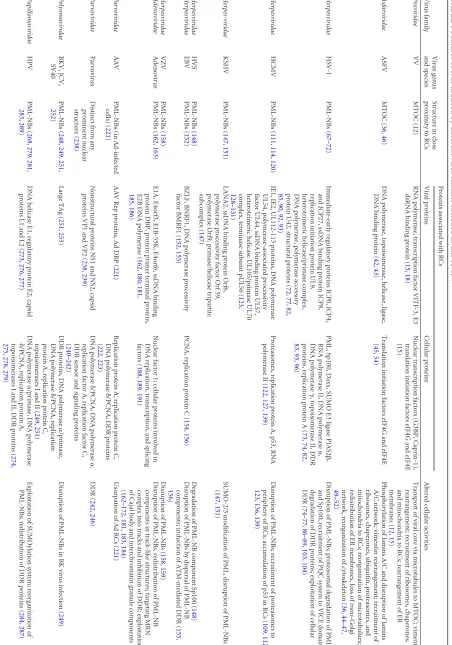
TABLE 1 Features of RCs induced by different virus families included in this review a Virus family Virus genus and species Structure in close proximity to RCs Proteins associated with RCs Altered cellular activities Viral proteins Cellular proteins Poxviridae VV MTOC ( 12 ) RNA polymerase, transcription factor VITF-3, E3 dsRNA binding protein ( 15 , 18 ) Nuclear transcription factors (G3BP, Caprin-1), translation initiation factors eIF4G and eIF4E ( 15 ) Transport of viral core via microtubules to MTOC; vimentin rearrangement; recruitment of ribosomes, chaperones, and mitochondria to RCs; rearrangement of ER membranes ( 12 , 15 ) Asfarviridae ASFV MTOC ( 36 , 46 ) DNA polymerase, topoisomerase, helicase, ligase, DNA binding protein ( 42 , 43 ) Translation initiation factors eIF4G and eIF4E ( 45 , 54 ) Phosphorylation of lamins A/C and disruption of lamins A/C network; vimentin rearrangement; recruitment of ribosomes, chaperones, ubiquitin, proteasomes, and mitochondria to RCs; reorganization of microtubulues; redistribution of ER membranes, loss of trans -Golgi network, reorganization of cytoskeleton ( 36 , 44–47 , 49–52 ) Herpesviridae HSV-1 PML-NBs ( 67–72 ) Immediate-early regulatory proteins ICP0, ICP4, and ICP27, ssDNA binding protein ICP8, replication initiation protein UL9, heterotrimeric helicase/primase complex, DNA polymerase, polymerase accessory protein U42, structural proteins ( 72 , 77 , 82 , 85 , 90 , 92 , 93 ) PML, Sp100, Daxx, SUMO E3 ligase PIAS2  , RNA polymerase II, DNA polymerase ␣ , DNA polymerase ␥ ,topoisomerase II, DDR proteins, replication protein A ( 73 , 74 , 82 , 83 , 93 , 96 ) Disruption of PML-NBs; proteasomal degradation of PML and Sp100; recruitment of PQC system to VICE domain; degradation of DDR proteins; exploitation of cellular DDR ( 74–77 , 86–89 , 103 , 104 ) Herpesviridae HCMV PML-NBs ( 111 , 114 , 120 ) IE1, IE2, UL112-113 proteins, DNA polymerase UL54, polymerase-associated processivity factor UL44, ssDNA binding protein UL57, heterotrimeric helicase UL105/primase UL70 complex, terminase subunit pUL56 ( 123 , 128–131 ) Proteasomes, replication protein A, p53, RNA polymerase II ( 122 , 127 , 139 ) Disruption of PML-NBs; recruitment of proteasomes to periphery of RCs; accumulation of p53 in RCs ( 109 , 112 , 123 , 136 , 139 ) Herpes-viridae KSHV PML-NBs ( 147 , 151 ) LANA2, ssDNA binding protein Orf6, polymerase processivity factor Orf 59, polymerase Orf9, primase/helicase tripartite subcomplex ( 147 ) SUMO-2/3 modification of PML, disruption of PML-NBs ( 147 , 151 ) Herpesviridae HVS PML-NBs ( 148 ) Degradation of PML-NB component Sp100 ( 148 ) Herpesviridae EBV PML-NBs ( 152 ) BZLF, BNRF1, DNA polymerase processivity factor BMRF1 ( 152 , 153 ) PCNA, replication protein C ( 154 , 156 ) Disruption of PML-NBs by dispersal of PML-NB components; induction of ATM-mediated DDR ( 155 , 156 ) Herpesviridae VZV PML-NBs ( 158 ) Disruption of PML-NBs ( 158 , 159 ) Adenoviridae Adenovirus PML-NBs ( 162 , 163 ) E1A, E4orf3, E1B-55K, E4orf6, ssDNA binding protein DBP, protein primer terminal protein, E2B DNA polymerase ( 162 , 180 , 181 , 185 , 186 ) Nuclear factor 1; cellular proteins involved in DNA replication, transcription, and splicing factors ( 188 , 189 , 191 ) Disruption of PML-NBs, redistribution of PML-NB components in track-like structures; targeting MRN complex into tracks and inhibition of DDR; exploitation of Cajal body and interchromatin granule components ( 162–172 , 181 , 183 , 184 ) Parvoviridae AAV PML-NBs (in Ad-infected cells) ( 221 ) AAV Rep proteins, Ad DBP ( 221 ) Replication protein A, replication protein C, DNA polymerase ␦ /PCNA, DDR proteins ( 222 , 223 ) Usurpation of Ad RCs ( 221 ) Parvoviridae Parvovirus Distinct from any prominent nuclear structure ( 238 ) Nonstructural proteins NS1 and NS2, capsid proteins VP1 and VP2 ( 238 , 239 ) DNA polymerase ␦ /PCNA, DNA polymerase ␣ , replication factor A, replication factor C, DDR sensor and signaling proteins ( 240–242 ) DDR ( 242 , 246 ) Polyomaviridae BKV, JCV, SV40 PML-NBs ( 248 , 249 , 251 , 252 ) Large TAg ( 251 , 253 ) DDR proteins, DNA polymerase ␣ /primase, DNA polymerase ␦ /PCNA, replication protein A, replication protein C, topoisomerases I and II ( 249 , 251 ) Disruption of PML-NBs in BK virus infection ( 249 ) Papillomaviridae HPV PML-NBs ( 268 , 279 , 281 , 283 , 289 ) DNA helicase E1, regulatory protein E2, capsid proteins L1 and L2 ( 273 , 276 , 277 ) DNA polymerase ␣ /primase, DNA polymerase ␦ /PCNA, replication protein A, topoisomerases I and II, DDR proteins ( 274 , 275 , 278 , 279 ) Exploitation of SUMOylation system; reorganization of PML-NBs; redistribution of DDR proteins ( 284 , 287 ) a The reference(s) supporting the reported information in each cell is provided in parentheses for each virus species.
on November 7, 2019 by guest
http://jvi.asm.org/
[image:2.585.68.520.70.715.2]Poxviridae. Poxviridae possess the unusual characteristic of replicating exclusively in the cytoplasm. Upon membrane fusion and release into the cytoplasm, the poxvirus core is transported via microtubules (MT) toward the MTOC. Such transport is associ-ated with vimentin rearrangement and recruitment of chaperones and mitochondria to perinuclear sites, where replication com-partments are finally formed (12). Early gene transcription, how-ever, is already initiated within the virus core by the viral DNA-dependent RNA polymerase (Pol) and viral transcription factors packaged into the virus particles together with the genome. Thus, a set of about 100 early mRNAs is transcribed and subsequently translocated into the cytoplasm where, while still associated with MT, they are engaged by ribosomes for translation. Meanwhile, viral cores accumulate close to the rough endoplasmic reticulum (rER). In the case of vaccinia virus (VV), the prototype and most-studied member ofPoxviridae, early proteins are required to un-coat the viral core and to release the viral DNA, as soon as the viral core reaches the ER (13). The disassembly steps for genome un-coating depend on ubiquitin-mediated proteasomal degradation of previously ubiquitinated capsid proteins, and a Cullin3-based ubiquitin ligase is required for genome replication (14). The ge-nome released from the core associates with a set of viral proteins
that participate in DNA replication and organization of the RC precursor (reviewed in reference12), as DNA replication pro-ceeds, intermediate and late mRNA, as well as ribosomes and translation factors, accumulate in expanding DNA factories (15), which are later released from the rER when virion assembly starts (16,17).
Poxviruses encode over 130 proteins, including factors in-volved in transcription (18), encompassing more than 20 viral proteins involved in RNA synthesis (reviewed in reference19), as well as DNA replication (reviewed in reference 20). Although these include a nearly complete repertoire of viral proteins re-sponsible for viral gene expression and DNA replication, the ex-tensive rearrangement of ER membranes is likely to recruit and control many cellular proteins. Indeed, a recent RNA interference screen identified 188 cellular factors required for VV infection, including both nuclear and cytoplasmic functions (14). During intermediate and late times of infection, cellular protein synthesis is downregulated (21–23) and cellular mRNA is degraded by a viral decapping enzyme (24,25). Furthermore,Poxviridaehave been suggested to promote selective and augmented translation of viral mRNAs inin vitro studies (26). The highly complex ER-surrounded RCs have been compared to mininuclei, where viral
mRNA translation
Adenoviridae
Viral ds DNA
PML pRB MRN sp100 P53 P53 PML-NB BLM DAXX
+ DBP PML
sp100 E4orf3PML E4orf3 E4orf3 E4orf3 sp100PML PML sp100 sp100 E4orf3 Transcription & splicing zone track-like structures early ICG late ICG transcription & splicing factors E1A E1B-55K E4orf6 Autonomous Parvoviridae DNA replication Late Transcription APAR body poly-Ub proteasomal degradation Viral transactivators Cellular factors NS1
DNA polα /primase
DNA polδ /PCNA
RPA RPC on late mRNA NS1 NS2 S-phase induction Formation of novel nuclear structures
DDR sensor & signalling proteins
NS1
Promotion of virus replication
Papillomaviridae
E1/E2 DNA Cell replication
machinery
Factors provided by Cajal bodies Activator/Repressor
directed to nucleus & PML-NBs by L2
Rolling circle Polyomaviridae Late Transcription Murine PyV SV40 JC BK PML pRB MRN sp100 P53 P53 PML-NB BLM DAXX Viral ds DNA + Histones
Localization to PML-NBs Disruption of PML-NBs No disruption of PML-NBs Transcription factors PML pRB MRN sp100 P53 P53 PML-NB BLM DAXX Virus Progeny Particle assembly adjacent to PML-NBs Early Transcription DNA replication
DNA polα /primase
DNA polδ /PCNA
RPA, RPC Topoisomerase I & II DDR proteins
Herpesviridae
Circularization
Linkage of DNA replication & packaging
Accumulation of ubiquitinated proteins
& the PQC system HSV-1
HSV-1 pol, ssDBP ICP8, heterotrimers helicase/primase complex DNA pol/primase DNA pol topoisomrase II DDR proteins Late Proteins + Chaperones VICE proper folding & storage site of viral proteins
20S proteasomes HCMV
Proteasomes relocalized to periphery of RCs (by active DNA replication)
↓ enhancing of early & late gene expression Replication foci
(active replication) DBP Ad DNA pol
PML pRB MRN sp100 P53 P53 PML-NB BLM DAXX NCLDV MTOC Nucleus Cytoplasm rER Translation nuclear transcription factors Assembly of virions Early Transcription viral DNA-dependent RNA polymerase
Viral ds DNA
Viral core DNA replication Transcription Transport to MTOC & Accumulation at rER Mitochondria Chaperones Early viral proteins viral transcription factors Dispersal of viral factory Asfarviridae Precursor DNA Initiation of DNA replication viral transcription/ translation factors
E8R A40R
AXX DNA Damage Response 100 Antiviral Response pRB Gene Transcription sp P53 Tumor Suppresion Apoptosis BLM post-transcriptional Modifications poly-Ub proteasomal degradation sp100 DAXX Cellular RNA pol II LT
DNA Cell replication machinery PML pRB MRN sp100 P53 P53 PML-NB BLM DAXX PML-NB PML-NB PML pRB MRN sp100 P53 P53 PML-NB BLM DAXX
Creation of host cell environment for genome replication
sTAg mTAg LTAg
early mRNA LM early mRNA LTAg sTAg LTAg LTAg LTAg Assembly of virions late mRNA Mre11 poly-Ub proteasomal degradation poly-Ub proteasomal degradation Late Transcription E1A E1A E1A E Early Transcription late mRNA DNA replication dsDNA early mRNA E4orf3 E1B-55K E4orf6 Mre11 P53 E4orf3 E1B-55K MRN STAT-1 ATR E1B-55K E4orf6 MRN DAXX BLM P53 Perinuclear Aggresome PML pRB MRN sp100 P53 P53 PML-NB BLM DAXX
Viral ds DNA + Histones V
Early Transcription
early mRNA
a sc p o
early mRNA L2 Assembly of virions Assembly of virions Late Transcription E1 ATM components recruitment & activation late mRNA
DNA polα /primase
DNA polδ /PCNA
RPA Topoisomerase I & II L1
DDR
E1 E2
DDR promotes DNA replication
DNA replication
E1 E2
E2 L2 E6 Degradation of PML-IV & reorganization of PML-NBs by L2 E6 L2 PML pRB MRN sp100 P53 P53 PML-NB BLM DAXX P pRB p P53 AT PML pRB MRN sp100 P53 P53 PML-NB BLM DAXX
Viral ds DNA
Late mRNA Export
DNA replication
Assembly of virions
Early HSV-1 induced foci
ICP0
Late Transcription
RNA pol II early mRNA
p
A p
A pop A po A po A po
A popo A po A popo A popoppopopopopopoppppol IIl IIlIIll IIl IIl IIl IIl II
Early Transcription
ICP4 ICP0
HCMV IE1 ICP27 pUL56 HCMV pp71 PML PML PML ICP4 PML pRB MRN sp100 P53 P53 PML-NB BLM DAXX HCMV HCMV Localization of UL112-113 to RCs
↓ Recruitment of DNA pol UL44 ssDBP heterotrimers Disruption of PML-NBs Disruption of PML-NBs Rolling circle PML sp100 DAXX late mRNA 7 Degradation of PML-NB components pp71 ICP0 Gene transcription
RNA pol II IE2 ICP8 ICP4 DAXX Uncoating of viral cores early mRNA early mRNA P53 MRN poly-Ub proteasomal degradation poly-Ub proteasomal degradation Tubular structures
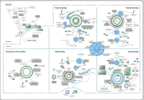
FIG 1Scheme of DNA virus replication compartments, indicating localization within the cytoplasm or nucleus. In particular, the virus familiesPolyomaviridae,
Papillomaviridae,Adenoviridae, andHerpesviridaeas well as NCLDV and autonomous parvoviruses are shown. Viral factors and proteins are indicated in green
and cellular ones are gray, with PML-NBs, which play a prominent role during replication ofPolyomaviridae,Papillomaviridae,Adenoviridae, andHerpesviridae,
shown in blue.
on November 7, 2019 by guest
http://jvi.asm.org/
[image:3.585.46.542.69.416.2]DNA as well as transcription and translation factors are enclosed inside a membranous compartment. However, although linked transcription and translation has recently been demonstrated (15), compartmentalization of the virus factories leads to separa-tion of gene expression and genome replicasepara-tion, which occur in distinguishable subcompartments within or on the surface of vi-rus factories. Katsafanas and Moss showed that viral RNA tran-scription occurs within the factory, as both the viral G8R RNA and poly(U) RNA localized to distinct compartments that those au-thors called cavities or tunnels of the virus factories. Moreover, poly(U) labeling showed reduced cytoplasmic staining compared to uninfected cells, consistent with cellular mRNA degradation during intermediate times of infection (15). The viral E3 dsRNA binding protein that counteracts activation of the dsRNA-sensing kinase (protein kinase R [PKR]) was also observed in RCs, as was the viral intermediate transcription factor 3. Nuclear factors re-quired for transcription of the viral genome are also recruited to these sites (18), as are the cellular G3BP and Caprin-1, both known to be required for viral intermediate transcriptionin vitro
(15). The cellular translation initiation factors eukaryotic initia-tion factor 4G (eIF4G) and eIF4E, as well as ribosomes, are also present in viral RCs; however, a large pool of ribosomes remains in the cytoplasm, suggesting that preferential translation of viral mRNA could be achieved by sequestering initiation factors. Translation of viral mRNA occurs within viral RCs, as -galacto-sidase expressed from a recombinant VV was confined to these compartments (15). This compartmentalization of translation factors on the one hand would be relevant for efficient expression of viral genes and on the other hand for simultaneous suppression of cellular protein synthesis.
During the late phase of infection, when virus assembly is ini-tiated, the replication sites are released from the rER and crescent-shaped membranes associate with viral DNA to initiate a complex assembly process (reviewed in reference27).
Asfarviridae.LikePoxviridae, ASFV, the only member of the
Asfarviridaefamily at this time, replicates in virus factories that are
distributed in perinuclear cytoplasmic sites. However, in contrast to poxviruses, ASFV not only recruits nuclear factors to these fac-tories but also initiates genome replication within the cell nucleus
(28–31). It is believed that short precursor DNA fragments are
subsequently exported from the nucleus to the cytoplasmic RCs, where they are used as primers for full-length genome replication
(30–32). Interestingly, cytoplasmic DNA synthesis is independent
of the nucleus (33). Given its large genome, it is thought that the virus encodes proteins that actively transport the viral genome into the nucleus as well as factors that direct the precursor DNA exit to the cytoplasm. While two viral structural proteins, p37 and p14, might be involved in nuclear entry (34), the viral matrix protein p37, which exhibits nucleocytoplasmic shuttling activi-ties, might trigger nuclear egress and transport of viral precursor DNA to RCs (34,35).
Upon viral entry and virion release from the endosome-lyso-some system into the cytoplasm, the ASFV core is transported to the MTOC (36). Meanwhile, transcription of early genes and post-transcriptional modification of the mRNA occur inside the virion; this is ensured by packaged proteins such as the viral RNA poly-merase and, hence, independent of cellular enzymes (37–41). Late gene expression, however, depends on viral DNA replication and viral early proteins. For DNA synthesis, the ASFV genome en-codes a set of proteins responsible for cytoplasmic DNA
replica-tion, including DNA polymerase, topoisomerase, helicase, ligase, and DNA binding proteins (42,43).
ASFV induces major changes in the organization of the in-fected cell. During the early phase, lamins A/C are phosphorylated and the nuclear membrane network next to sites where newly synthesized viral DNA accumulates is disrupted (44). Compo-nents of the nucleolus (B23) and splicing speckles (SC35) are also redistributed within the nucleus, and RNA polymerase II dephos-phorylation and degradation are induced and contribute to dis-rupt cellular transcription (44). Furthermore, ASFV induces shut-off of host protein synthesis by recruiting eIFs and ribosomes to RCs (45). At later time points, when viral DNA accumulates in cytoplasmic factories adjacent to the outer nuclear membrane, the lamin A/C network is further disrupted and sequestered to nuclear and cytoplasmic foci. The latter are recruited to the RCs along with other nuclear membrane components (44). Similar to other viruses, cytoplasmic ASFV factories have been compared with ag-gresomes, since emerging viral factories form at the MTOC and become surrounded by vimentin and recruit cellular chaperones, ubiquitin, proteasomes, and mitochondria. Furthermore, similar to aggresomes, ASFV factories require microtubules and dynein motor proteins for their formation (36,46).
Early ASFV replication sites appear as punctuate foci of ex-tranuclear viral DNA, while structural proteins are spread throughout the cytoplasm. Later on, these proteins are actively transported to the MTOC via microtubules, where they fuse with ASFV factories (47,48). Interestingly, the reorganization of mi-crotubules seems to be critical for DNA replication, as well as late gene transcription (46, 47,49), presumably by stabilizing viral RCs and concentrating cellular and viral proteins required for replication at the MTOC (46,47,49). Further reorganization of cellular components is induced as replication progresses and in-volves the relocation of mitochondria and chaperones as well as the redistribution of ER membranes to RCs, the loss of thetrans -Golgi network, and extensive reorganization of the cytoskeleton
(47,50–52). ASFV encodes a viral homologue of cellular
ubiqui-tin-conjugating enzymes (UBCs), suggesting that ASFV might manipulate ubiquitin-mediated host cell responses or modifica-tions of viral or host proteins (53).
Subdivision of poxviral factories allows the separation of viral gene expression and DNA replication (15). Similarly, during ASFV infection, host initiation factors are recruited to viral RCs, whereas viral RNA and host ribosomes are localized to the facto-ries’ peripheries (54).
The ASFV replication cycle primarily takes place in these virus factories, which are first observed 6 to 8 h postinfection (p.i.). During ongoing infection, amorphic and circular membranous material as well as increasing numbers of immature and mature viral particles accumulate in ASFV factories 12 to 24 h p.i. and follow a complex morphogenetic process analogous to that for poxviruses (reviewed in reference55).
Similar to poxviruses and ASFV, other members of the remain-ing NCLDV families also form cytoplasmic factories that share many of the features described above; however, the genomes of the iridoviruses (56) and phycodnaviruses (57) are initially shuttled into the nucleus, where replication begins, whereas genome rep-lication of mimiviruses seems to take place entirely in the cyto-plasm (6).
on November 7, 2019 by guest
http://jvi.asm.org/
NUCLEAR DNA VIRUS REPLICATION CENTERS
Like the NCLDV, DNA viruses that replicate in the nucleus use aggresome-like structures as sites for assembly of nuclear facto-ries. However, the ultrastructure and detailed architecture of the nuclear replication and assembly sites has not been studied in as much detail as cytoplasmic factories. The accumulated evidence and recent biochemical studies have found multiple interactions between viral genomes and RCs with nuclear components and domains, and the architecture of viral replication compartments has been revealed by a variety of fluorescence and electron micros-copy studies.
Upon internalization of virus particles, viral cores are trans-ported to the MTOC by cellular dynein/dynactin motors, and vi-ral genomes are delivered into the nucleus by disassembly of the viral capsid at the nuclear pore (reviewed in references5,58,59, and60).
Inside the nucleus, viral genomes colonize specific nuclear sites by utilizing cellular components to ensure efficient viral gene ex-pression and replication. These so-called replication centers or compartments (RCs) often form adjacent to PML-NBs, nuclear structures that have been implicated in DNA repair, transcrip-tional regulation, cell senescence, apoptosis, and the interferon-induced antiviral state. The major structural component of PML-NBs is the PML protein, also known as TRIM19, a member of the tripartite motif (TRIM) family that functions as the essential or-ganizer of the PML-NB and regulates protein transit by SUMO-mediated protein interactions (reviewed in reference61). In most cases, components of the PML-NB are targeted to the viral ge-nomes as part of a cellular defense mechanism. Subsequently, cel-lular factors relocalized to these sites are either utilized by the virus for its replication (DNA Pol, RNA Pol, transcription factors, post-transcriptional processing, and mRNA export) and/or inhibited by the virus to prevent/control the cellular antiviral defense (PML, p53, ATM, ATR, Daxx, STAT, interferon regulatory factors) (62–
64) (Fig. 1;Table 1).
The association of PML-NBs with nuclear aggresomes pro-vides a link for RCs to the storage as well as removal of (misfolded) proteins (63,65). In contrast to cytoplasmic replicating viruses, which assemble progeny particles in close association with cellular membranes, virions of DNA viruses replicating in the nucleus are often assembled in close proximity to viral transcription and DNA replication centers devoid of a surrounding membrane (1,5).
Herpesviridae.Upon infection with different members of the
Herpesviridaefamily, similar replication centers are formed (Fig.
1). Proteins of the PML-NB are recruited to the viral genome, and viral RCs containing the parental genome form adjacent to PML-NBs. However, RCs are established by different mechanisms, as described here, in particular, for herpes simplex virus 1 (HSV-1) and human cytomegalovirus (HCMV).
HSV-1 (alphaherpesvirus).Shortly after the HSV-1 genome
has entered the nucleus, the host cell antiviral defense is activated and components of PML-NBs are delivered to sites associated with the parental genome (66–71). The recruitment of PML, Sp100, and Daxx depends on their SUMO interaction motifs. Fur-thermore, SUMOylated proteins as well as the cellular SUMO E3 ligase PIAS2are found in these HSV-1-induced foci. Thus, it has recently been proposed that the cellular defense against foreign DNA is regulated by the SUMO pathway (72). The viral regulatory protein ICP0 colocalizes with these foci and induces the
protea-some-mediated degradation of PML and Sp100 by acting as an E3 ubiquitin ligase (73–77), thus leading to the disruption of PML-NBs. Interestingly, ICP0 specifically targets SUMO-1-modified forms of the PML-NB proteins for proteasomal degradation (78– 80). Ongoing replication requires the recruitment of different vi-ral and cellular proteins to the virus-induced foci, which mature to viral replication centers (81–83). As the name implies, these com-partments provide an optimal environment for viral gene expres-sion, DNA synthesis, and assembly of progeny nucleocapsids (84,
85). Additionally, aggresome-like structures are formed adjacent to RCs early during infection. These so-called virus-induced chap-erone-enriched (VICE) domains contain molecular chaperones, especially the Hsp70 chaperone family member Hsc70, and 20S proteasomes that are sequestered from a diffuse nuclear distribu-tion, as well as ubiquinated proteins (86–89). Both molecular chaperones as well as the ubiquitin proteasome system are key components of cellular protein quality control (PQC). Initially, ubiquitylation of cellular proteins (see below) during viral infec-tion leads to the formainfec-tion of the VICE domains, an addiinfec-tional nuclear aggresome-like structure (86). Hence, the cellular PQC system recruited to these structures can be exploited for different processes during HSV-1 replication. Thus, molecular chaperones allow assembly and proper folding of viral multimeric protein complexes, including the RC scaffold, the helicase/primase com-plex, and the viral capsids. VICE domains additionally serve as storage sites for structural proteins of the viral capsid that have been made in excess compared to those packaged into capsids. Thus, it has been suggested that VICE domains are assemblons that form late during infection (90,91). Viral gene expression in nuclear RCs starts immediately after entry of the viral genome into the nucleus. The linear DNA is circularized, and immediate-early (IE), early (E), and late (L) viral gene products are sequentially expressed. Active herpesviral gene expression in RCs requires the translocation of viral ICP0, ICP4, and ICP27 proteins as well as RNA polymerase II to these sites (92,93). Both ICP0 and ICP4 stimulate transcription of E and L genes, whereby ICP4 functions as a major transcriptional activator and is required for the pro-gression beyond the immediate-early phase. In contrast, ICP27 mediates export of the majority of herpesviral mRNAs, interacting with the cellular export receptors Tap/Nxf and Aly/Ref (94). The accumulation of viral proteins and viral template DNA as well as the translocation of cellular proteins to the RCs allow efficient gene expression as well as viral genome replication. Among the herpesviral proteins associated with RCs are the single-stranded DNA (ssDNA) binding protein ICP8, heterotrimeric helicase/pri-mase complex, and the HSV-1 polymerase. Furthermore, cellular DNA polymerases␣and␥and topoisomerase II, as well as DNA repair and recombination proteins, are recruited to the RCs (82,
83,95,96).
Progeny virion assembly, including capsid formation and DNA packaging, is a tightly regulated process that takes place in the nuclear RCs (97,98). Typically, this process involves proteins forming an internal scaffold where the capsid shell is assembled and the genome is subsequently incorporated (99–101). Thereaf-ter, the assembled nucleocapsid exits the nucleus by a unique bud-ding mechanism through the nuclear membrane. Finally, the vi-rion is transported via secretory transport vesicles to the plasma membrane and is released upon membrane fusion (102).
Common features of DNA viruses are the formation of E3 ubiquitin ligases, as described above for HSV-1 ICP0, as well as the
on November 7, 2019 by guest
http://jvi.asm.org/
deregulation of the cellular DNA damage response (DDR). Most interestingly, both the degradation of cellular proteins as well as the modulation of the DDR are linked to a considerable extent. Already in the early phase of HSV-1 infection, proteins involved in homologous recombination (HR) (83) as well as nonhomologous end joining (NHEJ) of DNA (95,103) are recruited to viral RCs. Interestingly, in contrast to adenoviruses, which inhibit this in-trinsic antiviral mechanism (see below), the cellular DDR appears to be beneficial for HSV-1. Nevertheless, HSV-1 also manipulates the DDR by inactivating some of its components and utilizing others. ICP0 interferes with both DDR pathways by inducing the degradation of different target proteins, among these, the catalytic subunit of the DNA-dependent protein kinase DNA-PK (104,
105), which is involved in sensing of NHEJ, as well as the E3 ligases RNF8 and RNF168 (106,107), which promote ATM tethering to sites of DNA damage, i.e., the sensing mechanism of HR. In con-trast, the Mre11-Rad50-Nbs1 (MRN) complex as well as ATM kinase activation, both of which are involved in NHEJ, seem to be beneficial for HSV-1 replication (103). Interestingly, although phosphorylated replication protein A (RPA) is excluded from RCs and sequestered to adjacent VICE domains, thereby preventing normal ATR signaling (108), ATR and ATRIP localize in RCs and have been shown to be conducive to HSV-1 replication (73).
HCMV (betaherpesvirus).Replication centers of human
cy-tomegaloviruses form adjacent to PML-NBs. Different studies have suggested that the recruitment of PML proteins to the viral nucleocapsid shortly after its delivery into the nucleus is part of the antiviral host defense. However, this is subsequently hampered by different HCMV proteins. The cellular transcriptional repressor Daxx inhibits HCMV gene expression from the major IE pro-moter. This is circumvented by the viral tegument protein pp71
(109–111). Interestingly, pp71 stimulates proteasomal
degrada-tion of Daxx by a mechanism that is independent of a funcdegrada-tional ubiquitin pathway (112). In addition, pp71 was found to trigger Daxx SUMOylation, although so far no function for this modifi-cation has been described (113). Furthermore, the viral transacti-vator IE1 colocalizes with PML-NBs and disrupts these structures (114) by interfering with SUMO modification of PML (115). Thus, both pp71 and IE1 cooperate in the disruption of cellular PML-NBs and antagonize host defense mechanisms. In addition, studies on these HCMV proteins revealed a link to the cellular SUMOylation pathway, as has been observed for other DNA vi-ruses. Consistent with these effects on cellular PML-NBs, down-regulation of Daxx and/or PML as well as of the PML-NB compo-nent Sp100 was found to enhance viral replication (116–119). Moreover, the HCMV kinase pUL97 seems to be an additional viral protein that interferes with PML function. pUL97 inhibits the complex formation between PML and the retinoblastoma pro-tein (Rb) by hyperphosphorylating Rb (120,121). Interestingly, the kinase activity of pUL97 also inhibits the formation of nuclear aggresomes (121). In contrast to the viral tegument protein pp65, which induces inappropriate aggregation and sequestration of vi-ral tegument and capsid proteins, pUL97 prevents this undesir-able effect. Thus, pUL97 is proposed to play an important role in progeny virus assembly.
Similar to HSV-1, HCMV expresses its genes in a temporally regulated cascade, with the replication cycle being divided into IE, E, and L phases. The immediate-early proteins IE1 and IE2 stim-ulate viral E and L gene expression, with IE2 the driving force of HCMV replication in general. As would be expected, RNA Pol II
and splicing factors colocalize with IE genes in RCs (122). While IE2 colocalizes with the viral RCs by interacting with the viral genome, IE1 localization to PML-NBs has been reported to de-pend on interaction with PML (123).
Recently, it was reported that efficient viral replication requires active cellular proteasomes (124–126). HCMV infection specifi-cally leads to enhanced proteasomal pathway activity. Further-more, active viral DNA replication induces the recruitment of proteasomes to the periphery of viral RCs, where active RNA tran-scription takes place. This is consistent with the observation that the cellular proteasomal pathway is exploited to enhance early and late gene expression (127).
Already early during HCMV infection, the UL112-113 pro-teins are localized together with IE2 to prereplication foci and are found in RCs throughout the viral life cycle (128–130). UL112-113 encodes four phosphoproteins, which are supposed to recruit proteins involved in DNA replication, to viral RCs (128, 131). These viral replication core proteins include the DNA polymerase UL54, the polymerase-associated processivity factor UL44, and the ssDNA binding protein UL57, as well as a heterotrimer con-sisting of the DNA helicase UL105, the primase UL70, and the primase-associated factor (132,133). Upon entry into the nucleus, the HCMV genome is circularized, and a rolling circle mechanism of replication produces concatemers late in infection (136,135). The HCMV terminase subunit pUL56 potentially links DNA rep-lication with packaging. Interestingly, this protein is involved in cleavage and packaging of progeny viral genomes (136) and also localizes to viral RCs (137).
As described for HSV-1, DNA viruses often interfere with the cellular DDR machinery. HCMV infection induces the accumu-lation of the tumor suppressor p53 (138–140). Simultaneously, the virus compromises the ability of p53 to transactivate cellular downstream targets (138, 140–142). Thus, HCMV may exploit p53 functions to fully activate the infected cells, in parallel inhib-iting its ability to regulate DDR as well as apoptosis. Interestingly, p53 as well as RPA are both relocalized to viral RCs (143). It could therefore be that both cellular proteins are utilized to help in viral DNA damage control and/or to promote efficient viral DNA syn-thesis, as has been reported for other DNA viruses (144–146).
Other herpesviruses. Interestingly, apart from HSV-1 and
HCMV, various herpesviruses counteract the antiviral properties of PML-NBs and lead to the disruption of these bodies and/or relocalization of specific PML-NB components. Thus, members of the gammaherpesvirus family, like Kaposi’s sarcoma-associated herpesvirus (KSHV) (147), herpesvirus saimiri (HVS) (148), and Epstein-Barr virus (EBV) (152), express proteins with properties similar to HSV-1 ICP0. Furthermore, viral replication compart-ments develop in association with PML-NB remnants (147,148,
152).
For KSHV, it has been shown that shortly after infection, PML-NB components localize to viral genomes and prereplica-tion compartments (147). Interestingly, PML is SUMO-2 modi-fied by LANA2, the latency-associated nuclear antigen 2. Since SUMO-2/3 modification of PML has been described to induce PML degradation via ubiquitination by the cellular RNF4 ubiqui-tin ligase (150), this modification most likely leads to proteasome-mediated degradation of PML and hence to PML-NB disruption. Thereby, LANA2 seems to interfere with PML-mediated tran-scriptional repression (151). DNA replication during the lytic cy-cle of KSHV infection requires six viral proteins, including the
on November 7, 2019 by guest
http://jvi.asm.org/
ssDNA binding protein Orf6, the polymerase processivity factor Orf59, and the polymerase Orf9, as well as a primase/helicase tri-partite subcomplex. Interestingly, transient cotransfection of these factors results in the formation of nuclear structures resem-bling replication compartments. Furthermore, these pseudo-RCs are surrounded by PML-NBs (147).
As mentioned above, HVS also antagonizes the PML-NB-me-diated, intrinsic host cell defense. Specifically, the HVS tegument protein Orf3 induces degradation of the PML-NB component Sp100, whereas the distribution and/or functionality of PML and Daxx remains unchanged. Although PML restricts HVS replica-tion, it might play an important role for the establishment of latent infection (148).
Herpesviruses can cause both latent and lytic infection of the host cell. During EBV latency, PML-NBs remain intact, and rep-lication of the virus is not associated with these nuclear structures. However, the switch from latent to lytic EBV replication immedi-ately disrupts PML-NBs by dispersing Sp100, Daxx, and NDP55. In contrast, PML is dispersed only after the onset of lytic replica-tion (149). For the disruption of PML-NBs, the EBV immediate-early protein BZLF alone is sufficient. Interestingly, BZLF com-petes with PML for free SUMO-1, which leads to a decrease of SUMO-1-modified PML (152). Furthermore, EBV expresses a tegument protein that specifically targets the intrinsic host cell defense, thereby activating viral early gene transcription. This pro-tein, BNRF1, disrupts the cellular Daxx-ATRX chromatin remod-eling complex, which seems to be required for the switch from latent to lytic infection by alteration of viral chromatin (153). Further proteins localizing to replication compartments and in-teracting with the EBV DNA polymerase processivity factor BMRF1 include the proliferating cell nuclear antigen (PCNA) and replication factor C (RFC), as well as several proteins of the cellu-lar mismatch repair system. Thereby, EBV might prevent recom-bination events between homologous stretches in its genome se-quences (154). Interestingly, active EBV DNA replication induces an ATM-mediated DDR (155), which is modified by the viral BGLF4 kinase. Ultimately, this results in an optimal environment for viral genome amplification, while host cell DNA synthesis is blocked (156,157).
Similar to HSV-1, the varicella-zoster virus (VZV) protein Orf61, a homologue of HSV-1 ICP0, disrupts cellular PML-NBs. The SUMO-interacting motifs (SIM) present in Orf61 are critical for both the interaction and dispersal of these nuclear structures (158). A three-dimensional (3D) reconstruction using serial sec-tion array scanning electron microscopy confirmed that PML-NBs are disassembled by the interaction of Orf61 SIM with PML and revealed that VZV capsids become entrapped in large PML cages (159).
Taken together, replication of herpesviruses occurs adjacent to cellular PML nuclear bodies, which are known to negatively re-strict viral replication. Interestingly, herpesviruses evolved vari-ous mechanisms to counteract the PML-NB function. Therefore, it is most likely that different herpesviruses target similar cellular pathways, involving DDR, chromatin remodelling, cell cycle con-trol, and transcription machinery, to foster their replication. So far, these mechanisms have been best studied for HSV-1 and HCMV; however, recent results revealed the first detailed insights into replication of other members of theHerpesviridae.
Adenoviridae.Upon infection of the cell, adenoviral (Ad)
par-ticles are transported toward the MTOC and disassembled at the
nuclear pore. Immediately after the viral genome is released into the nucleus, it is targeted by the host cell repression machinery. PML-NB components, like Daxx, thereby mediate transcriptional repression of the Ad genome, specifically, of the immediate-early E1A promoter (reviewed in references64and68). Interestingly, this is counteracted by the capsid protein VI, which enters the nucleus in association with the Ad genome (160). In the course of an adenoviral infection, Daxx is targeted for proteasomal degra-dation by the early E1B-55K protein (161).
Another early protein, E4orf3, associates with PML nuclear bodies and induces their disruption from punctuate foci into track-like structures (162,163). Specifically, PML, Sp100, Daxx, SUMO-1, and TIF1␣are relocated into these tracks by E4orf3
(163–166), which might resemble nuclear aggresomes (167).
These PML-containing structures are thereafter localized adjacent to viral RCs. Thus, the antiviral response is inhibited, whereas specific components of these nuclear bodies could be exploited by the virus for efficient replication (162,163).
Apart from the interferon-dependent antiviral defense medi-ated by PML-NB components, E4orf3 targets the Mre11-Rad50-Nbs1 (MRN) DNA damage-sensing complex into tracks (168). Ultimately, the MRN complex is redistributed to cytoplasmic ag-gresomes by E4orf3 (169). Furthermore, Mre11 and other cellular proteins are ubiquitinated by E1B-55K and E4orf6, which assem-ble a Cullin-dependent E3 ubiquitin ligase complex and thus tar-get proteins for proteasomal degradation (reviewed in reference
170). Besides Mre11, other proteins of the cellular DNA damage response, including the DNA ligase IV and Bloom helicase (BLM), are conducted to E1B-55K/E4orf6 complex-dependent degrada-tion to prevent concatenadegrada-tion of viral dsDNA genomes mediated by this pathway (168,171,172).
Another target protein of the E3 ubiquitin ligase complex is the tumor suppressor protein p53. During the early phase of expres-sion, p53 is induced by E1A (see below). Since p53 transactivates several downstream proteins, p53 induction by E1A is potently proapoptotic (173). Besides host cell proteins like the tumor sup-pressor p53, E1A also activates the transcription of viral early genes. Both are mediated by the exclusive binding of E1A to the retinoblastoma tumor suppressor (pRB) (174–177). This leads to dissociation of pRB from the E2F transcription factor and subse-quently to the constitutive activation of the E2F-responsive cellu-lar and the viral E2 early promoter (178,179). Thus, E1A econom-ically induces cell cycle progression as well as expression of viral proteins required for viral DNA replication.
The transition to the late phase of Ad infection is marked by the onset of genome replication and subsequent expression of genes from the major late transcription unit (MLTU). This switch coin-cides with formation of the viral RCs (180) and further reorgani-zation of nuclear bodies (181–185). Adenoviral DNA replication is initiated by a unique priming mechanism that involves a viral protein primer, the terminal protein (TP), and is accomplished by the viral E2B DNA polymerase. Additionally, viral and cellular proteins involved in host cell DNA replication are recruited to sites of viral DNA synthesis (186, 187). Single-stranded DNA products are associated with the ssDNA binding protein (DBP) and form nuclear inclusions that are variable in size and appear as crescents or spheres (180,185). Surrounding these sites of ssDNA accumulation is the peripheral replicative zone, where both repli-cation and transcription take place. Punctate sites of active replica-tion are arranged as a ring surrounding ssDNA. The replicated
on November 7, 2019 by guest
http://jvi.asm.org/
dsDNA, however, is displaced from these replication foci to the pe-riphery, where it serves as a template for the transcription of viral late genes (185). Active transcription leads to the formation of a ring-shaped transcription and splicing zone surrounding ssDNA and rep-lication foci (188). This zone contains dsDNA and nascent viral RNA, as well as transcription and splicing factors. These factors, which play an important role in enhancing Ad gene expression, are recruited from CB, which progressively disassemble during the late phase, and replication centers coalesce as they occupy regions of interchromatin granules (181,183,185,189,191).
Transcription and splicing, on the other hand, are tightly linked to the preferential export of Ad transcripts in the late phase of infection. Ongoing Ad replication leads to the concentration of splicing factors and viral RNA in interchromatin granules, which become progressively larger (182,190). This might enhance pro-cessing or trafficking, as well as transport of RNAs. The preferen-tial export of Ad late transcripts is mediated by the early Ad pro-teins E1B-55K and E4orf6. Interestingly, both propro-teins are present in viral RCs (191). The underlying mechanism, however, is still unclear, although recent results point to different scenarios. First, the activity of the E3 ubiquitin ligase complex is necessary for efficient export (189,192). Since during Ad replication the trans-port of bulk cellular mRNA is simultaneously blocked (193,194), it is likely that one or more proteins implicated in their export are targeted for proteasomal degradation, which might, in turn, pro-mote preferential export of viral transcripts. Second, further stud-ies suggested that the cellular export receptor of bulk cellular mRNA, TAP/NXF1, participates in the export of Ad late tran-scripts (195). An interesting hypothesis is that cellular export fac-tors are relocalized to viral transcription and replication centers and thus promote efficient export of newly transcribed mRNAs out of the nucleus. Simultaneously, these proteins would be de-pleted in the rest of the nuclear compartment, which could also account for the blockage of cellular mRNA transport. However, this only applies to the late phase of infection, when RCs are formed. During the early phase Ad exploits the CRM1-dependent export pathway for its transcripts (196).
Similar to theHerpesviridae, Ad replication centers form adjacent to PML-NBs and are therefore likely to both counteract PML-depen-dent antiviral activities and benefit from components and the scaffold from the PML-NB. Interestingly, the E1B-55K and E4orf3 proteins, are known to participate in SUMOylation of cellular substrates (197,
198), and both associate with PML-NBs (199).
Parvoviridae. Members of the family Parvoviridae differ in
some interesting aspects from other viruses that replicate in ver-tebrates. In contrast to DNA tumor viruses, parvoviruses are not capable of inducing S-phase entry of the host cell; rather, the virus remains inactive unless the host cell itself enters the S phase. Fur-thermore, parvoviruses that infect vertebrates can be distin-guished into two groups: autonomously replicating viruses and viruses that depend on coinfection of a helper virus and therefore are classified as dependoviruses (200).
Adeno-associated viruses.Members belonging to the
Depen-dovirusgenus are the so-called adeno-associated viruses (AAV),
which were first described as requiring adenovirus coinfection (201,202). More recently, HSV-1 and -2, CMV, and pseudorabies virus have been identified as helper viruses (203–206). In the ab-sence of a helper virus, the AAV genome integrates into the host cell genome (207,208). In the event of helper virus infection of
such latently infected cells, however, AAV gene expression is re-activated and the replication cycle progresses (209).
AAV contain two open reading frames that encode four non-structural (Rep) and three non-structural (Cap) proteins (210–212), as well as a recently identified protein involved in capsid assembly, the assembly-associated protein AAP (213). Rep78 and Rep68 are known to play a key role during AAV DNA amplification (211,
212), whereas Rep52 and Rep40 are assumed to be involved in DNA packaging (214,215).
Furthermore, AAV utilizes helper viral as well as cellular pro-teins for its own replication. So far, adenoviruses have been most extensively studied in the context of AAV helper virus, and thus will be mainly discussed here. Various Ad early gene products involved in regulating different processes during Ad infection are required for complete Ad helper virus function (208). Thus, Ad E1A enhances transcription of both Ad early genes as well as AAV Rep and Cap genes (216), and it activates the cellular gene expres-sion required for S-phase entry and synthesis of DNA replication proteins (208). Additionally, the Ad-dependent E3 ubiquitin li-gase regulates AAV gene expression, most likely by facilitating mRNA transport and promoting AAV DNA replication (217). The adenoviral DNA binding protein DBP stimulates AAV gene expression and DNA elongation, as well as AAV particle forma-tion (218,219). Thus, DBP is the only Ad protein, which has been proposed to be directly involved in AAV genome amplification (219), in contrast to the Ad protein primer and DNA polymerase, which are both dispensable for AAV replication.
Intriguingly, AAV further usurps Ad replication compart-ments for its own DNA replication in PML-NB-associated RCs (220). Therefore, AAV genome and Rep proteins are relocalized to Ad RCs (221). To ensure AAV DNA amplification, apart from Ad DBP, cellular components are also required in these nuclear struc-tures, including RPA, the replication factor C (RFC), PCNA, and DNA polymerase␦(222,223). Thus, an adenovirus coinfection not only provides AAV with essential Ad proteins and factors but also reprograms the host cell accordingly. However, assembly of AAV progeny virions is not conducted in the RCs but instead takes place in the nucleolus (213,224).
Interestingly, host cell functions can compensate for a helper virus infection under specific conditions that induce a cellular stress response (225,226). In agreement, the celluar DNA damage response is also induced during Ad coinfection. Recently, two in-dependent studies found that DDR proteins become activated and in part relocalized to RCs, e.g., DNA-PK, phosphorylated Nbs1, Ku70/Ku86, and/or ATM (227,228). Inhibition experiments have suggested that there is a distinct, yet unclear, requirement for cel-lular DDR proteins to foster or inhibit AAV replication (227).
Autonomous parvoviruses.In contrast to the dependoviruses,
autonomous parvoviruses are strictly dependent on host cell func-tions, especially on progression of the cell into the S phase (229,
230). The genome of autonomous parvoviruses comprises two transcription units, encoding the nonstructural proteins NS1 and NS2 and the capsid proteins VP1 and VP2 (231). NS1 is a multi-functional phosphoprotein that acts as a transcriptional activator of viral promoters as well as initiator and helicase in parvoviral DNA replication (232–237). Furthermore, NS1 colocalizes with replicating viral DNA in virus-induced nuclear foci (238,239).
These compartments represent parvovirus replication centers termed autonomous PV-associated replication (APAR) bodies. Besides NS1 and parvovirus DNA, host cell replication proteins
on November 7, 2019 by guest
http://jvi.asm.org/
that facilitate viral genome amplification, like PCNA, RPA, and RFC, as well as DNA polymerase␣and␦, accumulate in these bodies (238–241). Interestingly, the parvovirus RCs are distinct from any prominent nuclear structure, such as PML-NBs, nucle-oli, coiled bodies, and/or speckled domains (238). Thus, parvovi-rus induces the formation of novel structures in the host cell nu-cleus and simultaneously recruits required cellular proteins to these compartments. Accumulation of viral DNA and capsid components leads to the expansion of these RCs, as they ulti-mately occupy most of the host cell nucleus (242). To date, con-troversial data have been collected on whether capsid assembly occurs in the nuclear (224,243,244) or cytoplasmic (245) com-partment. It seems likely that the capsid protein VP2 triggers cy-toplasmic assembly of VP1/VP2 oligomers and, additionally, me-diates nuclear import of these complexes by its nuclear import signal. Sequentially, viral capsids might be assembled in the nu-clear compartment (246).
Similar to AAV, minute virus of mice (MVM), an autonomous parvovirus, has recently been described to induce a robust DNA damage response and to exploit this cellular pathway for its own benefit. DDR sensor proteins (Nbs1, Mre11, ATM, DNA-PKs, and RPA) and signaling proteins (␥H2AX, Ku70, and Ku86) ac-cumulate in APAR bodies in response to active viral DNA replica-tion. The virus-regulated modifications of cellular DDR include proteasome-dependent degradation of Mre11 and, seemingly, utilization of ATM, since inhibitors of this kinase restrict MVM replication. In contrast to AAV, DDR signaling in MVM-infected cells is mediated by this ATM kinase (247).
Polyomaviridae. Binding of cellular transcription factors to
the polyomaviral genome mediates its import into the nucleus. Subsequently, the genome is delivered to specific nuclear sites, namely, PML-NB (248).
During progression of the viral life cycle, various viral and cel-lular proteins colocalize with the polyomaviral (PyV) genome at PML-NBs (Fig. 1). These factors include proteins involved in gene expression and DNA replication as well as viral capsid proteins. Altogether, this suggests that these nuclear sites may be important for PyV replication. Intriguingly, studies on various PyV types have drawn different scenarios. Thus, the absence of PML, the main structural component of PML-NBs, either has no effect on or even enhances viral replication (249,250). Specifically, BK vi-ruses (BKV) seem to counteract PML-NB functions by dramatic reorganization of these structures and dispersal of two of its major components: Sp100 and Daxx (249). In contrast, although PML restricts JC virus replication, this virus does not directly modulate PML-NBs (250). Similarly, simian virus 40 (SV40) neither relo-calizes PML-NB components nor disrupts these structures. Inter-estingly, transfection of the viral large T antigen TAg alone already induces its localization to PML-NBs. Thus, the deposition of viral RCs at PML-NBs seems not only to be a passive effect mediated by a PML-dependent antiviral defense mechanism, but also PyV seems to actively induce RCs at these sites (249,250). The most detailed study on PyV factories was performed recently using a murine polyomavirus, and the investigators showed that although PyV RCs are located adjacent to PML-NBs, the establishment of these centers as well as effective virus growth did not depend on the PML protein (251).
In general, PyV DNA replication has been localized adjacent to PML-NBs (248,249,252,253). Interestingly, SV40 gene expres-sion also takes place in PyV RCs at PML-NBs. However, the spatial
restriction to PML-NBs only accounts for DNA amplification, while transcription seems to be an indirect consequence following genome delivery to these nuclear sites (254).
Concomitant with the onset of viral DNA synthesis, late gene transcription is initiated. Following transcription of late genes, the structural proteins VP1, VP2, and VP3 are transported into the nucleus via their nuclear localization signal (NLS) (255,256,257). However, capsid proteins are not imported as monomers but as capsid subunits, especially as VP1 pentamers and VP2/3 com-plexes (258). Since large TAg and VP1 have been found to colo-calize adjacent to PML-NBs, a spatial coupling of DNA replication and capsid assembly has been proposed (248,252,253). Intrigu-ingly, Garcea and coworkers established a new and unanticipated mechanism of virion assembly (251) that is contrary to the assem-bly process of larger DNA viruses, including herpes- and adeno-viruses, namely, the encapsidation of the viral genome into pre-formed capsids (98, 259). Already by 1980, polymerization of capsid subunits onto the viral genome had been proposed (260). However, VP1 pentamers seem to first assemble tubular struc-tures, in which the viral genome is integrated, and finally icosahe-dral virion particles are formed by a budding mechanism (251).
Similar to parvoviruses and papillomaviruses (see below), polyomaviruses actively recruit cellular DDR proteins to the RCs and exploit their functions (261–264). Specifically, PyV recruit proteins involved in homologous recombination, such as ATM kinase and the MRN complex (261,263), and activate these path-ways (265). Both relocalization and activation are essential for PyV growth (261–265). Interestingly, the multifunctional large TAg induces replication foci containing PML and relocalizes DDR proteins to these structures, although this has been shown to be PML independent (251,263). Similar to adenoviruses, Mre11 is recruited to SV40 replication sites and is later degraded (263).
Papillomaviridae.Compared toHerpesviridaeand
Adenoviri-dae, little is known about productive replication of papillomavi-ruses, as studying these viruses has been hindered by the absence of an appropriate cell culture system.
Papillomaviruses specifically infect squamous epithelial cells. The productive replication of papillomaviruses can be divided into an early and late phase, with the late phase, including viral DNA synthesis, production of capsid proteins, and virion assem-bly, being exclusively restricted to differentiated epithelial cells. It is likely that, upon infection, genome transport toward the nu-cleus is regulated by the L2 capsid protein via interaction with microtubules (266). Since cell division is required for nuclear ge-nome translocation and expression (267), the viral genome may enter the cell nucleus only during mitotic membrane breakdown. Comparable to other nuclear DNA viruses, the papillomavirus genome localizes to PML-NBs (Fig. 1). Intriguingly, these nuclear structures foster papillomavirus transcription (268), in contrast to most DNA viruses, which developed mechanisms to antagonize the restrictive function of PML-NBs.
In general, viral DNA replication is achieved by two early viral proteins, E1 and E2 (269–272). In contrast to E2, which is only required for initiation, E1 possesses ATPase and DNA helicase activities, thereby also triggering elongation of DNA synthesis (273,274). Except for E1 and E2, the residual replication initiation machinery, including DNA polymerase␣/primase, DNA poly-merase␦/PCNA, RPA, and topoisomerases I and II, is provided by the host cell (275,276).
Interestingly, like other DNA viruses, papillomavirus also
on November 7, 2019 by guest
http://jvi.asm.org/
seems to exploit the cellular SUMOylation system. Thus, E1 inter-acts with the cellular SUMO-conjugating enzyme Ubc9, seem-ingly triggering SUMO-1 modification of this viral protein. It has been shown that these functions are essential for the intranuclear accumulation of the protein as well as the efficient origin-depen-dent replication of the viral genome (277,278).
Furthermore, papillomavirus DNA replication is tightly linked with the cellular DNA damage response. E1 and E2 both activate and relocalize the cellular DDR to viral RCs. The targeting of the cellular DDR leads to growth suppression of the host cell. While E2 recruits ATM DDR components to the viral RCs, E1 specifi-cally activates this cellular ATM DDR pathway (279). Both growth arrest and ATM activation depend on the ATPase activity of E1 and its binding to the viral origin of replication. Interestingly, the cellular DDR is activated to facilitate viral DNA amplification, thus stimulating papillomavirus replication (280).
Prior to initial gene expression, the viral genome is localized to PML-NBs by the viral capsid protein L2, supporting viral early gene transcription (268). The presence of the HPV genome in basal cells leads to an increased total number of PML-NBs as well as elevated levels of posttranslationally modified PML protein (281). The HPV E6 oncoprotein has been described to colocalize with PML-NBs (282) and to induce proteasomal degradation of PML-IV, seemingly overcoming restrictive functions of these nu-clear bodies (283). Interestingly, these cellular structures are also associated with papillomaviral RCs (269,284–288). It has been proposed that L2 localizes to PML-NBs, where it associates with E2 and recruits the viral genome to sites of assembly. Subse-quently, L1 could be recruited to RCs for progeny virion assembly (268,284). Although, different studies observed no requirement of PML-NBs for papillomavirus DNA replication (281,288), it is evident that these nuclear structures become reorganized by L2 (289) and are target sites for virion assembly.
CONCLUSIONS
As obligate intracellular parasites, viruses have coevolved with the host cell. Consequently, the changes in cellular activities and architec-ture that are induced in a virus-infected cell are likely to reflect a “tug-of-war” between the opposing cellular defenses and the mecha-nisms viruses employ to divert or utilize the cellular machinery and metabolic pathways. Assembly of virus inclusions or aggregates is an essential step for productive replication of RNA and DNA viruses from a wide variety of different families, and although these struc-tures display different subcellular localizations, architecstruc-tures, and compositions, they seem to share several fundamental similarities: (i) virus factories function as scaffolds that anchor viral genomes, con-centrate viral and cellular macromolecules, and facilitate the assem-bly of replication complexes. RNA and DNA viruses that replicate in the cytoplasm make use of cellular membranes as scaffolds, whereas nuclear DNA viruses mostly rely on the nature and organization of nuclear domains, in particular PML-NB, for assembly of replication compartments that employ protein scaffolds. (ii) Although it is often not clear how viral genomes are directed to specific subcellular loca-tions, the assembly of replication compartments is accompanied by the reorganization of the cytoskeleton and associated motor proteins. Often viral genomes are directed to the MTOC, where perinuclear aggresomes assemble, as has been found for togaviruses, flaviviruses, bunyaviruses, coronaviruses, and arteriviruses, as well as for several of the NCLDV. Interestingly, the genomes of DNA viruses are targeted to or by PML-NBs and also make use of nuclear and in some cases
cytoplasmic aggresomes. This suggests that a cellular response dedi-cated to counter invasion of foreign/misfolded proteins may be re-sponsible for targeting viral genomes and structural proteins, thus initiating an aggresome response that is coopted by viruses to gener-ate assembly sites. Viruses from many different families either employ or modulate the ubiquitin-proteasome pathway for degradation of cellular substrates, making aggresomes appropriate sites where chap-erones, ubiquitin, and 26S proteasomes are concentrated. (iii) For-mation of cytoplasmic viral factories functions to conceal the viral genome and transcripts from an arsenal of cellular defense mecha-nisms that include the cytoplasmic dsRNA-sensing kinase (PKR), as well as the Toll-like receptors (TLRs) and cytoplasmic retinoic acid-inducible gene I (RIG-I) helicase receptors, which trigger the produc-tion of type I interferon. In the nucleus, PML-NBs are primary sites of the interferon-dependent as well as the intrinsic response to infection (64,67,290). Many proteins encoded by DNA and RNA viruses co-localize and perturb PML-NBs, suggesting that, as in the case of cy-toplasmic aggresomes, alteration of PML-NBs may be a viral strategy to evade a cellular defense mechanism. Cellular activities that are reg-ulated by PML-NBs include DNA damage repair, apoptosis, the ubiquitin pathway, and gene expression (136). Interestingly, as de-scribed in the previous sections, DNA viruses that replicate in the nucleus modulate, relocate, or induce the degradation of cellular fac-tors implicated in each of these activities.
Progress in understanding the molecular mechanisms that un-derlie the formation of replication centers formed by DNA viruses is likely to continue to advance rapidly in the near future. For example, three-dimensional analyses of infected-cells via electron tomography will help in the study of their architecture, while bio-chemical studies should help determine their protein and nucleic acid compositions, as well as the interactions that are established between viral and cellular components.
ACKNOWLEDGMENTS
R.A.G. and T.D. received support from the Research Group Linkage Pro-gram of the Alexander von Humboldt Foundation. The Heinrich Pette Institute is supported by the Freie und Hansestadt Hamburg and the Bundesministerium für Gesundheit. R.A.G. received grants from CONACyT-SEP (CB-2011-01-168497) and Promep-SEP.
REFERENCES
1.Netherton C, Moffat K, Brooks E, Wileman T.2007. A guide to viral
inclusions, membrane rearrangements, factories, and viroplasm
pro-duced during virus replication. Adv. Virus Res.70:101–182.http://dx
.doi.org/10.1016/S0065-3527(07)70004-0.
2.Paul D, Bartenschlager R.2013. Architecture and biogenesis of
plus-strand RNA virus replication factories. World J. Virol.2:32– 48.http://dx
.doi.org/10.5501/wjv.v2.j2.32.
3.Dreux M, Chisari FV.2010. Viruses and the autophagy machinery. Cell
Cycle9:1295–1307.http://dx.doi.org/10.4161/cc.9.7.11109.
4.Netherton CL, Wileman T. 2011. Virus factories, double membrane
vesicles and viroplasm generated in animal cells. Curr. Opin. Virol.
1:381–387.http://dx.doi.org/10.1016/j.coviro.2011.09.008.
5.Novoa RR, Calderita G, Arranz R, Fontana J, Granzow H, Risco C.
2005. Virus factories: associations of cell organelles for viral replication
and morphogenesis. Biol. Cell 97:147–172. http://dx.doi.org/10.1042
/BC20040058.
6.Colson P, De Lamballerie X, Yutin N, Asgari S, Bigot Y, Bideshi DK,
Cheng X-W, Federici BA, Van Etten JL, Koonin EV, La Scola B, Raoult
D.2013. “Megavirales,” a proposed new order for eukaryotic
nucleocy-toplasmic large DNA viruses. Arch. Virol.158:2517–2521.http://dx.doi
.org/10.1007/s00705-013-1768-6.
7.Iyer LM, Aravind L, Koonin EV.2001. Common origin of four diverse
families of large eukaryotic DNA viruses. J. Virol.75:11720 –11734.http:
//dx.doi.org/10.1128/JVI.75.23.11720-11734.2001.