This is a repository copy of
Escape from adamantane: Scaffold optimization of novel P2X7
antagonists featuring complex polycycles
.
White Rose Research Online URL for this paper:
http://eprints.whiterose.ac.uk/110732/
Version: Accepted Version
Article:
Barniol-Xicota, M, Kwak, S-H, Lee, S-D et al. (5 more authors) (2017) Escape from
adamantane: Scaffold optimization of novel P2X7 antagonists featuring complex
polycycles. Bioorganic & Medicinal Chemistry Letters, 27 (4). pp. 759-763. ISSN
0960-894X
https://doi.org/10.1016/j.bmcl.2017.01.039
© 2017 Elsevier Ltd. Licensed under the Creative Commons
Attribution-NonCommercial-NoDerivatives 4.0 International
http://creativecommons.org/licenses/by-nc-nd/4.0/
[email protected] https://eprints.whiterose.ac.uk/
Reuse
Unless indicated otherwise, fulltext items are protected by copyright with all rights reserved. The copyright exception in section 29 of the Copyright, Designs and Patents Act 1988 allows the making of a single copy solely for the purpose of non-commercial research or private study within the limits of fair dealing. The publisher or other rights-holder may allow further reproduction and re-use of this version - refer to the White Rose Research Online record for this item. Where records identify the publisher as the copyright holder, users can verify any specific terms of use on the publisher’s website.
Takedown
If you consider content in White Rose Research Online to be in breach of UK law, please notify us by
Accepted Manuscript
Escape from adamantane: Scaffold optimization of novel P2X7 antagonists
fea-turing complex polycycles
Marta Barniol-Xicota, Seung-Hwa Kwak, So-Deok Lee, Emily Caseley, Elena
Valverde, Lin-Hua Jiang, Yong-Chul Kim, Santiago Vázquez
PII:
S0960-894X(17)30051-3
DOI:
http://dx.doi.org/10.1016/j.bmcl.2017.01.039
Reference:
BMCL 24615
To appear in:
Bioorganic & Medicinal Chemistry Letters
Received Date:
29 December 2016
Revised Date:
7 January 2017
Accepted Date:
12 January 2017
Please cite this article as: Barniol-Xicota, M., Kwak, S-H., Lee, S-D., Caseley, E., Valverde, E., Jiang, L-H., Kim,
Y-C., Vázquez, S., Escape from adamantane: Scaffold optimization of novel P2X7 antagonists featuring complex
polycycles, Bioorganic & Medicinal Chemistry Letters (2017), doi:
http://dx.doi.org/10.1016/j.bmcl.2017.01.039
Bioorganic & Medicinal Chemistry Letters
Escape from adamantane: scaffold optimization of novel P2X7 antagonists featuring
complex polycycles
Marta Barniol-Xicota
a, Seung-Hwa Kwak
b, So-Deok Lee
b, Emily Caseley
c, Elena Valverde
a, Lin-Hua
Jiang
c, Yong-Chul Kim
b,*, and Santiago Vázquez
a,a
Laboratori de Química Farmacèutica (Unitat Associada al CSIC), Facultat de Farmàcia i Ciències de l’Alimentació, and Institute of Biomedicine (IBUB), Universitat de Barcelona, Av. Joan XXIII 27-31, Barcelona, E-08028, Spain
b
School of Life Sciences, Gwangju Institute of Science and Technology (GIST), Gwangju 500-712, Republic of Korea c
School of Biomedical Sciences, University of Leeds, Leeds, United Kingdom
———
Corresponding author. Tel.: +34-934-024-533; e-mail: [email protected]; Tel.: +82-62-715-2502; e-mail: [email protected]
The P2X7 receptor is an ionotropic ligand-gated purinergic receptor which is activated by extracellular ATP.1 Upon the application of physiological concentrations of this messenger, the P2X7 receptor acts as a cation selective ion channel, allowing the passage of Na+, K+ and Ca2+ through the plasma membrane, activating several signal transduction pathways for the correct function of the organism.2 However, when this receptor is exposed to high concentrations of ATP for a long period of time, in the high micromolar range, it induces the formation of a non-selective large pore3 permeable to molecules up to 900 Da, which ultimately triggers cell apoptosis.4
This ion channel is present in virtually all tissues of the body, being especially abundant in cells of hematopoietic lineage.5 Due to the widespread presence of the P2X7 receptor in the mammalian organism, its malfunction has been linked to a vast number of pathological conditions,6 including rheumatoid arthritis (RA), osteoporosis, Parkinson’s disease, multiple sclerosis and cancer, among other relevant inflammatory, immune or musculoskeletal disorders.7 Taking into account that modulation of the P2X7 offers an ample possibility of therapeutic intervention, it is no surprise that in recent years several academic and non-academic groups have directed their research efforts to the development of potential P2X7 antagonists.8
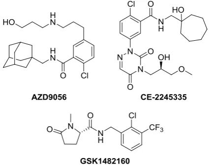
[image:3.595.319.532.543.711.2]Resulting from this endeavour several structurally diverse small molecule inhibitors have come to light, placing the most promising ones into clinical trials (Figure 1). Unfortunately, all the candidates to date have failed in this process, resulting in a void in the P2X7 therapeutic pipeline.
Figure 1. Structures of unsuccessful P2X7 clinical candidates. Two
additional drugs (AFC-5128 and EVT-401, of undisclosed structures, also failed in clinical trials).
A R T I C L E I N F O A B S T R A C T
Article history: Received
Received in revised form Accepted
Available online
The adamantane scaffold, despite being widely used in medicinal chemistry, is not devoid of problems. In the recent years we have developed new polycyclic scaffolds as surrogates of the adamantane group with encouraging results in multiple targets. As an adamantane scaffold is a common structural feature in several P2X7 receptor antagonists, herein we report the synthesis and pharmacological evaluation of multiple replacement options of adamantane that maintain a good activity profile. Molecular modeling studies support the binding of the compounds to a site close to the central pore, rather than to the ATP-binding site and shed light on the structural requirements for novel P2X7 antagonists.
2016 Elsevier Ltd. All rights reserved. Keywords:
One of the most promising chemical classes of P2X7 antagonists is found in the adamantyl derivatives. Indeed, this class of compounds had its most auspicious example in the AZD9056 (Figure 1), which successfully progressed to phase II clinical trials; regrettably it failed to show significant efficacy in several inflammatory diseases (osteoarthritis, RA, COPD and
Chrohn’s disease).9 Unexpectedly after this failure, the adamantane core was not only not abandoned, but regained interest as a scaffold for P2X7antagonists. Proof of this are the several patents filled by AstraZeneca, Lundbeck, Renovis and Abbot.10 Even more curious is the case of the academic groups that also resisted abandoning this scaffold, eagerly pursuing the preparation of adamantyl derivatives as P2X7antagonists. Worth mentioning are the compounds developed by Lee et al.,11 such as
2, and the C-2 disubstituted adamantanes developed by
[image:4.595.36.293.331.458.2]Battilocchio et al., such as 3.12 Despite the aforementioned combination of industry and academic efforts, modifying other moieties of the adamantyl derivatives has not met the expectation to overcome the drawbacks found in the past and send a compound to clinic. Surprisingly, little to no effort has been dedicated to exploring the possibilities of the adamantane moiety replacement. Among the few examples reported so far, we find only weakly active compounds such as norbornane 4,11 and cubane 5 (Figure 2).13
Figure 2. Selected adamantane derivatives (1-3) with activity as P2X7
receptor antagonists and examples of adamantane replacements (4-5).
Taking into account the vast experience of our group in successfully replacing the adamantane with other tailor made polycyclic moieties and the capability of the aforementioned compounds to undergo structural modification we decided to explore this understudied path.14
[image:4.595.308.556.441.705.2]For this purpose, we envisaged four types of suitable scaffolds that may act as adamantane replacement for their assessment in terms of P2X7 receptor activity. The aliphatic polycyclic scaffolds were judiciously chosen taking into account several factors, first of all the maintenance of the desirable drug-like properties shown by the adamantane compounds. Secondly, the fact that they already proved as suitable adamantane replacements in our hands, yielding to potent compounds.14 Finally, considering that when this work was carried out no crystal structures of the P2X7 were available,15 we aimed to explore the chemical space in the drug-binding site. To this aim, starting from the known nanomolar inhibitor 1, the bulkier substitution possibilities were embodied by I, its saturated analogue II, which display a similar shape index as adamantyl compounds (see below), and III, which exhibited a greater length alternative with restricted free rotation of the phenylhydrazine moiety. On the other hand, taking into account that smaller analogues of adamantane such as the norbornane and the cubane scaffolds exemplified by 4 and 5 led to poor antagonists, just one smaller scaffold (IV) was envisaged (Figure 3).
Figure 3. Adamantane replacement scaffolds. R = CONHNHAr, R’ = NHAr.
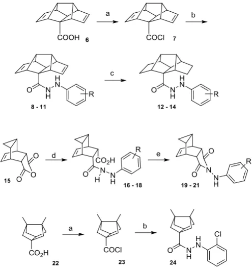
Diene 6, envisaged as a suitable precursor of I and II, was accessed through a concise 7-step synthetic route previously developed by our group.16 Upon treatment of 6 with thionyl chloride, the resulting acid chloride 7 was obtained and readily reacted with the desired hydrazine, to yield hydrazides 8-11 in good yields. The saturated analogues 12-14 were successfully prepared through a double bond reduction with hydrazide monohydrate under oxygen atmosphere.17 The compounds with the general structure III were easily accessed through the reaction of 1,3,5-cyclohepatriene with maleic anhydride, which furnished the polycyclic core in good yield.18 Upon reaction of 15 with the corresponding phenylhydrazine, the compounds 16 - 18 were dehydrated under Dean-Stark conditions to yield the cyclized adducts 19 – 21 (Scheme 1).
As the pharmacology trends for the already tested series I and
II placed the 2-Cl substitution among the preferred one (see
below), for the scaffold IV, only compound 24 was prepared. Hence, the hydrazide 24 was accessed from the known bisnoradamantane monoacid precursor19 and, paralleling the procedure for the preparation of derivatives I, was treated with thionyl chloride and readily reacted with 2-chlorophenylhydrazine (Scheme 1).
The structure of all new compounds was confirmed by accurate mass measurement, IR, 1H and 13C NMR spectroscopy.
Scheme 1. Reagents and conditions: (a) SOCl2, ; 2 h, quantitative yield. (b)
All new compounds were evaluated for their antagonistic
effects on 2’(3’)-O-(4-benzoylbenzoyl)-ATP (BzATP)-induced ethidium bromide uptake in human embryonic kidney 293 (HEK 293) cells stably expressing the human P2X7 receptor, using compound KN62 as a positive control (Table 1).20
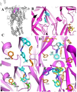
In order to rationalize the results, the binding mode of all the compounds was analysed by docking in the extracellular domain of the homology models of the receptor hP2X7and rP2X7, using AutoDock version 4.2.21 In agreement with the very recent crystal structures of five P2X7 antagonists in complex with a mammalian P2X7 receptor,15 our calculations supported the ability of the surrogate adamantane scaffolds to work as allosteric inhibitors. They fill a hydrophobic cavity close to the central pore of hP2X7, localized deeper in the receptor than the ATP binding site of hP2X7 (Figure 4A-B). The calculations also provided some clues about the observed structure-activity relationship (SAR).
Regarding the aromatic moiety substitution, we took advantage of the SAR previously developed by Abbott around the adamantane derivative 1 that revealed electron-withdrawing substitutents in the ortho position of the phenyl ring as the preferred group.22 In fact, within our series, the ortho position clearly showed to be the preferred one for the activity (compare 9 vs 10 and 13 vs 14, table 1). Interestingly, the introduction of a substituent in the para position was not only detrimental for the activity of the monosubstituted derivative (e.g., 10), but was able to abolish the benefitial effect of the ortho group (e.g., 11). This observation is explained by the steric clash of the para-trifluoromethyl group with the residue Phe95 of the receptor that has been previously shown to play a key role by forming -stacking interactions with distinct inhibitors (Figure 4C).23
Within this series, the most potent compound, 8 (IC50 = 96 nM), bearing a chlorine atom in the ortho position, was nearly 9 times less potent than its adamantane counterpart, 1 (IC50 = 11.5 nM). Notwithstanding, 8 was much more potent than previously studied replacements for adamantane (e.g., 4 and 5). Moving from dienes I to alkanes II led to higher IC50 values (compare 8 vs 12 and 9 vs 13), so full saturation of the structure is detrimental for the activity. This is rationalized as the dihedral angle formed by the amide and the aromatic ring, suffers a slight tilt in the saturated compound, compared to its more active unsaturated analogue (Figure 4D).
As the hydrogen atom of the CONH group did not seem to play an essential role in the binding, we drew our attention to the conformationally restricted series III. Neither the unsubstituted phenyl derivative, 19, nor the ortho substituted analogues 20 and
21, displayed significant activity, a fact that indicates that a
degree of conformational freedom in the aromatic moiety is required when designing P2X7 inhibitors.
Fully unexpectedly, taking into account the weak inhibitory activity displayed by known smaller replacement of adamantane, such as 4 and 5 (see above), compound 24 was revealed as the most potent analogue synthesized in this work, displaying an activity in the low nanomolar range (IC50 = 18 nM) which pairs the one displayed by its adamantane analogue 1 (IC50 = 11.5 nM). Strikingly, upon docking in the receptor, hydrazide 24 showed to adopt a completey different orientation (Figure 4E-F) than the one shown by its adamantane analogue 1 and 8, which parallels the adamantane compound in the binding site. Moreover, upon docking in the rP2X7 receptor, no unfavourable interactions were observed for 24, suggesting good antagonist potency. In view of the fact that several classes of inhibitors display significantly reduced the binding affinity in rP2X7 receptor, complicating
[image:5.595.302.564.116.302.2]further in vivo studies, our docking results further highlight the potential interest of bisnoradamantane derivatives as surrogates of adamantane.
Table 1. P2X7 receptor antagonist activity of novel compounds.a
Compound R IC
50 (nM)
8 2-Cl 96 ± 8
9 2-CF3 132 ± 20
10 4-CF3 <20%
b
11 2-Cl, 4-CF3 1557 ± 500
12 2-Cl 933 ± 156
13 2-CF3 366 ± 37
14 4-CF3 <20%
b
19 H <20%b
20 2-Cl <20%b
21 2-F <20%b
24 2-Cl 18 ± 2
KN-62 2-Cl 96 ± 11
a
Experiments were assessed in the ethidium accumulation assay using hP2X7R-expressing HEK293 cells. The IC50 values were obtained from concentration-response curves. Data are expressed as the means SD of at least 3-6 experiments. bPercent inhibition at concentration 10 M.
E
Figure 4. Docking of selected compounds and adamantane derivative 1 into
the human P2X7 receptor. A. Compounds binding site in the whole extracellular domain. B. Compounds binding site in relation with the ATP binding site. C. Steric clash of the trifluoromethyl group of 10. D. Superposition of 8 (light blue) and 12 (grey) in the binding site. E. Bisnoradamantane derivative 24 in the receptor binding pocket. Position 95 being Leu in the rP2X7 and Phe in the hP2X7. F. Superposition of 1 (yellow), 8 (light blue) and 24 (purple) in their binding site.
A
B
C
D
I
F
[image:5.595.306.559.358.663.2][image:6.595.32.293.192.289.2]
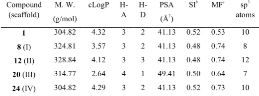
To further explore the suitability as an adamantane replacement we predicted their physicochemical properties using DataWarrior.24 Of note, 24 and its isomer 1 displayed very similar physicochemical properties (Table 2).One of the main concerns when moving adamantane derivatives further into drug discovery programs is its easy metabolization by oxidation at the bridgehead positions. Taking into account that scaffold IV features two of the bridgehead positions substituted its metabolization by oxidation might be more difficult.
Table 2. Relevant physicochemical properties for a representative example of
each scaffold predicted by DataWarrior.a
Compound (scaffold)
M. W.
(g/mol)
cLogP H-A H-D PSA (Å2 ) SIb MFc sp3 atoms
1 304.82 4.32 3 2 41.13 0.52 0.53 10
8 (I) 324.81 3.57 3 2 41.13 0.48 0.74 8
12 (II) 328.84 4.12 3 3 41.13 0.48 0.74 12
20 (III) 314.77 2.64 4 1 49.41 0.50 0.64 7
24 (IV) 304.82 4.29 3 2 41.13 0.52 0.73 10
a
All the series were predicted not to be mutagenic or tumorogenic agents. bShape index. cMolecular flexibility.
In conclusion, eleven new compounds have been synthesized from four available polycyclic compounds as P2X7antagonists. Several of these novel compounds displayed reasonable activity with 24, being the most potent with low nanomolar potency. To the best of our knowledge this is the first time, within the P2X7 antagonist context, that the adamantane scaffold has been successfully replaced by a different polycyclic structure. On top of that, 24 is predicted to have physicochemical properties that parallel those of adamantane, reinforcing its suitability as a scaffold to develop drug-like compounds. Taken together, we can conclude that the smaller bisnoradamantane scaffold IV revealed as a very promising surrogate of adamantane regarding P2X7 inhibition, in contrast to a hit-to-lead study reported by AstraZeneca that mentioned that no replacements for adamantane were found.25
Research towards the development of novel polycyclic scaffolds to function as adamantane replacement and the exploration of further analogues of 24 are in progress to reach derivatives with optimum P2X7 activity and enhanced ADME properties.
Acknowledgments
M. B.-X. and E. V. thank the Institute of Biomedicine of the University of Barcelona (IBUB) for a PhD Grant. S. V. thanks financial support from Ministerio de Economía y Competitividad (Spain) (SAF2014-57094-R) and the Generalitat de Catalunya (2014-SGR-00052).
References and notes
1. Burnstock, G. Pharmacol. Rev. 1972, 24, 509–581.
2. (a) Burnstock, G. Pharmacol Rev. 2006, 58, 58–86. (b) Coddou, C.; Yan, Z.; Obsil, T.; Huidobro-Toro, J.P; Stojilkovic, S. S. Pharmacol. Rev. 2011, 63, 641–683.
3. Yan, Z; Li, S; Liang, Z; Tomic, M.; Stojilkovic, S. S. J. Gen. Physiol. 2008, 132, 563–573.
4. Khadra, A.; Tomic, Melanija; Y., Zonghe; Z., H.; Sherman, A.; Stojilkovic, S. S. Biophys. J. 2013, 104, 2612–2621.
5. Parvathenani, L. K.; Tertyshnikova, S.; Greco, C. R.; Roberts, S. B.; Robertson, B.; Posmantur, R. J. Biol. Chem. 2003, 278, 13309–13317.
6. (a) De Marchi, E.; Orioli, E.; Dal Ben, D.; Adinolfi, E. Adv. Protein Chem. Struct. Biol. 2016, 104, 39–79. (b) Tewari, M; Seth, P. Ageing Res. Rev. 2015, 24, 328–342. (c) Skaper, S. D.; Debetto, P.; Giusti, P. FASEB J . 2010, 24, 337–345. (d) North, R. A.; Jarvis, M. F. Molec. Pharmacol. 2003, 83, 759–69. (e) Roger, S.; Pelegrin, P. Expert Opin. Investig. Drugs. 2011, 20, 875–80. 7. (a) Arulkumaran, N.; Unwin, R. J.; Tam, F. W. Expert Opin.
Investig. Drugs. 2011, 20, 897–915. (b) Di Virgilio, F.; Ceruti, S.; Bramanti, P.; Abbracchio, M. P. Trends Neurosci. 2009, 32, 79– 87.
8. (a) Friedle, S. A.; Curet, M. A.; Watters, J. J. Recent Pat. CNS Drug Discov.2010, 5, 35–45. (b) Bartlett, R.; Stokes, L.; Sluyter, R. Pharmacol. Rev. 2014, 66, 638–75.
9. (a) Keystone, E.C.; Wang, M.M.; Layton, M.; Hollis, S.; McInnes, I. B. Ann. Rheum. Dis. 2012. 71, 1630–1635. (b) Eser, A.; Colombel, J. F.; Rutgeerts, P.; Vermeire, S.; Vogelsang, H.; Braddock, M.; Persson, T.; Reinisch, W. Inflamm. Bowel Dis.
2015, 21, 2247-2253. (c)
https://openinnovation.astrazeneca.com/azd9056.html, accessed on Dec. 12th, 2016.
10. (a) Gunosewoyo, H.; Kassiou, M. Expert Opin. Ther. Pat. 2010, 20, 625–646. (b) Park, J.-H.; Kim, Y.-C. Expert Opin. Ther. Pa t. doi: 10.1080/13543776.2017.1246538
11. Lee, W.-G.; Lee, S.-D.; Cho, J.-H.; Jung, Y.; Kim, J.-H.; Hien, T. T.; Kang, K.-W.; Ko, H.; Kim, Y.-C. J. Med. Chem. 2012, 55, 3687–3698.
12. Battilocchio, C.; Guetzoyan, L.; Cervetto, C.; Di Cesare Mannelli, L.; Frattaroli, D.; Baxendale, I. R.; Maura, G.; Rossi, A.; Sautebin, L.; Biava, M.; Ghelardini, C.; Marcoli, M.; Ley, S. V. ACS Med. Chem. Lett. 2013, 4, 704–709.
13. (a) Gunosewoyo, H.; Guo, J. L.; Bennett, M. R.; Coster, M. J.; Kassiou, M. Bioorg. Med. Chem. Lett. 2008, 18, 3720–3723. (b) Wilkinson, S. M.; Gunosewoyo, H.; Barron, M. L.; Boucher, A.; McDonnell, M.; Turner, P.; Morrison, D. E.; Bennett, M. R.; McGregor, I. S.; Rendina, L. M.; Kassiou, M. ACS Chem. Neurosci. 2014, 5, 335–339.
14. (a) Camps, P.; Duque, M. D.; Vázquez, S.; Naesens, L.; De Clercq, E.; Sureda, F. X.; López-Querol, M.; Camins, A.; Pallàs, M.; Prathalingam, S. R.; Kelly, J. M.; Romero, V.; Ivorra, D.; Cortés, D. Bioorg. Med. Chem. 2008, 16, 9925–9936. (b) Duque, M. D.; Camps, P.; Profire, L.; Montaner, S.; Vázquez, S.; Sureda, F. X.; Mallol, J.; López-Querol, M.; Naesens, L.; De Clercq, E.; Prathalingam, S. R.; Kelly, J. M. Bioorg. Med. Chem. 2009, 17, 3198–3206. (c) Duque, M. D.; Camps, P.; Torres, E.; Valverde, E.; Sureda, F. X.; López-Querol, M.; Camins, A.; Prathalingam, S. R.; Kelly, J. M.; Vázquez, S. Bioorg. Med. Chem. 2010, 18, 46– 57. (d) Duque, M. D.; Ma, C.; Torres, E.; Wang, J.; Naesens, L.; Ju re - im ne , ; Camps, P ; u ue, ; e rado, ;
am , R A ; Pinto, H ; ue , S. J. Med. Chem. 2011, 54,
2646−2657 (e) Torres, E.; Duque, M. D.; López-Querol, M.;
Taylor, M. C.; Naesens, L.; Ma, C.; Pinto, L. H.; Sureda, F. X.; Kelly, J. M.; Vázquez, S. Bioorg. Med. Chem. 2012, 20, 942–948. (f) Rey-Carrizo, M.; Torres, E.; Ma, C.; Barniol-Xicota, M.; Wang, J.; Wu, Y.; Naesens, L.; DeGrado, W. F.; Lamb, R. A.; Pinto, L. H.; Vázquez, S. J. Med. Chem. 2013, 56, 9265–9274. (g) Torres, E.; Leiva, R.; Gazzarrini, S.; Rey-Carrizo, M.; Frigolé-Vivas, M.; Moroni, A.; Naesens, L.; Vázquez S. ACS Med. Chem. Lett. 2014, 5, 831–836. (h) Rey-Carrizo, M.; Barniol-Xicota, M.; Ma, C.; Frigolé-Vivas, M.; Torres, E.; Naesens, L.; Llabrés, S.; Juárez-Jiménez, J.; Luque, F. J.; DeGrado, W. F.; Lamb, R. A.; Pinto, L. H.; Vázquez, S. J. Med. Chem. 2014, 57, 5738–5747. (i) Valverde, E.; Sureda, F. X.; Vázquez, S. Bioorg. Med. Chem.
2014, 22, 2678–2683. (j) Rey-Carrizo, M.; Gazzarrini, S.; Llabrés,
S.; Frigolé-Vivas, M.; Juárez-Jiménez, J.; Font-Bardia, M.; Naesens, L.; Moroni, A.; Luque, F. J.; Vázquez, S. Eur. J. Med. Chem. 2015, 96, 318–329. (k) Valverde, E.; Seira, C.; McBride, A.; Binnie, M.; Luque, F. J.; Webster, S. P.; Bidon-Chanal, A.; Vázquez, S. Bioorg. Med. Chem. 2015, 23, 7607–7617.
15. During the writing of this manuscript, the crystal structures of a mammalian P2X7 receptor complexed with five structurally-unrelated antagonists was reported, see: Karasawa, A.; Kawate, T. eLIFE 2016, 5, 322153.
16. Camps, P., Pujol, X., Rossi, R. A., Vázquez, S. Synthesis. 1999, 85458.
17. Menges, N.; Balci, M. Synlett. 2014, 25, 671–676.
18. Abou-Gharbia, M.; Patel, U. R.; Webb, M. B.; Moyer, J. A.; Andree, T. H.; Muth, E. A. J . Med. Chem. 1988, 31, 1382−1392. 19. (a) Camps, P.; Iglesias, C.; Rodríguez, M. J.; Grancha, M. D.;
Linares, A. A. Chem. Ber. 1988, 121, 647–654. (b) Camps, P.; Font-Bardia, M.; Pérez, F.; Solans, X.; Vázquez, S. Angew. Chem. Int. Ed. Engl. 1995, 34, 912–914. (c) Camps, P.; Lukach, A. E.; Rossi, R. A. J . Org. Chem. 2001, 66, 5366–5373.
20. P2X7 receptor antagonist activity. Dye uptake using the ethidium ion assay. HEK293 cells stably expressing P2X7 receptor were maintained in Dulbecco's modified Eagle's medium (DMEM) supplemented with 10% fetal bovine serum (FBS), 2 mM L-glutamine, and antibiotics (50 U/mL penicillin and 50 mg/mL streptomycin) in a humidified 5% CO2 atmosphere at 37 ºC. We used Lipofectamine as a transfection reagent with a pcDNA3.1 vector-based plasmid harbouring hP2X7 receptor (Invitrogen). After diluting to 2.5 x 10⁶ cells/mL, buffer composed of 10 mM HEPES, 5mM N-methyl-glutamine, 5.6 mM KCl, 10 mM D-glucose, and 0.5 mM CaCl2 (pH 7.4) supplemented with either 280 mM sucrose or 140 mM NaCl, and an 80 L aliquot was added to each well of 96-well culture plates. The test com- pounds and
2’(3’)-O-(4-benzoylbenzoyl)-ATP (BzATP) were then added, and
the cells were incubated for 2 h in a humidified 5% CO2 atmosphere at 37ºC. After incubation, a Bio-Tek FL600 fluorescence plate reader was used to measure the absorbance at an excitation wavelength of 530 nm and an emission wavelength of 590 nm. The inhibition (percent) of ethidium ion uptake was expressed as a relative value of the maximum accumulation when stimulated with BzATP only. To calculate the IC50 values, we calculated a series of dose-response data using nonlinear regression analysis (i.e., per-centage accumulation of ethidium bromide vs compound concentration).
21. Homology modelling. Structural models of the P2X7 receptor were produced based on the zebrafish P2X4 receptor crystal structure in the closed and ATP-bound states (Protein Data Bank code 4DW0 and 4DW1, respectively).26 Modeller version 9.12 was used to generate one hundred versions of each model.27 The five with the lowest energy were further analysed with MolProbity and those with the greatest percentage of residues in allowed regions of the Ramachandran plot were selected for use in this study.28 The ModLoop server was used to model the non-conserved loop region between the 2 and 3 strands.29 Molecular docking simulations. Molecular docking was carried out using AUTODOCK version 4.2.30 The target cavity file for each docking simulation consisted of the P2X7 receptor extracellular domain. The auxiliary program AutoGrid was used to generate the affinity grid files.
22. Nelson, D. W.; Sarris, K.; Kalvin, D. M.; Namovic, M. T.; Grayson, G.; Donnelly-Roberts, D. L.; Harris, R.; Honore, P.; Jarvis, M. F.; Faltynek, C. R.; Carroll, W. A. J. Med. Chem. 2008, 51, 3030–3034.
23. Caseley, E. A.; Muench, S. P.; Baldwin, S. A.; Simmons, K.; Fishwick, C. W.; Jiang, L.-H. Bioorg. Med. Chem. Lett. 2015, 25, 3164–2167.
24. DataWarrior version 4.4.4.
25. Baxter, A.; Bent, J.; Bowers, K.; Braddock, M.; Brough, S.; Fagura, M.; Lawson, M.; McInally, T.; Mortimore, M.; Robertson, M.; Weaver, R.; Webborn, P. Bioorg. Med. Chem. Lett. 2003, 13, 4047–4050.
26. Hattori, M.; Gouaux, E. Nature 2012, 485, 207-212.
27. Webb, B.; Sali, A. Curr. Protoc. Bioinformatics 2006, unit 5.6. DOI: 10.1002/cphi.3
28. Davis, I. W.; Leaver-Fay, A.; Chen, V. B.; Block, J. N.; Kapral, G. J.; Wang, X.; Murray, L. W.; Arendall, W. B.; Snoeyink, J.; Richardson, J. S. Nucleic Acids Res. 2007, 35(suppl 2), W375-W383.
29. Fieser, A.; Do, R. K. G.; Šali, A. Protein Sci. 2000, 9, 1753-1773. 30. Morris, G.M.; Huey, R.; Lindstrom, W.; Sanner, M.F.; Belew,
R.K.; Goodsell, D.S.; Olson, A.J. J. Comput. Chem. 2009, 30, 2785–2791.
Supplementary Material
Graphical Abstract
To create your abstract, type over the instructions in the template box below. Fonts or abstract dimensions should not be changed or altered.
Escape from adamantane: scaffold
optimization of novel P2X7 antagonists
featuring complex polycycles
Marta Barniol-Xicota, Seung-Hwa Kwak, So-Deok Lee, Emily Caseley, Elena Valverde, Lin-Hua Jiang,
Yong-Chul Kim* and Santiago Vázquez*
scaffold
hopping