R E S E A R C H A R T I C L E
Open Access
Partial loss of CovS function in
Streptococcus
pyogenes
causes severe invasive disease
Ichiro Tatsuno, Ryo Okada, Yan Zhang, Masanori Isaka and Tadao Hasegawa
*Abstract
Background:CovRS (or CsrRS) is a two-component regulatory system that regulates the production of multiple virulence factors inStreptococcus pyogenes.covSmutations are often found in isolates recovered from mice that have been experimentally infected withS. pyogenesandcovSmutations enhance bacterial virulence in an invasive infection mouse model. In addition,covSmutations were detected more frequently in a panel of clinical isolates from severe invasive streptococcal infections than those from non-severe infections. Thus,covSmutations may be associated with the onset of severe invasive infections.
Results:KnowncovSmutations were divided into two groups: (i) frameshift mutations that caused a deletion of functional regions and (ii) point mutations that caused single (or double) amino acid(s) substitutions. Frameshift mutations are frequent in mouse-passaged isolates, whereas point mutations are frequent in clinical isolates. The functions of CovS proteins with a single amino acid substitution in clinical isolates were estimated based on the streptococcal pyrogenic exotoxin B (SpeB) production and NAD+-glycohydrolase (NADase) activity, which are known to be regulated by the CovRS system. Point mutations partially, but not completely, impaired the function of thecovSalleles. We also investigated some of the benefits that a partial loss of function incovSalleles with point mutations might confer on clinical isolates. We found thatcovSknockout mutants (ΔcovSstrains) had an impaired growth ability in a normal atmosphere in Todd Hewitt broth compared with parental isolates having wild-type or point-mutatedcovS.
Conclusions:The loss of CovS proteins inS. pyogenesmay confer greater virulence, but bacteria may also lose the ability to respond to certain external signals recognized by CovS. Therefore, point mutations that retain the function of CovS and confer hypervirulence may have natural selective advantages.
Background
Streptococcus pyogenesis a Gram-positive bacterium that infects the upper respiratory tract, including the tonsils and pharynx, which is responsible for post-infection dis-eases such as rheumatic fever and glomerulonephritis. S. pyogenesalso causes severe invasive diseases including necrotizing fasciitis [1-5].
S. pyogenes is exclusively a human pathogen and it
possesses many virulence factors that help it to resist host defense systems. The production of these factors is thought to be precisely regulated in response to host environmental conditions such as different infection sites or host immune system induction levels [6-8]. In
prokaryotes, the regulation of protein production in re-sponse to fluctuating environmental conditions depends primarily on two-component regulatory systems, which consist of a sensor histidine kinase and its cognate res-ponse regulator [9]. Thirteen two-component regulatory systems have been described inS. pyogenes, of which the CovRS system (also known as the CsrRS system) mediates the control of several virulence factors [10-15]. Specific isolates from mice infected withS. pyogenesexhibited en-hanced virulence in mice owing to spontaneous covR or covS mutations [10,15,16]. In addition, covS mutations were detected more frequently in a panel of clinical iso-lates from severe invasive streptococcal infections than in a panel of clinical isolates from non-streptococcal toxic shock syndrome [10,16-18]. Thus, Ikebe et al. [18] sug-gested thatcovSmutations are closely associated with the onset of streptococcal toxic shock syndrome.
* Correspondence:[email protected]
Department of Bacteriology, Nagoya City University Graduate School of Medical Sciences, 1 Kawasumi Mizuho-cho Mizuho-ku, Nagoya 467-8601, Japan
The strains used for experimental murine infections [10,15,16] and clinical isolates [18] frequently have the M1 serotype, which is the most widely disseminated glo-bal serotype [19-21]. Engleberg et al. [15] showed that mostcovSmutations were frameshift or nonsense muta-tions in isolates from mice infected with the M1 strain. In contrast, all of the spontaneous changes in CovS detected in clinical M1 isolates [22] resulted from single amino acid substitutions. Thus, we were interested in why this difference occurred and we hypothesized that it was related to the use of animal-passaged isolates in the first study whereas the latter used clinical isolates. Sev-eral covSmutations have been reported in other studies [10,16,18] in addition to the two mentioned previously [15,22]. In the current study, we first reviewed the differ-ent types ofcovSmutations. This suggested that most of the spontaneous changes in CovS detected in clinical M1 isolates resulted from single amino acid substitu-tions, whereas most of the covS mutations detected in animal-passaged isolates were frameshift mutations. We also showed thatcovSmutations comprising single amino acid substitutions in the clinical isolates partially, but not completely, impaired the functions of CovS. Finally, we present some new findings and discuss why covS muta-tions in clinical isolates are preferentially single amino acid substitutions, whereas animal-passaged isolates tend to have frameshift mutations.
Results and discussion
Classification ofcovSmutations
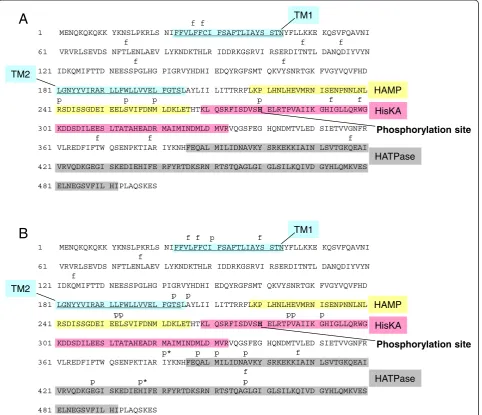
We investigated all previously reported covS mutations. The covS mutations were found most frequently in M1 isolates rather than any other serotypes [10,15,18,22, and our unpublished data]. The covS mutations detected in M1 strains were divided into two groups: (i) frameshift or nonsense mutations that caused a deletion in func-tional regions and (ii) point mutations that caused single (or double) amino acid(s) substitutions. Of the 34 covS mutations 25 were detected in isolates from mice in-fected with the M1 strain were frameshift mutations (Figure 1A and Additional file 1). In contrast, 16 of 29 covS mutations detected in a panel of clinical isolates comprised single (or double) amino acid(s) substitutions (Figure 1B and Additional file 2). Thus, significantly more frameshift mutations were detected in mouse-passaged isolates, whereas point mutations were most frequent in clinical isolates (P< 0.05, Fisher’s exact test).
Assessment of the function of CovS with an amino acid substitution using two-dimensional gel electrophoresis (2-DE)
Theoretically, it is possible that a single amino acid sub-stitution has no effect on CovS function, whereas a large deletion may affect domains that are critical for its
function. However, we previously observed that all four clinical isolates (strains GT01, K2, AP04, and AP06) with covS alleles comprising single amino acid substitutions had lower SpeB production than a clinical isolate having the wild-type covS when the culture supernatant pro-teins were analyzed by 2-DE [22]. It is known that covS positively regulatesspeBexpression [11,13,14], which sug-gests that mutated covS alleles degrade the function of CovS [22]. Thus, we were interested in how the functions of CovS proteins with single amino acid substitutions were degraded in clinical isolates. Thus, we deleted thecovSGT01 allele encoding CovSGT01A206S (a substitution of Ala206
with Ser) from the GT01 isolate. As shown in Figure 2, the resulting GT01ΔcovShad lower SpeB production than the parental GT01 isolate, suggesting that CovSGT01A206S
did not completely lose its function.
Evaluation of the function of CovS with an amino acid substitution based on its NADase activity
S. pyogenes secretes NAD+-glycohydrolase (NADase) as
one of its virulence factors. According to a previous study [10], mouse-passaged derivatives of a strain car-rying wild-type CovS exhibited high levels of NADase activity. A comparison of the entire genome of these strains also showed that one of the mouse-passaged de-rivatives had only one genetic change relative to the par-ental strain carrying the wild-type CovS, which consisted of a 7-bp insertion in covS. Therefore, we were inter-ested in the NADase activity of clinical isolates withcovS alleles containing single amino acid substitutions. Table 1 shows that strain 1529ΔcovShad a NADase activity level of 93.5 U, which was higher than the level of 3.4 U in parental isolate 1529 with wild-typecovSreported in our previous study [23]. In addition, the activity level of strain 1529ΔcovS(93.5 U) was higher than the levels of 62.9 U, 57.0 U, 59.8 U, 60.5 U, and 59.4 U found in the clinical isolates K2, GT01, AP04, AP06, and FI01, re-spectively, which had point-mutated covS alleles, in this study and a previous study [23], although isolate CR01 had a level of 114.3 U. The level of GT01ΔcovS was 105.0 U, which was higher than that of parental strain GT01 that carried CovSGT01A206S. Thus, the NADase
activity levels in isolates with point-mutated covSalleles were between those of isolates with wild-type covSand isolates with a complete deletion ofcovS. This was con-sistent with previous reports [22,23], where the levels of NADase in isolates KN01, MDYK, and MUY with wild typecovSwere 6.2 U, 3.0 U, and 3.0 U, respectively.
Next, we attempted to complement 1529ΔcovS with wild-typecovS1529or derivatives, which were cloned into plasmid vector pLZ12-Km2. Wild-type covS1529 from isolate 1529 was cloned into pLZ-covS1529 and it
1529ΔcovS(pLZ-covS1529). In contrast, the NADase
ac-tivity levels in pLZ-covS1529I30L, pLZ-covS1529E428G,
and pLZ-covS1529A206S encoding mutated covS alleles
from isolates K2, AP04 (or AP06), and GT01 were re-duced by 39.2 U, 17.1 U, and 15.2 U, respectively (Table 1 and Figure 2). Thus, the pLZ-covS1529I30L, the
pLZ-covS1529E428G, and the pLZ-covS1529A206S certainly
retained their abilities to reduce NADase activity, but the abilities were lower than that of the pLZ-covS1529
with wild-typecovS.
These results suggest that amino acid substitutions, such as I30L, E428G, and A206, partially impaired the function of CovS.
Benefits of partially impaired CovS
According to previous studies [10,16-18], CovS nega-tively regulates the expression of certain virulent genes; therefore, a mutation incovSmay increase the virulence in mouse models of infection where it plays a crucial role in the onset of severe invasive infections. However,
TM1
TM2
HisKA HAMP
HATPase
A
Phosphorylation site
TM1
TM2
HisKA HAMP
HATPase
B
Phosphorylation site
f f
1 MENQKQKQKK YKNSLPKRLS NIFFVLFFCI FSAFTLIAYS STNYFLLKKE KQSVFQAVNI f f f 61 VRVRLSEVDS NFTLENLAEV LYKNDKTHLR IDDRKGSRVI RSERDITNTL DANQDIYVYN
f f
121 IDKQMIFTTD NEESSPGLHG PIGRVYHDHI EDQYRGFSMT QKVYSNRTGK FVGYVQVFHD
181 LGNYYVIRAR LLFWLLVVEL FGTSLAYLII LITTRRFLKP LHNLHEVMRN ISENPNNLNL p p p p f f 241 RSDISSGDEI EELSVIFDNM LDKLETHTKL QSRFISDVSH ELRTPVAIIK GHIGLLQRWG
301 KDDSDILEES LTATAHEADR MAIMINDMLD MVRVQGSFEG HQNDMTVLED SIETVVGNFR f f f 361 VLREDFIFTW QSENPKTIAR IYKNHFEQAL MILIDNAVKY SRKEKKIAIN LSVTGKQEAI
421 VRVQDKGEGI SKEDIEHIFE RFYRTDKSRN RTSTQAGLGI GLSILKQIVD GYHLQMKVES
481 ELNEGSVFIL HIPLAQSKES
f f p f
1 MENQKQKQKK YKNSLPKRLS NIFFVLFFCI FSAFTLIAYS STNYFLLKKE KQSVFQAVNI f
61 VRVRLSEVDS NFTLENLAEV LYKNDKTHLR IDDRKGSRVI RSERDITNTL DANQDIYVYN f
121 IDKQMIFTTD NEESSPGLHG PIGRVYHDHI EDQYRGFSMT QKVYSNRTGK FVGYVQVFHD p p
181 LGNYYVIRAR LLFWLLVVEL FGTSLAYLII LITTRRFLKP LHNLHEVMRN ISENPNNLNL
pp pp p
241 RSDISSGDEI EELSVIFDNM LDKLETHTKL QSRFISDVSH ELRTPVAIIK GHIGLLQRWG
301 KDDSDILEES LTATAHEADR MAIMINDMLD MVRVQGSFEG HQNDMTVLED SIETVVGNFR p* p p p f
361 VLREDFIFTW QSENPKTIAR IYKNHFEQAL MILIDNAVKY SRKEKKIAIN LSVTGKQEAI f
p p* p
421 VRVQDKGEGI SKEDIEHIFE RFYRTDKSRN RTSTQAGLGI GLSILKQIVD GYHLQMKVES
[image:3.595.59.542.90.505.2]481 ELNEGSVFIL HIPLAQSKES
Table 1 NADase activity ofS. pyogenesstrains
Strain covS covR NADase (Ua) Reference
1529 wt wt 3.4 ± 0.7 [23]
1529ΔcovS ΔcovS wt 93.5 ± 3.5 this study
K2 I30L wt 62.9 ± 4.6 this study
GT01 A206S wt 57.0 ± 3.6 [23]
CR01 M391R wt 114.3 ± 8.7 [23]
AP04 E428G wt 59.8 ± 2.6 this study
AP06 E428G wt 60.5 ± 5.4 this study
FI01 I381T+H437R wt 59.4 ± 4.8 [23]
1529ΔcovR wt ΔcovR 106.7 ± 3.7 this study
GT01ΔcovS ΔcovS wt 105.0 ± 3.2 this study
GT01ΔcovR wt ΔcovR 103.5 ± 6.7 this study
1529ΔcovS(pLZ12-km2) ΔcovS wt 201.9 ± 2.8 this study
1529ΔcovS(pLZ-covS1529) wt wt 130.4 ± 3.4 this study
1529ΔcovS(pLZ-covS1529I381L) I381T wt 176.7 ± 8.9 this study
1529ΔcovS(pLZ-covS1529H437R) H437R wt 114.5 ± 6.8 this study
1529ΔcovS(pLZ-covS1529I30L) I30L wt 162.7 ± 11.0 this study
1529ΔcovS(pLZ-covS1529E428G) E428G wt 184.8 ± 6.9 this study
1529ΔcovS(pLZ-covS1529A206S) A206S wt 186.7 ± 4.2 this study
1529 (pLZ12-km2) wt wt 2.4 ± 0.16 this study
a
NADase activity (Units) ± standard error are indicated. One unit of NADase activity is defined as the amount (μg) ofβ-NAD cleaved per hour perμl culture supernatant, as described previously [23,31].
C (SF370ΔcovS)
D (1529ΔcovS)
pH3 pH10
175 62 47.5 32.5 83
25 16.5 kDa
B (GT01ΔcovS)
SpeB
A (GT01 wild)
[image:4.595.57.541.454.715.2]SpeF
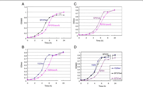
the loss of CovS function means thatS. pyogenescan no longer adjust to environmental fluctuations. For ex-ample, environmental Mg2+is thought to be recognized by the CovS sensor protein [24]. Therefore, the partial loss of CovS function may be favorable in nature, but not under laboratory conditions. This hypothesis led us to investigate the benefits of covSin S. pyogenes. Previ-ously, Trevino et al. [25] showed that a covS mutated strain had a lower growth ability than the parental wild-type strain in human saliva, but not in Todd Hewitt broth, which is the standard broth used to culture S. pyogenes. We were interested in the factor present in human saliva that is recognized by CovS; therefore, we repeated this experiment using the isolates 1529, SF370, and GT01. Bacteria were cultured under essen-tially the same conditions as those described previously [25]. However, the CFU (colony forming units)/ml for overnight THY broth cultures of strains 1529ΔcovS, SF370ΔcovS,and GT01ΔcovSwere lower than or similar to those of their parental strains; i.e., 1529, SF370, and GT01, respectively (Figure 3A), whereas the CFU/ml for overnight THY broth cultures of the isogenic mutant 2221covS::7 bp was four times that of the parental strain MGAS2221 reported in the previous study [25]. Thus, we observed two discrepancies: (i) between our results using isolates 1529, SF370, and GT01 (Figure 3A), and the previous results based on isolate MGAS2221 [25] and (ii) between our results with isolate 1529 (or GT01) and SF370. These discrepancies may be because of strain spec-ificities. We did not have strain MGAS2221, so we further investigated the discrepancy between strains SF370 and GT01 or 1529. First, we analyzed the growth curves of the covS mutated strains. SF370ΔcovS, 1529ΔcovS, and GT01ΔcovS all showed delayed growth compared with
that of their parental strains; i.e., SF370, 1529, and GT01, respectively (Figures 4A–C). Thus, there was no disc-repancy between strains SF370, GT01, and 1529 in terms of their growth kinetics. In addition, GT01 exhibited de-layed and advanced growth compared with strain 1529 (or SF370) and strain GT01ΔcovS (Figures 4D and C), which was consistent with our hypothesis that the A206S substitutions partially impaired the function of CovS.
The growth abilities of SF370 and its isogenic covS mutant SF370ΔcovS in THY broth differed from each other when evaluated on the basis of, but not the CFU/ml in overnight cultures (Figure 3A), the growth curves (Figure 4A). This new discrepancy may have occurred be-cause the overnight culture, but not the growth curve, was conducted in 5% CO2, which was the condition described
in a previous study [25]. Therefore, we prepared overnight cultures of wild-type SF370 (SF370wt) and SF370ΔcovS in natural atmosphere (NA) conditions. As shown in Figure 3B, the CFU/ml for SF370ΔcovS was lower than that of its parental strain SF370. Finally, we performed supplementary and supporting experiments to test the reliability of this study. covS and covRS cloned into a plasmid vector complemented the delayed growth of 1529ΔcovS (Figure 5). pLZ-covS1529 and pLZ-covRS1529
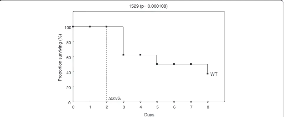
increased the CFU/ml for overnight THY broth cultures of strain 1529ΔcovS (Figure 6). As shown in Figure 7, 1529ΔcovS was hypervirulent in a mouse infection model compared with the parental strain 1529 (P<0.01), as shown with other strains and their isogenic ΔcovS mutants in previous studies [10,16].
Conclusions
On the basis of our results and those from previous studies we concluded that the loss of covSincreases the
CFU (x
10
7)
0 5 10 15 20 25 30 35 40 45 50
2
[image:5.595.57.541.509.665.2]A5% CO BNA
virulence ofS. pyogenes (which is advantageousin vivo). However, the loss ofcovSalso impaired the growth abil-ity of this organism in THY broth (which is disadvanta-geousin vitro). Therefore, the CovRS system may confer benefits in stages when virulent gene expression is not required. The expression of many genes is precisely reg-ulated so they are expressed only when required; e.g.,
catabolite repression. Therefore, partial attenuation of the CovRS system to promote resistance to the host defense system appears to be a wise choice for survival in nature.
We did not determine the components recognized by the CovS sensor proteins in our experimental conditions; i.e., THY broth, natural atmosphere, or 5% CO2.
How-ever, it was probably not the Mg2+ion, which was sug-gested previously [24], because we did not add Mg2+ion to THY broth. Therefore, we propose that CovS can sense other signals in addition to the Mg2+ion.
Methods
Bacterial strains
Streptococcal strains were isolated as the causative organisms in patients from Japan [22,23]. S. pyogenes (GAS) strain SF370, which was the most prevalent data-base reference isolate (accession number NC_002737), was provided by J. J. Ferretti [26,27]. Streptococcal strains were cultured in brain–heart infusion (E-MC62, EIKEN Chemical Co., Tokyo, Japan) supplemented with 0.3% yeast extract (BD, Sparks, MD, USA), (BHI-Y) broth or Todd Hewitt broth (BD, Sparks, MD, USA) supplemen-ted with 0.2% yeast extract broth (THY) unless other-wise stated.
0 0.2 0.4 0.6 0.8 1 1.2
0 2 4 6 8 24
SF370wt
SF370ΔcovS
Time (h)
OD
660
0 0.1 0.2 0.3 0.4 0.5 0.6 0.7 0.8 0.9 1
0 2 4 6 8 24
1529wt
1529ΔcovS
Time (h)
OD
660
0 0.1 0.2 0.3 0.4 0.5 0.6 0.7 0.8 0.9
0 2 4 6 8 24
GT01wt
GT01ΔcovS
0 0.1 0.2 0.3 0.4 0.5 0.6 0.7 0.8 0.9 1
0 2 4 6 8 24
Time (h)
OD660
GT01
1529
SF370
1529wt
SF370wt
GT01wt
A
B
C
D
OD660
[image:6.595.59.538.89.381.2]Time (h)
Figure 4Growth curves ofcovSnull mutants in THY broth.Three independent experiments produced essentially the same results. Representative data from three independent experiments are shown. (A-C) SF370ΔcovS, 1529ΔcovS, and GT01ΔcovSall exhibited delayed growth compared with that of their parental strains SF370, 1529, and GT01, respectively. (D) GT01 exhibited delayed growth compared with strains 1529 and SF370.
0 0.2 0.4 0.6 0.8 1 1.2
0 2 4 6 8 23
Time (h)
OD
660
1529ΔcovS(vector) 1529ΔcovS(pLZ-covRS1529)
1529Δcov (pLZ-covS1529)
[image:6.595.55.292.545.683.2]Production ofcovSknockout strains
We constructedS. pyogenesstrain 1529ΔcovSas described previously [28]. Strains GT01ΔcovSand SF370ΔcovSwere constructed using the same strategy [28].
Two-dimensional gel electrophoresis (2-DE)
Each bacterial isolate was cultured in BHI-Y at 37°C overnight without agitation. Exoproteins from the cul-ture supernatant were prepared as described previously [22]. In brief, all sample pellets derived from bacterial culture supernatant were dissolved in dehydration solu-tion, which consisted of 7.8 M urea, 2 M thiourea, 2% CHAPS, 0.6% dithiothreitol, and 0.5% IPG buffer. The samples were loaded onto 13 cm Immobiline DryStrip gels (pH 3–10, GE Healthcare Biosciences Co. Piscataway, NJ, USA). The first-dimensional electrophoresis conditions
were carried out according to the manufacture’s in-struction. Second-dimensional SDS-PAGE separation was performed as described previously [22]. The expe-riments were repeated at least 3 times to confirm their reproducibility.
Production ofcovRknockout strains
To construct the plasmid for thecovRknockout mutant, the 50 end ofcovR (fragment 1) was amplified using the oligonucleotide primer covR-n6 (50-GGCTAGCCTTT
AGAGAATATGGTTACT-30) with an NheI restriction
site and primer covR-c2 (50-TCCCCCGGGCTTTGTCA
TTTATACCAACC-30) with anSmaI restriction site, while the 30 end of covR (fragment 2) was amplified using
the primer covR-n7 (50-TCCCCCGGGGAGAAATAAGTC
ATATGGAA-30) with an SmaI restriction site and primer
CFU
(x
10
7)
0 5 10 15 20 25 30 35 40 45
CFU
(x
10
7) 5% CO2
0 5 10 15 20 25 30
[image:7.595.59.539.89.264.2]NA
Figure 6Growth ofcovSmutantS. pyogenesin THY broth with 5% CO2or NA.These experiments were performed as described in
Figures 3. The error bars indicate the standard errors of the means.
0 20 40 60 80 100
0 1 2 3 4 5 6 7 8
Proportion
surv
iv
ing
(%)
Days 1529 (p= 0.000108)
WT
ΔcovS
[image:7.595.58.540.506.704.2]covS-c10 (50-GGACTAGTATGTAAAATTAGAGTCCAC C-30) with an SpeI restriction site. Fragment 2 was di-gested with SmaI and SpeI before its insertion into multicloning site 2 in the plasmid pFW12 [29]. The resulting plasmid was digested usingNheI andSmaI, and the spc1 DNA fragment containing aad9 (promoterless spectinomycin resistance gene), which was obtained from aSmaI-digested fragment of pSL60-1 [29], and theNhe I-SmaI-digested fragment 1 were inserted. This plasmid, covR::aad9/pFW12, was a suicide vector for S. pyogenes. To prepare competent cells, strains 1529 and GT01 were harvested in the early to mid-log phase (OD660, 0.4) and
washed twice with 0.5 M sucrose buffer. The suicide vec-tor construct, covR::aad9/pFW12, was transformed into strains 1529 and GT01 via electroporation. The conditions for electroporation were 1.25 kV/mm, 25-μF capacitance, and 200-Ω resistance, and it was performed using a GenePulser II instrument (Bio-Rad, Hercules, CA). After incubation at 37°C for 3 h, competent cells were spread onto BHI agar plates containing 0.3% yeast extract and spectinomycin (final concentration, 100 μg/ml). Selected colonies were cultured from the plates. The cultured bac-teria were washed once with saline, resuspended in 10 mM Tris-1 mM EDTA, and boiled for 10 min. Gen-omic DNA was obtained from the supernatant of the boiled bacteria. The double-crossover replacement was analyzed by PCR using genomic DNA. Successful double-crossover replacement was further confirmed by DNA sequencing.
Quantification of the NADase activity in the bacterial supernatant
NADase activity was determined using the method of Stevenset al. [30] as described previously [31].
Plasmids
pLZ-covS1529, pLZ-covS1529I30L, and pLZ-covS1529E428G
were constructed as described previously [22]. To
con-struct pLZ-covS1529A206S, the DNA fragment was
amplified using the oligonucleotide primers covR-n2 (50-CTTTAGAGAATATGGTTACT-30), covS-c2 (50-GT
AATTACATTTTGGACAAC-30), and GT01 genomic
DNA as templates with TaKaRa Ex Taq DNA
poly-merase (Takara, Ohtsu, Japan). The fragment consisted of covRGT01, covSGT01, and their 50-noncoding region, which possibly contained the promoter region. This fragment was cloned into the pGEM-T vector (Promega, Madison, WI, USA). The resultant plasmid was digested withEcoRI and ligated into the same site in the pLZ12-Km2 plasmid [32] (pLZ-covRSGT01). To construct a plasmid containing
only the covSGT01 region, inverse PCR was conducted
using two primers, covR-c2Sma (50-TCCCCCGGGC
TTTGTCATTTATACCAACC-30) and covR-n7Sma (50
-TCCCCCGGGGAGAAATAAGTCATATGGAA-30), with
pLZ-covRSGT01 plasmid DNA as template and
Prime-STAR HS DNA polymerase (Takara) to eliminate thecovR region. This blunt-ended PCR product was treated with T4 polynucleotide kinase (Takara) and self-ligated. The re-sultant plasmid was pLZ-covS1529A206S. pLZ-covRS1529
encoding the covRS1529 operon of isolate 1529 was constructed as described previously [22]. All of thecovRS DNA sequences were confirmed by sequencing.
Mouse model of invasive skin tissue infection
All animal studies conducted comply with federal and institutional (the Committee on the Ethics of Animal Experiments of the Nagoya City University) guidelines. The protocol was approved by the Committee on the Ethics of Animal Experiments of the Nagoya City Uni-versity (Permit Number: H23M-07). All efforts were made to minimize suffering.
The ability of S. pyogenes to cause local skin lesions and necrosis in mice after skin inoculation was assessed using a similar procedure to that described previously [23,33]. Three-week-old female ICR mice (10–12 g) were anesthetized with sevoflurane and the skin of the left flank was laid bare by separating the hair with an alcohol swab, unless indicated otherwise. Bacteria (0.2 ml; 2 × 10⁷CFU/mouse) grown in BHI-Y were injected immedi-ately beneath the surface of the skin using a 27-gauge needle so a superficial bleb appeared below the skin sur-face. The number of CFU injected was verified in each experiment by plating bacteria on BHI-Y or sheep blood agar plates and counting the CFU.
Statistical analysis
The survival times were assessed using a log-rank com-parison. The R program was used for the statistical ana-lysis http://bioinf.wehi.edu.au/software/russell/logrank/ webcite.P≤0.05 was considered significant.
Availability of supporting data There are two supplementary tables.
Additional files
Additional file 1: Table S1.csrSmutations from mouse-passaged isolates of M1S. pyogenes.
Additional file 2: Table S2.csrSmutations from human clinical isolates of M1S. pyogenes.
Abbreviations
NADase:NAD+-glycohydrolase; NA: Natural atmosphere; THY: Todd Hewitt
yeast.
Competing interests
Authors’contributions
IT conceived the study. IT, RO, and TH designed and performed the experimental work with help by YZ and MI. All authors contributed to the data analysis. IT wrote the original manuscript. TH helped to produce the final manuscript. All authors approved the final manuscript.
Acknowledgments
We thank Hideyuki Matsui for technical assistance, and Drs. M. Ohnishi, M. Ato, T. Ikebe for their helpful advices. This study was supported by JSPS KAKENHI Grant number 21790425 and 24590531, a grant from Ohyama Health Foundation, and a grant from the 24thGeneral Assembly of the
Japanese Association of Medical Sciences (Medical Science Promotion Fund). The authors would like to thank Enago (www.enago.jp) for the English language review.
Received: 7 January 2013 Accepted: 20 March 2013 Published: 28 March 2013
References
1. Cone LA, Woodard DR, Schlievert PM, Tomory GS:Clinical and bacteriologic observations of a toxic shock-like syndrome due to
Streptococcus pyogenes.N Engl J Med1987,317:146–149.
2. Hoge CW, Schwartz B, Talkington DF, Breiman RF, MacNeill EM, Englender SJ:The changing epidemiology of invasive group A streptococcal infections and the emergence of streptococcal toxic shock-like syndrome. A retrospective population-based study.JAMA1993,
269:384–389.
3. Schwartz B, Facklam RR, Breiman RF:Changing epidemiology of group A streptococcal infection in the USA.Lancet1990,336:1167–1171. 4. Stevens DL:Invasive group A streptococcal infections: the past, present
and future.Pediatr Infect Dis J1994,13:561–566.
5. Hasegawa T, Hashikawa SN, Nakamura T, Torii K, Ohta M:Factors determining prognosis in streptococcal toxic shock-like syndrome: results of a nationwide investigation in Japan.Microbes Infect2004,
6:1073–1077.
6. Agarwal S, Agarwal S, Pancholi P, Pancholi V:Role of serine/threonine phosphatase (SP-STP) inStreptococcus pyogenesphysiology and virulence.J Biol Chem2011,286:41368–41380.
7. Musser JM, Shelburne SA 3rd:A decade of molecular pathogenomic analysis of group A Streptococcus.J Clin Invest2009,119:2455–2463. 8. Kreikemeyer B, McIver KS, Podbielski A:Virulence factor regulation and
regulatory networks inStreptococcus pyogenesand their impact on pathogen-host interactions.Trends Microbiol2003,11:224–232. 9. Stock AM, Robinson VL, Goudreau PN:Two-component signal
transduction.Annu Rev Biochem2000,69:183–215.
10. Sumby P, Whitney AR, Graviss EA, DeLeo FR, Musser JM:Genome-wide analysis of group A streptococci reveals a mutation that modulates global phenotype and disease specificity.PLoS Pathog2006,2:e5. 11. Levin JC, Wessels MR:Identification ofcsrR/csrS, a genetic locus that
regulates hyaluronic acid capsule synthesis in group A Streptococcus. Mol Microbiol1998,30:209–219.
12. Bernish B, van de Rijn I:Characterization of a two-component system in
Streptococcus pyogeneswhich is involved in regulation of hyaluronic acid production.J Biol Chem1999,274:4786–4793.
13. Federle MJ, McIver KS, Scott JR:A response regulator that represses transcription of several virulence operons in the group A streptococcus. J Bacteriol1999,181:3649–3657.
14. Heath A, DiRita VJ, Barg NL, Engleberg NC:A two-component regulatory system, CsrR-CsrS, represses expression of threeStreptococcus pyogenes virulence factors, hyaluronic acid capsule, streptolysin S, and pyrogenic exotoxin B.Infect Immun1999,67:5298–5305.
15. Engleberg NC, Heath A, Miller A, Rivera C, DiRita VJ:Spontaneous mutations in the CsrRS two-component regulatory system of
Streptococcus pyogenesresult in enhanced virulence in a murine model of skin and soft tissue infection.J Infect Dis2001,183:1043–1054. 16. Walker MJ, Hollands A, Sanderson-Smith ML, Cole JN, Kirk JK, Henningham
A, McArthur JD, Dinkla K, Aziz RK, Kansal RG, Simpson AJ, Buchanan JT, Chhatwal GS, Kotb M, Nizet V:DNase Sda1 provides selection pressure for a switch to invasive group A streptococcal infection.Nat Med2007,
13:981–985.
17. Ato M, Ikebe T, Kawabata H, Takemori T, Watanabe H:Incompetence of neutrophils to invasive group A streptococcus is attributed to induction of plural virulence factors by dysfunction of a regulator.PLoS One2008,
3:e3455.
18. Ikebe T, Ato M, Matsumura T, Hasegawa H, Sata T, Kobayashi K, Watanabe H:
Highly frequent mutations in negative regulators of multiple virulence genes in group A streptococcal toxic shock syndrome isolates.PLoS Pathog2010,6:e1000832.
19. Rogers S, Commons R, Danchin MH, Selvaraj G, Kelpie L, Curtis N, Robins-Browne R, Carapetis JR:Strain prevalence, rather than innate virulence potential, is the major factor responsible for an increase in serious group A streptococcus infections.J Infect Dis2007,195:1625–1633.
20. Aziz RK, Pabst MJ, Jeng A, Kansal R, Low DE, Nizet V, Kotb M:Invasive M1T1 group A Streptococcus undergoes a phase-shift in vivo to prevent proteolytic degradation of multiple virulence factors by SpeB. Mol Microbiol2004,51:123–134.
21. Aziz RK, Kotb M:Rise and persistence of global M1T1 clone of
Streptococcus pyogenes.Emerg Infect Dis2008,14:1511–1517.
22. Hasegawa T, Okamoto A, Kamimura T, Tatsuno I, Hashikawa SN, Yabutani M, Matsumoto M, Yamada K, Isaka M, Minami M, Ohta M:Detection of invasive protein profile ofStreptococcus pyogenesM1 isolates from pharyngitis patients.APMIS2010,118:167–178.
23. Tatsuno I, Isaka M, Minami M, Hasegawa T:NADase as a target molecule of
in vivosuppression of the toxicity in the invasive M-1 group A Streptococcal isolates.BMC Microbiol2010,10:144.
24. Gryllos I, Grifantini R, Colaprico A, Jiang S, Deforce E, Hakansson A, Telford JL, Grandi G, Wessels MR:Mg(2+)signalling defines the group A
streptococcal CsrRS (CovRS) regulon.Mol Microbiol2007,65:671–683. 25. Trevino J, Perez N, Ramirez-Pena E, Liu Z, Shelburne SA 3rd, Musser JM,
Sumby P:CovS simultaneously activates and inhibits the CovR-mediated repression of distinct subsets of group A Streptococcus virulence factor-encoding genes.Infect Immun2009,77:3141–3149.
26. Ferretti JJ, McShan WM, Ajdic D, Savic DJ, Savic G, Lyon K, Primeaux C, Sezate S, Suvorov AN, Kenton S, Lai HS, Lin SP, Qian Y, Jia HG, Najar FZ, Ren Q, Zhu H, Song L, White J, Yuan X, Clifton SW, Roe BA, McLaughlin R:
Complete genome sequence of an M1 strain ofStreptococcus pyogenes. Proc Natl Acad Sci U S A2001,98:4658–4663.
27. Suvorov AN, Ferretti JJ:Physical and genetic chromosomal map of an M type 1 strain ofStreptococcus pyogenes.J Bacteriol1996,178:5546–5549. 28. Sawai J, Hasegawa T, Kamimura T, Okamoto A, Ohmori D, Nosaka N,
Yamada K, Torii K, Ohta M:Growth phase-dependent effect of clindamycin on production of exoproteins byStreptococcus pyogenes. Antimicrob Agents Chemother2007,51:461–467.
29. Lukomski S, Hoe NP, Abdi I, Rurangirwa J, Kordari P, Liu M, Dou SJ, Adams GG, Musser JM:Nonpolar inactivation of the hypervariable streptococcal inhibitor of complement gene (sic) in serotype M1Streptococcus
pyogenessignificantly decreases mouse mucosal colonization.Infect Immun2000,68:535–542.
30. Stevens DL, Salmi DB, McIndoo ER, Bryant AE:Molecular epidemiology of
ngaand NAD glycohydrolase/ADP-ribosyltransferase activity among
Streptococcus pyogenescausing streptococcal toxic shock syndrome. J Infect Dis2000,182:1117–1128.
31. Tatsuno I, Sawai J, Okamoto A, Matsumoto M, Minami M, Isaka M, Ohta M, Hasegawa T:Characterization of the NAD-glycohydrolase in streptococcal strains.Microbiology2007,153:4253–4260.
32. Okada N, Tatsuno I, Hanski E, Caparon M, Sasakawa C:Streptococcus pyogenesprotein F promotes invasion of HeLa cells.Microbiology1998,
144:3079–3086.
33. Ashbaugh CD, Warren HB, Carey VJ, Wessels MR:Molecular analysis of the role of the group A streptococcal cysteine protease, hyaluronic acid capsule, and M protein in a murine model of human invasive soft-tissue infection.J Clin Invest1998,102:550–560.
doi:10.1186/1756-0500-6-126