inactivation model for hepatitis A virus and
its application in shellfish processing
by
Stephen Grove, B. App. Sci (Hons).
Submitted in fulfilment of the
Requirements for the Degree of
i
Doctor of Philosophy
School of Agricultural Science
University of Tasmania
I, Stephen Francis Grove, declare this thesis contains no material which has been accepted for a degree or diploma by the University of Tasmania or any other institution, except by way of background information and which is duly acknowledged in the thesis, and to the best of my knowledge and belief the thesis contains no material previously published or written by another person, except where due acknowledgement is made in the thesis, nor does the thesis contain any material that infringes copyright.
Signature: ___
Stephen Francis Grove
This thesis may be made available for loan and limited copying in accordance with the Copyright Act 1968.
Signature:
Stephen Francis Grove
1. Grove, S.F., A. Lee, T. Lewis, C.M. Stewart, H. Chen, and D.L. Hoover.
2006. Inactivation of foodborne viruses of significance by high pressure and
other processes. J. Food Prot. 69:957-968.
2. Grove, S.F., S. Forsyth, J. Wan, J. Coventry, M. Cole, C.M. Stewart, T.
Lewis, T. Ross, and A. Lee.
Inactivation of foodbome viruses by high pressureThe following people contributed to the publication of work undertaken as part of
this thesis:
1. Grove etal., 2006.
Candidate: 30%, A. Lee: 10%, T. Lewis: 10%, C.M. Stewart: 10%, H.
Chen: 10%, D.L. Hoover: 30%
• The candidate contributed by preparing the original review and editing
drafts prior to submission. A. Lee, T. Lewis, C.M. Stewart and H. Chen
assisted with manuscript preparation and editing, and D.L Hoover
formulated the idea, combined contributions and conducted final editing.
2. Grove et al., in press.
Candidate: 20%, S. Forsyth: 6%, J. Wan: 6%, J. Coventry: 8%, M. Cole:
6%, C.M. Stewart: 8%, T. Lewis: 8%, T. Ross: 8%, and A. Lee: 30%
• The contributions by the candidate and A. Lee included the laboratory
investigations, manuscript writing and final editing. J. Wan, J. Coventry
and M. Cole contributed to the project idea and with data analysis, C.M.
Stewart, T. Lewis, and T. Ross contributed to experimental plan, data
analysis and manuscript editing, and S. Forsyth contributed laboratory
assistance.
We the undersigned agree with the above stated "proportion of work undertaken"
for the above published peer-reviewed manuscript contributing to this thesis:
Signature:
Tom Ross, Supervisor
University of Tasmania Date:
Signature:
Tom McMeekin, Co-Supervisor
University of Tasmania Date:
Signature:
Peter Lane, Head of School
Af Accuracy factor B1 Bias factor bp Base pair
BSA Bovine serum albumin Ct Cycle threshold FCV Feline calicivirus HAV Hepatitis A virus
HPP High pressure processing IMS Inununo-magnetic separation
LogS Survival ratio (logarithm of the remaining infectious fraction of HAV at a particular treatment time)
MNV-1 Murine norovirus MPa Megapascal
NBS Newborn bovine serum PBS Phosphate-buffered saline PFU Plaque-forming units ppt Parts per thousand PV Poliovirus
qRT-PCR Quantitative reverse transcription-polymerase chain reaction R2 Coefficient of determination
RFU Relative fluorescence units SS Sum of squared residuals
TCID50 50% tissue culture infective dose TM Melting temperature
Hepatitis A virus (HAV) has been responsible for many large outbreaks of illness
throughout the world, often resulting from consuming raw or minimally cooked
filter-feeding shellfish contaminated with human faecal effluent.
High pressure processing (HPP) is an alternative food preservation technique to
heat, preserving the flavour, appearance and nutritional value of high quality
foods, including oysters, often with extended shelf life. In this study, the
effectiveness of HPP in inactivating HAV was assessed.
HAV, suspended in buffered tissue culture media containing either 15 parts per
thousand (ppt) or 30 ppt salt (NaCl), was treated with 300, 400 and 500 MPa for
between 60 and 600 s. A log-linear function was developed in Microsoft® Excel
to model the kinetic inactivation data with the effects of NaC1, pressure and
treatment time. The model can be used to predict HAV inactivation by
interpolation at processing parameters not actually tested for in the laboratory.
For the model to be validated in oysters contaminated with HAV, methods for
HAV extraction and purification from spiked oyster homogenate were first
evaluated. Methods evaluated included the crude extraction method, modified
from Kingsley and Richards (2003), and the PEG precipitation method, modified
from the glycine-polyethylene glycol (PEG)-Tri reagent-poly(dT) extraction
(GPTT) method described by Kingsley and Richards (2001). The PEG
was improved by up to 27.3%. In comparison, the crude extraction technique,
which did not include a virion concentration step, recovered on average more than
40% of the initial spiked titre, and was chosen as a reliable method to extract
HAV from oysters for cell culture quantitation.
Commercially grown and harvested Pacific oysters (Crassostrea gigas) were
contaminated with HAV by natural accumulation, when immersed for up to 24 h
in seawater contaminated with 1.1x10 7 TCID50/m1 HAV. Infectious HAV was
detected in only two of the six oysters tested, and less than 1% of the initial
contaminating HAV titre was recovered in positive oysters, possibly due to the
association of viruses and microalgae with oyster shells and aquarium surfaces
throughout the trials.
A quantitative real-time reverse transcription PCR (qRT-PCR) method was
developed as an alternative method for HAV detection in contaminated oysters to
the infectivity assay. HAV was detected by qRT-PCR in all contaminated
oysters, including those negative by infectivity assay. An immuno-magnetic bead
separation technique was also developed, which additionally purified and
concentrated virions, improving the sensitivity of detection by qRT-PCR.
The log-linear inactivation model was validated in homogenised oyster meat
artificially inoculated with known titres of HAV. Salinity and temperature of
samples were adjusted to that of buffered samples, while intermediate times and
tended to be greater in spiked oyster homogenate compared to pure culture in
treatments at higher pressures (400 — 500 MPa). The validated model may be a
useful reference for Australian oyster processors wishing to implement HPP into
their post-harvest processing regime.
My sincere gratitude goes to my supervisory team of Assoc Prof Tom Ross, Dr
Alvin Lee, Dr Cynthia M. Stewart, Dr Tom Lewis and Prof Tom McMeekin for
your guidance, patience and hard work across the time zones.
Tom Ross created the log-linear mathematical model described in Chapter 6, and
assisted in model analysis; I am indebted for the invaluable support provided. Mr
Stephen White and Dr Michelle Bull co-ordinated and performed all high pressure
processing experiments described in the thesis, often on short notice. I sincerely
appreciate the long hours spent processing. Alvin Lee imparted much technical
knowledge and advice throughout my candidature, and spent several stressful
hours printing and binding this thesis on my behalf, for which I am very grateful.
Thank-you to Food Science Australia and its staff for providing the facilities and
training required during my candidature, enabling me to conduct this research
off-campus; to Avure Technologies, Inc. for project and scholarship funding; to the
University of Tasmania for scholarship funding; and to the Australian Food
Safety Centre of Excellence for providing project funding and training.
To my family, thank-you for your support, and for having me back home during
my candidature; it made life so much easier. And to my wise and
Chapter 1: Literature review
1.1
1.1 INTRODUCTION 1.1
1.2 VIRAL ENTERITIS 1.2
1.3 HEPATITIS A VIRUS 1.4
1.3.1 Difficulties of laboratory study
1.6
1.3.2 Illness and transmission of infection
1.7
1.3.3 Outbreaks of interest
1.9
1.4 BIVALVE MOLLUSCAN SHELLFISH 1.10
1.4.1 Procedures employed to improve shellfish safety 1.11
1.4.1.1 Purification methods 1.11
1.5 INACTIVATION OF HUMAN ENTERIC VIRUSES 1.13
1.6 NONTHERMAL PRESERVATIVE PROCESSES 1.14
1.6.1 High pressure processing (HPP)
1.15
1.6.1.1 Pressure effects on microorganisms 1.18
1.6.1.2 Oyster processing 1.19
1.6.1.3 Pressure effects on viruses 1.20
1.7 PREDICTIVE MICROBIOLOGY 1.24
1.7.1 Model development
1.24
1.7.2 Model validation
1.26
2.1 GENERAL PROCEDURES 2.1
2.2 PREPARATION OF SOLUTIONS 2.2
2.3 GENERAL TISSUE CELL CULTURE METHODS 2.6
2.3.1 Maintenance of tissue culture cell lines 2.6
2.3.2 Storage of cell lines 2.6
2.3.3 Quantification of infectious hepatitis a virus 2.7
Chapter 3: Evaluation and comparison of methods for
high-throughput cultivation of hepatitis A virus
3.1
3.1 INTRODUCTION 3.1
3.2 METHODS 3.3
3.2.1 Tissue culture cells and hepatitis A virus 3.3
3.2.2 Hepatitis A virus cultivation 3.3
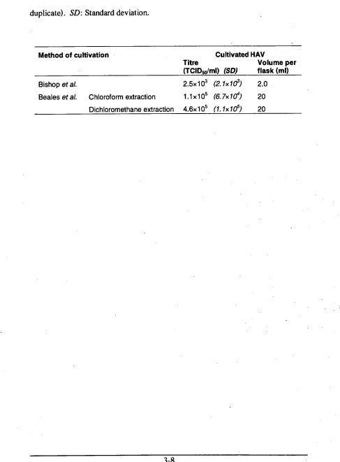
3.3 RESULTS 3.5
3.4 DISCUSSION 3.12
Chapter 4: Development of a method for extraction and
purification of hepatitis A virus from contaminated oysters 4.1
4.1 INTRODUCTION 4.1
4.2 METHODS 4.4
4.2.1 Tissue culture cells and hepatitis A virus 4.4
4.2.2 PEG precipitation method 4.4
4.3 RESULTS 4.6
4.3.1 Original PEG precipitation method 4.6
4.3.2 Further modifications 4.8
4.3.3 Crude extraction 4.13
4.4 DISCUSSION 4.15
Chapter 5: Accumulation of hepatitis A virus by oysters in a
laboratory aquarium
5.1
5.1 INTRODUCTION 5.1
5.2 METHOD 5.3
5.2.1 Aquarium, seawater and oysters 5.3
5.2.2 First trial 5.4
5.2.3 Second trial 5.5
5.2.4 Crude virus extraction method 5.6
5.2.5 RNA extraction 5.7
5.2.6 Quantitative RT-PCR 5.7
5.2.7 Standard curve 5.8
5.3 RESULTS 5.9
5.3.1 Quantitative RT-PCR 5.11
5.3.1.1 Standard curve 5.11
5.3.2 First trial 5.11
5.3.3 Second trial 5.15
inactivation model for hepatitis A virus
6.1
6.1 INTRODUCTION 6.1
6.2 METHODS 6.4
6.2.1 Sample preparation — Buffered samples
6.4
6.2.2 Sample preparation — Spiked oyster homogenate
6.5
6.2.3 High pressure treatment
6.6
6.2.4 HAV quantitative analysis
6.7
6.2.5 Model development
6.8
6.2.6 Development of a Secondary Model
6.9
6.2.7 Tests for model fit
6.12
6.3 RESULTS
6.14
6.3.1 Buffered samples
6.14
6.3.2 Fit and comparison of predictive models
6.16
6.3.3 Model validation in spiked oyster homogenate
6.24
6.4 DISCUSSION
6.30
Chapter 7: Rapid and Quantitative detection of hepatitis A virus
by quantitative real-time reverse-transcription PCR 7.1
7.1 INTRODUCTION 7.1
7.2 METHODS 7.3
7.2.1 Quantitative RT-PCR
7.3
7.2.2 HAV dilution series and standard curve
7.5
7.2.3 RNA extraction
7.5
7.2.6 Qiagen method 7.7
7.3 RESULTS 7.8
7.3.1 Primer optimisation 7.8
7.3.2 Standard curve 7.10
7.3.3 Detection and quantification of hepatitis A virus in spiked oyster
homogenate 7.17
7.4 DISCUSSION 7.24
Final Discussion
D-1
References
R-1
Appendix
A-1
Chapter 1: Literature review.
1.1 INTRODUCTION
All foodborne viruses that are detrimental to human health emanate from the
human intestinal tract (Blackwell
et al.,
1985). Faecal-oral transmission canoccur by indirect routes or direct personal contact. Those foods and beverages
susceptible to faecal contamination and lacking an intervention step such as
cooking prior to consumption usually carry a greater risk of causing viral illness.
Foodborne viruses are considered to be responsible for the majority of foodborne
illness in the United States of America (U.S.A.) by a wide margin (Mead
et al.,
1999), but viruses are often the least or last studied in process development, and
are not routinely tested in food and environmental samples due to technical and
cost issues associated with their extraction, observation and culture.
Viruses differ greatly from the bacterial agents of foodborne disease. They have
been described as extracellular organelles evolved to transfer nucleic acid from
one cell to another (Harrison
etal.,
1996). Viruses have no cellular structure andcontain either RNA or DNA enclosed in a protein coat or capsid (Madigan
et al.,
2000). The capsid functions as the primary protective barrier for the viral particle
or virion. The capsid of some viruses is additionally enveloped in an outer lipid
membrane. All human enteric viruses are non-enveloped, as enveloped viruses
tend to be susceptible to adverse environmental conditions and are generally
The diameters of virus particles range between 25 to 300 nm, so most cannot be
visualized with a light microscope. Furthermore, viruses are obligate intracellular
parasites, meaning they can only replicate in a suitable living host cell, and not in
the environment (Cliver and Matsui, 2002). However, the usually low infectious
dose of enteric viruses (believed to be far less than 100 virions), means that even
a small amount of contamination in food may result in a significant threat to
public health (Jaykus, 2000; Cliver and Matsui, 2002). This
is
coupled with ahigh level of persistence in foods and the environment, with select enteric viruses
being able to withstand conditions such as high acidity (Scholz
et al.,
1989), freezing temperatures and reduced water activity without loss of infectivity forextended periods of time, as reviewed by Koopmans and Duizer (2004).
1.2 VIRAL ENTERITIS
Over the last several years there has been a growing awareness of the significant
role viruses play in foodborne disease. While it is uncertain how many different
viral diseases have been or can be spread by contaminated foods and beverages,
the number of different viruses acknowledged as primary agents of foodborne
illness is actually quite low. Noroviruses are now recognized as the most
common cause of all foodborne disease in the U.S.A., estimated to be responsible
for 23 million cases annually (Mead
et al.,
1999). Approximately 40% or 9.2 million cases per year are estimated to be foodborne, and this number correspondsto 67% of all cases, 33% of hospitalizations and 7% of deaths due to foodborne
The incidence of hepatitis A in Australia and the U.S. is declining, but outbreaks
continue to occur despite an increase in sanitation and hygiene standards
(Grohmann and Lee, 2003). Due to the serious nature of the disease it can cause,
hepatitis A virus (HAV) is usually ranked second on the list of important
foodborne viruses, and is described as the only common vaccine-preventable
foodborne disease in the U.S. (Fiore, 2004).
Human enteric viruses are transmitted by the faecal-oral route via contamination
with human faecal matter. Foodborne transmission of human enteric viruses most
frequently occurs in those foods requiring little or no intervention (e.g. heat
processing) prior to consumption, and/or ready-to-eat foods that are prepared by a
food handler immediately before consumption. As their name suggests,
'filter-feeding' shellfish, such as oysters feed by filtering small particles such as algae
from the surrounding water. Oysters in particular can accumulate
microorganisms to concentrations higher than that in the surrounding water.
Fresh produce may also transmit enteric viruses following irrigation with
contaminated water, or food may be contaminated by infected food handlers with
poor personal hygiene. These foods may benefit from more stringent farming
practices and improved education for food handlers, as well as an intervention
1.3 HEPATITIS A VIRUS
Hepatitis A virus (HAV) belongs in the genus Hepatovirus, within the family
Picornaviridae. Seven distinct genotypes of HAV have been described (I to VII),
with viruses from four genotypes (I, II, III and VII) isolated from humans, and
viruses from the remaining three genotypes (IV, V and VI) classified as simian
strains (Lemon et al., 1992; Robertson et al., 1992).
The HAV genome, of single-stranded, positive-sense (i.e., translatable) RNA, is
approximately 7.5 kb in length, and like all picornaviral genomes, consists of
three parts: Two noncoding regions (NCR) and an open reading frame (ORF),
which contains the regions P1 (for the capsid proteins VP1, VP2 and VP3), and
P2 and P3 (for nonstructural proteins) (Cuthbert, 2001; Hollinger and Ticehurst,
1996; Rueckert and Wimmer, 1984). The nucleic acid is packaged within a
protein capsid of icosahedral symmetry (faces of the capsid are equilateral
triangles). The intact virion is approximately 27-30 nm in diameter without
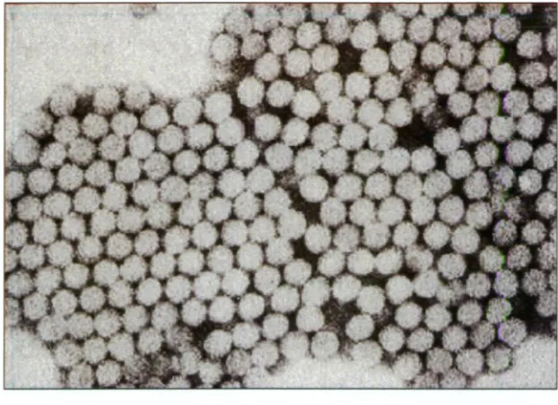
distinctive surface features (Cliver and Matsui, 2002; Biichen-Osmond, 2003)
(Figure 1.1). As reviewed previously (Cliver and Matsui, 2002; Grohmann and
Lee, 2003; Hollinger and Ticehurst, 1996), HAV possesses notable stability to
environmental conditions, particularly to heat and drying, and is more resistant to
low pH (pH 1.0), gamma rays, UV light and low levels of chlorine and ozone than
Figure 1.1. Electron micrograph of hepatitis A virus (HAV) virions.
Individual virions are 27-30 nm diameter.
(Source: Centers for Disease Control and Prevention (CDC),
1.3.1 Difficulties of laboratory study
Tissue culture is an effective method for the propagation and quantification of a
variety of viruses, including those pathogenic to humans, but propagation of HAV
in vitro using mammalian tissue culture cells can best be described as only
'moderately successful' (Koopmans et al., 2002), especially for environmental
isolates or wild-type strains which can prove exceptionally difficult to propagate.
Enumeration of viable viruses traditionally requires the development of plaques
and cytopathic effects in infected tissue culture cells. Although virions are
released from infected cells into the surrounding liquid medium where they may
be recovered for further analysis, only a relatively small proportion of HAV is
released from infected tissue culture cells (Hollinger and Ticehurst, 1996).
Instead, most of the infectious virions remain in the cell cytoplasm (Bishop et al.,
1994). Tissue culture methods for analysing and enumerating HAY remain
lengthy and labour-intensive, thereby encouraging the use of rapid detection
methods including PCR techniques. However, PCR-based techniques, whilst
considerably faster to perform, are compromised by insufficient sample
purification prior to assay, the presence of food-related inhibitory compounds,
and the detection of the sequence of interest in genetic material from any intact.
1.3.2 Illness and transmission of infection
Human HAV is shed exclusively in human faeces; therefore infection is initiated
when the virus is ingested, commonly via contaminated water or food (Cliver and
Matsui, 2002). Transmission may also occur by exposure to HAV-contaminated
blood or blood products (Fiore, 2004). After penetrating and replicating in the
intestinal epithelial cells, the virus infects the liver where replication also takes
place inside hepatocytes (Cuthbert, 2001; Hollinger and Ticehurst, 1996). When
an immune response is evoked, cytotoxic T cells destroy infected liver cells,
severely disrupting regular body functions controlled by the liver (Cliver and
Matsui, 2002). Following secretion of the virus from the liver in bile, hepatitis A
virions are excreted from the body in faeces. Consequently, if treatment
processes for human faecal effluent are inadequate to eliminate HAV prior to
release into the environment, a significant risk may be posed to drinking and
irrigation water, waters from which shellfish are harvested, and waters for
recreational use (Cliver and Matsui, 2002).
The incubation period of hepatitis A averages 28-30 days, and during this time the
virus is shed from the body. Virions continue to be shed in low numbers for up to
2 weeks following the onset of symptoms which include fever, loss of appetite,
nausea and abdominal discomfort, often followed by jaundice lasting several days
(Cliver and Matsui, 2002). In infants and children under five years of age,
infection is often mild or asymptomatic. Immunity to hepatitis A is usually
From 1987 to 1997 an average of 120,000 acute hepatitis A cases were estimated
to occur annually in the U.S.A., but following the introduction of an effective
vaccine in the mid-1990's, cases of hepatitis A have been decreasing since 1997,
with almost 6,000 reported and 24,000 estimated cases of acute hepatitis A in
2004 (Centers for Disease Control and Prevention (CDC), 2006; Food Safety
Network, 2005). The occurrence of hepatitis A in Australia has also declined
over the last 30 years; however, the occurrence in some indigenous human
communities remains high (Lin et al. , 2002). Between 1991 and 1999, the annual
number of hepatitis A infections in Australia averaged 2,115, but in 2000, the
number of hepatitis A cases declined by 48% from the previous year. This
reduction was thought to be due to effective control measures such as
vaccinations of susceptible populations and improvements in hygiene (Lin et al.,
2002).
Hepatitis A can cause particular public health problems in areas lacking proper
sewage treatment facilities, as well as in locations where an adequate. level of
hygiene may be difficult to maintain, such as child care centres, prisons, and
camp grounds (Grohmann and Lee, 2003). The disease is endemic in developing
regions such as Southeast Asia, the Indian subcontinent and Africa (Koopmans et .
al., 2002). In such areas, many children under the age of six acquire a mild or
subclinical disease, and thus immunity to re-infection. Consequently, the
occurrence of hepatitis A outbreaks in these areas is rare (Koopmans et al., 2002).
In developed countries, however, improved sanitary conditions have resulted in
fewer cases of hepatitis A, but left large populations susceptible to a greater risk
1.3.3 Outbreaks of interest
In 1997, a highly publicised hepatitis A outbreak in Australia resulted from the
consumption of contaminated shellfish (Conaty
et al.,
2000). More than 400 hepatitis A cases, including one death, were reported following the consumption, of infected oysters harvested from the Wallis Lakes area in New South Wales
(N.S.W.), Australia (Conaty
et al.,
2000). Whilst the precise cause of contamination in this outbreak is unknown, it was suggested that unusually highrainfalls resulted in a turbid flow of water to the oyster harvesting area. Heavy
rain can widely distribute a source of contamination in a lake or estuarine area,
and probably ensured the pollution was well dispersed almost lake-wide (Conaty
et al.,
2000). This outbreak occurred despite no apparent breach of microbiological safety regulations, including a mandatory period of shellfishpurification following harvest, industry safeguards of ceasing shellfish harvests
following rainfall, and the regular testing of shellfish meat for indicator
microorganisms (Conaty
et al. ,
2000).In 1988, an outbreak of hepatitis A linked to contaminated raw clams in China
killed 32 people and infected nearly 300,000 (Halliday
et al.,
1991). This outbreak may well be the largest recorded viral food poisoning outbreak in theworld to date. Clams were contaminated by the release of untreated human faecal
effluent from a nearby residential area which had reported an epidemic of
hepatitis A in the preceding months, and from boats dumping human waste
In October and November 2003, a large hepatitis A outbreak originated in a
restaurant in Pennsylvania, U.S.A. and received nationwide media coverage, as
more than 600 people demonstrated symptoms consistent with hepatitis A
infection, and three fatalities were recorded (Wheeler
et al.,
2005). Green onions imported from Mexico and exposed to human faeces were confirmed as thevector, and were an ingredient in a mild salsa that was served to all patrons in the
restaurant during the outbreak.
1.4 BIVALVE MOLLUSCAN SHELLFISH
Bivalve molluscan shellfish such as oysters, clams, mussels and scallops are
soft-bodied animals protected by a shell that is divided in two halves (valves).
Powerful adductor mussels near the hinge of each valve close the shell for
protection, and open to allow feeding and respiration via the gills (Campbell,
1996). Bivalve molluscs are filter feeders, inducing a current of water (up to 20 L
water per hour under ideal conditions) to pass over gills (Grohmann and Lee,
2003). As shellfish pump water, strands of mucous are continuously secreted,
trapping food particles such as algae and microorganisms, which are then carried
by cilia to the mouth area to be either ingested or eliminated as pseudofaeces
(DiGirolamo
et al,
1970).Filter-feeding shellfish are at particular risk of human enteric virus transmission.
They are usually cultivated in estuaries where the sheltered waters contain high
nutrient levels; however, these shallow inlet waters may also be contaminated
The microbiological profile of oysters is directly related to that of the surrounding
water (Son and Fleet, 1980). Pathogenic microorganisms bioaccumulated by
oysters are often detected in oyster flesh at concentrations greater than that in the
surrounding water. Because oysters and other bivalve molluscs are often
consumed whole and raw or lightly cooked, their consumption results in an
increased risk of illness (Goyal
et al.,
1979; Lees, 2000).In the environment, viruses are afforded protection from environmental sources of
inactivation, such as UV rays and warm temperatures, through association with
particulate matter (Grohmann and Lee, 2003). Populations of faecal coliforms
tend to be higher in sediment than in the overlying water, and during rainfall,
sediment is disrupted, increasing the water's turbidity (Metcalf
et al.,
1995). Virus survival is enhanced in turbid water, and shellfish growing areas ofnormally good quality water are therefore at risk of contamination during periods
of heavy rainfall (Grohmann and Lee, 2003).
1.4.1 Procedures employed to improve shellfish safety,
1.4.1.1 Purification methods
Purification techniques are employed to reduce the microbiological load of
filter-feeding shellfish. Shellfish are transferred from their normal harvesting
environment to a clean supply of seawater to purge contaminating
microorganisms via the natural filter-feeding process (Son and Fleet, 1980). This
method is commonly practiced throughout the world to purify oysters, and is
either performed in tanks in a land-based setting (depuration), or in clean waters
Water used in depuration is typically disinfected by treatment with UV. Unlike
sanitizers such as chlorine or ozone, UV light leaves no residual effect to inhibit
the biological processes of shellfish or shellfish taste, and is effective against both
bacteria and viruses (Richards, 1988). Efficient operation of this disinfection
method requires water of low to moderate turbidity, adequate flow rate, and
regular cleaning of UV lamps to ensure optimal light penetration.
Whilst depuration is useful for reducing the bacteriological contamination of
bivalve molluscs, viruses are removed at a slow rate. Kingsley and Richards '
(2003) exposed oysters to seawater contaminated with 180 plaque-forming units
(PFU) per ml HAV, then performed depuration in UV-treated seawater that was
changed daily for 6 weeks. In one trial, four weeks of depuration was required
before HAV reached nondetectable levels in oyster tissue by tissue cell culture
plaque assay, and in the second trial, HAV was still detected in oysters by reverse
transcription-polymerase chain reaction (RT-PCR) 6 weeks post-contamination.
The results demonstrated that the popular practice of depuration for between 36-
48 h may be insufficient for removing HAV from shellfish.
Relaying is an alternative purification process that does not require a water
pumping facility. Bivalve molluscs of poor microbiological quality are
transferred to clean shellfish growing water, often for a longer duration than that
of depuration (at least 14 days; Bird, 1994). The process may he applied if the
bacterial counts in shellfish cannot be sufficiently reduced in the relatively short
Dore et al. (1998) reported that levels of male-specific bacteriophages, used in
their study as indicators of viral contamination, were not eliminated from oysters
after 4 weeks of relaying followed by depuration. Factors thought to affect the
rate that contaminants are purged from oysters include oyster species, water
temperature and the initial level of contamination (Dore etal., 1998).
1.5 INACTIVATION OF HUMAN ENTERIC VIRUSES
Most viruses are inactivated by heating to temperatures typically used in cooking,
and complete inactivation of HAV is reportedly achieved within 4 min exposure
to 70°C, 30 s at 75°C, 5 s at 80°C and immediately after exposure to 85°C (Parry
and Mortimer, 1984). Strong oxidizing agents, such as chlorine, ozone, and UV
light in water or on surfaces are effective against viruses (Blackwell etal., 1985);
however, HAV is quite resistant to drying (Conaty etal., 2000) and can survive in
the environment for an extended period of time (Hewitt and Greening, 2004;
Kingsley and Richards, 2003; Croci et al., 2002; Grohmann and Lee, 2003).
Inactivation by exposure to UV, hypochlorite (1.2-1.25 ppm) or 72°C is due to
conformational change of capsid proteins affecting the function of antigenic sites
and receptor attachment sites. Virus inactivation almost always accompanies loss
of virus attachment (Nuanualsuwan and Cliver, 2003). Therefore, in order to
inactivate viruses in foods using a processing method, either the protective capsid
layer must be denatured or disrupted so the virion cannot attach to a host cell, or
the nucleic acid contained in the particle must be damaged to an extent preventing
1.6 NONTHERMAL PRESERVATIVE PROCESSES
It is difficult to educate a population on the health benefits of changing their
eating habits from traditionally consuming raw shellfish to thoroughly cooking
shellfish before consumption (Halliday
et al.,
1991; Salamina and D'Argenio, 1998). An epidemiological survey conducted in Naples, Italy, concluded that thecommon consumption of shellfish by the population was not affected by the
awareness of the high incidence of hepatitis A infection in the region and
knowledge of its route of transmission, and that the common conditions used in
shellfish cooking were frequently insufficient to inactivate viruses prior to
consumption (Salamina and D'Argenio, 1998). Thus an alternative non-thermal
or low-heat preservative process would be valuable to reliably improve the
margin of safety associated with consumption of raw product. Due to the
traditional consumption of oysters raw or minimally cooked, this process must not
only ensure a microbiologically-safe food, but also provide a product that is
almost identical to the raw product in organoleptic quality. To ensure that harvest
water quality and good manufacturing practices are not compromised, the
introduction of such a process must add to, and not replace, current standard
procedures.
The application of heat has long been recognized as a process that prolongs the
shelf-life of foods while improving food safety, but in some products heating can
cause undesirable changes affecting product organoleptic and nutritional qualities.
For example, food textures are usually altered, some vitamins are known to
degrade, and vegetable tissues are often softened during thermal processing and
Consumers are increasingly demanding food products that are fresh-tasting,
nutritious and convenient. At the same time, consumer concerns about food
safety have steadily increased as the incidence of reported foodborne illnesses has
continued to rise. These trends have fuelled interest in non-thermal processing
technologies, such as high hydrostatic pressure processing, irradiation, pulsed
electric fields, and high-intensity pulsed light. In comparison to traditional
thermal processing methods that often cause detrimental changes in foods, these
non-thermal processing techniques offer benefits such as the potential for
minimising or eliminating extensive thermal processing and chemical
preservatives, and limiting unfavourable effects on food quality. Preservation of
freshness and protection of flavour, appearance and nutritional value results in a
high quality food product, often with extended shelf-life. For these reasons,
non-thermal processing technologies offer the ability to produce foods with improved
quality, increased consumer appeal and a value-added premium price. Although
commercialisation of these technologies has been slow to date, the above trends
plus improvements in efficiency and reductions in cost mean that the rate of
adoption of non-thermal processes is likely to increase.
1.6.1 High pressure processing (HPP)
Today, high pressure pasteurisation has become a commercial reality with several•
high pressure-treated fruit- and vegetable-based refrigerated food products
currently on the international market, including a range of juices and fruit
smoothies, jams, applesauce-fruit blends, guacamole and other avocado products,
tomato-based salsas and fajita meal kits containing acidified sliced capsicum and
Additionally, ready-to-eat meat products and seafood, including oysters, are on
the market in the U.S.A. and Europe (Smelt, 1998; Stewart and Cole, 2001). A
batch processing system is typically used where the product is placed into a final
flexible consumer package before pressurization. The packages are loaded into a
basket and placed into the pressure vessel, where they are submerged in a liquid
of low compressibility (typically potable water). Once loaded and closed,
pressures ranging from 100 to 700 MPa are normally generated by pumping
additional water into the vessel. The process is relatively energy-efficient,
requiring a similar amount of energy to raise the pressure to 400 MPa, as required
to heat to 30°C (Cheftel, 1995). Once the desired target pressure is achieved, no
further energy is required to sustain that pressure (Farr, 1990). Unlike thermal
processing, pressure is distributed instantaneously and uniformly throughout
foods, ensuring a homogenous treatment regardless of the size or shape of the
product (Hoover, 1993).
The treatment of foods with HPP involves compressing the water surrounding the
food (Barbosa-Canovas et al., 1998.). Although its compressibility is low, the
volume of water is decreased by 15% at 600 MPa and 22°C (Farr, 1990). The
compression of water causes a moderate increase in temperature (commonly
referred to as adiabatic heat or the heat of compression), the extent of which is
dependent on the initial temperature of the vessel and the rate of compression.
Decompression of the vessel reverses this effect at an equivalent rate (Cheftel,
The primary advantages of HPP over thermal processing are the minimal
chemical and physical effects exerted on most foods while imparting a microbial
kill step. High pressure does cause a range of effects on the molecular
interactions in foods. Ionic bonds and at least a proportion of hydrophobic
interactions are broken or distorted by high pressure, whereas hydrogen bonds_ are
strengthened (Hoover
et al.,
1989), and covalent bonds are unaffected (Ledward, 1995). As a. result of the pressure-induced changes to ionic bonds andhydrophobic interactions, proteins start to denature at room temperature above
pressures of 100-200 MPa (Cheftel, 1995). Oligomeric structures dissociate into
their subunits, monomeric structures partially unfold and denature, and proteins
aggregate and gel. The conformation of proteins is altered by an increase in
pressure due to irreversible changes to the secondary, tertiary, quaternary and
supramolecular structures (Palou
et al.,
1999). Denaturation may result when proteins are exposed to pressure beyond that of the individual protein-specificpressure threshold (Cheftel, 1995). The structure and function of lipids and
polysaccharides are altered by HPP (Ledward, 1995); however, pressure effects
on lipids are usually reversible which is often not the case for polysaccharides and
proteins. Smaller molecules such as vitamin C and B-carotene are not affected by
high pressure (Bull
et al.,
2004; Cheftel, 1995). Oxidative reactions in foods and enzymatic browning in some fruits are reportedly enhanced by HPP, while partialdiscoloration has been reported in treated red meats (Cheftel, 1995; Ledward,
1.6.1.1 Pressure effects on microorganisms
The required pressure treatment for microbiologically safe and stable products is
dependent on the target microorganism to be inactivated. Bacterial vegetative
cells, yeasts, moulds and some viruses are sensitive to pressures between 200-700
MPa; bacterial spores may survive pressurization above 1000 MPa (Arroyo
et al.,
1999; Cheftel, 1992; Saleet al.,
1970). Spoilage of food and/or food safety issues due to the outgrowth of bacterial spores can be controlled via complementarymeans such as refrigeration and acidification. Various common factors influence
the pressure resistance of microorganisms, including the target microorganism
and its physiological state, the intrinsic properties of the menstruum, the
processing temperature, and the time and magnitude of pressure treatment
(Hoover
et al.,
1989). Considerable variation in susceptibility to high pressure has been observed among various microbial species, strains, and onmicroorganisms in different substrates (Patterson
et al.,
1995). Certain foods provide microorganisms protection from inactivation or injury from highpressure. For example, milk is said to be more protective to bacteria during HPP
than a buffered solution (Cheftel, 1995) or meat (Patterson
et al. ,
1995).The critical site of pressure damage leading to inactivation of bacteria and fungi is
the cytoplasmic membrane. Cell permeability is altered and ion exchange is
disrupted due to crystallization of membrane phospholipids and protein
denaturation (Cheftel, 1995; Yuste
et al. ,
2001). Pressure-sensitive bacteria begin to lose viability at approximately 180 MPa (Lado and Yousef, 2002). Between200-400 MPa, irreversible changes such as cell leakage, which leads to cell death,
has been demonstrated by the release of UV-absorbing material from E. coli
1.6.1.2 Oyster processing
The application of HPP to whole oyster processing has been attractive for a
variety of reasons. Oysters (and other shellfish) are high-value foods traditionally
consumed raw throughout the world (Kingsley et al., 2002). Pathogens
associated with raw oysters, notably Vibrio spp. and HAV, are sensitive to
inactivation by HPP (Calci et al., 2005; Kingsley etal., 2002; Styles et al. , 1991).
The refrigerated shelf-life of harvested oysters is limited, so any extension of
shelf-life without altering sensory quality is highly desirable. An extension of
oyster shelf-life can be achieved by pressure treatment. Additionally,
Lopez-Caballero et al. (2000) described pressure-treated oysters as 'slightly more
voluminous with a very pleasant appearance', and reported that oysters were more
appealing following treatment at chilled temperatures than at room temperature
and above. Flavour may be enhanced, possibly by pressure infusion of the salty
liquor within the oyster shell into the meat (Hoover et al., 1989).
Adductor muscles holding oyster shells tightly closed are cleaved during high
pressure treatment, ensuring convenient manual shucking of the whole oyster
without the need for shucking knives (Kingsley et al., 2002). This allows for
higher yields, as there is both a full release of the muscle from the shell and no
damage to the tissue from the shucking knife. At 275 MPa, nearly 100% of Whole
shell oysters are opened. Usually a hold time of 1 to 2 minutes is used. Once
shucked, the oyster meat can be manually shaken off the shell and further
processed in semi-rigid containers at 415 MPa for several minutes which extends
Pressure treatment has been shown to speed removal of lobster meat from its
surrounding shell as well.
1.6.1.3 Pressure effects on viruses
The work of Giddings
et al.
(1929) was the first documented attempt to estimate the pressure sensitivity of viruses by studying tobacco mosaic virus (TMV).TMV was found to be extremely resistant to pressure; pressurization at 920 MPa
was necessary to show any measurable inactivation. Fortunately, the pressure
resistance of most human and animal viruses is lower than that of TMV. Most of
these viruses can be inactivated at pressures <450 MPa (Table 1.1). An exception
is poliovirus, which appears to be the most pressure resistant amongst the human
and animal viruses studied thus far, capable of surviving an hour at 600 MPa with
only modest reductions in infectivity (Wilkinson
et al.,
2001).Kingsley
et al.
(2002) reported treatment with 450 MPa for 5 min at ambient temperature reduced HAV in isotonic tissue culture medium by approximately 7-logio PFU/ml. The pressure required to inactivate HAV within 5 min increased
when treated in seawater of 27.4 ppt salinity (Kingsley
et al.,
2002). Salt may act to stabilise viral capsid proteins at high pressure; but more information detailingthe effect of salt on the stability of viruses during HPP will be important for -future
Loss in Temperature infectivity (0C) (logio) 22 -15 22 25 25 25 21 0
20 • 6.85
22 7
20 <1
25 8
22 5
not specified
20
References 21 no reduction Kingsley etal., 2004
21 7 Kingsley et aL, 2004
21 no reduction Kingsley etal., 2004
Kingsley etal., 2004
Oliveira et al, 1999
Kingsley et aL, 2002
Nakagarni etal., 1992
Nakagami et aL, 1992
Nakagami et at, 1996
• Kingsley et at, 2004
Tian et at, 2000
Kingsley et at, 2007
Chen et aL, 2004
Wilkinson etal., 2001
Khadre and Yousef, 2002
Jurkiewicz et aL, 1995
Gaspar et at, 1997
-Silva et aL, 1992
[image:36.559.50.550.89.806.2]7 6 7 7 4 >3 Table 1.1. Pressure inactivation of viruses.
Virus name Enveloped? Pressure (MPa) Time (min)
Aichivirus A846/88 no 600 5
Coxsackievirus A9 no 500 5
Coxsackievirus B5 no 600 5
Feline calicivirus no 275 5
Foot and mouth disease virus no 240 120
Hepatitis A virus no 450 5
Herpes simplex virus type 1 yes 400 10
Human cytomegalovirus yes 300 10
Human immunodeficiency virus yes 350 10
Human Parechovirus-1 no 500 5
Infectious bursal disease virus no 230 120
Murine norovirus no 450 5
Phage 400 .20
Poliovirus no 600 60
Rotavirus no 300 2
Simian immunodeficiency virus yes 250 60
Sindbis virus no 250 480
Vesicular stomatitis virus yes 260 720
The HAV capsid reportedly remains intact following inactivation by HPP
(Kingsley
et al.,
2002). HPP may therefore denature the capsid proteins essential for host cell attachment to initiate infection, but not release RNA from virions(Khadre and Yousef, 2002; Kingsley
et al.,
2002). RT-PCR performed on the RNA from non-infectious virions still yielded a positive result, demonstrating itsunreliability for determining the viability of pressure-treated virus.
HAV could not be recovered from strawberry puree or sliced green onions
following 5 min exposure to 375 MPa, a reduction of 4.32-logio and 4.75-logio
PFU, respectively (Kingsley
et al.,
2005). Rotavirus titre was found to decline by 5-logio 50% tissue culture infective dose units (TCID50)/m1 within a 70-sexposure to 300 MPa in tissue culture media at 25°C, but 1-logio TCID50/m1 still
remained after a 10-min treatment (Khadre and Yousef, 2002). Herpes simplex
type 1 virus and human cytomegalovirus were inactivated by >7-logio and >4-
logio after 10-min exposure to more than 400 MPa in tissue culture media at 25°C
(Nakagami
et al.,
1992). Damage to the viral envelopes prevented virions from binding to host cells and subsequently initiating infection.A 7-logio TCID50/m1 culture of the norovirus surrogate, feline calicivirus (FCV),
was completely inactivated (detection limit 10" TCID50) in isotonic tissue culture
medium after 5 min exposure to 275 MPa or more (Kingsley
et al.,
2002). This highlights the potential for inactivating human norovirus with HPP, but cannot berelied upon to guarantee the susceptibility of norovirus to the process. For
example, HAV and poliovirus are both members of the picornavirus family, but
More recently, a murine norovirus (MNIV-1) was found to possess greater
stability to HPP than FCV, with a 5 min treatment at 450 MPa required for a 6.85-
logio PFU reduction in titre (Kingsley et al., 2007). MNV-1 has more
biochemical, pathological and molecular similarities to human noroviruses than
FCV (Wobus et al., 2006; Cannon et al., 2006), and is therefore likely to also be a
better indicator of the sensitivity to HPP of human norovirus.
The extent of virus inactivation is dependent upon treatment pressure duration and
temperature. Jurkiewicz et al. (1995) studied the pressure sensitivity of simian
immunodeficiency virus (Sly), and observed •a 5-logio reduction in Sly
infectivity after a 1-h exposure to 250 MPa at 21.5°C. Treatments at 200 and 150
MPa required 3 and 10 h, respectively, to attain equivalent reductions of 5-10gio
infectious units.
A number of reports have indicated that the dissociation and denaturation of
proteins and viruses by pressure is promoted by low temperatures (Bonafe et al.,
1998; Foguel etal., 1995; Gaspar et al. , 1997; Kunugi and Tanaka, 2002; Tian et
al., 2000, Weber, 1993). The explanation for this phenomenon is that low
temperatures promote the exposure of polar side chains to water. The
non-polar interactions are more affected by pressure because they, are more
compressible. Oliveira et al. (1999) examined the combined effect of pressure
and low temperature on the stability of foot-and-mouth disease virus (FMDV), an
animal virus that can cause devastating losses in the meat and dairy industries.
FMDV was found to be sensitive to pressure: exposure to 240 MPa for 2 h
6-logio units at -15°C. Interestingly, this effect was not noted for HAV by
Kingsley
et al.
(2006), who reported enhanced inactivation from high pressure treatment at 50°C compared to treatment at -10 and 20°C.1.7 PREDICTIVE MICROBIOLOGY
HPP may have the potential to improve the microbiological safety of shellfish
consumption, but its implementation, as is the case for the commercialisation of
any novel food process, is reliant on obtaining systematic inactivation kinetic data
of specific target microorganisms (Stewart and Cole, 2001; Lado and Yousef,
2002). This data, applicable over a wide range of conditions, forms the basis of a
kinetic inactivation model.
1.7.1 Model development
In predictive microbiology, microbial growth, survival or death in a specific food
may be described or predicted by a mathematical model formed from data
collected from microbial responses to defined and controlled conditions. By
interpolation, a model can predict the microbial response to specific conditions
not actually tested in the laboratory, and be a cost-effective substitute for
performing traditional shelf life and food safety studies (Ross and McMeekin,
The first consideration when designing a model is identifying its intended use
(Legan
et al.,
2002). Inactivation models predict the response of a microorganism over a defined range of a lethal process, such as heat or high pressure. Otherfactors, such as pH and water activity, act in combination with the lethal process
to influence microbial death, and must also be identified for study within a
realistic range. Three or four factors should be investigated over ranges that will
allow coverage of all possible and likely responses in the intended process.
Replication assists in minimising the impact of the variability in the microbial
response (Legan
et al.,
2002). To identify any curvature and to gain a true understanding of the microbial response during the treatment, 10-15 data pointsshould be measured throughout the process (McMeekin
et al.,
1993). This is known as a kinetic study, and is preferred to an end point measurement becausemore information is gained, and the failure point in the process can be identified
(Stewart and Cole, 2001).
The need for more kinetic inactivation data prior to the commercialisation of HPP
for pathogen reduction has already been highlighted (IFT, 2000). Thermal
processing has long been used in food production, and the effect of heat on most
known microorganisms and foods is known. In this case, the 'processing
continuum' has provided food processors with an abundance of literature with
which to base decisions regarding changes in food constituents or production
regime (Legan
et al.,
2002). For processors wishing to implement a novel non-thermal technique into food production, however, a great deal of kineticCreation of a model should involve experimentation in a homogeneous medium,
for example, liquid culture media, which is less complex than the food to which
validation of the model will occur (Legan
et al.,
2002). In this way, the number of controlling factors can be limited as required. The controlling factors used tocreate a model, and the range over which each factor exists must be carefully
chosen, as a model may only be applied within these conditions. If not applied
within these specific conditions, the underlying principle for obtaining the data is
lost because the action of interacting factors outside the model's determined range
will be unknown. This is because currently available models are empirical, not
mechanistic, and so there is no theoretical basis for extrapolation of models
beyond the bounds of the data used to generate them.
1.7.2 Model validation
Validation of a predictive model is achieved by comparing data gained in a real
food system with predictions made by the original model, and making appropriate
modifications, where required (McMeekin
et al.,
1993). Validation of the model is specific to each particular food. Process conditions can be adjusted accordingto the microbial protection afforded by the food, allowing optimised processes for
1.8 PROJECT AIMS
Evaluate and compare cultivation methods for HAV to produce moderately large
volumes of high titre virus to be used in challenge testing
Evaluate and compare methods for extraction and purification of HAV from
oysters to ensure high percentage recovery
Develop a method to artificially inoculate live oysters with HAV, using the
natural filter-feeding process of oysters
Collect HPP kinetic inactivation data for HAV suspended in buffered media, and
develop a predictive inactivation model for HAV that may be validated in oysters
naturally contaminated with HAV
Develop a quantitative real-time reverse transcription-PCR assay for HAV, and
determine its ability to detect and accurately quantitate HAV extracted from
Chapter 2: General materials and methods
All laboratory work described in this thesis was conducted at the Food Science
Australia laboratory in Werribee (Melbourne), Victoria, Australia, unless
otherwise specified.
2.1 GENERAL PROCEDURES
Glassware used during tissue culture protocols was acid-washed prior to
autoclaving, by rinsing thoroughly with 1 M hydrochloric acid followed by 5
rinses with Milli-Q® water (Millipore, Australia). Acid-washed glassware and
autoclavable plasticware were sterilised by autoclaving at 121°C for 25 min, and
solutions were sterilised by autoclaving at 121°C for at least 20 min.
Centrifugation of volumes less than 2 ml was performed in a Sigma 1-15
microcentrifuge; volumes between 2 and 10 ml were centrifuged in a Sigma 2-5
centrifuge; and volumes greater than 10 ml were centrifuged in a Beckman J2-21
M/E centrifuge. Where centrifugal force greater than 15,000xg was required, a
Beckman Coulter Optima L-90 K ultracentrifuge was used.
Weighing of substances more than 2 g was performed on a Mettler PM 6100
balance, and substances less than 2 g were weighed on an Sartorius A210 P
Socorex calibra 822 micropipettes were used for all micropipetting of liquids
from 1 to 1 ml. A Genex Delta pipette was used to transfer volumes of liquid
from 1 ml to 25 ml.
Tissue culture cell lines were incubated in a Sanyo MC0-20A1C model CO2
incubator at 37°C and 95% humidity in an atmosphere containing 5% CO2.
22
PREPARATION OF SOLUTIONS
All solutions were prepared with Milli-Q ® water and sterilised by autoclaving
unless otherwise stated.
Acetone and methanol: A 1:1 (v/v) mixture of acetone (Sigma-Aldrich, U.S.A.)
and methanol (BDH, Australia). This solution was not autoclaved.
Antibiotic/antimycotic solution: 100x solution (Sigma-Aldrich); added at 1:50 or
1:100, as specified. This solution was not autoclaved.
Anti-HAV monoclonal antibody in skim milk: A 1:2000 (v/v) dilution of mouse
anti-HAV monoclonal antibody (CSL Ltd., Australia) in 1% (w/v) skim milk.
This solution was not autoclaved.
Citrate/phosphate buffer: 0.05 M buffer prepared by mixing sterile 0.1 M citrate
buffer (BDH, England) with sterile 0.1 M phosphate buffer (BDH, Australia) and
Conjugated anti-mouse antibody: Anti-mouse antibody conjugated with
horseradish peroxidase (Sigma-Aldrich), diluted 1:1000 (v/v) in PBS. This
solution was not autoclaved.
Dulbecco's modification of Eagle's minimum essential medium (DMEM): 500 ml
DMEM was prepared by mixing 100 ml sterile 5x DMEM (Thermo Electron
Corp., Australia) with 372 ml sterile Milli-Q ® water, 10 ml sterile 1 M HEPES
(Thermo Electron Corp.), 13.5 ml sterile 7.5% sodium bicarbonate (Thermo
Electron Corp.) and 4.5 ml sterile liquid L-glutamine (Thermo Electron Corp.).
Ethidium bromide (EtBr): Prepared by diluting 0.625 mg/ml EtBr (Mercury,
U.S.A.) to 0.5 Kg/m1 in deionised water. This solution was not autoclaved.
Glycine buffer: 0.1 M glycine (Calbiochem, U.S.A.), 0.3 M NaC1 (Chem-Supply,
Australia); pH 9.5.
GNT: 80% (v/v) glycerol (BDH, Australia), 100 InM NaC1 (Chem-Supply), 100
mM Tris (Sigma-Aldrich); pH 7.4.
Growth media: DMEM with 10% (v/v) newborn bovine serum (NBS; Thermo
Electron Corp.). This solution was filter-sterilised through a 0.2 [tin filter before
use.
Non-ionic detergent: Igepal® CA-630 (Sigma-Aldrich) diluted to 10% in Milli-Q ®
NT buffer: 100 mM NaC1 (Chem-Supply), 10 mM Tris (Sigma-Aldrich); pH 7.4.
PEG solution: 16% (w/v) polyethylene glycol (PEG) 8000 (Sigma-Aldrich), 0.525
M NaC1 (Chem-Supply).
Phosphate-buffered saline (PBS): Prepared by dissolving one PBS tablet (Oxoid,
Australia) in 100 ml Milli-Q ® water; pH: 7.3.
PBS and Tween 20: Prepared by mixing PBS with 1% (v/v) Tween 20
(Sigma-Aldrich). This solution was not autoclaved.
Seawater: 30 parts per thousand (ppt) prepared unless otherwise specified, by
dissolving 34 g Red Sea salt (Red Sea Fish Pharm, Israel) per litre of deionised
water. Salinity was calculated using a hydrometer, thermometer, and conversion
table (Bird, 1994). This solution was not autoclaved.
Skim milk: Prepared by dissolving skim milk powder. (Nestle, Australia) in PBS.
This solution was not autoclaved.
Sodium lauryl sulfate (SDS): Prepared by dissolving SDS (Sigma-Aldrich) in
water. This solution was not autoclaved.
Substrate solution: 0-phenylaminediamine (OPD, 0.6 mg/ml; Sigma-Aldrich) and
hydrogen peroxide (30% (w/v); BDH) diluted 1:2400 (v/v) in 0.05 M
citrate/phosphate buffer. This solution was not autoclaved.
TBE buffer: Made to 10x stock solution and diluted to lx prior to use. To make
10x stock solution, 10.8% (w/v) Tris (Sigma-Aldrich), 5.5% (w/v) boric acid
(Calbiochem) and 3.7% (w/v) ethylenediaminetetraacetic acid (EDTA;
Sigma-Aldrich) were dissolved in Milli-Q
.
water. This solution was not autoclaved.
Trypsin: EDTA: Pre-prepared sterile 1:250 mixture (Thermo Electron Corp.
2.3 GENERAL TISSUE CELL CULTURE METHODS
2.3.1 Maintenance of tissue culture cell lines
A continuous African Green Monkey kidney cell line (BSC-1) was grown to
confluence in a 25 or 75 cm2 flask twice weekly, and passaged by removing
growth media and then washing cells twice with sterile PBS. Cells were detached
from the base of the flask by trypsination; where cells were incubated at 37°C for
5 min with 1 or 2 ml trypsin:EDTA (for 25 or 75 m12 flasks, respectively). Five
millilitres DMEM was then added to the flask and the entire contents decanted
into a sterile 10 ml centrifuge tube. The cells were pelleted by centrifugation at
500xg for 2 min, then resuspended in 4 ml DMEM. A new 25 or 75 cm2 tissue
culture flask was seeded with 1 ml resuspended cells, and then filled with fresh
growth media. For virus quantification in 96-well microtitre plates, 1 ml of
resuspended cells was diluted in 9 ml of growth media, and 0.1 ml seeded into
each well. BSC-1 cells between passage numbers 59 and 79 were used in all
experiments.
2.3.2 Storage of cell lines
Trypsinised and pelleted cells derived from a 25 cm2 flask were resuspended in
0.9 ml DMEM and transferred to a sterile 2 ml cryotube containing 0.1 ml
dimethylsulphoxide (DMSO; Sigma-Aldrich) Tubes were mixed gently by
inversion and transferred to -70°C for 24 h prior to long-term storage in liquid
Cells recovered from long term liquid nitrogen storage were thawed quickly under
warm water, transferred to 5 ml DMEM slowly (dropwise), and centrifuged for 2
min at 500xg. The supernatant was then removed and the pelleted cells
resuspended gently in DMEM and seeded into a 25 cm2 tissue culture flask with
fresh growth media.
2.3.3 Quantification of infectious hepatitis a virus
An enzyme immunoassay, modified from Borovec and Uren (1997), was used to
determine infectious HAV titre. Serial 10-fold dilutions of HAV (strain HM:175,
kindly donated by Prof. David Anderson, MacFarlane Burnet Institute, Australia)
were made in DMEM + 2% NBS, and 0.1 ml of each dilution was transferred to 5
replicate wells containing a monolayer of BSC-1 cells (approximately 50%
confluent) on a 96-well microtitre plate. In low titre and/or low volume samples, a
1:2 (v/v) or 1:4 (v/v) dilution of sample was analysed in the assay to increase the
assay's detection limit.
After 7 days incubation, media was removed from wells, and cells fixed to the
base of wells by addition of a 1:1 mixture of acetone and methanol (pre-cooled to
4°C). The fixative was removed after 5 min and plates allowed to dry by standing
uncovered for at least 1.5 h at room temperature. One hundred microliters of a
1:2000 dilution of anti-HAV monoclonal antibody in skim milk was added to
each well to detect HAV antigen. The antibody solution was removed after 1 h
incubation at 37°C, and wells were washed three times with 0.1 ml PBS and
Tween 20. One hundred microliters of conjugated anti-mouse antibody was
visualised upon the addition of 0.1 ml substrate solution. The colour change was
monitored visually until the reaction was stopped by addition of 0.1 ml 1 M
sulphuric acid. Wells that produced visible colour to the naked eye were scored
positive for HAV presence. The titre of infectious HAV was then determined by
the 50% tissue culture infective dose (TCID50) method, described by Reed and
Muench (1938), in the units TCID50/rn1 of undiluted sample. Detection limit of
Chapter 3: Evaluation and comparison of
methods for high-throughput cultivation of
hepatitis A virus
3.1 INTRODUCTION
For the purpose of microbiological challenge testing and vaccine production, high
concentrations of microorganisms in large volumes are often required. Although
HAV is cultivatable in the laboratory, it can be difficult to propagate in tissue
culture cell lines, requiring lengthy incubation times (1-2 weeks) to produce a
sufficiently high titre suitable for challenge testing.
The difficulty of HAV propagation is predominantly due to a large proportion
(approximately 80%) of virions remaining cell-associated during infection
(Nasser and Metcalf, 1987). As a result, the spread of infection throughout a
tissue cell culture monolayer is relatively slow. Quantification by infectivity
assay in cell culture is made more difficult by the fact that not all HAV isolates
form plaques or cause cytopathic effects in cells, and those that do require long
incubation of more than 10 days (Beales
et al.,
1996; Borovec and Uren, 1997).During cultivation of HAV, the host cell line is infected with the virus and
incubated for a number of days to promote the generation of viral progeny.
Virions are released from infected cells by mechanical lysis, for example, with
sequential freezing and thawing. Additional purification of the virus stock may
be required, especially for vaccine production, and this may be achieved with
solvent extractions, ultracentrifugation and gel-exclusion or ion-exchange
chromatography. These procedures often require specialised equipment, can be
time consuming to perform and may result in loss and/or dilution of virus (Bishop
et al., 1994). Additionally, the use of hazardous solvents and chemicals during
virus purification may be restricted in certain laboratories due to the increased
occupational health and safety risks involved in carrying out the procedure.
Whilst preparation of a pure virus stock is always important, the level of
purification required for production of human vaccines is much higher than that
required for laboratories performing challenge studies with, for example, HPP.
For example, virions tend to associate with each other in suspension, forming
'clumps', and with particulate matter in the environment, which may confer
increased resistance to environmental conditions or treatment processes (Metcalf
et al. , 1979; Landry et al. , 1983).
The aim of this study was to compare throughput and modify if necessary,
established methods of HAV cultivation and to establish a simple and effective
method for cultivating moderately large quantities of infectious HAV in high
titres for challenge testing with HPP.
Two well established cultivation methods described by Bishop et al. (1994) and
Beales et al. (1996) were compared in this study. The freeze-thaw method was
cytopathic quantitative assay for HAV, and involved sequential freeze-thaw
cycles to release virions from infected cells followed by a solvent extraction step
to purify virions. The alternate method, described by Bishop
et al.,
involved an ultracentrifugation step through discontinuous sucrose/glycerol density gradientsthat could consistently yield highly purified infectious HAV particles suitable for
use in vaccines and diagnostic tests.
3.2 METHODS
3.2.1 Tissue culture cells and hepatitis A virus
BSC-1
cells at 50% confluency in a 75 cm2 flask were infected withapproximately 105 TCID50 purified HAV stock. Twenty millilitres of DMEM
with 2% (v/v) NBS was added to the flask and incubated for 7 days.
3.2.2 Hepatitis A virus cultivation
The cultivation method described by Bishop
et al.
(1994) was initially investigated, with 75 cm2 flasks used for cell growth instead of the 6000 cm2 cellfactories described in the original protocol. Following the 7-day incubation
described above, growth media was decanted from the flask and infected cells
were washed twice with 2 ml PBS. Cells were trypsinised with 2 ml trypsin:
EDTA and incubated for 5 min at 37°C. Trypsin was then inhibited by the
addition of 5 ml DMEM with 5% (v/v) NBS. Cells were decanted into a sterile
centrifuge tube (Nunc, Denmark) and pelleted by centrifugation at 500xg for 5