
Copyright 0 1974 AmericanSociety for Microbiology Printed in U.S.A.
Proteins
Specified by
Herpes
Simplex
Virus
XII. The Virion Polypeptides of Type 1 Strains
JOCHEN W. HEINE, ROBERT W. HONESS, ENZO CASSAI, AND BERNARD ROIZMAN Departments ofMicrobiology and Biophysics, The University of Chicago, Chicago, Illinois 60637, and Institut
diMicrobiologia, Universita di Ferrara, Ferrara, Italy 44100
Receivedforpublication27March 1974
The polypeptides from purified virions of a herpes simplex 1 (human
herpes-virus 1) strain, Fl, which had been passaged a limited number of times in cell
culture after isolation, formed 33 bands on electrophoretic separation in
poly-acrylamide gels cross-linked with N,N'-diallyltartardiamide in contrast to a
maximum resolution of only 24 to 25 bands in gels cross-linked with
N,N'-methylenebisacrylamide. This increase in the number of bands was due chiefly
to an improved separation of glycosylated polypeptides from nonglycosylated
polypeptides with which they co-electrophoresed on methylenebisacrylamide
cross-linkedgels.Purified virions of HSV-1 [Fl ]had aprotein/DNA mass ratio of
10.7 i 0.96,andbased on a DNA molecular mass of 85 x 106 to 100 x 106 the
estimatedweight of virion polypeptides ranges from 16.4 to 19.4 x 10⁻¹⁶ g. The
number ofmolecules ofeachpolypeptide per virion ranged from less than 50 to
1,500. Comparison ofthevirion polypeptides of two HSV-1 strains with similar
isolation and limited passage history with those of four HSV-1 strains with
histories of numerous passages outside the human host showed a number of
nonrandomvariations in virionpolypeptides. Thus, although thevirion
polypep-tidesof two strains withsimilar isolation and limited passage history could not be
differentiated, strains with extended passage histories differed markedly from
each other and from the limited passage strains in the number and
electropho-retic mobility ofnoncapsid polypeptides and notably inthose of the envelope.
An earlier publication from this laboratory
(22) reported the protein composition of
puri-fied virions oftheFl strain ofherpessimplex 1
(human herpesvirus 1, HSV-1). From an
elec-trophoretic analysis of virion polypeptides on
polyacrylamide gels
cross-linked withmethyl-enebisacrylamide (MBA)
it was concludedthat HSV-1 [F1] virions contained at least 24
virionpolypeptides (VP) numberedfromVP1 to 24, and that trace amounts of host proteins
remaining in
purified
virus preparations wereadventitious contaminants and not structural
constituents ofthe HSV-1 virion. Inthat
study
itwasalsoshown thatmany virion
polypeptides
were glycosylated; however, attempts to
iden-tify glycosylated
polypeptides by
autoradiogra-phy of gels containing separated
polypeptides
labeled with [4C
]glucosamine
were notentirelysatisfactory. At least two amino acids labeled
bands, VP7 and 8, were resolved in the region
occupied byasinglebroad glucosamine labeled
band.
Similarly,
twoadditional broad bands ofglucosamine labelwerelocatedatlower
molecu-lar weights in the region of VP10-16 and
VP17-19 even though nine separate
polypep-tides bands were readily differentiated in the
Coomassie
brilliantblue-stained
gels fromwhich theautoradiogramswereprepared as well as in autoradiograms of gels containing
sepa-rated
"4C-amino
acid-labeled polypeptides.Theratio ofglucosamine label to protein therefore
varied across each of these regions and was
much lower for the region of bands VP10-16
than forVP7-8orVP17-19. Theseobservations
suggested
either the existence ofprotein bandswithwidely different degreesofglycosylationor
a failure to separate
glycosylated
fromnon-glycosylatedcomponents, notably in the region
ofVP10-16.Thesealternativeexplanationshad
verydifferent implications for the biosynthesis
ofglycosylated components and thetopologyof
the virion. In addition, we have previously
noted that virus strains which have been
pas-sageda limited number oftimes incell culture
differ from a number of strains with extensive passage histories outside the human host with respect to immunologic specificity (5), virus
growthand development (21), and effectsonthe
social behavior of infected cells (5, 10, 15, 16).
We therefore wished to determine whether 640
on November 10, 2019 by guest
http://jvi.asm.org/
PROTEINS SPECIFIED BY HSV
strains differing in their passage history had systematic differences in their virion polypep-tide compositions.
In this paper we present additional data on
the protein composition of HSV-1 virions. This data includes the separation of glycosylated and
nonglycosylated polypeptides in the region of
VP10-19 on polyacrylamide gels cross-linked
withdiallytartardiamide (DATD), estimates of
the number of copies of virion polypeptides, and
results ofcomparisons ofthe polypeptide
com-position of virions from HSV-1 strainspassaged
a limited number of times in cell cultures with
those ofHSV-1 strains with histories of
numer-ous passages outside the human host.
MATERIALS AND METHODS
Cells. Human Epidermoid Carcinoma no. 2 (HEp-2) cells were grown at 37 C in Eagle minimal essential medium (EMEM) supplemented with 10%
calf serum, 0.001% ferric nitrate, and 1% sodium pyruvate.
Viruses. The HSV-1 strains usedinthesestudies were as follows:HSV-1 [Fl].This strain wasisolated in 1967 at the University ofChicago from a patient with a herpetic infection of the face and under the designation of HSV-1 [F] served as the prototype of HSV-1 strains in this laboratory in all publications subsequent to that of Ejercito et al. (5). The viruswas
isolated in HEp-2 cells and is being passaged in these cells at low multiplicities of infection a maximum of four times. The properties of the virus and its DNA have beenpublished elsewhere (5, 6, 7, 11, 22).
HSV-1 [F5]. The isolation and passage history of
this strain are similar to that of HSV-1 [Fl]. By plaque morphology (5), it cannot be differentiated from HSV-1 [F1].
HSV-1 [mP]. This virus, originally designated as
strainNT, was obtained in1958 as aninfected third passagechorioallantoic membrane. It was isolated in 1955 at the Johns Hopkins Hospital from a patient with eczema herpeticum. It was passaged some 20 times inFLcells andsubsequently some 40 timesin
HEp-2 cells. Its plaque morphology is similar to that ofHSV-1 [Fl].Some differences apparent on electron microscope studies of the development of this virus as compared to that of HSV-1 [Fl]have been reported (21).
HSV-1 []arose spontaneously in FL cells in-fected with HSV-1 [mP] (8). It was differentiated fromthe parent strain by the fact that it caused fusion rather than clumping of infected cells. The virus was plaque purified and passaged some 20 times in FL
cells andnearly 60 times in HEp-2 cells. It has been previously shown to differ from its parent HSV- 1
[mP]virus and from HSV-1 [Fl] not only in its effect
on the social behavior of cells (5, 15) but also in
certainfeatures of its development, sedimentation of
its DNA (2), chromatographic behavior on brushite columns (19), immunologic specificity (5, 19),
buoy-ant density in CsCl solutions (18), and in the virus-specific polypeptides binding to the membranes of infected cells (10, 16, 21).
HSV-l [13] wasobtained from R. Monservigi and M. Terni.The virus wasisolated in Italy and passaged five times in HEp-2 cells prior to use in the experi-mentsdescribed in this paper. The virus was classi-fied as a strainofHSV-1onthebasis ofthebuoyant density of its DNA, as determined by isopycnic banding in a Beckmanmodel Eanalytical ultracentri-fuge, and byserum neutralization tests asdescribed
byEjercitoetal. (5).
HSV-1 [13 v B4] is a plaque morphology variant obtained after BUdR mutagenesis ofHSV-1 [13]. It was isolated and grown in HEp-2 cells andon these cells gives large syncytial plaques incontrast to the clumps of rounded cells produced by the parental virus.
HSV-1 HFEM] was obtained from Peter Wildy by way ofGordon Plummer. Itisderived from the HF strainoriginally isolated at the Rockefeller Institute in1922and was subsequently passagedineggs,mice, HeLa, and inBHK-21 cells.
The procedure for the propagation of the HSV strains were asdescribedpreviously (20).
Radiolabeling and purification of enveloped
virus particles. HEp-2 cells infected at multiplicity of5 to 10 PFU per cell were incubated at 34 C and labeled from 4 to 24 or 40 h postinfection with
['4C]leucine, ['4C] isoleucine, and [14C]valine (0.3
MCi/ml), with [14C]glucosamine (0.3 4Ci/ml) or with
[14C]fucose (0.3 uCi/ml), in 100 ml of labeling
me-dium per 2 x 10⁸ cells. For "4C-amino acids, the labeling medium was mixture 199 containing one-fourth of the normal concentration ofleucine, isoleu-cine, and valine andsupplemented with 1%dialyzed
calf serum. For
["4C]glucosamine
the labeling me-dium was EMEM containing one-half of the normal concentration ofglucose andsupplemented with 1%dialyzed calf serum. The labeling medium for
[I4C]fucose
was the same as that for glucosamineexcept that it contained the normal amount of
glu-cose. The purification of the virions essentially fol-lowed theprocedure previously described (22), except that virions obtained after dextran bandingwereused directly. Virions of comparable purity to those ob-tained previously by isopycnic banding of dextran-purified virions (22) could be obtaineddirectly from
dextran gradients providing that: (i) the Dounce homogenization was minimal, consistent with effi-cientcell breakage, and (ii) the cytoplasmic fraction from no morethan 2 x 10⁸ to 3 x 10⁸ cells was loaded ontoeach 35-mldextran-10 gradient. Electron micros-copy of such virion preparations negatively stained with neutral phosphotungstate showed at least 95% unpenetrated enveloped virus particles (normally
>98%).
Analyticalprocedures. Proteinand DNA deter-minations. Proteinconcentration was determined by theprocedureofLowry et al. (13) with bovine serum albumin ascalibrating standard. DNA concentration was determined by the diphenylamine technique as describedby Burton (3) with either salmon sperm or MicrococcuslysodeikticusDNAs as calibrating stan-dards. In the measurement of DNA, color
develop-mentwith thediphenylamine reagent was allowed to proceed at 28C overnight. Under these conditions sialicacid,apotential reactant in the diphenylamine
641
VOL.14,1974
on November 10, 2019 by guest
http://jvi.asm.org/
procedure, did not affect the measurements when added in amounts sufficient to yield a ratio of sialic acid to virus protein of 0.02(wt/wt)inthe experimen-tal reaction mixtures.
Polyacrylamide gel electrophoresis. The proce-dures for sample preparation and electrophoresis on sodium dodecylsulfate-containing polyacrylamide gel slabs wereas previously described (4, 12, 22) except that gels werepolymerized at26 to 28C with DATD (1, 7) in place of N,N'-MBA. A somewhat higher gel strength was necessary in order to achieve the same migration ofstandard protein DATD cross-linked gels as compared with MBA cross-linked gels (compare Fig. 1A and E of this paper and our Fig. 2 with data of ref. 22). It should also be noted that gels cross-linked with DATD also expand more than MBA cross-linked gels under acid conditions of fixation and stain-ing.
Quantitation of virionpolypeptides.Virion
poly-peptides were quantitated by computer-aided pla-nimetry ofabsorbance tracings of virionpolypeptides separated on polyacrylamide gels and stained with Coomassie brilliantblue,orfromabsorbance tracings ofautoradiographic images ofelectrophoretically sep-arated polypeptides labeled with "4C-amino acids. The methods employed have been described and illustrated elsewhere (9).
RESULTS
Separation
of HSV-1[Fl]
virionpolype-ptides on
polyacrylamide
gels cross-linkedwith DATD and with MBA. Figure 1 shows
autoradiogramsofelectrophoretically separated
HSV-1 [Fl] virion polypeptides labeled with
"4C-amino
acid in polyacrylamide gelscross-linked with DATD and MBA, and
autoradio-grams of virion polypeptides labeled with
["C ]fucose and [4C
]glucosamine,
respectively,and separated onDATD cross-linked gels.
Comparisons
of the autoradiograms of"4C-amino acid-labeled polypeptides show that,
although similar profiles are obtained in both
types of gels, the resolution of the DATD
cross-linked gels is somewhat superior. For
example, whereas VP15 and 16 are resolved in
bothsystems
they
are moreclearly
separatedonDATD cross-linked gels (compare Fig. IAwith
B and E.) Moreover, another polypeptide
(VP15.8) is resolved only in the DATD
cross-linked gels (Fig. 1B and E). A minor
polypep-tidedesignated VP6.5becameapparent inboth
bisacrylamideand DATD cross-linked
gels.
Theelectrophoretic mobility of this polypeptide is
greater than VP6 but not as high as VP7 in
bisacrylamide cross-linked gels. In gel
cross-linked with DATD VP6.5 is obscured by VP8
and is visible only in virions lacking VP8 as
described below (Fig. 4 andrelated text).
In general, the behavior of
nonglycosylated
polypeptides on DATD and MBA gels do not
differsignificantly, as evident from a
compari-son of the molecular weights of viral
polypep-tides determined on DATD gels (Fig. 2) with
thosepreviously (22) determinedonMBA
cross-linkedgelsandlisted inTable 1. Thedifference
between DATD and MBApolyacrylamide gels
is in the sieving of glycosylated polypeptides.
Specifically:
(i) Thepolypeptidesformingbands7and 8 in
MBA gels separated into three bands in gels
cross-linked with DATD. In this instance VP8,
characterized by a higher level ofglycosylation,
lower ratio of peak height to band width, and more rapid migration than VP7 in MBA, ac-tually migrated moreslowly than VP7 in DATD cross-linked gels containing less than 10%
acryl-amide. The new band, designated VP8.5, mi-grated more rapidly than VP7. On DATD gels VP7 was differentiatedfrom VP8 and 8.5 by its lower ratio ofglucosamine to amino acids and its higher ratio of peak height to width.
(ii) Comparisons of viral polypeptides in HSV-1 [Fl] virions and capsids on MBA cross-linked gels (6) led to the prediction that virion band 19 consisted of two polypeptides, i.e., a nonglycosylated polypeptide designated 19C
and contained in thecapsid and aglycosylated polypeptide 19E contained in the virion pro-teins. Thesepolypeptideswere notseparatedon MBAgelsbut werereadily separatedon DATD gels of appropriate concentration (Fig. 1A and
B).
(iii) In DATD cross-linked gels the minor bands ofglucosamine-labeled polypeptides mi-grating between VP11 and VP15 did not comi-grate with the major protein bands in this region, i.e., VP12, 13, 14, 15, and 16. These minor bands designated VP12.3, 12.6, 14.5, 15.5, and 16.5 (Fig. 1B, C, and D)are therefore minor glycoproteins. It is noteworthy, however,
that the ratio ofglucosamineto aminoacids for
thesepolypeptides wasapproximatelythe same
asthat for themajorglycoproteins (VP8, 8.5, 17, 18, and 19E) andthatthis ratio wasmore than
4,000-fold higher than that for themajorvirion
nonglycosylated polypeptides (e.g., VP5). The
latter observationis in accord with theprevious
report from this laboratory (10) that infected
FIG. 1. Autoradiograms ofHSV-1 [Fl]polypeptidesseparatedonpolyacrylamide gels.A,Polypeptidesfrom virions labeled with "4C-amino acidsseparatedona 7.75%gelcross-linked with MBA.B, Polypeptidesfrom
virions labeled with"4C-aminoacidsseparatedon an8.5%
gel
cross-linked with DATD. CandD,Polypeptides from virions labeled withD-["4C]glucosamine
andL-["4CIfucose,
respectively, subjectedtoelectrophoresison an J. VIROL.on November 10, 2019 by guest
http://jvi.asm.org/
A
B
C
1-2
3
D
.C
.
a*...
A/
1-2
3
6
6.5
7
_8
11
12
_13
14
15
-b 16
16
17
vi4-~A
18
40fk**
19E,C
22
23
4
65
8
857
10
1211
13
14
5.8 o5
16-1 7
19E
19C
20
21
I
.... .,;. ;
.--urnD
I
8
7
8.5
9
11
' 12.6
145
15.5
176.5
918
E
1-2
3
_
~5
8
7
w_
895
-
12
13
14
15
-15.8
M
-16
17-18
1=
19E,C
21
with
~22,23
8.5%gel slab cross-linked with DATD.B, C, and D arefrom adjacentsampleslots ofasingleslabgel, but different exposure intervals were used in printing thephotographs for this figure. Coomassie brilliant blue-stainedpolypeptides inB, C,and Dwereidentical inamountandelectrophoreticmobilities.E, Polypeptides
from virions labeled with
"4C-amino
acids andseparatedon a 7.75%gel cross-linked with DATD. Thetracebands between VP1-2 and3and between VP3 and 4 ofFig. lB were seenonly in occasional preparations; togethertheyaccountforonly 0.3%of the total absorbanceofthisautoradiogram, i.e.,well within therangeof estimated amountsof hostcontaminantsinourpreparationsof purifiedvirions(23).Bands15.8and16 wereonly
separatedon DATDcross-linkedgels, theprolonged exposurenecessarytorevealfainterbands hasobscured VP15.8 by VP16 in IB, but they can both be seenclearlyinFig.IE.AcomparisonofFig. lBandJEalsoserves toillustratetheimportance of gelstrengthfor theseparationof glycosylated fromnonglycosylated polypeptides
of similarmobility. Thus, 19E and 19Cwereseparatedonthe8.5%DATD cross-linkedgelbutnot onthe7.75%
DATD cross-linked gel. We have found the 8.5% DATD cross-linked gel optimal for the separation of glycosylatedandnonglycosylated polypeptides.
64:3
22
23
on November 10, 2019 by guest
http://jvi.asm.org/
C.,
- v..utaaisegu - {c
° Heavy'chains 21 21 21
Ovalbumin 22222
22
231e-.75%
X
Alcohol-40
2323
dehydrogenase
X
~~~~~~~~~~~~~8.5
%20
95
%Lysozyme 10.0 %
10
0
0.2
0.4
0.6
0.8
1.0
Relative migration
FIG. 2. Molecularweights ofHSV-1
[Fl]
virionpolypeptides determinedfrom theirmigrationrelative to proteinstandards ofknown molecularweight (opencircles, identifiedat theappropriatemolecularweighton theordinate) on7.75,8.5, 9.5, and 10.0%polyacrylamide gelscross-linked with DATD.Thepositionofvirionpolypeptidesonthe variousgels is indicatedbyshortvertical linesintersectingthecurveconnectingstandard proteins and annotated with theappropriatenumericaldesignation.Thepositions ofVP1-3are notshown,since atallof thegelstrengths employedtheir mobilitieswerelessthan thatofmyosinandtherelationshipbetween distance ofmigrationandlogmolecularweightwas notlinear in this rangeofmobilities. Thepositionofminor glycosylated polypeptides 9, 12.3, 12.6, 14.5, and 15.5 are not shown to
simplify
thefigure; however,
their molecular weights were estimated in an identicalfashion to those shown andare summarized in Table 1togetherwith the estimates obtainedforotherpolypeptides. Themigrations ofstandardproteinsandofvirion
polypeptides wereexpressedasafraction ofthedistance migrated by bromophenol bluedye.
HEp-2 cells donot convertsignificantamounts
of exogenous labeled glucosamine and fucose into amino acid precursors. The ratio of
major
to minorcomponents wasreproducible andwasnotchanged byfurtherpurification of virions
by
isopycnic bandinginsucrose density
gradients.
It is also significant that allglucosamine-labeledglycoproteins in the virion were also labeled
with fucose and that the ratio of fucose to
glucosamine label was similar for both minor
(i.e., VP12.3, 12.6, 14.5) and
major (i.e.,
VP8,
8.5, 17, 18) virion
glycoproteins.
The current designations, molecular weights,
and properties of the virion
polypeptides
arepresentedin Table 1.
Determination ofvirion
protein:DNA
massratio and estimation oftotal virion protein
mass. Table 2 shows the results of chemical
determination of protein and DNA content of fivedifferent virion preparations. Basedonthe
average ratio ofprotein toDNAobtained from
these determinations (10.7 s 0.96) and the
previously determined values which are in the
range from 85 x 106 to 100 x 106 for the
on November 10, 2019 by guest
http://jvi.asm.org/
[image:5.493.67.447.55.430.2]TABLE 1. Enumeration, quantitation, and glycosylation of virion polypeptides of HSV-1
[Fl
]MolWtx 10-l %Virionproteinmasst Moleculesperparticled Glycosylation
Virionpolypeptidea I e j f C | e
ab c d e f Glucos-
Fucose~~~~~~~~~~~~~~~~~~~~~~~~~~~~~~~~~~~~~~~~~~~~~~~~~~~
1-2 260-275 >260 3.3 (10) 3.5 (5) <110-150 <120-150 _ _
3 260 >260 1.0 (8) 1.3 (4) <30-40 <50-60 _ _
4 184 177 1.1 (8) 0.9 (7) 60-64 50-60 _ _
5 155 157 10.9 (10) 13.5 (7) 690-810 850-1,000 - _
6 146 149 1.0 (10) 65-80 _ _
6.5 130 0.4 (2) 30-40 _ _
7 126 4.8 (8) 380-450 +? +
8 129 6.5 (8) 500-590 + +
8.5 119 4.1 (8) 340-400 + +
(7+ 8 +8.5) 18.3 (10) 11.8(7) 1,440-2,140 930-1,100
9 112 115 0.8 (5) 70-86 + +
10 98 100 0.9(3) 80-100 -? -?
(9 + 10) 2.0(6) 1.7(5) 190-220 150-180
11 93 94 2.5(2) 260-310 + +
12 87 91 2.2(2) 240-280 _
-(11+ 12) 3.8 (9) 4.7(5) 410-480 500-600
12.3 88 ND ND + +
12.6 86 ND ND + +
13 78 82 5.0 (6) 600-710 _ _
14 78 80 6.5 (6) 810-950 _
-(13+14) 11.5 (10) 13.0 (7) 1,400-1,660 1,590-1,880
14.5 76 ND ND + +
15 71 73 2.9(8) 3.6(2) 390-460 490-580 - _
15.5 70 ND ND + +
15.8 69 +? +?
16.0 65 68
(15.8 + 16.0) 6.9 (8) 5.9 (2) 1,000-1,200 850-1,010 (15 + 15.8 +16.0) 9.8 (10) 11.1 (7)
17 59 62 3.6(7) 3.5 (2) 580-680 560-660 + +
18 57 59 5.4(7) 4.1 (2) 860-1,020 690-810 + +
19E 53 57 3.2 (7) 2.5 (2) 560-660 430-510 + +
(17+ 18+ 19E) 12.3 (10) 10.3 (5)
19C 53 55 3.8 (8) 4.2 (2) 680-808 760-890 - _
20 50 51 1.5 (6) 1.8 (7) 300-350 350-410 + +
21 44 47 1.1 (4) 230-270 - _
21h 2.2 (4) 480-570 - _
22 37 39 4.9 (6) 8.8 (2) 1,240-1,470 2,230-2,640 -
-23 33 36 5.3 (6) 4.5 (2) 1,460-1,720 1,240-1,460 - _
(22 + 23) 9.9 (8) 11.8 (7)
24 25
aColumn a: Molecular weights of virion polypeptide as determined by Spear and Roizman (23) by
electrophoresisonpolyacrylamide gelscross-linked with MBA and calibrated with proteins of known molecular weight.
Column b: Molecular weights of virion polypeptides determined by electrophoresis on calibrated polyacrylamide gels cross-linked with DATD (Fig. 2).
cColumns c and d:Contribution of each polypeptide to total virion protein mass determined by
computer-aidedplanimetry (9) of absorbance scans of (c) autoradiograms and (d) absorbance scans of Coomassie brilliant blue-stained gels. The number of determinations on different gels contributing to each mean value are indicated in parentheses. Note that in some instances for groups of closely spaced polypeptides (e.g., VP7, 8, 8.5 and VP13, 14) thecontribution of the group of components could be estimated under conditions where resolution was inadequate to allow accurate estimation of the amounts of each species within the group. Therefore, the mean values for thecontribution of these groups of components were derived from a greater number of estimations than were the number of estimations of each polypeptide within the group. ND, Not accurately estimable; all thesevalues were <1% ofprotein mass.
dColumns eand f: Thenumber of molecules of eachpolypeptide or group of polypeptides per virion was calculated from theirpercentage in columns c (e) and d (f), respectively, based on the relationship:
Total protein weight of virion x percentagecontribution Moleculespervirion = of
polypeptide
tototal virionprotein
Polypeptide molecular weight
Forgroupsofmore than onepolypeptide the total number of molecules of all polypeptides within the group was calculated using theindependently estimated values for the percentage contribution of the group to total virion protein andavalue for themolecular weight which was the average of the molecular weights of the separated polypeptides. Upper and lower values for the number of molecules refer to values calculated for extremes of the range(16.4 to 19.4) x 10-16 g per virion for the weight of virion protein determined as described in the text.
645
on November 10, 2019 by guest
http://jvi.asm.org/
TABLE 2. Determinationofprotein/DNA ratio of purified virions ofHSV-1 [Fl ]
Virion preparation
Determinatioi 1
1 2 3 4 5
Protein 0.877 0.547 0.930 1.130 0.803
(mg/mi)
DNA(mg/ 0.087 0.049 0.080 0.124 0.070 ml)
Protein/ 10.0 11.1 11.6 9.1 11.5
DNAa
aMeanprotein/DNAratio SD = 10.7 + 0.96.
molecular weight of viral DNA (11, 23), we
calculated that theaggregate massof the virion
protein is 9.9 x 10⁸to 11.7 x 10⁸, i.e., that the
total weight of proteinpervirion isbetween 16.4
x 10-16 and 19.4 x 10⁻¹⁶ g. Recent values for
the molecular weight of herpes simplex virus DNA determined by measurements ofcontour
length with the electron microscope (P. Sheld-rick, personal communication; G. Heyward, personal communication) or by
co-sedimenta-tion withT4phage DNA (24)arecloser to 100 x
106 and thereafter favor theuppervalue for the
protein mass of the virion. This estimate is in
agreementwith determinations basedonratios
of protein weight to particle count obtained with the aid of the electron microscope.
How-ever, the scatterindeterminations of proteinto
particle ratios by electron microscopywaslarger
than that observed in thecourse of
determina-tions ofprotein/DNA ratios.
Numberof copies of virion polypeptidesper
particle. The averagenumber ofcopiesofeach
polypeptide in the virion may be computed from the molecular weight of the polypeptide, the total virionproteinmass,and thefraction of the total virion protein mass constituted by
eachpolypeptide. The estimates ofthe
percent-age oftotal virion protein contributed by each
polypeptidearebasedoncomputer-aided
plani-metricanalysis bothofautoradiogramsof poly-acrylamide gels containing electrophoretically separated "4C-amino acid-labeled polypeptides (Table 1, column c) and ofCoomassie brilliant
blue-stained gels (Table 1, column d). The
computed number of copies based on each of
these estimates are shown in columns e and f, respectively. Twocommentsshould be madein
connection with these data. A priori, both
methods of estimation suffer from systematic
errors. Radiochemical analyses suffer from two
sources oferror, i.e., errorsdue tothe fact that
thestructural polypeptidesdonot allbelongto
the samecoordinatelyregulated groupsand, as
documented elsewhere, they belong to classes
whose temporal patterns of synthesis differ (7.
9). This source of error can be minimized by
beginning the labeling interval early in
infec-tion. The second error would arise if
polypep-tideshad large differencesinthe molar
percent-age ofthe amino acidsused for labeling. How-ever, nolargedifferenceswereobserved between
autoradiogramsofelectrophoreticallyseparated
viral polypeptides labeled with a mixture of 13
"4C-amino
acids and those labeled with thethreeamino acids used in this study (9).
Analy-sesofCoomassie brilliant blue-stained
gels
arefree of these errors but suffer from the fact,
documented elsewhere (6, 7, 22), that virion
polypeptides differ in their affinity for this stain. For most polypeptides the agreement
between the estimates obtained by the two
techniques is quite good and it is notable that
the exceptionsare polypeptides7, 8, 8.5, and 22
which, as documented elsewhere (6, 7, 22),
differ from other virion polypeptides with
re-spect to their affinity for Coomassie brilliant
blue.
Ingeneralthese estimates provide an explicit
statement of the molecular complexity of the
herpesvirion. Inparticular, they indicate thata
number ofmajor virion components are present
in comparable molarities, i.e., VP5, VP19C,
VP14, and VP15.8 + 16.0, and that although
manycomponents are present in more than 400 copies pervirion,with the possibleexceptions of VP22 and 23, none arerepresentedin more than
about 1,000 copiesperparticle. Basedoneither
absorbance of autoradiograms or ofCoomassie
brilliant blue-stained gels (Table 1), we
calcu-lated that the polypeptidesknown to be inthe
virus capsid, VP5, 19C, 21, 23, and 24 (ref. 6, 7),
makeup 20 to 23%ofthe protein content ofthe
virion. Ofthe remainder, 33 to 39% is made up
ofglycosylated polypeptides presumed to be in
the virion envelope and 33 to 39% of
non-glycosylated noncapsid, polypeptides whose
lo-cation is uncertain
(B.
Roizman and D.Fur-long,
Comprehensive Virology,
vol. 3, inpress).Comparison
of virionpolypeptides
ofsev-eral strains of HSV-1. The structural and
biochemical studies
reported
from thislabora-tory in recent years have been done with
strain HSV-1 [F1] annotated in Materials and
Methods. Previousstudies from our
laboratory
have shownthatHSV-1
[F1]
differs from manyvirus strains propagated serially inthe
labora-tory for many years with respect to
immuno-logic specificity, certain physical properties,
viraldevelopment, and effect ofthe virusonthe
social behaviorofinfected cells. It wastherefore
ofinterest todetermine theextentofdifferences in the structural polypeptides of virus strains
on November 10, 2019 by guest
http://jvi.asm.org/
PROTEINS SPECIFIED BY HSV
HSV-l
F5
F1
F1
- ~
r
mi
-
mu
.c
.u..
f
V.,
.-.NW
_I
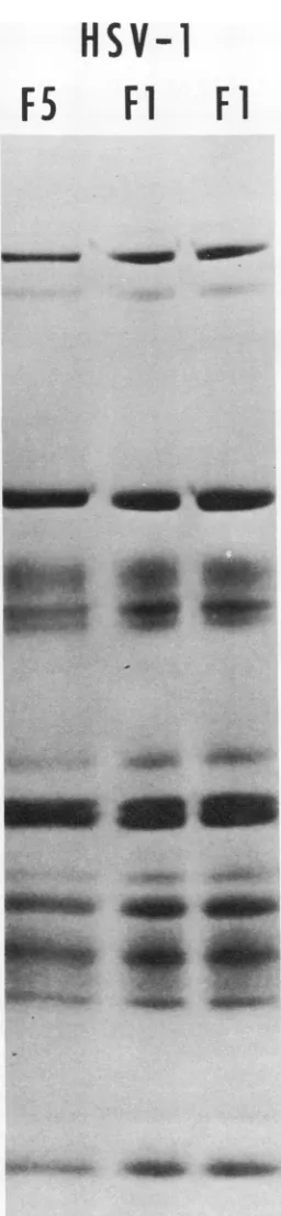
FIG.3. Autoradiogram ofapolyacrylamide gel slab containing electrophoretically separated polypeptides fromtwoindependently purified preparations of HS
V-I [Fl
I
virions and from purified virions of a virus strain with similar isolation and limited passage history,differing in their isolation and passage histories.
The basis for thiscomparison isthe observation
that the electrophoretic profiles of virion
poly-peptides obtained from numerous independ-ently purified preparations of HSV-1 [F1 ] inthe
same type ofgel are identical. Figure 3 shows
autoradiogramsofelectrophoretically separated
virion polypeptides of two independently puri-fied preparations of HSV-1 [Fl] virions and
those of strain HSV-1 [F5] characterized by a
similar isolation andpassage history. Themain
feature ofthe data presentedinFig.3isthatthe
three autoradiograms cannot be differentiated
with respect to the relative amounts and
elec-trophoretic mobility of thevirionpolypeptides.
In contrast, the autoradiogram of
electro-phoretically separated virion proteins of five
HSV-1 strains (HSV-1 [13], HSV-1 [13 v B4]
HSV-1 [HFEM], HSV-1
[mP],
and HSV-1[MP]), Fig. 4 and 5, demonstrate that the
structural polypeptides of strainscharacterized
by long histories of passage in cell culture, and
in some instances in experimental animals,
differextensively fromthose oftheHSV-1 [Fl]
strain. The autoradiograms in Fig. 4 are
pre-sented as they appeared on the intact gel slab
and separately to aid in pinpointing those
polypeptides which areeithermissing in one or
more strains orwhich donotcoincidein
electro-phoretic mobility with those of HSV-1 [Fl].
Specifically, HSV-1
[mP]
differed from HSV-1[F1] inthat VP13 and 15.8 wereabsent, whereas
anewpolypeptide appeared betweenVP15and
VP16. HSV-1 [MP] was identical to HSV-1
[mP]from whichitwasderivedexceptthatVP8
was absent or drastically reduced. In the
ab-sence of VP8 the minor virion polypeptide
VP6.5 (VP6A of ref. 9) can be observed on
DATD cross-linked gels (see HSV-1 [MP] and
HSV-1 [13 ts B4] electropherograms of Fig. 4).
This trace component is demonstrable in all virions so far examined by electrophoresis on
MBA gels, where it migrates slower than VP7
(see Fig. 1A). Virions of HSV-1 [MP] also
lacked VP15.8 and thereappearedtobe
quanti-tative differences in the region ofVP17 and 18
as compared with corresponding regions of
HSV-1 [mP] and of HSV-1 [Fl]. HSV-1
[HFEM
]
differed in several respects fromHSV-1 [Fl]. The polypeptides in the mobility
range of VPll and 12 were more widely sepa-rated and neither comigrated with those of
HSV-1 [Fl]; the amount of VP13 was reduced
and an additional polypeptide was present in
theregion between VP16 andVP17. Inaddition,
HSV-1 [F5]. Allpreparations werelabeled with 14C-amino acids.
647
VOL.14,1974
on November 10, 2019 by guest
http://jvi.asm.org/
[image:8.493.74.202.66.619.2]648
B4 HFEM MP m P
.-.~A *A
14
HEINE ET AL.
HSV-1
F1
'-U
I'M'
A_-go
>~~~~ >* ~~~~~~~8 *-~~~~=
-412
___l12e*p4
I13
1314
~~15
1111
3>1=<3>n£<>-
_~~~~~~~~~~415.8
1-5<1
158
_
>_22£__.-!_
~~~~~~~~~~~~~16
417-18
17-18
-~3
id
w
_
19C21
"REgEl -som-_ f"woI3-I- - 2223
FIG. 4. Autoradiograms ofapolyacrylamidegelslabcontaining electrophoretically separatedpolypeptides
from purified virions of HSV-i Fl],HSV-1 [mP],HSV-1 [MP], HSV-1 [HFEM],andHSV-1 [13tsB4]labeled with "4C-aminoacids. The intact autoradiogram is shownonthe right together with the designations ofHSV-1
[Fl]virion polypeptides. To facilitate comparisons, the autoradiogramsof each gelarealso shownontheleft.
Thearrows onthe (-) side of each gel pinpoint the positionofHSV-l [FlIpolypeptides absent from thevarious
HSV-1 strains examined. Thearrows onthe(+)side indicate thepolypeptides in virions of these otherstrains
for which there isnoelectrophoretic analogue in the HSV-1 [Fl]virions. Thearrowsagainstthe gelcontaining
the electrophoretically separated HSV-1 [Fl] polypeptides indicate all polypeptides whose electrophoretic analogueisabsentinone or moreof the other HSV-1 strains tested.
VP18 was either reduced in amount orabsent
from the HFEM. Theelectrophoretic profile of HSV-1 [13 v B4] proteins diverged more from
thatofHSV-1 [F1] polypeptides than did those of theother virus strains.Thus, VP8, 13 and14
weremissing, polypeptidesinposition 11and12
wereeven more widely separated than those in
thecorresponding region of the HSV-1
[HFEM
]electropherogram, and two additional polypep-tideswereobserved in the region between VP12
and 13 ofHSV-1 [Fl]. The additional polypep-tideobserved inthe region between VP15 and16
intheother strainswasalsopresent;and lastly, VP22 and 23 were separated by asignificantly
larger distance than the corresponding polypep-tides ofHSV-1 [F1]. Figure 5shows
autoradio-gramsof thepolypeptides ofthe parentHSV-1 [13] strain and of HSV-1 [Fl] subjected to
electrophoresis in different slots of a
single
polyacrylamide gel slab, but with an
interven-ing sample removed to permitidentification of
the polypeptides. It is noteworthy that HSV-1 [13
]and
HSV-1 [13vB4]differonly in theregion
of the major virion glycoproteins, VP7-8.5. These differences may be the consequences of mutagenesis of the viral DNA with BUdR.
DISCUSSION
Thispaperdeals with threeaspectsof
protein
composition of HSV-1 virions. It is convenienttodiscuss each of thesetopics
separately.
Number of polypeptide species in theB4
HFEM
MP
J.VIROL.
mP
Fl
go-N
,+
wv*+.J~~
I_- . 1 245
8
78.5
48
48.5
_N_r
"own
on November 10, 2019 by guest
http://jvi.asm.org/
[image:9.493.44.471.65.411.2]PROTEINS SPECIFIED BY HSV
HSV-1 virion. In anearlierpaper inthis series,
(22) our
laboratory' reported
thatpolypeptides
containedinHSV-1 [F]virionsformed24bands
onelectrophoresis through polyacrylamide gels
cross-linked with MBA. In this paper we are
reporting that we have resolved several
addi-tional species of
polypeptides
by
the use ofpolyacrylamide gels cross-linked with DATD.
The separations of nonglycosylated
polypep-tides obtained in those gels were in
general
comparable to those previously obtained on
MBA cross-linked gels, and the agreement
be-tween the molecular weights estimated in this
and preceding studies were excellent
(Table
1,columns a and b). The main
advantage
oftheDATD cross-linked gels was their
ability
toseparate a number ofminor,
glycosylated
pro-teins from thenonglycosylated polypeptides
with which they comigrated on MBA
cross-linked gels. These analysesonDATD gels have
shown that the HSV-1 virion contains at least
33 electrophoretically distinct species of
poly-peptides. Several comments should be madein
connection with this observation.
Although
the33 polypeptides do not exceed the potential
informational content ofHSV-1 DNA, we
can-notexclude thepossibility that notall
polypep-tide moieties of the 33 glycosylated and
non-glycosylated polypeptides are primary gene
products. It may be that the virion contains
both a nonglycosylated precursor and its
gly-cosylatedproductorthe precursorandproducts
from cleavage of a glycosylated polypeptide.
However, the improved separation of minor
glycosylated polypeptides has shown that
they
donot differ
significantly
intheextent oftheirglycosylation from that of the major
glyco-proteins. Thus, if minorglycosylated
polypep-tides represent a
glycosylated
subset of othernonglycosylatedvirionpolypeptides,it is
appar-ent that
glycosylation
is as extensive on thissubset as on themajor glycoproteins.
Molar concentrations of virion
polypep-tides. The data presented in Table 1 are of
interest from several points of view. First, they
illustrate the large disparities between the
molar concentrations of certain polypeptides.
These are evident, for example, from
compari-sons of the number of molecules per virion of
VP4 or VP6 as with those ofVP5, 14, 16, 22, and
23. Second, it is ofinterest to note that at least
two capsid (VP5 and
19C)
and twonon-glycosylated, noncapsid (VP14, 16)
polypep-tides are present in nearly identical molarities
suggesting possible interaction between each
pair in the corresponding substructures of the virion.On the otherhand the data point to some ofthe problems encountered in theattempts to
13
+
FlI
3
4
5
8
7.5
VP
o-12
J~~~~
13*
14
>
..4
158
''1 5Mow
*15.8-16
_UP
17-18
19E
fi-im
19C
21
4_.
in~
22
23
FIG. 5. Autoradiograms oftwo slots from a single
polyacrylamidegel slab containingelectrophoretically separated polypeptides from purified virions of
HSV-1 [13]and HSV-1 [Fl ] labeled with 14C-amino acids. Theprint oftheautoradiogram was labeled as in Fig.
4.
estimate the molar concentrations of certain
polypeptides. We refer specifically to the wide
differences in theestimates of VP8, 8.5, and 22
obtained from analyses of the stained gels and
from autoradiograms of such gels. We have
al-649
VOL. 14, 1974
on November 10, 2019 by guest
http://jvi.asm.org/
[image:10.493.250.441.73.555.2]ready reported the anomalous staining of these
polypeptides (7, 6, 22) and it is likely that the
discrepancies between the two methods of
esti-mation are largely due to differences in the
binding ofCoomassiebrilliant blue. Ingeneral,
however, the buildingofmodelsofthestructure
of the herpesvirion will probably require the
purification and characterization of subviral
structures.
Variability in the structure of the HSV-1
virions.Theobservedvariation inthe polypep-tide composition of the virions from these
strainshasanumberofimplications. First,it is
evident that the observed variations are not
random, but rather that certain polypeptides
varyamong the virus strains, whereas others are invariant in all strains so far examined. The
variable polypeptides are chiefly noncapsid
components. This is perhaps not surprising
since the constraints on the conformation of
noncapsid polypeptides, which would include
thoseofthe envelope, may belowerthan those
ofcapsidpolypeptides. Although it isapparent
from these studies that the pressures selecting
thesevariations are not uniform forall
polypep-tides, the nature ofthe selection processes are
not presently known. The observation that a
number of laboratory strains differ from two
isolates ofHSV-1 passagedalimitednumberof
times in culture may mean that the initial
isolates were different or that the laboratory
strains represent variants selected for some
advantageous property in the cells in which
they were passaged. The first hypothesis
im-plies that wild strains ofHSV-1 vary both in
timeandgeographically. Although comparisons
ofHSV-1 [13]andHSV-1
[Fl]
suggestthat thismight be the case, other isolates from Italy
resembled HSV-1 [Fl] rather than HSV-1
[13]
and definitive data that would substantiateor
refute this hypothesis are lacking. However,
several observations support the alternative.
Thus, HSV-1 [MP] arose spontaneously in a
culture infected with HSV-1
[mP]
and has aselective advantage in cultures infected at low multiplicities in that it spreads frominfectedto uninfected cells more rapidly than the parent strain (8). Another example is the selection of spontaneous mutants (HSV-1
[MPdk+lp]
andHSV-1 [MPdk+sp]) capable of multiplying in
dog kidney cells from cultures of these cells
abortively infected with the HSV-1 [MP] virus (17). The mutants differed from the parent
HSV-1 [MP] with respect toimmunologic
spec-ificity, buoyant density in CsCl
solution,
andstability at 40C-all of which are consistent withaltered virion structure.
Finally, the data havesomeimportant
practi-cal implications since concurrent with, or
re-latedto, thevariations in the noncapsid
compo-nents are two other properties of the virus. Thus, the virion may exhibit altered
immuno-logic specificity as has been clearly
demon-strated for HSV-1 [MP] which is neutralized
equally well by both anti HSV-1 and HSV-2
sera, whereas the parent HSV-1 [mP] virus is
morereadilyneutralized by antiHSV-1thanby
antiHSV-2sera(5, 14).Moreover,cellsinfected
with the laboratory strain differ from those
infected with HSV-1 [Fl] in details pertaining
to development asevident fromelectron
micro-scope studies (21) and from the nature of the
social interaction (i.e., tight clumps, loose
clumps, polykaryocytosis) of infected cells
among themselves(5, 16).The obvious implica-tions are that virus strains used as antigenic reagents in seroepidemiologic studies must be
shown tobe identical withthestrains
circulat-ing in the population being analyzed since
misleading data may otherwise result.
More-over, biochemical studies should include suffi-cient information onthevirus strainbeing used
to assess the validity of comparisons of data
generated in different laboratories.
ACKNOWLEDGMENTS
These studies were aided by grants from the National Cancer Institute (Public Health Service grant CA-08494), Chicago Cancer Research Center Grant (CA-14599), the AmericanCancer Society (VC 1031), and the National Science Foundation (GB-38270), and by C. N. R. Contract No. 73.
00688.65 with the University of Ferrara, Italy.J. W. Heine
was afellowoftheNewYorkCancer Institute. R. W. Honess
was a fellowofthe Jane Coffin Childs Memorial Fund. We thank Herbert Anker for the initial sample of the N,N'-dial-lyltartardiamide.
LITERATURE CITED
1. Anker, H. S. 1970. A solubilizable acrylamide gel for electrophoresis. FEBSLett. 7:293.
2. Bachenheimer, S. L., E. D. Kieff, L. Lee, and B. Roizman. 1972. Comparativestudies of DNAsof Ma-rek's disease andherpessimplexvirus, p.74-81.In P. A. Briggs, G. de The, and L. N. Payne (ed.), Onco-genesis and herpesviruses. International Agency for ResearchonCancer, Lyon.
3. Burton, K. 1956. Astudy ofthe conditions and mech-anisms forthediphenylamine reaction forthe colori-metricestimation ofdeoxyribosenucleicacid. Biochem. J. 62:315-323.
4. Dimmock, N. J., and D. H. Watson. 1969. Proteins specifiedbyinfluenzavirus ininfected cells: analysis by polyacrylamidegelelectrophoresisofantigensnot
presentinthe virusparticles.J.Gen.Virol. 5:499-509. 5. Ejercito, P. M., E. D. Kieff, and B. Roizman. 1968.
Characterizationofherpessimplexvirusstrains
differ-ingintheir effect onsocial behaviorofinfected cells.J.
Gen.Virol. 2:357-364.
6. Gibson,W.,andB. Roizman. 1972. Proteinsspecifiedby' herpessimplexvirus. VIII.Characterization and
com-positionofmultiple capsidforms ofsubtypes1and 2.J.
Virol. 10:1044-1052.
on November 10, 2019 by guest
http://jvi.asm.org/
PROTEINS SPECIFIED BY HSV
7. Gibson, W., and B. Roizman. 1974. Proteins specified by herpes simplex virus. X. Staining and radiolabeling properties of B capisd and virion proteins in polyacryl-amide gels. J. Virol. 13:155-165.
8. Hoggan, M. D., and B. Roizman. 1959. The isolation and properties of a variant of herpes simplex producing multinucleated giant cells in monolayer cultures in the
presenceofantibody. Amer. J. Hyg. 70:208-219.
9. Honess, R.W., and B. Roizman. 1973. Proteins specified by herpes simplex virus. XI. Identification and relative molar rates ofsynthesis of structural and
non-struc-turalherpesvirus polypeptidesin the infected cell.J.
Virol.12:1347-1365.
10. Keller, J.M., P. G. Spear, and B. Roizman. 1970. The proteins specified by herpes simplex virus. III. Viruses differing in their effects on the social behavior of
infected cells specify different membrane glyco-proteins.Proc. Nat. Acad. Sci. U.S.A. 65:865-871.
11. Kieff, E.D., S. L. Bachenheimer, and B. Roizman.1971.
Size, composition and structure of the DNA of
sub-types1and2herpes simplex virus. J.Virol.8:125-132.
12. Laemmli, U. K. 1970. Cleavage of structural proteins during the assembly of the head of bacteriophage T4. Nature(London) 227:680-684.
13. Lowry, 0. H., N. T. Rosebrough, A. L. Farr, andR. J.
Randall. 1951. Protein measurement with the Folin phenolreagent.J.Biol. Chem. 193:265-275.
14. Plummer, G., J. L. Waner, A. Phuangrab, and C. R. Goodheart. 1970. Type 1 and type 2herpes simplex viruses:serological and biological differences. J. Virol. 5:51-59.
15. Roizman, B.1962.Polykaryocytosis. Cold Spring Harbor Symp. Quant. Biol. 27:327-342.
16. Roizman, B. 1971. Herpesviruses, membranes and the social behavior of infected cells, p. 37-72. Proc. 3rd
International Symposium on Applied and Medical
Virology (Fort Lauderdale, Fla.). Warren Green Pub-lishers, St. Louis, Mo.
17. Roizman, B., and L.Aurelian. 1965. Abortive infection of caninecells by herpes simplex virus. I. Characteriza-tion of viral progenyfromcooperative infection with
mutantdiffering in abilitytomultiply in canine cells. J. Mol.Biol. 11:528-538.
18. Roizman, B., and P. R. Roane, Jr. 1961. A physical difference betweentwostrains ofherpes simplex virus
apparentonsedimentationincesium chloride.Virology 15:75-79.
19. Roizman, B., and P. R.Roane, Jr. 1963. Demonstration ofasurface difference between virions oftwostrains of herpes simplex virus.Virology 19:198-204.
20. Roizman, B., and P. G. Spear. 1968. Preparation of herpes simplex virus ofhigh titer. J. Virol. 2:83-84.
21. Schwartz, J., and B. Roizman. 1969. Similarities and differences in the development of laboratory strains andfreshly isolated strains of herpes simplex virus in HEp-2 cells: electron microscopy. J. Virol. 4:879-889. 22. Spear, P. G., and B. Roizman. 1972. Proteins specified by
herpes simplex virus. V. Purification and structural proteins of theherpesvirion. J. Virol.9:143-159.
23. Wagner, E. K., K. K. Tewari, R. Kolodner, and R. C. Warner.1974.Themolecular size of the herpes simplex virustype1genome.Virology 57:436-447.
24. Wilkie, N. M. 1973. Thesynthesis and substructure of
herpesvirus DNA: the distribution of alkali-labile
single strand interruptions in HSV-1 DNA. J. Gen.
Virol. 21:453-467.
VOL. 14, 1974
![FIG. 2.glycosylatedpolypeptidesproteinpolypeptidesproteinsatdistancemoleculartogetherthe all Molecular weights of HSV-1 [Fl] virion polypeptides determined from their migration relative to standards of known molecular weight (open circles, identified at th](https://thumb-us.123doks.com/thumbv2/123dok_us/1581270.110754/5.493.67.447.55.430/glycosylatedpolypeptidesproteinpolypeptidesproteinsatdistancemoleculartogetherthe-molecular-polypeptides-determined-migration-standards-molecular-identified.webp)
![TABLE 1. Enumeration, quantitation, and glycosylation of virion polypeptides of HSV-1 [Fl]](https://thumb-us.123doks.com/thumbv2/123dok_us/1581270.110754/6.493.41.439.42.467/table-enumeration-quantitation-and-glycosylation-virion-polypeptides-hsv.webp)
![TABLE 2. Determination of protein/DNA ratio ofpurified virions of HSV-1 [Fl ]](https://thumb-us.123doks.com/thumbv2/123dok_us/1581270.110754/7.493.61.255.70.201/table-determination-protein-dna-ratio-ofpurified-virions-hsv.webp)
![FIG. virions of HSV-i of a Fl], HSV-1 [mP], HSV-1 [HFEM], [MP], HSV-1 separated polypeptidesfrom polyacrylamide gel slab containing purified 4](https://thumb-us.123doks.com/thumbv2/123dok_us/1581270.110754/9.493.44.471.65.411/fig-virions-hfem-separated-polypeptidesfrom-polyacrylamide-containing-purified.webp)
![FIG. 5.polyacrylamideseparated1The [13] Autoradiograms of two slots from a single gel slab containing electrophoretically polypeptides from purified virions of HSV- and HSV-1 [Fl ] labeled with 14C-amino acids](https://thumb-us.123doks.com/thumbv2/123dok_us/1581270.110754/10.493.250.441.73.555/polyacrylamideseparated-autoradiograms-containing-electrophoretically-polypeptides-purified-virions-labeled.webp)
