0022-538X/96/$04.00
1
0
Copyright
q
1996, American Society for Microbiology
Reversible Dissociation of the Poliovirus Replication Complex:
Functions and Interactions of Its Components in
Viral RNA Synthesis
DENISE EGGER, LUIS PASAMONTES,† ROGER BOLTEN, VITALY BOYKO,
ANDKURT BIENZ*
Institute for Medical Microbiology, University of Basel, Basel, Switzerland
Received 8 July 1996/Accepted 5 September 1996
Membrane-bound replication complexes containing transcriptionally active replicative intermediates (RI)
can be isolated from poliovirus-infected HEp-2 cells and consist of rosette-like structures of virus-induced
vesicles surrounding the replicating viral RNA. At low ionic strength and low temperature, the rosettes
reversibly dissociate into individual tubulated vesicles. As determined by immunoelectron microscopy and
immunoprecipitation, the vesicles carry a set of viral structural and nonstructural proteins as well as RI RNA.
At 30
&
C, the vesicles reassociate into rosettes synthesizing plus-strand RNA in the RI. The in vitro
transcrip-tional activities of rosettes and vesicles kept separated by high dilution were assessed by an RNase protection
assay. The synthesis of the first 178 nucleotides at the 5
*
end of the plus strand was considered to reflect
initiation, and the detection of a 530-nucleotide fragment in the P2 genomic region was considered to reflect
elongation. It could be shown that the initiation and elongation of plus strands on individual vesicles are
comparable to those in rosettes, with initiation proceeding in de novo-assembled initiation complexes. By use
of detergent treatment it was found that initiation, but not elongation, is dependent on vesicular membranes.
During replication of poliovirus RNA, plus-strand genomic
RNA is copied by the viral polymerase 3D
pol(4, 19, 55) into
minus-strand RNA, which acts as a template for the synthesis
of progeny plus strands in the replicative intermediate (RI)
(21). Plus-strand synthesis depends on specialized cellular
membraneous structures (12, 16, 18, 39), whereas the
intracel-lular structural prerequisites for minus-strand RNA synthesis
are not known. Virtually all noncapsid proteins, i.e., the P2 (7,
14, 26, 30, 36, 52, 56) as well as the P3 proteins (2, 3, 20, 23, 27,
35, 41, 55), in addition to cellular factors (2, 33, 47), seem to be
involved in plus-strand synthesis.
The RI-dependent plus-strand RNA synthesis proceeds in a
viral replication complex (reviewed in reference 10) which was
identified by electron microscopy (9, 13) and found to be
lo-cated on the surfaces of virus-induced cytoplasmic vesicles.
The intracellular formation of these vesicles has been
attrib-uted to the viral protein 2BC (13). These vesicle-inducing and
membrane-altering properties of 2BC have been recently
con-firmed by expressing recombinant P2 proteins 2BC and 2C in
cultured cells (1, 17). The altered membranes of
poliovirus-infected cells have been found to be of multiorganelle origin,
including the rough endoplasmic reticulum as well as the Golgi
complex (49).
Upon isolation from infected cells, the virus-induced vesicles
were found to form rosettes which surround the replication
complex (12). The vesicular rosettes can be isolated in a
func-tional state, and the rosettes continue to initiate (51) and
elongate and release (11, 14, 51) progeny plus-strand RNA in
an in vitro transcription system. Such systems rely on the
pre-formed structures isolated from infected cells and thus are
different from the recently described cell-free
translation-tran-scription systems, which are reconstituted from uninfected
cells and programmed with virion RNA (5, 6, 37, 38).
In investigating the structure of the rosette and the nature of
the association of the viral RNA with components of the
ro-sette, it was found that only mature progeny plus-strand RNA
is present on the surface of the rosette (11), whereas the actual
RNA-synthesizing machinery is tightly enclosed and protected
in the interior of the rosette (11). This arrangement impedes to
a large extent the in situ investigation of the architecture of the
rosettes. In the present study, in trying to characterize the
individual components of the functional replication complex,
we found that rosettes can reversibly be dissociated into their
components, i.e., the virus-induced vesicles, and reassociated
into rosettes. This proved to be helpful in elucidating some of
the structural and functional features of the viral replication
complex. It was found that the vesicles undergo extensive
tu-bulation on their side facing the center of the rosette. They
carry sets of nonstructural and capsid proteins as well as an RI
which was found to be attached to the surface of individual
vesicles. Vesicles reassociate into rosettes which synthesize
viral RNA in the RI. By using an RNA protection assay, it was
possible to demonstrate that initiation of plus-strand RNA
synthesis as well as elongation of viral RNA can take place not
only in rosettes but also on single vesicles. Individual vesicles,
therefore, are equipped for initiation and elongation and can
sustain the transcriptional activity of the viral RI.
MATERIALS AND METHODS
Cells, virus, and isolation of the replication complex containing rosettes. HEp-2 cells and poliovirus type 1 (Mahoney) were grown in suspension cultures. The multiplicity of infection for the experiments was 30 PFU per cell. The preparation of cytoplasmic extracts and the isolation of the vesicle-attached replication complexes (rosettes) was as published previously (14). In short, cy-toplasmic extracts were centrifuged onto a double sucrose cushion (30 and 45% sucrose), and the band on the 30% sucrose was harvested (30% sucrose fraction). In vitro RNA synthesis and dissociation and reassociation of rosettes.The 30% sucrose fractions were stored at2708C. For the experiments, suitable aliquots were thawed, and 7 to 10ml was added directly to 40ml of an in vitro RNA-synthesizing system as described previously (14, 51). To dissociate rosettes into single vesicles, rosettes were diluted 1:3 with water before or 1:10 with 10
* Corresponding author. Mailing address: Institute for Medical
Mi-crobiology, University of Basel, Petersplatz 10, CH-4003 Basel,
Swit-zerland. Phone: 41-61 267 3290. Fax: 41-61-267 3298. Electronic mail
address: [email protected].
† Present address: F. Hoffmann-La Roche AG, Vitamin Research
Biotechnology 64/7, CH-4070 Basel, Switzerland.
8675
on November 9, 2019 by guest
http://jvi.asm.org/
transcription with SP6 or T7 polymerase (Boehringer) from the corresponding DNA template which had the SP6 or T7 promoter added by PCR (50, 58). The probe was detected with anti-DIG antibody coupled to alkaline phosphatase (Boehringer) with nitroblue tetrazolium–5-bromo-4-chloro-indolylphosphate toluidinium as a color reagent.
RNase protection assay to demonstrate initiation and elongation.To test for synthesis of newly initiated viral plus-strand RNA and for elongation of plus strands in rosettes or vesicles, viral RNA was pulsed with 0.5 to 1.75mCi of [35S]UTP (Amersham) perml, phenol-chloroform-isoamyl alcohol extracted, and
subjected to an RNase protection assay. For this assay, unlabeled riboprobes of minus polarity, spanning nucleotides (nt) 1 to 178 and 4124 to 4653, were hybridized to the labeled viral RNA at 458C overnight in Ambion hybridization buffer (Ambion Inc., Austin, Tex.). After hybridization, the RNA was digested with a mixture of RNases A (0.045 U/ml) and T1(1.8 U/ml) for 30 min at 378C
and then digested with proteinase K (130mg/ml) for 15 min at 378C (all enzymes from Boehringer), before being analyzed on a 2.4% Nusieve (FMC, Rockland, Maine) denaturing agarose gel. The gel was blotted as described above and analyzed on a PhosphorImager (Molecular Dynamics). The presence of a radio-labeled band of 178 nt indicated that the very 59end of the viral plus-strand RNA was synthesized during the pulse time and thus that plus-strand RNA synthesis had been initiated, whereas the presence of a band of 530 nt, originating from the middle region of the genome, indicated ongoing plus-strand RNA synthesis. As negative controls, hybridization was performed without added probe and after RNase digestion of the nascent plus strands of the RI.
MAb. The preparation and specificities of monoclonal antibodies (MAb) against 2C (40) and 14S (43) were described earlier. MAb against proteins 3Dpol
and 2B were prepared as described previously (40). As an antigen immunizing against protein 3D, a fusion protein ofb-galactosidase and 3D expressed by the thermoinducible plasmid pEX2 in Escherichia coli NF1 was used. For induction of the MAb against 2B, the appropriate poliovirus sequence was PCR amplified, introduced in the IPTG (isopropyl-b-D-thiogalactopyranoside)-inducible vector
DHRF, and expressed in E. coli M15. The expressed proteins were purified by polyacrylamide gel electrophoresis (PAGE) followed by electroelution (40) and Centriprep (Amicon, Danvers, Mass.) concentration. CBA mice and BALB/c mice were immunized with proteins 3D and 2B, respectively. The isolated spleen cells were restimulated in vitro with 1 to 5mg of the appropriate antigen per ml during 3 days in the presence of interleukins 2, 4, 5, and 6 (a kind gift of P. Erb, Institute for Medical Microbiology, University of Basel) before being fused to SP2/0 cells with polyethylene glycol. The hybridoma supernatants were screened for MAb binding to their target proteins on Western blots (immunoblots) in order to select for sequence-specific MAb, which were previously found to be most suitable for immunocytochemistry (8).
Immunoprecipitation and detergent treatment of vesicles.For immunopre-cipitation of native, non-detergent-treated vesicles, MACS beads (Miltenyi Bio-tec GmbH, Bergisch Gladbach, Germany) were used, since they allow direct examination of the precipitate by negative-staining EM and immuno-EM (IEM) because of their diameter of only 30 nm. For immunoprecipitation, 60ml of rosettes was dissociated in 4 volumes of Tris (10 mM) and kept at 08C for 15 min. Seventy microliters of MAb (hybridoma supernatant) was added, and after 30 min at 48C, 10ml of MACS (rat anti-mouse immunoglobulin G1-coated mi-crobeads) was added and the incubation at 48C was continued for another 15 min. Adsorption to, washing of, and elution from the magnetic column into 500 to 800ml of 10 mM Tris buffer were done as indicated by the manufacturer.
For immunoprecipitation of detergent-treated vesicles, sheep anti-mouse an-tibody-coated magnetic beads (M 280; Dynal, Oslo, Norway) were used as de-scribed previously (42). For immunoprecipitation, detergents were added to the vesicles 15 min before the addition of the Dynabeads, and detergents were included in all incubation and washing steps. Final concentrations of detergents were 0.1 and 0.5% for Nonidet P-40 (NP-40) (Boehringer), 0.7% for Triton X-100 (Boehringer), and 0.2% for Na-deoxycholate (DOC) (Difco, Detroit, Mich.).
transcription buffer by gel filtration through Bio-Spin 6 columns (Bio-Rad, Rich-mond, Calif.). MAb against 2B and 2C had the same concentration as measured by titration on dot blots. For some experiments, the MAb against 2C was concentrated 16 times by lyophilization.
RESULTS
Dissociation of rosettes and morphology of their
compo-nents.
Upon isolation by cell fractionation and sucrose
gradi-ent cgradi-entrifugation, the poliovirus replication complex presgradi-ents
itself as an RNA-synthesizing structure tightly enclosed in a
rosette of virus-induced vesicles (14). In an attempt to
inves-tigate the architecture of such rosettes and the interplay of
their components, we found the rosettes (Fig. 1a) to be
disso-ciable into their components (Fig. 1b) by lowering of the
tem-perature to 0
8
C and decreasing of the salt concentration from
150 mM to 30 to 40 mM by diluting the rosette-containing
subcellular fraction with 10 mM Tris-HCl buffer or water. A
low temperature was found to be more important for the
dissociation process than a low salt concentration, since partial
dissociation was obtained at 150 and 80 mM salt at 0
8
C. The
dissociation of the rosettes was found to be reversible in that
increasing the temperature to 30
8
C leads to reformation of
rosettes (see below).
Negatively stained preparations of dissociated rosettes
con-tain individual vesicles which carry pieces of a granular
struc-ture with an appearance very similar to that found in the center
of the intact rosette and identified previously as the actual
replication complex (11). These pieces are either capping the
vesicles or covering small protrusions on the surface of the
vesicles. The replication complex-like caps are also found to be
associated with long tubules, of about 40 nm in diameter and
200 to 500 nm in length, emerging from many of the vesicles
(Fig. 1c).
A much less frequently occurring population of vesicles has
a diameter of approximately 50 nm (not shown). They
corre-spond to the small, compact vesicles within the center of intact
rosette described earlier (11). These small vesicles never were
observed to carry any caps. Isolated larger replication
complex-like structures complex-like those obtained after DOC treatment of
rosettes (11) are virtually never found.
Immunocytochemical localization of viral proteins on the
components of dissociated rosettes.
Previous IEM studies (10,
11, 14) have shown that the P2 proteins 2B and 2C and their
precursors are located exclusively on the rosettes and thus can
serve as marker proteins for the replication complex. IEM of
dissociated isolated rosettes showed that all replication
com-plex-like caps on the surface of the vesicles and tubules contain
P2 proteins (Fig. 2a and b). 14S pentamer capsid precursors,
which were previously shown to be abundant in the replication
complex (42), were also found by IEM in many, albeit not all,
on November 9, 2019 by guest
of the caps (Fig. 2c). Labeling with the anti-3D MAb, however,
was inconsistent and weak (not shown). It is not clear whether
only some caps contain proteins with 3D epitopes or whether
3D is not easily accessible for immunolabeling. Alternatively,
the caps could contain very small (i.e., catalytic) amounts of
3D.
The IEM experiments indicate that the caps consist of, or at
least contain substantial amounts of, replication
complex-de-rived material. The observations also indicate that dissociation
of the rosette in the cold is fundamentally different from the
dissociation of rosettes by guanidine (14). Guanidine, by
inter-fering with protein 2C (44), detaches the replication complex
from the vesicular membrane, thus giving rise to “naked”
ves-icles (14), whereas dissociation in the cold seems to break up
the replication complex into pieces, which are still attached to
the vesicular surface. In the following experiments, the protein
and RNA contents of the caps were investigated.
Immunoprecipitation of dissociated rosettes.
Native,
disso-ciated rosettes were immunoprecipitated with the MACS
sys-tem, and the precipitate was analyzed by IEM. The MAb
directed against 2C, 2B, 3D, and 14S pentamers precipitated
the same tubulated vesicles as observed in
nonimmunoprecipi-tated dissociated rosettes (Fig. 3; compare with Fig. 1b and c
and 2). Analysis of such precipitates on Western blots with a
cocktail of our noncapsid MAb showed that all MAb
precipi-tated, in addition to the proteins containing their own epitope,
the entire set of P2 and P3 proteins recognized by the detection
system (Fig. 4).
To test whether viral proteins are associated directly with
each other or rather by virtue of the vesicular membrane,
dissociated rosettes were detergent treated with either NP-40,
Triton X-100, or DOC, and immunoprecipitation was carried
out with Dynabeads. Under these conditions, the anti-P2 MAb
no longer coprecipitated protein 3D (Fig. 5) and, inversely, the
MAb against 3D did not coprecipitate the P2 proteins (Table
1), whereas the capsid proteins were found to remain
associ-ated with P2 proteins as well as with 3CD whether
immuno-precipitation was done with MAb against 2B, 3D, or 14S
(Ta-ble 1). This latter observation might also reflect an interaction
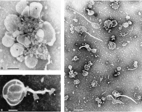
FIG. 1. (a) Rosette of virus-induced vesicles (V) surrounding a granular structure (RC) containing the replicating viral RNA, thus representing the actual replication complex. Such rosettes are isolated from infected cells by sucrose gradient centrifugation and synthesize viral plus-strand RNA in the RI in vitro. Bar, 100 nm. (b) Upon incubation at 0 to 48C in low-ionic-strength buffer, rosettes dissociate into single vesicles. Bar, 500 nm. (c) Many of the vesicles carry tubules and pieces (caps [arrowheads]) of the granular replication complex. Bar, 100 nm.
on November 9, 2019 by guest
http://jvi.asm.org/
[image:3.612.63.552.69.455.2]of soluble 3CD (recognized by MAb against 3D) and 14S
capsid proteins, as described previously (29).
[image:4.612.66.551.68.342.2]Viral RNA species associated with components of
dissoci-ated rosettes.
To test whether the viral RNA would be set free
by the process of dissociation of the rosette, RNase was added
to dissociated rosettes. The nascent plus strands on the RI
FIG. 2. IEM of single vesicles obtained by dissociation of rosettes in the cold. All caps were found to be labeled with MAb against 2C (a) and 2B (b), but only a subpopulation was labeled with MAb VP1, recognizing the 14S pentamer configuration (c). The non-vesicle-associated gold label in panel c represents free 14S pentamers. Bars, 100 nm.
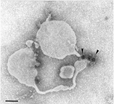
[image:4.612.346.522.407.664.2]FIG. 3. IEM preparation of immunoprecipitate obtained from single vesicles of dissociated rosettes. MAb against 2B and MACS magnetic beads (arrow-heads) precipitated tubulated vesicles identical to those shown in Fig. 2b. Bar, 100 nm.
FIG. 4. Western blot of immunoprecipitate obtained with MACS magnetic beads as in Fig. 3 and MAb against 2B, 2C, and 3D. To test for coprecipitation of P2 and P3 viral proteins not recognized by the precipitating antibodies, the blot was developed with a cocktail of MAb against 2B and 2C and two MAb against 3D, one recognizing the N-terminal part of 3D, including 3C9, and one recognizing the C-terminal part of 3D, including 3D9. (M), poliovirus marker proteins.
on November 9, 2019 by guest
[image:4.612.64.293.482.690.2]were found to be RNase sensitive, and the RI was converted
into core (18), whereas the entire RI was RNase sensitive only
after DOC treatment of the vesicle preparation (Fig. 6). This
suggested an association of the RI, or its core, with
mem-branes.
Therefore, we wanted to know whether the RI would remain
associated with individual capped vesicles when the rosette was
in the dissociated state. To test this, dissociated rosettes were
subjected to MACS immunoprecipitation with the MAb
against 2B (Fig. 3). As shown above, endogenous RNase in the
immunoprecipitation system can attack the nascent plus
strands in the RI as soon as the rosette is dissociated. Thus, the
RI was identified in 2B immunoprecipitates of dissociated
ro-settes by visualizing its 7.5-kb negative strand by Northern
(RNA) blot hybridization with a minus-strand-detecting
DIG-labeled riboprobe (Fig. 7, lane 3).
To confirm that the negative-strand RNA originated from
RI molecules, we tested the RNase accessibility of the 7.5-kb
minus strand before and after DOC treatment of dissociated
rosettes (Fig. 6). Figure 7, lane 5, shows that the minus strand
became RNase sensitive only after DOC treatment of single
vesicles, which indicates that it was of RI origin associated with
a single vesicle.
Reassociation of vesicles into rosettes showing initiation
and elongation activities.
Incubation of dissociated rosettes at
30
8
C causes a reassociation of the vesicles into rosettes as
shown by IEM of such preparations. The vesicles become
grad-ually tighter rosettes (Fig. 8), and in not yet completely
re-formed rosettes, the vesicles still carry their tubules, which
seem to become interwoven, forming a tight network.
Figure 9A shows, on a nondenaturing RNA gel, that
reas-sociated rosettes synthesize RNA, producing the same species
of viral RNA as rosettes which have not been dissociated. To
test whether the reassociated rosette not only elongates
al-ready initiated plus strands but also initiates viral plus-strand
RNA synthesis, an RNase protection assay was performed (see
Materials and Methods). Figure 9B, lane 1, shows that
initia-tion as well as elongainitia-tion is readily demonstrable after
reasso-ciation of vesicles into rosettes.
To test whether the initiation occurred on preformed
initi-ation complexes, e.g., RNA-P3 protein-containing complexes
as described previously (2, 3, 23), or whether such complexes
were newly formed after rosette reassembly, reassociated
ro-settes were preincubated in an in vitro transcription system
with unlabeled nucleoside triphosphates (NTPs) for 0, 30, or
60 min to use up preformed complexes. After that time,
[
35S]UTP was added, and incubation was continued. Figure 9B,
[image:5.612.83.269.68.292.2]lanes 1, 3, and 4, show that with or without preincubation,
FIG. 5. Western blot of immunoprecipitates obtained from single vesicles of dissociated rosettes with Dynabeads and MAb against 2B. The vesicles were either not treated (lane 1) or detergent treated with 0.1% NP-40 (lane 2), 0.5% NP-40 (lane 3), 0.7% Triton X-100 (lane 4) or 0.2% DOC (lane 5) before and during immunoprecipitation. The blot was developed with the same cocktail of MAb as used for Fig. 4. Protein 3D no longer coprecipitated with the anti-2B MAb after detergent treatment. H and L, heavy and light chains of the precip-itating MAb, respectively.
FIG. 6. After incubation for 60 min in an in vitro transcription system sup-plemented with [3
H]UTP, rosettes were dissociated at 08C and either not treated (lane 1) or treated with RNase A (lane 2) or DOC and RNase A (lane 3). RNA was extracted, analyzed on a nondenaturing gel, En3
[image:5.612.58.561.645.718.2]Hance impregnated, and fluorographed. Lane 1, UTP incorporation into the RI and genome-length RNA (vRNA); lane 2, vRNA and the nascent plus strands on the RI are RNase sensitive, so that only the core of the RI remains; lane 3, DOC renders all freshly synthesized RNA RNase sensitive.
TABLE 1. Immunoprecipitation of native and detergent-treated vesicles
Precipitating MAb Proteins precipitated from:
Native vesicles Detergent-treated vesicles
Anti-2B
2BC, 2C, 3D, VP0, VP1, VP3
2BC, 2C, VP0, VP1, VP3
Anti-2C
2BC, 2C, 3D (VP NT
a)
2BC, 2C (VP NT)
Anti-3D
2BC, 2C, 3CD, 3D, VP0, VP1, VP3
3CD, 3D, VP0, VP1, VP3
Anti-14S pentamers
2BC, 2C, 3CD, 3D, VP0, VP1, VP3
2BC, 2C, 3CD, VP0, VP1, VP3
aNT, not tested.
on November 9, 2019 by guest
http://jvi.asm.org/
syntheses of the 5
9
end and central part of the plus strands
were comparable. This argues for newly formed initiation
com-plexes in the in vitro transcription system.
After the membranes were dissolved by DOC, however,
digest the nascent plus strands on the RI (Fig. 6). Upon raising
of the temperature, the vesicles reassociated, as monitored by
EM, and synthesized RNA (not shown). These results point to
protein-lipid or protein-protein interactions as the driving
force for the reassociation of vesicles into rosettes. However,
since the functionality of the RI resides with individual
vesi-cles, as shown below, reassociation and ongoing RNA synthesis
are likely to be independent activities.
Role of individual vesicles in RNA synthesis.
To test which
steps in viral RNA synthesis (initiation or elongation) can be
exerted by individual vesicles, 7
m
l of rosettes was diluted,
dissociated at 0
8
C, and then further diluted 20-fold into an in
vitro transcription system containing [
35S]UTP. The vesicles
were brought to 30
8
C to allow RNA synthesis to proceed. The
purpose of the dilution of the vesicles was to prevent their
reassociation during the rewarming and testing of their
syn-thetic activity. This was confirmed by EM (not shown). Figure
10, lane 1, shows that viral plus-strand RNA was initiated and
elongated. Analysis on native gels showed that [
35S]UTP was
incorporated into the RI and genome-length RNA (not
shown).
In contrast to the results of experiments in which vesicles
were allowed to reassociate into intact rosettes (Fig. 9b),
ini-tiation but not elongation was sensitive to ATA in vesicles kept
separated in the in vitro transcription system (Fig. 10, lane 2).
This experiment indicates de novo formation of initiation
com-plexes on individual vesicles. Thus, our findings show that
individual vesicles are autonomous RNA-replicating units.
DISCUSSION
[image:6.612.88.264.70.245.2]The multicomponent structure synthesizing poliovirus
prog-eny plus-strand RNA in the infected cell is composed of
virus-induced vesicles associated with the actual replication complex
(14). Upon isolation, the vesicles are found to be arranged in
rosettes, forming such a tight structure that it is difficult to get
more insight into its architecture and functioning.
Further-more, the architectural integrity of the replication complex is
generally considered necessary for its ability to replicate viral
RNA (45). This notion stems from experiments in which the
formation of vesicles was blocked by inhibitors such as
Brefel-din A (25, 32) or Cerulenin (22) or in which the replication
complex was disassembled with guanidine (14) or partially
dissolved with detergents (11, 18, 54). The findings reported in
this paper, i.e., not only that the rosette can be dissociated into
single vesicles but also that the vesicles will spontaneously
reform a rosette and that individual vesicles are capable of
initiation and elongation of plus-strand RNA on their own,
indicate that our protocol of dissociation of rosettes in the cold
does not produce artifactual breakdown products but produces
FIG. 7. Northern blot of immunoprecipitate obtained from single vesicles with MAb against 2B and MACS magnetic beads. The blot was probed with a DIG-labeled RNA probe (nt 163 to 443) of plus polarity. The blot shows that RI-derived minus strand is present in the 2B immunoprecipitate. Lane 1, marker; lanes 2 and 3, vesicles before (lane 2) and after (lane 3) immunoprecipitation contain 7.5-kb negative-strand RNA; lane 4, 7.5-kb negative-strand RNA in RNase-treated vesicles is still present; lane 5, in DOC- and RNase-treated vesicles, the 7.5-kb negative-strand RNA is RNase sensitive. Numbers on the left are sizes in kilobases.
FIG. 8. Single vesicles from dissociated rosettes were incubated at 308C. The IEM preparation (label, MAb against 2B) shows vesicles in the process of reassociation into a rosette. Note tubules (arrowheads) extending towards the center of the rosette. Bar, 100 nm.
on November 9, 2019 by guest
[image:6.612.61.297.408.692.2]biologically meaningful subunits of the rosette and the
repli-cation complex. The analysis of these subunits might help to
answer still-open questions about the structure and function of
the replication complex.
A structural analysis of dissociated rosettes showed that
their vesicles have tubular protrusions with parts of the
repli-cation complex attached to them. Upon reassociation, the
pro-trusions extend inwards into the replication complex in the
center of the rosette. It is not clear whether the tubules found
with our virus-induced vesicles play any role in poliovirus RNA
replication. Tubules with very similar structure and dimensions
were found in isolated Golgi complexes (57; for a review, see
reference 48), where their formation was inducible by the
ad-dition of NTPs and abolished by GTP
g
S. However, since
po-liovirus protein 2C was found to have ATP and GTP binding
properties (34, 46) and to be able to induce extensive
tubula-tion of membranes (17), it is tempting to speculate that the
formation of the tubules might depend on NTP-binding and
-hydrolyzing activities of protein 2C, satisfying structural needs
of the RNA replication or the encapsidation step.
Further-more, similarity in the reactions of the Golgi membranes and
of the virus-induced vesicles to GTP-binding proteins would
not be surprising, since Schlegel et al. (49) have found Golgi
membranes, as well as the endoplasmic reticulum, also to be
involved in the formation of virus-induced vesicles.
Membranes of the virus-induced vesicles are not necessary
for elongation of viral RNA but are required for initiation of
viral plus-strand RNA synthesis (Fig. 9b). The membranes may
not only act as a simple carrier for the initiation site, i.e., where
the 3
9
end of the minus-strand template comes together with
viral and cellular proteins and structures participating in
plus-strand initiation (2, 3, 23), but also organize and arrange all
components involved in initiation so that the events making up
initiation can proceed in a highly ordered way. Our approach
to monitor this multistep initiation process was to demonstrate
the synthesis of the very 5
9
end of the viral plus strand, which
is the consequence not only of the actual priming reaction but
of all the other steps of initiation as well. Thus, synthesis of the
5
9
ends of plus strands indicates that all initiation steps have
successfully been accomplished.
Our immunoprecipitation experiments showed that the
cap-sid proteins (14S pentamers), the P2 proteins 2C and 2BC, and
the P3 proteins 3D and 3CD are coprecipitated from isolated
native vesicles. It cannot be determined from this experiment
if every cap contains all proteins. By IEM, all caps contain P2
but only some carry 14S pentamers or 3D or neither. This
indicates that the vesicle population is heterogeneous in
pro-tein content. Preliminary experiments with an anti-3A MAb
are consistent with such a heterogeneity (data not shown).
The association of the P2 proteins with each other and with
the capsid proteins is detergent resistant, whereas the
associ-ation of 3D
polwith all other viral proteins tested in this assay
FIG. 9. (A) Reassociated rosettes (as in Fig. 8) were incubated for 60 min in an in vitro transcription system supplemented with [3
H]UTP and processed on a native gel as indicated in the legend to Fig. 6. UTP was incorporated into the RI and genome-length RNA. (B) Reassociated rosettes as in panel A. RNA was labeled with [35
S]UTP in an in vitro transcription system, extracted, and hybridized to unlabeled, minus-polarity riboprobes complementary to nt 1 to 178 to detect initiation and to nt 4124 to 4653 to detect elongation (see the schematic drawing and Materials and Methods). After RNase digestion, protected radiolabeled, and thus in vitro freshly synthesized, RNA bands were detected on blots of denaturing gels by PhosphoImager analysis. Lane 1, initiation and elongation in reassociated rosettes; lane 2, DOC added to vesicles during reassociation abolishes initiation but not elongation; lanes 3 and 4, reassociated rosettes were preincubated in an in vitro system with unlabeled NTPs for 30 and 60 min, respectively, and after preincubation, [35
S]UTP was added and incubation was continued. Lanes 1, 3, and 4 show comparable 59bands, used to detect initiation. Lane 5, single vesicles were treated with 100mM ATA before being allowed to reassociate at 308C in an in vitro system. In the rosette thus obtained, neither initiation nor elongation is blocked by ATA.
FIG. 10. After dissociation of rosettes, vesicles were kept single by high dilution in an in vitro transcription system at 308C, and an RNA protection assay was performed as described for Fig. 9B to monitor initiation and elongation. Lane 1, single vesicles show initiation and elongation; lane 2, ATA greatly inhibits initiation on single vesicles.
on November 9, 2019 by guest
http://jvi.asm.org/
[image:7.612.115.498.70.257.2]However, the precise location of the RI on the vesicular
sur-face still awaits further clarification.
If one assumes the distribution of viral proteins proposed
above and of the RNA on the vesicular surface to reflect
functional interactions, our findings would also indicate that
the P2 proteins, in addition to their role in viral plus-strand
RNA synthesis, participate in encapsidation (31).
Reassociation of vesicles into rosettes was not inhibited by
MAb against 2C and 2B, by RNase digestion of the nascent
plus strands on the RI, or by ATA. The ineffectiveness of the
MAb in the inhibition of reassociation, despite the possible
implication of the P2 proteins in the organization of the rosette
(14), could be explained by the P2 proteins being already
associated with their target, i.e., membranes. It is less likely
that the epitope recognized by the MAb would be hidden
under these conditions, since individual vesicles can be
immu-nolabeled and immunoprecipitated with anti-P2 MAb.
The failure of ATA to prevent reassociation and, in
ciated vesicles, initiation is puzzling. While ongoing
reasso-ciation with ATA could be explained by RNA-protein
in-teractions not being required for reassociation, initiation
unquestionably involves such interactions. This is also shown
by the finding that ATA greatly inhibits initiation in vesicles
kept separately. However, if we take into account that the
rosette protects the RI from RNase (11) and that Enviroxime
(24) and guanidine (unpublished data) have no effect if added
to in vitro RNA-synthesizing rosettes, a likely explanation for
the lack of ATA-mediated inhibition of transcription in
reas-sociated rosettes could be that early in reassociation the
ro-sette becomes impermeable also to ATA so that the amount of
the drug present within the rosette becomes insufficient to
block initiation.
A hypothetical model of the rosette’s architecture put
for-ward earlier (11) calls for an RI stretched out within a rosette
with its 5
9
ends of the nascent plus strands attached to the
surface of the rosette’s vesicles. However, the observation that
RNase digestion of the nascent 5
9
ends in the RI does not
inhibit reassociation indicates that nascent plus strands do not
seem to be involved in (re-)association of vesicles into rosettes.
Clearly, the vesicles seem to have a strong tendency to
re-form rosettes, even though the mechanism of reassociation is
not known. To form a rosette could be advantageous for virus
replication because it could greatly increase the effectiveness
and speed of plus-strand RNA synthesis. There are two main
reasons for this: first, macromolecular crowding, exerted by the
mass of vesicles, leading to an enhanced concentration of
fac-tors necessary for RNA synthesis; and second, the providing of
more membrane-bound initiation sites (20, 23) for the RI. This
allows the RI, or rather its minus-strand template RNA, to
easily move on within the rosette and to combine with its 3
9
2. Andino, R., G. E. Rieckhof, P. L. Achacoso, and D. Baltimore. 1993. Polio-virus RNA synthesis utilizes an RNP complex formed around the 59end of viral RNA. EMBO J. 12:3587–3598.
3. Andino, R., G. E. Rieckhof, and D. Baltimore. 1990. A functional ribonucle-oprotein complex forms around the 59end of poliovirus RNA. Cell 63:369– 380.
4. Baron, M. H., and D. Baltimore. 1982. In vitro copying of viral positive strand RNA by poliovirus replicase. Characterization of the reaction and its products. J. Biol. Chem. 257:12359–12366.
5. Barton, D. J., P. E. Black, and J. B. Flanegan. 1995. Complete replication of poliovirus in vitro: preinitiation RNA replication complexes require soluble cellular factors for the synthesis of Vpg-linked RNA. J. Virol. 69:5516–5527. 6. Barton, D. J., and J. B. Flanegan. 1993. Coupled translation and replication of poliovirus RNA in vitro: synthesis of functional 3D polymerase and infectious virus. J. Virol. 67:822–831.
7. Bernstein, H. D., P. Sarnow, and D. Baltimore. 1986. Genetic complemen-tation among poliovirus mutants derived from an infectious cDNA clone. J. Virol. 60:1040–1049.
8. Bienz, K., D. Egger, and L. Pasamontes. 1986. Electron microscopic immu-nocytochemistry. Silver enhancement of colloidal gold marker allows double labeling with the same primary antibody. J. Histochem. Cytochem. 34:1337– 1342.
9. Bienz, K., D. Egger, and L. Pasamontes. 1987. Association of polioviral proteins of the P2 genomic region with the viral replication complex and virus-induced membrane synthesis as visualized by electron microscopic im-munocytochemistry and autoradiography. Virology 160:220–226. 10. Bienz, K., D. Egger, and T. Pfister. 1994. Characteristics of the poliovirus
replication complex. Arch. Virol. Suppl. 9:147–157.
11. Bienz, K., D. Egger, T. Pfister, and M. Troxler. 1992. Structural and func-tional characterization of the poliovirus replication complex. J. Virol. 66: 2740–2747.
12. Bienz, K., D. Egger, Y. Rasser, and W. Bossart. 1980. Kinetics and location of poliovirus macromolecular synthesis in correlation to virus-induced cyto-pathology. Virology 100:390–399.
13. Bienz, K., D. Egger, Y. Rasser, and W. Bossart. 1983. Intracellular distribu-tion of poliovirus proteins and the inducdistribu-tion of virus-specific cytoplasmic structures. Virology 131:39–48.
14. Bienz, K., D. Egger, M. Troxler, and L. Pasamontes. 1990. Structural orga-nization of poliovirus RNA replication is mediated by viral proteins of the P2 genomic region. J. Virol. 64:1156–1163.
15. Blumenthal, T., and T. A. Landers. 1973. The inhibition of nucleic acid-binding proteins by aurintricarboxylic acid. Biochem. Biophys. Res. Com-mun. 55:680–688.
16. Caliguiri, L. A., and I. Tamm. 1970. The role of cytoplasmic membranes in poliovirus biosynthesis. Virology 42:100–111.
17. Cho, M. W., N. Teterina, D. Egger, K. Bienz, and E. Ehrenfeld. 1994. Membrane rearrangement and vesicle induction by recombinant poliovirus 2C and 2BC in human cells. Virology 202:129–145.
18. Etchison, D., and E. Ehrenfeld. 1981. Comparison of replication complexes synthesizing poliovirus RNA. Virology 111:33–46.
19. Flanegan, J. B., and D. Baltimore. 1979. Poliovirus polyuridilic acid poly-merase and RNA replicase have the same viral polypeptide. J. Virol. 29: 352–360.
20. Giachetti, C., and B. L. Semler. 1991. Role of a membrane polypeptide in strand-specific initiation of poliovirus RNA synthesis. J. Virol. 65:2647–2654. 21. Girard, M. 1969. In vitro synthesis of poliovirus ribonucleic acid: role of the
replicative intermediate. J. Virol. 3:376–384.
22. Guinea, R., and L. Carrasco. 1990. Phospholipid biosynthesis and poliovirus genome replication, two coupled phenomena. EMBO J. 9:2011–2016. 23. Harris, K. S., W. Xiang, L. Alexander, W. S. Lane, A. V. Paul, and E.
Wimmer.1994. Interaction of poliovirus polypeptide 3CDpro with the 59and 39termini of the poliovirus genome. J. Biol. Chem. 269:27004–27014.
on November 9, 2019 by guest
24. Heinz, B. A., and L. M. Vance. 1995. The antiviral compound Enviroxime targets the 3A coding region of rhinovirus and poliovirus. J. Virol. 69:4189– 4197.
25. Irurzun, A., L. Perez, and L. Carrasco. 1992. Involvement of membrane traffic in the replication of poliovirus genomes. Effects of Brefeldin A. Vi-rology 191:166–175.
26. Johnson, K. L., and P. Sarnow. 1991. Three poliovirus 2B mutants exhibit noncomplementable defects in viral RNA amplification and display dosage-dependent dominance over wild-type poliovirus. J. Virol. 65:4341–4349. 27. Kuhn, R. J., H. Tada, F. Ypma-Wong, J. J. Dunn, B. L. Semler, and E.
Wimmer.1988. Construction of a “mutagenesis cartridge” for poliovirus genome-linked viral protein: isolation and characterization of viable and nonviable mutants. Proc. Natl. Acad. Sci. USA 85:519–523.
28. Laemmli, U. K. 1970. Cleavage of structural proteins during the assembly of the head of bacteriophage T4. Nature (London) 227:680–685.
29. Lawson, M. A., and B. L. Semler. 1992. Alternate poliovirus nonstructural protein processing cascades generated by primary sites of 3C-proteinase cleavage. Virology 191:309–320.
30. Li, J.-P., and D. Baltimore. 1988. Isolation of poliovirus 2C mutants defec-tive in viral RNA synthesis. J. Virol. 62:4016–4021.
31. Li, J.-P., and D. Baltimore. 1990. An intragenic revertant of a poliovirus 2C mutant has an uncoating defect. J. Virol. 64:1102–1107.
32. Maynell, L. A., K. Kirkegaard, and M. W. Klymkowsky. 1992. Inhibition of poliovirus RNA synthesis by Brefeldin A. J. Virol. 66:1985–1994. 33. McBride, A. E., A. Schlegel, and K. Kirkegaard. 1996. Human protein Sam
68 relocalization and interaction with poliovirus RNA polymerase in infected cells. Proc. Natl. Acad. Sci. USA 93:2296–2301.
34. Mirzayan, C., and E. Wimmer. 1994. Biochemical studies on poliovirus polypeptide 2C: evidence for ATPase activity. Virology 199:176–187. 35. Molla, A., K. S. Harris, A. V. Paul, S. H. Shin, J. Mugavero, and E. Wimmer.
1994. Stimulation of poliovirus proteinase 3Cpro-related proteolysis by the genome-linked protein VPg and its precursor 3AB. J. Biol. Chem. 269: 27015–27020.
36. Molla, A., A. V. Paul, M. Schmid, S. K. Jang, and E. Wimmer. 1993. Studies on dicistronic polioviruses implicate viral proteinase 2A (pro) in RNA rep-lication. Virology 196:739–747.
37. Molla, A., A. V. Paul, and E. Wimmer. 1991. Cell-free, de novo synthesis of poliovirus. Science 254:1647–1651.
38. Molla, A., A. V. Paul, and E. Wimmer. 1993. Effects of temperature and lipophilic agents on poliovirus formation and RNA synthesis in a cell-free system. J. Virol. 67:5932–5938.
39. Mosser, A. G., L. A. Caliguiri, and I. Tamm. 1972. Incorporation of lipid precursors into cytoplasmic membranes of poliovirus-infected Hela cells. Virology 47:39–47.
40. Pasamontes, L., D. Egger, and K. Bienz. 1986. Production of monoclonal and monospecific antibodies against non-capsid proteins of poliovirus. J. Gen. Virol. 67:2415–2422.
41. Paul, A. V., X. Cao, K. S. Harris, J. Lama, and E. Wimmer. 1994. Studies with poliovirus polymerase 3Dpol. Stimulation of poly(U) synthesis in vitro by purified poliovirus protein 3AB. J. Biol. Chem. 269:29173–29181. 42. Pfister, T., D. Egger, and K. Bienz. 1995. Poliovirus subviral particles
asso-ciated with progeny RNA in the replication complex. J. Gen. Virol. 76:63–71. 43. Pfister, T., L. Pasamontes, M. Troxler, D. Egger, and K. Bienz. 1992. Im-munocytochemical localization of capsid-related particles in subcellular frac-tions of poliovirus-infected cells. Virology 188:676–684.
44. Pincus, S. E., and E. Wimmer. 1986. Production of guanidine-resistant and -dependent poliovirus mutants from cloned cDNA: mutations in polypeptide 2C are directly responsible for altered guanidine sensitivity. J. Virol. 60:793– 796.
45. Plotch, S. J., and O. Palant. 1995. Poliovirus protein 3AB forms a complex with and stimulates the activity of the viral RNA polymerase, 3D pol. J. Vi-rol. 69:7169–7179.
46. Rodriguez, P. L., and L. Carrasco. 1993. Poliovirus protein-2C has ATPase and GTPase activities. J. Biol. Chem. 268:8105–8110.
47. Roehl, H. H., and B. L. Semler. 1995. Poliovirus infection enhances the formation of two ribonucleoprotein complexes at the 39end of viral negative-strand RNA. J. Virol. 69:2954–2961.
48. Rothman, J. E. 1994. Mechanisms of intracellular protein transport. Nature (London) 372:55–63.
49. Schlegel, A., T. H. Giddings, Jr., M. S. Ladinsky, and K. Kirkegaard. 1996. Cellular origin and ultrastructure of membranes induced during poliovirus infection. J. Virol. 70:6576–6588.
50. Sitzmann, J. H., and P. K. Lemotte. 1993. Rapid and efficient generation of PCR-derived riboprobe templates for in situ hybridization histochemistry. J. Histochem. Cytochem. 41:773–776.
51. Takeda, N., R. J. Kuhn, C.-F. Yang, T. Takegami, and E. Wimmer. 1986. Initiation of poliovirus plus-strand RNA synthesis in a membrane complex of infected HeLa cells. J. Virol. 60:43–53.
52. Teterina, N. L., W. D. Zhou, M. W. Cho, and E. Ehrenfeld. 1995. Inefficient complementation activity of poliovirus 2C and 3D proteins for rescue of lethal mutants. J. Virol. 69:4245–4254.
53. Towbin, H., T. Staehelin, and J. Gordon. 1979. Electrophoretic transfer of proteins from polyacrylamide gels to nitrocellulose sheets: procedure and some applications. Proc. Natl. Acad. Sci. USA 76:4350–4354.
54. Toyoda, H., C. F. Yang, N. Takeda, A. Nomoto, and E. Wimmer. 1987. Analysis of RNA synthesis of type 1 poliovirus by using an in vitro molecular genetic approach. J. Virol. 61:2816–2822.
55. VanDyke, T. A., and J. B. Flanegan. 1980. Identification of poliovirus polypeptide p63 as a soluble RNA-dependent RNA polymerase. J. Virol. 35:732–740.
56. VanKuppeveld, F. J. M., J. M. D. Galama, J. Zoll, P. J. J. vandenHurk, and W. J. G. Melchers.1996. Coxsackie B3 virus protein 2B contains a cationic amphipathic helix that is required for viral RNA replication. J. Virol. 70: 3876–3886.
57. Weidman, P., R. Roth, and J. Heuser. 1993. Golgi membrane dynamics imaged by freeze-etch electron microscopy. Views of different membrane coatings involved in tubulation versus vesiculation. Cell 75:123–133. 58. Young, I. D., R. J. Stewart, L. Ailles, A. Mackie, and J. Gore. 1993. Synthesis
of digoxigenin-labeled cRNA probes for nonisotopic in situ hybridization using reverse transcription polymerase chain reaction. Biotech. Histochem. 68:153–158.