Scholarship@Western
Scholarship@Western
Electronic Thesis and Dissertation Repository
6-19-2012 12:00 AM
Characterization of EIF2Be and Its Interaction with RGS2
Characterization of EIF2Be and Its Interaction with RGS2
William H. Xue
The University of Western Ontario
Supervisor Dr. Peter Chidiac
The University of Western Ontario
Graduate Program in Pharmacology and Toxicology
A thesis submitted in partial fulfillment of the requirements for the degree in Master of Science © William H. Xue 2012
Follow this and additional works at: https://ir.lib.uwo.ca/etd
Part of the Pharmacology Commons
Recommended Citation Recommended Citation
Xue, William H., "Characterization of EIF2Be and Its Interaction with RGS2" (2012). Electronic Thesis and Dissertation Repository. 612.
https://ir.lib.uwo.ca/etd/612
This Dissertation/Thesis is brought to you for free and open access by Scholarship@Western. It has been accepted for inclusion in Electronic Thesis and Dissertation Repository by an authorized administrator of
CHARACTERIZATION OF EIF2Bε AND ITS INTERACTION WITH RGS2
(Thesis format: Monograph)
By
William Xue
Graduate Program in Physiology and Pharmacology
A thesis submitted in partial fulfillment of the requirements for the degree of
Master of Science
The School of Graduate and Postdoctoral Studies The University of Western Ontario
London, Ontario
ii
THE UNIVERSITY OF WESTERN ONTARIO
SCHOOL OF GRADUATE AND POSTDOCTORAL STUDIES
CERTIFICATE OF EXAMINATION
Supervisor
______________________________ Dr. Peter Chidiac
Supervisory Committee
______________________________ Dr. John Diguglielmo
______________________________ Dr. Stanley Dunn
Examiners
______________________________ Dr. Andy Babwah
______________________________ Dr. Sean Cregan
______________________________ Dr. Eric Ball
______________________________ Dr. Graham Wagner (GSR)
The thesis by
William Hui Xue
entitled:
CHARACTERIZATION OF EIF2Bε AND ITS INTERACTION WITH
RGS2
is accepted in partial fulfilment of the requirements for the degree of
Master of Science
iii ABSTRACT
RGS2 is a GTPase accelerating protein for Gαq and its expression can be upregulated in
response to different types of stress. We recently showed that RGS2 can bind to the
translation initiation factor, eIF2Bε, and decrease global protein synthesis. The objective
of this study is to characterize the RGS2 binding domain of eIF2Bε, investigate the effect
of phosphorylation on binding, and examine the functional consequences as a result of
their interaction. To identify the RGS2 binding domain of eIF2Bε, I generated various
truncated eIF2B-GST fusion proteins and used them in an in vitro pulldown assay. I
hypothesized that phosphorylation of eIF2Bε will increase its binding to RGS2 and that
eIF2Bε binding to RGS2 will decrease the GAP activity of RGS2. The results of my
study revealed that the binding domain lies within the C-terminal region of eIF2Bε,
which is consistent with our previous yeast two-hybrid data. A highly conserved amino
acid residue within this region (leucine 576) appeared to play an important role in binding
to RGS2. In addition, mutating the GSK3 phosphorylation site on eIF2Bε increased its
binding to RGS2. However in my hands, purified eIF2Bε did not inhibit GAP activity of
RGS2, contrary to my hypothesis.
iv
DEDICATION
TO MY FAMILY
v ACKNOWLEDGEMENTS
I would like to thank my supervisor, Dr. P. Chidiac for mentoring and guiding me
through the course of my research experience. You have taught me to not be afraid of the
unknown, encouraged me to have curiosity to explore, and to take risk and step of faith to
find out. What you have invested into me has been invaluable, and these years have
helped shape my character and have set me up for success in whatever I endeavor to do in
the future. I would also like to thank my colleagues Dr. C. Nguyen, Dr. E. Zhao, and
Alina Sobiesiak, who have taught me laboratory techniques, for being mentors and for
being patient with me when I was slow in learning and understanding. I would like to
acknowledge my committee members Drs. S. Dunn, J. Di Guglielmo, G. Wagner, L.
Coolen, and M. Tini who have taken time out of their busy schedules to help me and
guide me through the course of my research. Lastly my thanks go to Kevin Leung and
Jeff Law who have been good friends and support during the times of uncertainty and
failed experiments. You guys have encouraged me to not give up, but to persevere
vi TABLE OF CONTENTS
Certificate of Examination ii
Abstract and Keywords iii
Dedication iv
Acknowledgements v
Table of Contents vi
List of Figures viii
List of Abbreviations ix
Chapter 1
Literature Review 1
1.1G-protein coupled receptors (GPCR) 2
1.2G-Proteins 2
1.3RGS proteins 3
1.4Gαq/11 signaling 5
1.5RGS2 5
1.6RGS2 and its role in cardiovascular system 8
1.7Protein synthesis 10
1.8Translation initiation and eIF2B 10
1.9Regulation of eIF2B by eIF2 13
1.10 Interaction between eIF2 and eIF2B 15
1.11 Regulation of eIF2B by RGS2 18
1.12 Structure of eIF2Bε and putative RGS2 binding domain 19 1.13 Regulation of eIF2B by other eIF2 independent mechanisms 21
1.14 GSK3 23
1.15 Regulation of eIF2B by GSK3 25
1.16 Regulation of eIF2B by CK2 26
1.17 Regulation of eIF2B by serine 525 26
1.18 Rationale 27
1.19 Objective overview 28
Chapter 2 30
Methods
2.1 Reagents 31
2.2 Cloning 31
2.3 RGS2 purification 35
2.4 GST-eIF2B ε and GST-fusion mutant purification 37
2.5 Co-immunoprecipitation assay 40
2.6 GST pull down assay 40
2.7 Immunoblot analysis 41
2.8 GTP hydrolysis assay 41
vii
Chapter 3 43
Results
3.1 Region of Interaction on eIF2B ε 44
3.2 Purification of eIF2B ε and mutants from E-coli 49 3.3 Assessing the ability of purified eIF2B ε or truncation
mutants to bind to purified RGS2 52
3.4 Amino acid substitution of residues within the putative RGS2 binding
domain of eIF2B ε 55
3.5 Effects of mutating phosphorylation sties on eIF2B ε binding to RGS2 57 3.6 Mutating phosphorylation site on eIF2B ε and overexpression in
mammalian cells 62
3.7 Looking at functional consequence of eIF2B ε binding on RGS2 GAP
Activity 65
Chapter 4 70
Conclusion and Discussion
4.1 Overview 71
4.2 Significance of Research 71
4.3 Interaction between G-protein signaling and mRNA translation systems 72
4.4 Characterizing the region of binding 73
4.5 Phosphorylation sites 78
4.6 Functional consequences of binding 81
4.7 Future studies 83
References 85
Appendix 91
Permission to reprint figures 92
viii LIST OF FIGURES
1.1GPCR and G-proteins. 4
1.2 List of different RGS proteins known to date 7 1.3 Expression of RGS2 in cardiovascular system 9
1.4 Translation initiation 12
1.5 Regulation of protein synthesis by eIF2 14
1.6 Interaction between eIF2 and catalytic domain of eIF2B ε 17
1.7 Putative RGS2 binding domain 20
1.8 Regulation of eIF2B by eIF2 independent mechanisms 22
1.9 Glycogen synthase kinase 3β 24
2.1 List of primers used for mutagenesis (table 2.1) 33 2.1 Purification of His-RGS2 from E.coli BL21 (DE3) 36 2.2 Purification of GST-eIF2Bε and N/C-terminal truncation mutants 38 3.1 Co-immunoprecipitation of eIF2B ε transfected HEK293 lysates and
purified RGS2 45
3.2 Co-immunoprecipitation using HEK293 lysates transfected with
eIF2Bε/internal deletion mutants and purified RGS2 46
3.3 Densitometry analysis 48
3.4 GST fusion proteins. 51
3.5 GST-pulldown assay between N/C truncated GST-eIF2B ε and His RGS2. 53 3.6 Co-immunoprecipitation assay between eIF2B ε/substitution mutants
transfected HEK293 lysates and purified RGS2. 56 3.7 GST-pulldown assay between GST-300/substitution mutants at the GSK3β
site and His RGS2. 58
3.8 Densitometry analysis 60
3.9 Co-immunoprecipitation of eIF2B ε/substitution mutants transfected
HEK293 lysates and purified RGS2. 63
3.10 Effect of GST-300 on RGS2 GAP activity 67
3.11 Effect of GST-300 on RGS2 GAP activity version 2 69 4.1 Conserved amino acid residues within the putative RGS2 binding domain on
ix LIST OF ABBREVIATIONS
ANOVA- analysis of variance
BL21 (DE3)- E.coli strain lacking Lon and OmpT protease CK2- casein kinase 2
DAG- diacylglycerol
DTT- Dithiothreitol
DYRK- dual tyrosine regulated kinase eIF2- eukaryotic initiation factor 2
eIF2B- eukaryotic initiation factor 2B
ER- endoplasmic reticulum
ERK 1/2- extracellular signal regulated kinase
G proteins- guanine nucleotide binding proteins
GAP- GTP hydrolysis accelerating protein
GCN2- general control nondepressible
GDP- Guanosine diphosphate
GEF- guanine nucleotide exchange factor
GPCR- G-protein coupled receptor
GSK3- glycogen synthase kinase 3
GST- glutathione s-transferase
GTP- Guanosine triphosphate
Gα- alpha subunit of heterotrimeric G proteins
Gαq/11- subtype of Gα that activates phospholipase C Gβ- beta subunit of heterotrimeric G proteins
Gγ- gamma subunit of heterotrimeric G proteins
HEK293- human embryonic kidney
HEPES- 4-(2-hydroxyethyl)-1-piperazineethanesulfonic acid
HRI- heme regulated kinase
IP3- inositol trisphosphate
IPTG- Isopropyl β-D-1-thiogalactopyranoside JNK- cJun N-terminal kinase
MAPK- mitogen activated protein kinase
mRNA- messenger ribonucleic acid
Norit- activated charcoal
PERK- double stranded RNA-activated protein kinase-like ER kinase
PIP2- phosphatidylinositol 4,5-bisphosphate
PKC- protein kinase C
PKR- RNA activated protein kinase
PLC- phospholipase C
PMSF- phenylmethylsulfonyl fluoride
p38- class of MAPK
RGS- regulator of G-proteins SF9- Spodoptera frugiperda
TRIS- tris(hydroxymethyl)aminomethane
tRNA- transfer RNA
tRNAi- initiator transfer RNA
Chapter 1
1.1 G-protein Coupled Receptors (GPCRs):
G protein-coupled receptors (GPCRs) are a class of seven transmembrane domain
proteins found in many types of animal cells and tissues. They recognize and bind to
activating ligands and in turn activate heterotrimeric G proteins by promoting GTP
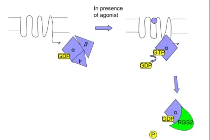
binding in exchange for GDP 1 (Figure 1.1). This leads to the activation of G proteins and initiation of downstream signaling cascades. GPCRs makes up the largest family of
membrane proteins with around 800 human GPCRs known to exist, of which over half
are olfactory receptors and about 340 of them non-olfactory receptors 2. GPCRs are multifaceted in that they are involved in a number of physiological processes such as
mediating signals for hormones, neurotransmitters, ions, photons, and other activators.
GPCRs in vertebrates are generally classified into five different families, each according
to structural and sequence similarities. They are the rhodopsin, secretin, glutamate,
adhesion and frizzled/taste2 receptor families. The largest of these, the rhodopsin family
is the most diverse and comprises several subfamilies, with each member possessing
conserved sequence motifs and hence structural similarities. However despite their
similarities, each GPCR exhibits its own expression pattern and unique signaling
pathways via different subtypes of G-proteins as well as G-protein independent functions
and regulatory processes 3.
1.2 G Proteins:
Heterotrimeric G-proteins are membrane bound proteins comprising three
subunits; alpha, beta and gamma (Gα, Gβ and Gγ) (Figure 1.1). The Gα subunit is able to
GDP, it is inactivated 14. The various Gα subunits are categorized into four classes; Gs which stimulates adenylyl cyclase, Gi which inhibits adenylyl cyclase, Gq which
activates phospholipase Cβ, and G12/G13 which regulates Rho GEFs 5. The Gβ and Gγ
subunits form a stable dimer (Gβγ) that binds and stabilizes Gα-GDP by slowing GDP
dissociation. Notwithstanding this, Gβγ is also required for the activation of Gα by
GPCRs. In addition, Gβγ can also bind to effectors and participate in signaling pathways
such as ion channels, PI-3 kinase, and guanine nucleotide exchange factors for (GEFs)
for small G-proteins 6.
1.3 Regulator of G-protein Signaling (RGS) Proteins:
Regulator of G-protein signaling (RGS) proteins are a class of proteins that
attenuate G-protein signaling by binding to G-proteins (Figure 1.1), and accelerating the
rate of GTP hydrolysis to GDP via a conserved 120 amino acid RGS domain (also known
as the RGS box). This function limits the duration of G-protein signaling and hence fine
tunes the degree to which the signaling pathway will elicit a response. To date over
twenty RGS proteins have been discovered, and they are categorized into five
subfamilies. They are B/R4, RZ, R7, R12, and RL; categorized based on similarities in
regions within the RGS domain as well as outside 7. Four of the five subfamilies are known to have GTPase accelerating activity, and of those most are able accelerate
GTPase activity of the Gi subfamily, and some being able to attenuate activity of the Gq
subfamily 8. No RGS proteins that have been discovered to date can accelerate Gs subfamily GTPase activity although RGS2 can interact with certain isoforms of adenylyl
Figure 1.1GPCR and G-proteins
A GPCR is activated upon binding to a ligand, which stabilizes the conformation favourable towards binding of Gα and catalyzes guanine nucleotide exchange. Upon binding to GTP, the Gα is activated and acts on downstream effectors, while Gβγ acts on its distinct effectors. Gα possesses intrinsic GTPase activity, however upon binding to a RGS protein the GTP hydrolysis is accelerated and the duration of the signaling is shortened.
1.4 Gq/11 Signaling:
Angiotensin II, catecholamines, and endothelin-1 are among the endogenous
agonists that bind to GPCRs that couple to Gαq/11 subfamily G-proteins. Upon
activation Gαq/11 in turn activates phospholipase Cβ (PLCβ), leading to cleavage of
phosphatidylinositol 4,5-bisphosphate (PIP2) and production of inositol trisphosphate
(IP3) and diacylglycerol (DAG) 10. Production of IP3 leads to release of stored
intracellular calcium mediated through the direct binding to IP3 receptor localized on the
endoplasmic reticulum. Intracellular calcium in turn activates various intracellular
proteins including calcineurin and NFAT transcription factors, while conventional
Protein Kinase C (PKC) is activated by calcium and DAG, and it affects MAPK signaling
through kinases such as ERK 1/2, JNK and p38 10.
1.5 RGS2:
RGS2 belongs to the B/R4 subfamily of RGS proteins and is unique among others
in that it selectively inhibits Gαq and Gαs signaling 11
. The human form contains 211
amino acids and its structure comprises a short N-terminal amphipathic region
responsible for cell localization 12 followed by an RGS box, which is responsible for its GTPase accelerating protein (GAP) activity, and a short C-terminus (Figure 1.2).
However in recent years, non-canonical functions of RGS proteins have been discovered
and characterized. Besides its GAP function, RGS2 can interact with the cation channel
TRPV6 and also with tubulin via its N-terminal region. Both of these novel RGS2
interactions map to amino acid residues that lie outside of the RGS box 1314. In addition RGS2 can bind to eIF2Bε and thereby inhibit the translational machinery 15
non-canonical function of RGS2 that our lab has taken recent interest in and the focus of
this thesis is to further understand the interaction between RGS2 and eIF2B.
The expression of RGS2 can be upregulated by Gαq and Gαs mediated signals but
its expression can also be induced by various forms of cellular stress including, heat
shock, DNA damage, viral infection, and oxidative stress 1617. The ability and the role of RGS2 in attenuating G protein signaling have been well characterized, however its role in
response to cellular stress has just begun to be deciphered. The working hypothesis in
our laboratory is that the upregulation of RGS2 in cellular stress serves to decrease the
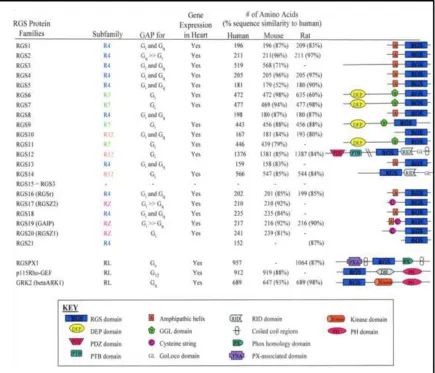
Figure 1.2 List of different RGS proteins known to date 18
RGS2 belongs to the R4 subfamily of RGS proteins, possessing short amphipathic motif near the N-terminus followed by the RGS box. Compared to other subfamily of RGS proteins, RGS2 is relatively small and seemingly has fewer motifs. However in recent years, more binding partners for RGS2 have been discovered, revealing its versatility and complexity beyond its known role as a GAP.
Figure obtained from Circulation Research (2005); 96(4):401-11. Article, Multi-tasking RGS proteins in the heart: the next therapeutic target?
1.6 RGS2 and its role in the cardiovascular system
RGS proteins are expressed throughout the body, with each having a different
expression profile depending on the tissue type 13 19. RGS2 especially is ubiquitously expressed throughout the cardiovascular system 20, where it plays an important role in mediating smooth muscle relaxation 21 and attenuating hypertrophic pathways in the heart, which is thought to occur via its interaction with Gq and Gs signaling pathways
(Figure 1.3) 22. RGS2-/- mice have been shown to be hypertensive with increased agonist dependent Gq signaling 23 2124. Studies with aortic banding in mice have shown that the procedure leads to an increase in expression of RGS2 25, while we have shown that overexpression of RGS2 attenuates hypertrophic responses in cultured cardiomyocytes 26. However others have observed an eventual decrease in RGS2 protein expression levels as
hypertrophy progresses, potentially due to desensitization of RGS2 upregulation due to
prolonged duration of Gq/11 signaling 27 28, although even then RGS2 would still be expected to be one of the predominant RGS proteins in the myocardium. Ultimately,
hypertrophic responses are the direct result of increased protein synthesis 29, and the attenuation of this process by RGS2 is thought to be mediated through its regulatory
effects on Gq and Gs pathways 26. Notably, it has been shown that eIF2B is required for β-adrenergic receptor mediated hypertrophy, and this can be regulated by glycogen
synthase kinase 3β (GSK3β) via phosphorylation on serine 540 29. However recently as mentioned above, our lab has demonstrated that RGS2 also may have a direct role in
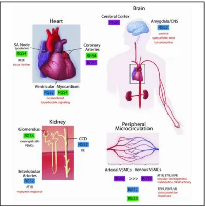
Figure 1.3Expression of RGS2 in cardiovascular system 30
Although RGS2 is found throughout the body, it is highly expressed in the cardiovascular system. RGS2 is acutely upregulated in response to α1-adrenergic activation in cultured ventricular myocytes, and in turn attenuates the hypertrophic response in these cells.
Figure obtained with permission from Clinical Science (2009) 116, (391–399) article, RGS proteins: Identifying new GAPs in the understanding of blood pressure regulation and cardiovascular function
1.7 Protein Synthesis:
Protein synthesis is a complex cellular process, which involves the coordination
of multiple factors like mRNA, tRNA, ribosomes, and accessory factors in order to
synthesize new proteins. Translation is categorized and divided into three steps;
initiation, elongation and termination. In each of steps there are many known protein
factors which help coordinate the positioning of ribosomes, tRNA and mRNA and help
execute the addition of amino acids to the nascent peptide chain 31. Of the three, initiation is considered to be the rate limiting step of translation and hence it is not
surprising that multiple initiation factors are involved in this process 32. Dysregulation of the protein synthesis machinery can contribute to the development of diseases like
cardiac hypertrophy, cancer, neurodegeneration and diabetes 33. Therefore it is important that the regulation of translation be explored and investigated in order to better
understand the complex physiology and pathophysiology of diseases where protein
synthesis is altered.
1.8 Translation initiation and eIF2B:
In translation initiation, methionine tRNAi binds to eukaryotic initiation factor 2
(eIF2), a heterotrimeric G protein comprising α, β, and γ subunits. Unlike membrane
bound heterotrimeric G-proteins, eIF2 resides in the cytosol and GTP binds to its γ
subunit. eIF2 is active when it is bound to GTP, and it escorts the initiator methionyl
tRNA to the 40s (small subunit) ribosome. The mRNA then is recruited to the 40s
promotes the hydrolysis of GTP by eIF2, leading to release of bound initiation factors
and recruitment of the 60s subunit to begin peptide elongation (Figure 1.4) 32.
GDP bound to eIF2 dissociates at a slow rate as it has higher affinity for GDP
than for GTP 34. Eukaryotic initiation factor 2B (eIF2B) is a heteropentameric protein that plays a key role in protein translation initiation in that it acts as a guanine nucleotide
exchange factor (GEF) for eIF2. Once GDP is exchanged for GTP, eIF2 can be activated
again to begin another round of initiation (Figure 1.4). eIF2B consists of five subunits;
α, β, γ, δ, and ε. α, β, and δ subunits share similar sequence motifs, which are known to
interact with α subunit of eIF2 and play a regulatory role in controlling eIF2B activity. γ
and ε on the other hand form their own subcomplex to facilitate its catalytic activity.
However, it is the ε subunit alone that possesses the ability to catalyze nucleotide
Figure 1.4 Translation initiation
1.9 Regulation of eIF2B by eIF2
The rate of nucleotide exchange on eIF2 is one of the key rate limiting steps in
translation, making eIF2B a crucial protein to regulate in order to control the overall rate
of protein synthesis 32. The most thoroughly studied and well understood mechanism of such regulation involves the phosphorylation of eIF2. During glucose and amino acid
starvation, oxidative stress, endoplasmic reticulum (ER) stress, or viral infection,
corresponding stress-activated kinases in the cell become activated in response. These
kinases include General Control Nondepressible (GCN2), Protein kinase-like ER kinase
(PERK), Heme-regulated inhibitor kinase (HRI), and Protein kinase R (PKR), all of
which converge to phosphorylate the highly conserved serine 51 residue on the alpha
subunit of eIF2 (Figure 1.5) 36 37 38 39. When eIF2 is phosphorylated, it becomes a competitive inhibitor of eIF2B by binding at an even higher affinity than that of
nonphosphorylated eIF2 34. Since eIF2 is usually found in greater abundance than eIF2B in the cell, phosphorylated eIF2s can easily sequester eIF2B and prevent it from
interacting with nonphosphorylated eIF2 and thereby decrease the rate of global protein
Figure 1.5 Regulation of protein synthesis by eIF2 41
In response to different kinds of stress, different stress kinases converge and phosphorylate serine 51 on eIF2α. The phosphorylated form of eIF2 binds to eIF2B with higher affinity and sequesters eIF2B from non-phosphorylated eIF2. eIF2 reactivation slows down as a result and thus the initiation rate is decreased.
Figure obtained with permission from Cell Cycle. 2008 May 1;7(9):1146-50. Article, PERK and PKR: Old kinases learn new tricks.
1.10 Interaction between eIF2 and eIF2B
eIF2 is a heterotrimeric G-protein comprising three subunits; α, β and γ, with the γ
subunit being able to bind to GTP. eIF2B is a heteropentameric protein containing 5
subunits; α, β, γ, δ and ε. As mentioned above, the α subunit contains the conserved
serine 51 which can be phosphorylated by different kinases under stress 42, and in turn the phosphoserine interacts with α, β and δ subunits of eIF2B 43
. This is in addition to the
interaction that already occurs under non-stressed conditions.
Under normal conditions, interaction between eIF2 and eIF2B involves distinct
motifs found in the β subunit of eIF2 and the ε subunit of eIF2B. eIF2 β possesses three
lysine rich motifs called the lysine boxes localized near its N-terminus (Figure 1.6)44. When these motifs were mutated to alanines, the interaction between eIF2β and eIF2B ε
was markedly decreased. Conversely, eIF2Bε possesses acidic and aromatic motifs near
its C-terminus and when these were mutated to alanines, the binding between the two was
decreased 44. Therefore it is thought that these lysine boxes on eIF2β form ionic interactions with the acidic and aromatic residues on eIF2Bε.
The catalytic region on eIF2Bε responsible for its guanine nucleotide exchange
ability contains highly conserved residues throughout human, mammalian, plant and
yeast species 45. Among all species, residues glutamic acid 577 and leucine 576 and tryptophan 709 (in human sequence) are especially important for interaction and function
(Figure 1.6). When L576 of eIF2Bε is mutated to alanine, interaction between the
catalytic portion of eIF2Bε and eIF2β and γ becomes impaired with the effect more
other hand almost completely destroyed the interactions of eIF2Bε with both eIF2β and
-γ. E577 of eIF2Bε also was mutated to different residues including alanine, aspartic acid,
arginine, and lysine, with all mutations significantly reducing the affinity of eIF2Bε to
eIF2 with the exception of the alanine mutation. Although the E577A mutant still
bound to eIF2 in yeast, the ability of the mutant to catalyze guanine nucleotide exchange
was significantly decreased, and the growth of yeast was also impaired compared to wild
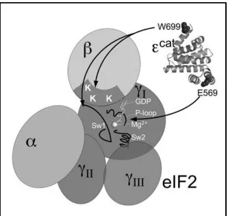
Figure 1.6 Interaction between eIF2 and catalytic domain of yeast eIF2Bε 46
eIF2 binds to the C-terminal domain of yeast eIF2Bε. Acidic motifs on eIF2Bε interact with lysine boxes on eIF2 β, while residue W699 (709 in humans) interacts with both eIF2β and -γ. Residue E569 (577 in humans) is important for interacting with GDP bound eIF2γ and plays an important role in nucleotide exchange.
Figure obtained from Molecular Cell Biology (2007) Jul;27(14):5225-34. Critical contacts between the eukaryotic initiation factor 2B (eIF2B) catalytic domain and both eIF2beta and -2gamma mediate guanine nucleotide exchange.
1.11 Regulation of eIF2B by RGS2
From previous work in our lab, a possible interaction between RGS2 and
eukaryotic initiation factor 2B (eIF2B), a heteropentameric protein, was identified based
on yeast two-hybrid screen results. The sequence that was obtained from the screen
contained 45 residues that matched exactly to a string of residues found near the
C-terminus of eIF2Bε, the catalytic subunit of the eIF2B 15. We confirmed this interaction at the protein level by co-immunoprecipitating overexpressed eIF2Bε and purified RGS2
as well as the endogenous proteins in osteoblast-like osteosarcoma (UMR-106) cells 15. In addition RGS2 was able to decrease GEF activity of eIF2Bε on eIF2 in a dose
dependent manner, suggesting competition between eIF2 and RGS2 for binding to
eIF2Bε 15. Experiments from in vitro translation assays revealed that RGS2 was able to decrease translation efficiency by approximately 75% compared to control, while
decrease in protein synthesis by 25-40% compared to control was observed in cell based
leucine incorporation assays.
The eIF2Bε binding domain on RGS2 was eventually mapped to thirty seven
amino acids that overlap with the beginning of the RGS box 15. Subsequently a peptide was synthesized according to this region, and it was able to exert inhibitory effect on
protein synthesis to a similar extent as full length RGS2 15. This discovery has led us to further research into the area of translation, and the role of RGS2 in regulating protein
1.12 Structure of eIF2Bε and putative RGS2 binding domain
To date there is no crystal structure of the entire eIF2Bε molecule available.
However, the structure of approximately the last two hundred amino acids of the
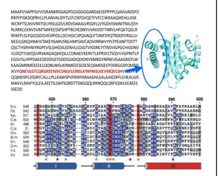
C-terminal domain has been elucidated 47. The protein structure comprises eight alpha helices that are compacted in a globule. The forty five amino acid putative RGS2 binding
domain on eIF2Bε coincidently lies within the first two and half helices of this structure
(Figure 1.7). In addition it overlaps with the catalytic region described previously by
Pavitt et al., which contains highly conserved residues that are important for binding to
Figure 1.7 Putative RGS2 binding domain 4547
Putative RGS2 binding domain lies near the C-terminal region of human eIF2Bε. The crystal structure of C-terminal domain eIF2Bε shows that this spans about 2 and half helices. Within this region there are conserved residues such as human E577(H.s.) and L576(H.s.) which are known to be important for interaction with eIF2 (residue markers of the bottom figure are based on yeast sequence).
Crystal structure obtained from Protein and Cell (2010) Jun;1(6):595-603. Crystal structure of the C-terminal domain of the ɛ subunit of human translation initiation factor eIF2B.
Wei J, Jia M, Zhang C, Wang M, Gao F, Xu H, Gong W.
1.13 Regulation of eIF2B by other eIF2-independent mechanisms:
The interaction between eIF2 and eIF2B and its regulation by eIF2 has been well
characterized by different labs over the years. However more recently, there have been
several discoveries of eIF2-independent mechanisms whereby eIF2B can be regulated as
well. There are at least six phosphorylation sites that are known to regulate the activity
of human eIF2B through its ε subunit. These include regulation by glycogen synthase
kinase 3 (GSK3) targeting serine 540, and its presumed priming kinase dual tyrosine
regulated kinase (DYRK) on serine 544, resulting in decreased eIF2B activity 49. Casein kinase 2 (CK2) targets two serines (717/718) situated at C-terminus, and the
phosphorylation of these is required for efficient interaction between eIF2B and eIF2 50. The absence of available amino acids for translation can result in an unknown kinase
phosphorylating serine 525, leading to a decrease in eIF2B activity 51. Lastly RGS2 can also regulate eIF2B by binding to the ε subunit and interfering with guanine nucleotide
exchange and translation efficiency (Figure 1.8) 15. It is not surprising that eIF2B is a target of control by various mechanisms since it is one of the rate limiting steps in
translation 32.
In this study, we were interested to see whether the changes in eIF2B activity
associated with these phosphorylation events are accompanied by changes in affinity
between eIF2B and RGS2. For example when eIF2 is phosphorylated at serine 51 on its
α subunit, its affinity for eIF2B is strengthened and hence turns it from being a
productive binding partner to a competitive inhibitor 34. Therefore it is of interest to us to investigate whether the phosphorylation state of these serines on eIF2Bε can affect
Figure 1.8 Regulation of eIF2B by eIF2 independent mechanisms
1.14 Glycogen synthase kinase 3 (GSK3)
GSK3 as its name suggests phosphorylates glycogen synthase which decreases its
activity during the absence of glucose and insulin signaling. However this enzyme is
known to play other roles in the cell including its inhibitory role in the Wnt/ β-catenin
pathway (promotes cell proliferation) as well as the Hedgehog pathway (promotes
angiogenesis and is anti-apoptotic). GSK3 is constitutively active in most cell types, and
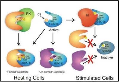
is generally a negative regulator of its substrates. It is unique compared to other kinases
in that it may require the phosphorylation of a priming site four residue downstream of its
target site before the substrate can be efficiently phosphorylated (Figure 1.9) 52. Substrates of this nature include glycogen synthase and eIF2Bε. In case of eIF2Bε, in
addition to the requirement of priming site, the presence of the last sixty residues are
essential in order for GSK3 to efficiently interact and phosphorylate eIF2Bε 50.
There are two mammalian isoforms of GSK3; GSK3α and GSK3β which are
encoded by two distinct genes. Both forms are structurally similar and comparable in
size with GSK3α being 51 kDa while GSK3β is a 47 kDa protein. They possess 98%
homology within their kinase domain in rats but are only 36% identical in the last 76
C-terminal residues. Although both isoforms are structurally similar they are not
functionally identical. For example GSK3β knockout mice are embryonically lethal due
to degeneration in liver from apoptosis in hepatocytes. This means that GSK3α could not
rescue the ablation of GSK3β and the phenotype is arising from the loss of the beta
Figure 1.9 Glycogen synthase kinase 3 52
GSK3 is constitutively active and usually phosphorylates its substrate to deactivate it. It is unique among kinases in that for certain substrates GSK3 will bind more efficiently if they are already primed by another kinase. For eIF2Bε, in addition to the priming site the presence of last 60 amino acids is essential for binding to GSK3.
Figure obtained with permission from Journal of Cell Science (2003) Apr 1;116(Pt 7):1175-86. GSK-3: tricks of the trade for a multi-tasking kinase.
1.15 Regulation of eIF2B by GSK3
In addition to the phosphorylation of eIF2, eIF2B can also be regulated by GSK3
when it phosphorylates a conserved serine residue on the ε subunit of eIF2B. In the
absence of insulin, GSK3 is constitutively active and is able to efficiently interact with
eIF2Bε and phosphorylate it provided that phosphorylation of the priming site four
residues downstream is already in place. In addition to the priming mechanism, it seems
that the presence of the last 60 amino acids of eIF2Bε is also essential in order for GSK3
to bind as truncation of the C-terminus impaired the interaction 50. This phosphorylation results in a decreased eIF2B activity and thus decreases the rate of translation 4953. This is thought to happen by decrease in the intrinsic GEF activity of eIF2Bε, rather than
altering its binding to a regulator. However this does not rule out a possibility whereby
the phosphorylation event promotes a favorable interaction with another binding partner,
resulting in stabilization of the less active conformation.
In functional assays, RGS2 also inhibited protein translation, consistent with the
observed co-immunoprecipitation of RGS2 and eIF2Bε and with results showing that
RGS2 can inhibit the guanine exchange activity of eIF2Bε on eIF2 in a dose dependent
manner 15. However the trigger or the mechanism by which RGS2 is recruited to eIF2B is unknown, which led to the idea of investigating the relationship between regulation of
1.16 Regulation of eIF2B by Casein Kinase 2 (CK2):
CK2 is a kinase that is ubiquitously expressed in cells. It is so far known to be
involved in cell proliferation, transformation, apoptosis and senescence 54. Wang et al. have discovered that eIF2B is also a target of phosphorylation by CK2, showing that
such phosphorylation is actually important and necessary for efficient interaction between
eIF2 and eIF2B50. The phosphorylation sites are located three amino acids from the C-terminus in humans, and they are surrounded by acidic motifs on both sides. Presumably
the phosphorylation of the two serines strengthens the acidic motif, hence allowing
eIF2Bε to better interact with the lysine boxes found on eIF2β. Based on the observed
ability of RGS2 to inhibit the GEF activity of eIF2B15, it is plausible that RGS2 could be competing for binding to eIF2B with eIF2. Therefore it would be interesting to
investigate whether phosphorylation of the CK2 site might have an effect on binding
between RGS2 and eIF2B.
1.17 Regulation of eIF2B by serine 525:
During amino acid starvation, translation can be inhibited via phosphorylation of
serine 51 on the α subunit of eIF2 and subsequent sequestration of eIF2B 55. However the ε subunit of eIF2B can also be regulated by amino acid availability independent of
eIF2α phosphorylation. Serine 525 is found adjacent to a known phosphorylation site
that is regulated by GSK3. Although the identification of the kinase and phosphatase
responsible for this site remains to be established, it is known that this site can regulate
in the absence of amino acids. This suggests that in the absence of sufficient amino acid
in the cell, this site is usually phosphorylated; leading to decreased activity of eIF2B
while in the presence of abundant amino acids this site is dephosphorylated 51.
This site also lies close to the putative RGS2 binding domain since it is adjacent to the
GSK3 site, which itself lies thirteen amino acid upstream of the binding domain.
Similarly to GSK3, the phosphorylation of this site results in decreased eIF2B activity. It
is of interest to investigate whether phosphorylation of this site may have any effect on
eIF2Bε-RGS2 interaction.
1.18 Rationale
RGS2 possesses a region whose amino acid sequence bear 40% homology with
that of the β subunit of eIF2 15. eIF2β interacts with eIF2Bε and the eIF2β binding domain on eIF2Bε lies near its C-terminus. In addition, eIF2γ interacts with the catalytic
region of eIF2Bε and hence mutation of key residues within this region not only impairs
GEF activity but also its interaction with eIF2 51. Since RGS2 seems to be able to compete with eIF2 and disrupt GEF activity in a dose dependent manner 15, we hypothesized that the RGS2 binding domain on eIF2Bε may be near or partially
overlapping with the eIF2 binding domain. Together with previous yeast two-hybrid data
we believe RGS2 binding domain on eIF2Bε comprises at least 45 amino acid region
near the C-terminus and perhaps further extends to region N and C terminally.
The binding between eIF2 and eIF2B has been investigated by other researchers,
and from their research it was identified that eIF2Bε (the subunit which catalyzes
have been shown to regulate eIF2B activity and binding to eIF2 50. Two of the phosphorylation sites are targeted by GSK3 and its priming kinase, while two others are
phosphorylated by kinase called CK2. GSK3 and its priming site are located about ten
amino acids upstream of 45 amino acid putative RGS2 binding domain, while the CK2
site is located at the C-terminus. Therefore, it would be worthwhile first to investigate
GSK3 site as it lies closer to the putative binding region.
In addition to these regulatory mechanisms, our lab has found that RGS2 can
interact with eIF2B and consequently reduce translational efficiency 15. Therefore we planned to investigate whether the reciprocal is true; whether the binding of eIF2B to
RGS2 may have an effect on the ability of RGS2 to promote GTP hydrolysis.
Therefore my hypothesis is that the phosphorylation of eIF2B will increase the
binding of RGS2 to eIF2B, and that eIF2B binding to RGS2 will decrease GAP activity
of RGS2.
1.19 Objectives Overview:
1. a) Identify the region(s) on eIF2Bε that interacts with RGS2 using an internal
deletion method in order to assess the ability of mutant eIF2Bε to interact with
RGS2
b) Express eIF2Bε and mutants in E.coli and purify the proteins in order to show
c) Use an amino acid substitution method to further assess which residue(s) are
important for binding.
2. Identify phosphorylation sites on eIF2Bε that are near the region of interaction
with RGS2, and assess their ability to influence the binding affinity by using point
substitutions
Chapter 2
2.1 Reagents
Plasmids encoding histidine-tagged wild-type human RGS2 were provided by Dr. J.
Hepler (Emory University, Atlanta, GA). pHM Plasmids encoding c-myc and
histidine-tagged human eIF2B subunits 56 were provided by Dr. C.G. Proud (University of British Columbia, B.C.). The baculovirus encoding mouse M1 muscarinic receptor–G11 fusion
protein 57 was provided by T. Haga (Hongo campus, University of Tokyo, Japan) 58. Protein A/G PLUS-agarose beads and anti-histidine antibody were purchased from Santa
Cruz Biotechnology, Inc. Anti c-myc antibody was purchased from Cell Signaling
Technology. HEK293 cells were used for transfection, E.coli XL1 blue for
transformation, and E.coli BL21 (DE3) for protein purification.
2.2Cloning
pHM eIF2Bε internal deletion and pGEX 4T-1 eIF2Bε N terminal and C-terminal
truncation constructs were made using inverse PCR using phosphorothioate-modified
primers 59. The primers (listed in Table 2.1) were designed flanking the sequences that needed to be excised. Four phosphorothioate bases were incorporated twelve bases from
the 5’ end, ensuring that the resulting overhang would be unique enough to complement
one another. PCR reactions were carried out using pfu DNA polymerase with annealing
temperatures at 55 °C for 1 min, while the extension was carried at 72 °C for 8 min for a
total of 30 cycles. Following PCR reaction, the resulting product was treated with
exonuclease, which creates a 3’ overhang by digesting the bases from the 5’ end. Internal
deletion mutants eIF2Bε Δ1,2,3 and Δ1 and Δ2 were made by excising amino acids
eIF2Bε GST-300 version1, GST-300 version2, GST-200, GST-80 were made by excising
amino acids 1-430, 6-436, 1-520, and 1-520/599-721 respectively. Substitution mutants
were made at L576W, Y585W, S540A, S544A, S540E and S544E. The mutant plasmids
were confirmed by DNA sequencing the region of deletion or substitution. Although
mutations from previous studies were numbered based on yeast eIF2Bε, we have used
human eIF2Bε for our present study and therefore the residues numbering corresponds
accordingly to human sequence.Subsequently, the plasmids were transformed into E.coli
XL1 blue and purified using a mini-prep kit (Qiagen). pHM and pGEX 4T-1 eIF2Bε
single substitution mutants were made using site-directed mutagenesis. Primers were
designed flanking the desired base(s) that needed to be changed. PCR reaction was
performed followed by plasmid transformation into E.coli XL1 blue and purification
Table 2.1 List of primers used for mutagenesis
2.3 RGS2 purification
N-terminal 10 polyhistidine tagged human RGS2 containing pET19b vector was
expressed in E.coli BL21 (DE3) strain and purified via nickel affinity chromatography.
First, 3 L of bacterial culture were incubated with vigorous shaking at 37°C to an OD600
of 0.6-0.8. Expression of RGS2 protein was induced by the addition of 0.5 mM Isopropyl
β-D-1- thiogalactopyranoside (IPTG) for 3 hrs before harvesting the bacteria by
centrifugation. Bacteria were resuspended in 50 ml of HEPES buffer (50mM HEPES,
pH 8.0, 500 mM NaCl, 20 mM β-mercaptoethanol, 0.1 mM PMSF, 10 μg/ml leupeptin, 1
μg/ml aprotinin, and 1% Triton X-100), and the suspension was sonicated at 10 Watts for
4 x 15 second intervals with 30 seconds of cooling on ice in between. The lysate was
centrifuged at 15000 g for 30 min, and the supernatant was extracted. A 50% slurry of
equilibrated nickel resin (1 ml) was added to the supernatant and mixed on a rocking
platform for 1.5 hrs at 4°C, followed by loading of the supernatant/slurry of beads onto a
column, allowing the supernatant to flow through using gravity. Then the beads were
washed with 20 ml HEPES buffer supplemented with 20 mM imidazole twice and the
protein was eluted with 500 μl HEPES buffer with 150 mM imidazole 4-5 times. The
eluates were pooled and dialyzed in 1L dialysis buffer (25 mM HEPES, pH 7.5, 500 mM
NaCl, 2 mM DTT) for 6 hours, followed by exchange of old for fresh dialysis buffer, and
then further dialyzed overnight. Purified proteins were run on SDS-PAGE gel (see
Figure 2.1) and found to be ≥75% pure with predicted size of ~26kDa and apparent size
Figure 2.1 Purification of His-RGS2 from E.coli BL21 (DE3)
2.4 Purification of GST-eIF2Bε wild type and mutant proteins
Human GST-eIF2Bε and mutant constructs containing pGEX vector were expressed in
E.coli BL21 (DE3) strain and purified via glutathione sepharose affinity chromatography.
First 4 L of bacterial culture were incubated with vigorous shaking at 37°C to an OD600
of 0.6-0.8. Expression of the GST-fusion proteins was induced by the addition of 0.5 mM
IPTG for 1 hr at 15°C before harvesting the bacteria by centrifugation. Bacteria were
resuspended in 50 ml of TRIS buffer (50 mM TRIS, pH 8.0, 100 mM NaCl, 20 mM
β-mercaptoethanol, 0.1 mM PMSF, 10 μg/ml leupeptin, 1 μg/ml aprotinin, and 1% Triton
X-100), and the suspension was sonicated at 10Watts for 4 x 15 second intervals with 30
second cooling on ice in between bursts. The lysate was centrifuged at 15000 g for 30
min, and the supernatant was extracted. A 50% slurry of equilibrated glutathione
sepharose resin (1 ml) was added to the supernatant and mixed on a rocking platform for
1.5 hrs at 4°C. Subsequently the beads were centrifuged at 500 g and supernatant were
removed, followed by five washes (5 ml) with TRIS buffer lacking Triton X-100 and
centrifugation and aspiration of the supernatant. The proteins were eluted with 500 μl
elution buffer (50 mM TRIS, pH 7.5, 100 mM NaCl, 50 mM glutathione) and stored in
aliquots at -80°C. Protein concentrations were determined using the Bradford assay.
A.
C.
D.
Figure 2.2 Purification of GST-eIF2Bε and N/C-terminal truncation mutants
2.5 Co-immunoprecipitation assay
10 cm round culture plates containing HEK 293 cells were transfected using
lipofectamine with plasmids (20 µg) encoding eIF2Bε or mutants thereof, followed by 48
hrs incubation at 37 °C with 5% carbon dioxide. The cells were lysed with 500 µl lysis
buffer (50 mM HEPES, pH 7.5, 150 mM NaCl, 1% Triton X-100, 0.1 mM PMSF, 10
μg/ml leupeptin, 1 μg/ml aprotinin) and centrifuged at 13,000 rpm (17,400 g) for 10 min
to pellet cell debris and lysates were extracted. 10 μg of purified histidine-tagged RGS2
were mixed with cell lysate containing the overexpressed protein and were incubated for
1 hr at 4 °C. Myc-antibody and protein A/G beads were added to the mixture and
incubated for 3 hrs, followed by three washes with lysis buffer (700 µl) consisting of
centrifugation, aspiration of supernatant and finally suspension in 2x Laemmli sample
buffer (50 mM Tris-Cl pH 6.8, 2% SDS, 10% glycerol, 2.5% β-mercaptoethanol, 0.01%
bromophenol blue).
2.6 GST pull down assay
150 nM or 300 nM of purified GST-fusion proteins were mixed with 1 μM of purified
RGS2 in 200 µl of binding buffer (50 mM TRIS, pH 7.5, 250 mM NaCl, 0.1% Tween,
0.1 mM PMSF, 10 μg/ml leupeptin, 1 μg/ml aprotinin) and incubated for 30 min at 4 °C
on a eppendorf tube rotator. Glutathione sepharose beads were then added to the mixture
for another 1 hr before washing with binding buffer three times by centrifuging the beads,
aspirating the supernatant and resuspension in buffer. Resulting beads were suspended in
2.7 Immunoblot analysis
Protein samples in Laemmli buffer were incubated at 99 °C for 5 min, followed by
SDS-PAGE and transfer for 1 hr using semi-wet transfer machine (Bio Rad) to polyvinylidene
fluoride membrane. Probing for the presence of the proteins was done using
anti-histidine or anti-GST primary antibodies with horse radish peroxidase conjugated
secondary antibodies. Blots were then visualized by using a chemiluminescent substrate
(LumiGLO Reserve; Kirkegaard & Perry Laboratories, Inc.) and a camera-based imaging
system (Fluorchem 8000; Alpha Innotech Corporation). Photoshop (CS2; Adobe) was
used to fine tune the brightness and contrast only.
2.8 GTP hydrolysis assay
Receptor- and RGS protein-stimulated G protein GTPase activity was assayed essentially
as described previously 1115. Briefly, Sf9 insect cells were grown to a density of 2 × 10⁶ cells/ml followed by infection with baculoviruses encoding Gβ1, Gγ2, and an M1
muscarinic receptor-Gα11 fusion protein. At 48 hrs after infection, cells were centrifuged
at 228 g for 5 min, resuspended in phosphate buffered saline (PBS), and centrifuged
again. The resulting pellet was resuspended in one third of the original volume of lysis
buffer (20 mM Tris, pH 8.0, 0.1 mM PMSF, 10 μg/ml leupeptin, and 1 μg/ml aprotinin)
and incubated on ice for 15 min. The cells were lysed using a homogenizer (Polytron;
Brinkmann Instruments) and centrifuged at 500 g for 10 min. to remove debris and
unbroken cells. The supernatant was retained and centrifuged for 30 min at 48,000 g. The
supernatant from this centrifugation was discarded, and the pellets were resuspended in
Membranes were assayed for 100 μM carbachol-stimulated GTP hydrolysis for 5 min at
30°C in the absence and presence of the indicated RGS proteins and GST-fusion proteins.
The reaction buffer contained 106 cpm/assay γ-[32P]GTP, 20 mM Hepes, pH 7.5, 1 mM EDTA, 1 mM DTT, 200 nM GTP, 0.5 mM ATP, 0.1 mM PMSF, 1 μg/ml aprotinin, 10
μg/ml leupeptin, 0.1 mM ascorbic acid, 10 mM NaCl, and 2 mM MgCl2 making up a
total reaction volume of 60 μl. The reaction was then incubated in a water bath at 30 °C
for 5 min. Nonspecific GTPase activity was defined as that in the presence of membranes
plus the inverse agonist, 10 μM tropicamide, and these values were subtracted from
carbachol stimulated activity to yield the specific agonist and receptor-dependent signal.
GTP hydrolysis reactions were terminated by the addition of 940 µl 5% Norit (activated
charcoal) in 50 mM NaH2PO4, pH 3.0, 4 °C. The reaction mixture was centrifuged at
3,000 g for 10 min, and 600 µl of supernatant containing 32Pi was recovered from the reaction tube. Radioactivity was measured on a liquid scintillation counter (Packard
Tri-Carb 2900TR; PerkinElmer).
2.9 Statistical analysis
Data are presented as means with standard error for (n) values as indicated in each figure.
Statistical significance was determined using unpaired student T-test or one-way
ANOVA test (GraphPad Prism 4, La Jolla, California) with Tukey’s post-test.
Concentration dependent GAP activity of RGS2 (Fig.3.11) was analyzed by nonlinear
regression using a sigmoidal curve fit with a variable slope (GraphPad Prism 4, La Jolla,
Chapter 3
3.1 Region of RGS2 interaction on eIF2Bε:
The interaction between RGS2 and eIF2Bε was first detected based on a yeast
two-hybrid screen from mouse brain library, where full length RGS2 was used as a bait to
probe for novel interaction partners 15. There were several positive results from this screen, and one that occurred several times was a forty five amino acid sequence that
matched exactly the C-terminal region of eIF2Bε. These stretches of sequence make up
about the first two and half alpha helices of the catalytic domain of eIF2Bε, and therefore
in the present study this region comprising forty five amino acids of human eIF2Bε was
either removed all together (Δ1,2,3) or each helix deleted individually (Δ1 or Δ2) (Figure
3.2).
To assess the ability of mutants to interact with purified RGS2, plasmids containing
eIF2Bε or internal deletion mutants were transfected into human embryonic kidney (HEK
293) cells. Subsequently, the cells were lysed, and purified RGS2 was added to the lysate
for co-immunoprecipitation assays. His/c-myc-tagged eIF2Bε was immunoprecipitated
using an anti c-myc antibody and the mutants were then assessed on a western blot for the
presence of eIF2Bε and RGS2 (both eIF2Bε and RGS2 are His-tagged so both can be
Figure 3.1 Co-immunoprecipitation using HEK293 lysates transfected with eIF2Bε and purified RGS2
Figure 3.2 Co-immunoprecipitation using HEK293 lysates transfected with eIF2Bε/internal deletion mutants and purified RGS2
A.
The result shows that the ability of all mutants to interact with RGS2 is visibly
reduced compared to wild type (Fig.3.2B) However there are technical limitations to this
assay in that purified RGS2 became entrapped and/or bound nonspecifically to the
protein A/G beads which was used to immunoprecipitate the protein complex (see lane 1
in Figure 3.2B). Therefore for densitometry, RGS2 levels were subtracted from the
pcDNA transfection control before calculating the percentage bound to eIF2Bε (Figure
3.3). In addition mutant proteins expressed at approximately one-third that of the wild
type (data not shown), making it difficult to control precisely the amount of eIF2Bε and
mutants being added to each experimental group. To compensate for the difference in
proteinexpression levels between wild type and the mutants, three times the the total
protein were added for mutant conditions. Therefore another assay was used to further
assess the physical interaction between RGS2-eIF2Be and also to address the issue of
nonspecific binding as well as expression level of eIF2Bε mutants.
3.2 Purification of eIF2Bε wild type and mutants variants from E.coli:
E.coli strain BL 21 (DE3) was used to express eIF2Bε and its mutants. Purifying
the protein instead of transfecting those into HEK cells allows us to better control the
amount of protein being added into each assay group. In addition, this would allow for
the confirmation that eIF2Bε directly interacts with RGS2 by performing pull down
assays.
From the co-immunoprecipitation assays shown above (Figures 3.2B and 3.3), it
domains that are crucial for interaction, as it is possible that the internal deletions may
have affected the folding of the protein. To address this issue, a series of N-terminal or
C-teminal truncation deletions was made and tested. This region of the protein is known
to function as a binding site for other subunits of eIF2B. Even with N-terminal or
C-terminal truncation, there still remains the possibility that the constructs may not fold
properly. However, if these truncation mutants are able to interact with RGS2, we can
confirm that the putative binding is sufficient for interaction and is likely in the proper
conformation..
For purification, cDNA encoding human eIF2Bε or a variant thereof was
subcloned into a pGEX vector to construct a fusion protein containing an N-terminal
Glutathione S Transferase (GST) tag. The plasmid was then transformed into E.coli
strain BL21 (DE3) and expression was induced with a lactose metabolite analog (IPTG).
The bacteria were lysed using sonication, and the protein of interest purified using
glutathione sepharose 4B beads. The purified proteins were visualized following
A.
B.
Figure 3.4 GST fusion protein expression
A) Wild type eIF2Bε and truncation mutants were cloned in pGEX –vector and expressed and purified from E.coli BL21 (DE3). Green colour indicates the 45 a.a. RGS2 binding domain. B)
3.3 Binding of purified eIF2Bε WT or truncation mutants to purified RGS2
The RGS2 binding capabilities of purified eIF2Bε proteins were assessed using
pull down assays via glutathione sepharose 4B beads. GST protein was used as a control
in order to verify that purified RGS2 bound to eIF2Bε and not to the GST tag. These
purified proteins were used to ascertain whether or not there is direct interaction between
eIF2Bε and RGS2. N-terminal and C-terminal truncations were made flanking the forty
five amino acid putative RGS2 binding domain, until about thirty five amino acids were
left N-terminally to the putative binding region (illustrated in Figure 3.4A). At least thirty
five amino acids were left between the N-terminal GST tag and the putative RGS2
binding domain as a precaution due to the possibility of steric hindrance arising from the
size of GST moiety, which consists of over two hundred amino acids. GST-pulldown
assay was used to assess the ability of GST-eIF2Bε and truncation mutants to bind to
Figure 3.5 GST-pulldown assay between N/C truncated GST-eIF2Bε and His RGS2
We found that RGS2 can bind to all of the different types of truncation mutants
that we generated, thereby confirming the yeast two-hybrid data that RGS2 binding
domain of eIF2Bε lies near the C-terminus. In comparison with wild-type eIF2Bε and
the other two truncation mutants, GST-300 a.a. binding to RGS2 appeared to be reduced
(Figure 3.5). From the blot however, it is evident that the lane showing GST-300 a.a. has
greater proportion of free GST present compared to other fusion constructs. This implies
that a portion of free 300 a.a. fragments was also present in the solution before the
pulldown took place. Since any RGS2 that may bind to this tag free form of GST-300
a.a. will not be recovered via glutathione sepharose beads, we would not expect to
observe this interaction on the western blot. Therefore, for subsequent pulldown assays
(Figure 3.7B) we generated a modified version of this construct where we attached a
different N- terminal amino acid to the GST tag in order to create greater stability during
pulldown assay. In the modified version of the GST-300 contruct, the first methionine
plus four subsequent amino acids of eIF2Bε were attached to the GST tag instead of
isoleucine 431.
This assay addresses the possibility of protein misfolding due to internal deletion
from Figure 3.2B and confirms that forty five amino acid putative RGS2 binding domain
is indeed important for interaction between eIF2Bε and RGS2. The results obtained from
Figure 3.2B and 3.5 suggest that eIF2Bε can directly interact with RGS2 in vitro and that
3.4 Amino acid substitution of residues within the putative RGS2 binding domain of
eIF2Bε:
From section 3.1 to 3.3 we confirmed that a 45 amino acid stretch near the eIF2Bε
C-terminus is important for binding to RGS2. Therefore we further investigated which
residues within this region might contribute to binding. To do this we employed a single
amino acid substitution method. From previous studies, we know that there are
conserved residues near the C-terminus of eIF2Bε that are involved in promoting guanine
nucleotide exchange on eIF2 45. Of those, it is known that eIF2Bε residue leucine 576 is conserved throughout different species, and in its substitution to other residues results in
stunted growth 46. Based on the binding data from Figures 3.3 and 3.5, it appears that the forty five amino acid stretch identified from the yeast two-hybrid data is indeed important
for binding to RGS2. This region evidently also coincides with the region responsible for
facilitating guanine nucleotide exchange 48. Therefore I investigated whether mutation of these specific residues within this region would have an effect on the binding to RGS2.
We chose residue leucine 576 and another conserved residue tyrosine 585, both of which
according to crystal structures are at the surface of the protein and are not involved in
folding of structure 47. We mutated both residues to tryptophan which is a relatively large residue that would be expected to serve as a steric hindrance for interaction. Other
investigators have used an alanine mutation for leucine 576 46, however we chose tryptophan instead because of its larger molecular size, and hence it would be easier to
see an effect if this site is indeed important for interacting with RGS2. Subsequently
these mutants were transfected into HEK 293 cells and the cell lysates were used to
Figure 3.6 Co-immunoprecipitation assay between eIF2Bε/substitution mutants in lysates from transfected HEK 293 cells and purified RGS2
Tryptophan substitution at eIF2Bε residue 576 seems to reduce binding while
mutation at residue 585 had no appreciable effect (Figure 3.6). Our previous functional
data indicate that eIF2 and RGS2 may compete for binding to eIF2Bε 15, and this experiment taken together with results from Figures 3.3 and 3.5 further suggests that
RGS2 and eIF2 may share common binding motifs.
3.5 Effects of mutating eIF2Bε phosphorylation sites on its binding to RGS2
Since the region of binding has been established, we looked next into the effect of
phosphorylation on ability of eIF2Bε to interact with RGS2. As mentioned above in
Section 1.5, there are at least five known phosphorylation sites on eIF2Bε, four of which
play a role in modulating its effects on eIF2 50. Two of the phosphorylation sites are targeted by GSK3 (Ser540) and an associated priming kinase (Ser544), while two other
residues near the C-terminus are phosphorylated by CK2. Phosphorylation by GSK3 is
known to decrease the activity of eIF2B 49 while phosphorylation by CK2 seems to be essential for eIF2Bε’s ability to interact with eIF2 50. In order to assess the effects of phosphorylation of the GSK3-associated residues on eIF2Bε binding affinity to RGS2,
point mutations were made to change these serine residues to alanine (to mimic
non-phosphorylated state) or glutamic acid (to mimic non-phosphorylated state). The mutants
were then tested using pull down assays and immunobloting to probe for the presence of
RGS2 (Figure 3.7 and 3.8). Since GSK3 is known to decrease protein synthesis, we
predicted that phosphorylation by GSK3 and its associated priming site would increase
Figure 3.7 GST-pulldown assay between purified eIF2Bε GST-300/substitution mutants at the GSK3 site and RGS2
A.
Figure 3.8 GST-pulldown assay between purified eIF2Bε GST-300/substitution mutants at the GSK3 site and RGS2