O R I G I N A L R E S E A R C H
Role of the Bronchodilator Test De
fi
ned by the
Forced Vital Capacity in Chronic Obstructive
Pulmonary Disease Phenotyping
This article was published in the following Dove Press journal:
International Journal of Chronic Obstructive Pulmonary Disease
Xiaoling Zhang1,*
Zhenchao Wu2–4,*
Yi Liu1,2,4
Shujuan Jiang1,4
1Department of Pulmonary and Critical
Care Medicine, Shandong Provincial Hospital Affiliated to Shandong University, Jinan 250021, Shandong, People’s Republic of China;2Department
of Pulmonary and Critical Care Medicine, Shandong Provincial Hospital Affiliated to Shandong First Medical University, Jinan 250000, Shandong, People’s Republic of China;3Department of Pulmonary and
Critical Care Medicine, The Third Affiliated Hospital of Shandong First Medical University, Jinan 250000, Shandong, People’s Republic of China;
4Shandong Key Laboratory of Infectious
Respiratory Disease, Jinan 250000, Shandong, People’s Republic of China
*These authors contributed equally to this work
Purpose:In clinical practice, some chronic obstructive pulmonary disease (COPD) patients experienced a remarkable increase in forced vital capacity (FVC) after bronchodilator
administration, whereas forced expiratory volume in the first second (FEV1) remains
sub-stantially unchanged. We assume this may relate to airway inflammatory type. We aim to
analyze the clinical characteristics and explore the usefulness of the bronchodilator test, especially FVC, in this new COPD phenotype.
Patients and Methods:A total of 346 COPD patients with exacerbation who underwent
bronchodilator tests, fractional exhaled nitric oxide (FeNO) measurements and blood eosi-nophil counts were analyzed. The characteristics, FeNO levels, and blood eosieosi-nophil counts
were compared between patients with and without significant bronchodilator responsiveness
in terms of FVC.
Results:Patients with significant FVC responsiveness displayed poorer lung function and higher FeNO levels compared with those without considerable FVC responsiveness (Z=
−5.042 to−0.375, p=0.000–0.022). There is a discernible linear relationship between FeNO levels and FVC responsiveness to bronchodilator use (r=0.251, P=0.001). The application of bronchodilator responsiveness of FVC for detecting high FeNO levels in COPD patients exhibited relatively high sensitivity (61.8%) and specificity (86.7%).
Conclusion:We demonstrated that COPD patients with significant FVC responsiveness had higher FeNO levels than non-responders and established a simple method for detecting high
FeNO values. FVC responders may be identified as a separate group of COPD patients.
Keywords: COPD, pulmonary function test, bronchodilator responsiveness, forced vital
capacity, fractional exhaled nitric oxide
Introduction
Chronic obstructive pulmonary disease (COPD) is a disease characterized by persistent airflow obstruction, which is not fully reversible and is usually progres-sive and is associated with elevated chronic inflammatory responses to noxious particles or gases in the airways.1The prevalence of COPD is skyrocketing, and it is expected to be the world’s third most fatal disease by 2030.2
COPD is heterogeneous in nature. COPD patients with similar forced expiratory volume in thefirst second (FEV1) display different clinical manifestations, quality of life, exacerbation frequencies, and responses to treatment.3 Nowadays, COPD phenotyping is increasingly becoming a topic of interest to more and more physi-cians aiming at classifying patients into distinct clinical subgroups to initiate Correspondence: Shujuan Jiang
Email [email protected]
International Journal of Chronic Obstructive Pulmonary Disease
Dove
press
open access to scientific and medical research
Open Access Full Text Article
International Journal of Chronic Obstructive Pulmonary Disease downloaded from https://www.dovepress.com/ by 118.70.13.36 on 24-Aug-2020
appropriate individualized care which may lead to a better prognosis and quality of life for patients.4,5
The bronchodilator test plays a crucial role in the management of COPD, eg, the diagnosis of COPD is confirmed in cases with FEV1/FVC ratios below 0.7 after bronchodilator administration. Flow response after bronchodilator inhalation is universally and frequently used as an indicator for assessing responsiveness in gen-eral practice. However, in moderate to severe COPD cases,
flow response may be poor, yet the response in terms of volume can be relatively favorable, which is probably due to a reduction in hyperinflation.6–9It appears that patients with significant FVC responsiveness are apt to progress to more severe COPD stages.
Airway hyperresponsiveness is associated with airway inflammatory, especially Th2 cell-induced eosinophilic inflammation. Recent research had proved eosinophilic inflammation dominated COPD is an important COPD phenotype. Its clinical characteristics are airway respon-siveness (the change of FEV1), higher eosinophil counts, and higher FeNO concentrations. Although some studies were focused on the role of the change of FEV1 value after bronchodilator inhalation, but there are rarely studies about the role of the change of FVC value in COPD, and we are not clear about the relationship between the change and FeNO or eosinophil.
On these grounds, this study’s objectives were as fol-lows: 1) to compare the characteristics of COPD patients classified as FVC responders and non-responders during the bronchodilator test and 2) to evaluate the benefits of the bronchodilator test in terms of FVC in COPD phenotype.
Patients and Methods
Patients
A total of 346 patients with COPD exacerbation were sequentially enrolled from June 2017 to August 2019. All patients were outpatients from Respiratory Clinic of Shandong Provincial Hospital. They were definitely diag-nosed with COPD by spirometry based on the Global initiative for chronic obstructive lung disease (GOLD 2017) criteria.1 COPD exacerbations are defined as an acute worsening of respiratory symptoms that result in additional therapy.1 COPD patients simultaneously diag-nosed with lung cancer, interstitial pulmonary disease, asthma, bronchiectasis, history of pulmonary resection, or significant comorbidities other than COPD that might
influence the ability to perform pulmonary function tests, fractional exhaled nitric oxide (FeNO) measurements and blood eosinophil counts were excluded.
Observational Indicators
Basic medical information: gender, age, height and weight, smoking history were recorded. Smoking history: patients who still smoking without quitting smoking were defined current smokers; patients who formerly had smoking his-tory, but have quitted smoking more than 3 months were defined ex-smokers.
Exhaled nitric oxide measurements (FeNO): FeNO levels were assessed according to the ATS/ERS guidelines using a NO analyzer (SV-eCO-01, Sunvon, China).10 The subjects were informed to deeply inhale NO-free air and immediately fully exhale via a mouthpiece at a constant
flow rate (50 mL/s) for 10 s. Every FeNO measurement was acquired before the reversibility tests in order to prevent spirometry from influencing the FeNO levels.
Pulmonary function test (PFT): The bronchodilator tests were conducted according to the American Thoracic Society (ATS) and the European Respiratory Society (ERS) guidelines.11–13 Patients received good technique-conducting and tested by a unified and qualified equipment (Master Screen Body, Jaeger, Germany). The second time FVC maneuver was performed, similar to thefirst one, 15 minutes after the administration of 400 μg of albuterol (Ventolin, GlaxoSmithKline) via a metered-dose inhaler. Changes in the FEV1 were expressed as 1) An absolute change relative to the baseline (ΔFEV1) and 2) A percentage change relative to the baseline (ΔFEV1% baseline). Changes in the FVC were expressed in a similar manner. Predicted values of spirometry were calculated from the reference equations published by Zheng and Zhong.14 The bronchodilator test was considered signifi -cant if an increase in either FEV1 or FVC≥12% was observed and ≥200 mL as an absolute value compared with the baseline.15
Blood Eosinophil counts (EOS): Blood eosinophil and total white blood cell counts were obtained by blood routine test using an Automated Cell Counter (Sysmex, Kobe, Japan).
Before the above tests, both physician and examination operator should inquire the patients smoking history and verify the patients had no medication history within 3 days as bronchodilators, corticosteroids. All three tests were performed on the same day. Otherwise, patients were excluded from this study.
International Journal of Chronic Obstructive Pulmonary Disease downloaded from https://www.dovepress.com/ by 118.70.13.36 on 24-Aug-2020
Study Design
Based on a previous study which is about Exhaled nitric oxide, systemic inflammation, and the spirometry response to inhaledfluticasone propionate in severe COPD patients.16 We assumed a population mean of pre-bronchodilator FVC is 2.61L and sample mean is 2.44L. The study was powered (alpha of 0.05 and beta of 0.20) for pre-bronchodilator FVC and the change in FVC after the administration of 400μg of albuterol to detect this difference with a level of confidence of 95%. This resulted in a sample size calculation of 169 patients, allowing for 20% of consented subjects to either fail spirometry screening.
All of the 346 COPD patients were divided into two groups according to the FVC changes in response to albu-terol administration including: (I) positive FVC group (PFVC): Patients with ΔFVC≥200 mL and Δ FVC%base-line≥12%; (II) negative FVC group (NFVC): referring to the rest of the patients without significant responsiveness in terms of FVC. Next, patients withΔFEV1≥200 mL and
ΔFEV1%baseline≥12% were excluded and the remaining 180 patients then were separated into two groups in a similar way: (I) pure positive FVC group (PPFVC): composed of patients with ΔFVC≥200 mL and ΔFVC% baseline≥12% without significant responsiveness with respect to FEV1; (II) negative FVC-negative FEV1 group (NNFVC): comprised of patients without significant responsiveness neither in terms of FVC nor FEV1.
The study was approved by the Shandong Provincial Hospital Affiliated to Shandong University Ethics Committee (No. 2016–23). This action did not produce any potential harm to patients. This operation was strictly obeyed with the Helsinki Declaration. All patients have signed an informed consent. All patient information was kept confidential.
Statistical Analysis
The data were analyzed using the SPSS 22.0 software (SPSS Inc., Chicago, IL, USA). Qualitative variables were expressed as proportions and numbers, whereas quantitative variables were expressed as median (interquartile range, IQR) (non-normal distribution). Comparisons between PFVC and NFVC, PPFVC and NNFVC were investigated using the Mann–Whitney U-test or χ2-square test where appropriate. The Spearman correlation was used to evaluate the relationships between the FeNO concentration and bronchodilator responsiveness vis-a-vis FVC and FEV1. Receiver operating characteristic curves were generated for selected cutoff values of bronchodilator responsiveness in
terms of FVC to detect high FeNO values. P<0.05 was considered statistically significant.
Results
Baseline Characteristics of COPD
Patients with or Without Signi
fi
cant
Responsiveness in Terms of FVC
All the 346 patients with COPD were recruited in this study, the mean age was 59.3±10.5 years, 232 (67.1%) were males and 114 (32.9%) were females. The mean age, sex, and body mass index (BMI) were similar within each subgroup. Patients with significant responsiveness related to the FVC had lower baseline FVC values, FEV1 values, FEV1/FVC ratios, forced expiratoryflows at 75% of the FVC (MEF75), forced expira-toryflows at 50% of the FVC (MEF50), forced expiratory
flows at 25% of the FVC (MEF25), maximal mid-expiratory
flows (MMEF), and higher baseline residual volume/total lung capacity (RV/TLC) ratios, compared with who were without significant FVC responsiveness (Z= −5.042 to -0.375, p=0.000–0.008) (Table 1).
FeNO Levels and Blood Eosinophil
Counts of Patients with or Without
Signi
fi
cant Responsiveness in Terms of
FVC
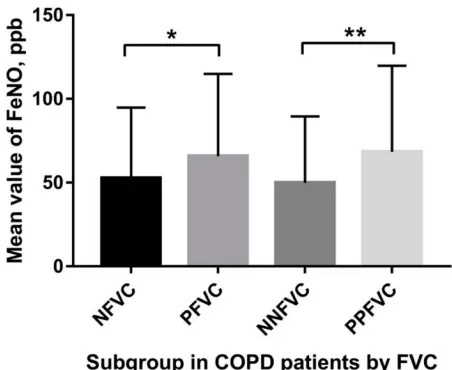
A significant difference in FeNO values was observed between the subgroups (Figure 1). For the entire cohort, FeNO levels in patients with significant FVC responsiveness were significantly higher than the NFVC group (Z=−2.284, p=0.022); Similar tendency on parameters was observed in the PPFVC and NNFVC groups (Z=−2.588, p=0.010) (Table 1).
There is no significant discrepancy observed regarding the blood eosinophil counts between the subgroups (Table 1).
Cut-off Values of FVC Responsiveness
Used to Detect High FeNO Levels
For the entire sample (NFVC and PFVC groups), positive correlation was depicted between FeNO andΔFVC (r=0.216, P=0.000), ΔFVC%baseline (r=0.154, P=0.004) (Figure 2). The correlation between FeNO and ΔFEV1 (r=0.071, P=0.186) orΔFEV1%baseline (r=0.040, P=0.459) were not significant. Excluded the patients with significant responsive-ness in terms of FEV1, concerning the NNFVC and PPFVC groups, linear relationships between FeNO levels andΔFVC (r=0.251, P=0.001), ΔFVC%baseline (r=0.190, P=0.011)
International Journal of Chronic Obstructive Pulmonary Disease downloaded from https://www.dovepress.com/ by 118.70.13.36 on 24-Aug-2020
(Figure 2) were equally noticed. Meanwhile, the relation-ships between FeNO and ΔFEV1 (r=−0.002, P=0.980) or
ΔFEV1%baseline (r=−0.047, P=0.535) were also no difference.
Regarding the NNFVC and PPFVC groups population, the area under the receiver operating characteristic curve (AUC) for detecting FeNO levels ≥25 parts per billion (ppb) withΔFVC was calculated as 0.701 (p=0.010), the AUC for detecting FeNO levels ≥50ppb withΔFVC was determined to be 0.623 (p=0.005) (Figure 3). TheΔFVC
cutoff values with the best sensitivity and specificity were selected according to the Youden index: 0.165 L ofΔFVC with a sensitivity of 61.8% and a specificity of 86.7% for detecting FeNO levels ≥25 ppb, 0.205 L of ΔFVC with a sensitivity of 61.1% and a specificity of 63.9% for detecting FeNO levels ≥50 ppb.
Discussion
This study’s principalfinding can be stated as follows: 1) FeNO concentrations in COPD patients were significantly
Table 1The Clinical Characteristics of COPD Patients
Variables NFVC PFVC p-value NNFVC PPFVC p-value
Sex (male/female) 112/49 120/65 0.353a 79/35 43/23 0.566a
Age, years 59.0(14.0) 61.0(12.5) 0.456b 62.0(14.3) 64.0(14.0) 0.075b
BMI, kg/m2 24.61(5.75) 24.04(4.67) 0.165b 23.26(5.33) 23.43(5.82) 0.149b
Smoking history (current/ex-/non-) 75/28/58 83/35/67 0.834b 62/21/31 37/13/16 0.750b
BBD FEV1, L 1.59(0.89) 1.17(0.74) <0.001b 1.54(1.10) 1.0(0.73) <0.001b
BBD FEV1, %pred 59.2(26.6) 47.65(24.2) <0.001b 60.35(26.5) 46.3(27.7) <0.001b
ABD FEV1, L 1.70(0.98) 1.45(0.87) 0.002b 1.63(1.06) 1.16(0.64) <0.001b
ABD FEV1, %pred 65.22(24.06) 57.80(28.15) 0.023b 65.20(23.98) 52.95(28.63) 0.001b
ΔFEV1, L 0.15(0.18) 0.24(0.17) <0.001b 0.10(0.12) 0.15(0.07) 0.001b
ΔFEV1, %baseline 8.60(10.50) 21.90(16.00) <0.001b 6.85(6.20) 12.05(10.90) <0.001b
BBD FVC, L 2.89(1.30) 2.47(0.98) <0.001b 2.73(1.53) 2.11(1.32) <0.001b
BBD FVC, %pred 86.00(21.80) 75.10(24.50) <0.001b 86.25(20.10) 69.35(30.40) <0.001b
ABD FVC, L 2.99(1.26) 2.97(1.15) 0.725b 2.86(1.43) 2.47(1.39) 0.025b
ABD FVC, %pred 88.29(23.41) 91.27(25.27) 0.083b 87.81(22.89) 83.18(35.39) 0.491b
ΔFVC,L 0.13(0.17) 0.47(0.27) <0.001b 0.13(0.17) 0.36(0.19) <0.001b
ΔFVC, %baseline 5.00(6.40) 17.75(11.00) <0.001b 4.35(6.50) 16.50(8.00) <0.001b
BBD FEV1/FVC, % 55.35(17.43) 49.13(17.98) <0.001b 57.04(18.65) 48.10(16.23) 0.002b
ABD FEV1/FVC, % 59.54(15.58) 51.53(18.89) <0.001b 60.06(17.06) 48.18(17.87) <0.001b
BBD MEF75, L/S 1.95(1.85) 1.25(1.34) <0.001b 1.98(2.44) 1.06(1.19) <0.001b
BBD MEF75, %pred 30.80(27.90) 20.70(19.60) <0.001b 33.15(33.50) 18.40(20.80) <0.001b
BBD MEF50, L/S 0.82(0.80) 0.61(0.59) <0.001b 0.84(1.00) 0.46(0.54) <0.001b
BBD MEF50, %pred 21.20(19.00) 16.15(15.40) <0.001b 21.60(22.30) 13.15(11.60) <0.001b
BBD MEF25, L/S 0.24(0.22) 0.24(0.18) 0.289b 0.23(0.27) 0.22(0.16) 0.290b
BBD MEF25, %pred 17.80(16.00) 18.00(13.00) 0.626b 19.90(18.20) 19.25(13.60) 0.707b
BBD MMEF, L/S 0.61(0.58) 0.48(0.47) 0.002b 0.62(0.66) 0.37(0.39) 0.001b
BBD MMEF, %pred 19.20(16.70) 15.85(14.00) 0.004b 19.45(19.80) 13.35(13.40) 0.003b
BBD RV/TLC % 46.85(11.10) 52.13(10.71) <0.001b 46.85(10.87) 55.58(8.21) <0.001b
BBD RV/TLC, %pred 127.60(25.20) 136.80(30.50) 0.008b 123.55(20.20) 140.80(27.20) 0.001b
FeNO, ppb 41.00(32.00) 46.00(48.80) 0.022b 41.00(29.50) 50.00(42.00) 0.010b
EOS, 109/L 0.14(0.18) 0.16(0.27) 0.160b 0.12(0.16) 0.13(0.23) 0.689b
EOS%, % 1.95(2.80) 2.10(3.60) 0.522b 1.90(2.70) 1.80(2.40) 0.988b
EOS≥2%/EOS<2% 81/80 100/85 0.487b 54/60 32/34 0.885b
Notes:Values are median (interquartile range).a
Analyzed by Chi-square test;b
Analyzed by Mann–Whitney’sU-test.
Abbreviations: BMI, body mass index; Smoking history (current/ex-/non-), current-smokers, ex-smokers, non-smoking; BBD, before bronchodilator; ABD, after bronchodilator; FVC, forced vital capacity; FEV1, forced expiratory volume in one second; MEF75, forced expiratoryflow at 75% of the FVC; MEF50, forced expiratory flow at 50% of the FVC; MEF25, forced expiratoryflow at 25% of the FVC; MMEF, maximal mid-expiratoryflow; RV, residual volume; TLC, total lung capacity;Δ, absolute change relative to baseline; Pred, predicted; FeNO, fractional exhaled nitric oxide; ppb, parts per billion; EOS, blood eosinophils absolute count; EOS%, blood eosinophils percentage count; PFVC, positive FVC group, COPD patients with significant responsiveness in terms of FVC (ΔFVC≥200 mL andΔFVC%baseline≥12%) regardless of FEV1; NFVC, negative FVC group, COPD patients without significant responsiveness in terms of FVC regardless of FEV1.PPFVC, pure positive FVC group, COPD patients with significant responsiveness in termx of FVC, but without significant responsiveness in term of FEV1 (ΔFEV1≥200 mL andΔFEV1%baseline≥12%); NNFVC, negative FVC-negative FEV1 group, COPD patients without significant responsiveness neither in term of FVC nor in terms of FEV1.
International Journal of Chronic Obstructive Pulmonary Disease downloaded from https://www.dovepress.com/ by 118.70.13.36 on 24-Aug-2020
and positively correlated with ΔFVC; 2) The application of bronchodilator responsiveness in terms of FVC to detect high FeNO levels in COPD cases is a comparatively sim-ple method with relatively high sensitivity and specifi -city; 3) FVC responders exhibited poorer baseline lung functions than non-responders. Chen et al reported that RV/TLC% predictions were positively correlated with
ΔFVC (r=0.386) and ΔFVC%baseline (r=0.495), using of FVC bronchodilator responsiveness in detecting
hyperin-flation in COPD patients demonstrated high sensitivities and specificities.8As showed in this study, RV/TLC values were elevated and were found to be higher in FVC respon-ders than non-responrespon-ders. This may be partly accounted for by the higher degree of hyperinflation, which airway obstruction made, in FVC responders.
COPD patients on the mild side of the severity spectrum differ from those on the severe side regarding the relation between their FEV1 and FVC responses to bronchodilator inhalation. Bronchodilator response of advanced lung function parameters depends on COPD severity.17In more severe cases of COPD patients, FVC remarkably rises after bronchodilator administration whereas FEV1 remains substantially constant,7 isolated volume response to bronchodilators is a characteristic of severe emphysema in COPD patients.6,9,18In other words, FVC responders may suffer from more severe airway hyper-responsiveness, airway obstruction, and emphysema. It is well known that COPD is an inflammatory disease affecting both the airways and the lungs, which induces the release of numerous proinflammatory cytokines and mediators. This
cascade of events might include the synthesis of induced nitric oxide synthase (iNOS) by macrophages, epithelium, and vas-cular smooth muscles in the airway.19We speculated that FVC responders with more severe airway hyperresponsiveness, way obstruction, and emphysema sustained more severe air-way inflammation or airway eosinophilic inflammation which in turn elevated their FeNO levels, considering that excessive inflammation plays an important pathophysiologic role in the progress of COPD. The magnitude of change in the FVC after bronchodilator administration may to some extent reflect the degree of the undergoing inflammatory process in COPD. Another possible explanation is the decreased pulmonary diffusing capacity of NO due to irreversible structural defects caused by emphysema.20,21
Airway inflammation is a consistent feature of COPD. Despite the fact that neutrophilic infiltration is the most common inflammatory phenotype in COPD, eosinophilic infiltration does exist in a subset of COPD patients, even after the careful exclusion of patients with any features of asthma.22Singh et al revealed that 37% of COPD patients have eosinophilic airway inflammation.23 One possible explanatory hypothesis is that smoking and other mechan-isms that recruit neutrophils into the airway mucosa in COPD also cause a certain degree of eosinophil influx.22 Furthermore, these patients exhibit the greater response to corticosteroid therapy.22,24 Eosinophilic COPD appears to be a distinct patient subgroup with an increased corticos-teroid response and better prognoses.23
Identifying COPD patients with an eosinophilic infl amma-tion may be of great help in COPD management. A positive correlation between FeNO levels and eosinophilia has been confirmed in induced sputum and bronchoalveolar lavage.25 Additionally, it has been considered a marker for eosinophilic airway inflammation, especially in asthma.25 For COPD, similar outcomes have been demonstrated in previous researches.26,27But considered convenience and comfort for patients, a safe and simple test to reflex the inflammatory type and level is necessary. So, the research about the relationship between FVC and FeNO had a practical meaning. Soter et al proved that FeNO values were reliable surrogate markers of eosinophilic inflammation in COPD patients. There was a significant positive correlation between the percentage of sputum eosinophils and FeNO concentrations both during exacerbations (r=0.593, p<0.001) and hospital discharge (r=0.337, p=0.044).26FeNO had a positive relationship with COPD severity and eosinophilic inflammation level.28,29 Moreover, COPD patients with higher levels of FeNO had a better responsiveness to corticosteroid therapy.27,30 In Figure 1The distribution of FeNO mean value among respondents.
Notes:*P<0.05; **P<0.05.
Abbreviations:NFVC, negative FVC group; PFVC, positive FVC group; NNFVC, negative FVC-negative FEV1 group; PPFVC, pure positive FVC group.
International Journal of Chronic Obstructive Pulmonary Disease downloaded from https://www.dovepress.com/ by 118.70.13.36 on 24-Aug-2020
summary, FeNO measurements in COPD patients may help to improve the classification of patients into the aforementioned subclinical categories and promote more personalized treat-ment regimens.31
Frankly, there some limitations in this study. FeNO levels in our data seem higher than in previous studies.32 One explanation for this disparity may be the variations in popu-lation samples. As high as 52.3% (181/346) of patients in this study had peripheral blood eosinophil counts≥2%. Due to this study’s shortage of clinical data, it is possible that its cohort included some patients with the so-called“Asthma– COPD overlap”phenotype.33It may be one reason for higher FeNO concentrations and higher eosinophil counts percent. One thing to note, there is no difference between NFVC and PFVC (or NNFVC and PPFVC) group in EOS indicators.
We think COPD is airway inflammatory disease, it would display the lesion locallyfirst and significant, compared with general lesion, so the change of FVC or FeNO would be more evident than peripheral blood eosinophil counts. In the next research we will cover this limitation and observe the relationship between airway eosinophilic inflammation level and airway obstruction and iNOS. Besides, FeNO measure-ments can be affected by a number of factors, including different smoking histories, inhaled corticosteroid use, diet, different methodologies for measurement of exhaled NO levels, anthropometric variables, and so on.34All the above factors might induce variability and limit the comparison between studies. We will pay more attention on the COPD rat model to detect the iNOS level, eosinophil counts in airway epithelium and smooth muscle, test the lung function Figure 2Scatter diagrams for FeNO levels versusΔFVC%baseline,ΔFVC: (A, B) entire cohort; (C, D) the population after the exclusion of patients with significant responsiveness in terms of FEV1 (ΔFEV1≥200 mL andΔFEV1%baseline≥12%).
Abbreviations:FVC, forced vital capacity; FEV1, forced expiratory volume in one second;Δ, absolute change relative to baseline;Δ%baseline, percentage change relative to baseline; FeNO, fractional exhaled nitric oxide; ppb, parts per billion.
International Journal of Chronic Obstructive Pulmonary Disease downloaded from https://www.dovepress.com/ by 118.70.13.36 on 24-Aug-2020
of COPD rat model, study the mechanism of role of iNOS in airway hyperresponsiveness, airway obstruction, and emphy-sema in further research.
Conclusion
In conclusion, as far as we know this is thefirst time that linear relationships were demonstrated between FeNO levels and FVC responsiveness to bronchodilators. Since FeNO is useful for identifying eosinophilic inflammation
responding better to corticosteroid therapy, these data may have pertinent implications for the management of COPD patients. Considering the relationship betweenΔFVC and emphysema, FVC responders may be identified as a different group of COPD patients. The usefulness of the bronchodilator test with respect to the FVC in the clinical management of COPD should be evaluated.
Disclosure
The authors report no conflicts of interest in this work.
References
1. Singh D, Agusti A, Anzueto A, et al. Global strategy for the diag-nosis, management, and prevention of chronic obstructive lung
dis-ease: the GOLD science committee report 2019. Eur Respir J.
2019;53(5):1900164. doi:10.1183/13993003.00164-2019
2. Mathers CD, Loncar D. Projections of global mortality and burden of disease from 2002 to 2030.PLoS Med.2006;3(11):e442. doi:10.1371/ journal.pmed.0030442
3. Vestbo J, Anderson W, Coxson HO, et al. Evaluation of COPD long-itudinally to identify predictive surrogate end-points (ECLIPSE).Eur Respir J.2008;31(4):869–873. doi:10.1183/09031936.00111707 4. Han MK, Agusti A, Calverley PM, et al. Chronic obstructive
pul-monary disease phenotypes: the future of COPD.Am J Respir Crit Care Med.2010;182(5):598–604. doi:10.1164/rccm.200912-1843CC 5. Burgel PR, Paillasseur JL, Caillaud D, et al. Clinical COPD phenotypes: a novel approach using principal component and cluster analyses.Eur Respir J.2010;36(3):531–539. doi:10.1183/09031936.00175109 6. O’Donnell DE, Forkert L, Webb KA. Evaluation of bronchodilator
responses in patients with“irreversible”emphysema.Eur Respir J.
2001;18(6):914–920. doi:10.1183/09031936.01.00216501
7. Schermer T, Heijdra Y, Zadel S, et al. Flow and volume responses after routine salbutamol reversibility testing in mild to very severe COPD.
Respir Med.2007;101(6):1355–1362. doi:10.1016/j.rmed.2006.09.024 8. Chen C, Jian W, Gao Y, Xie Y, Song Y, Zheng J. Early COPD
patients with lung hyperinflation associated with poorer lung function but better bronchodilator responsiveness. Int J Chron Obstruct Pulmon Dis.2016;11:2519–2526. doi:10.2147/copd.S110021 9. Quanjer PH, Ruppel GL, Langhammer A, et al. Bronchodilator
response in FVC is larger and more relevant than in FEV1 in severe airflow obstruction. Chest. 2017;151(5):1088–1098. doi:10.1016/j. chest.2016.12.017
10. Exhaled NO. ATS/ERS recommendations for standardized procedures for the online and offline measurement of exhaled lower respiratory nitric oxide and nasal nitric oxide, 2005.Am J Respir Crit Care Med.
2005;171(8):912–930. doi:10.1164/rccm.200406-710ST
11. Miller MR, Hankinson JA, Brusasco V, et al. Standardisation of spirometry. Eur Respir J. 2005;26(2):319–338. doi:10.1183/09031 936.05.00034805
12. Wanger J, Clausen JL, Coates A, et al. Standardisation of the mea-surement of lung volumes. Eur Respir J. 2005;26(3):511–522. doi:10.1183/09031936.05.00035005
13. Macintyre N, Crapo RO, Viegi G, et al. Standardisation of the single-breath determination of carbon monoxide uptake in the lung.
Eur Respir J. 2005;26(4):720–735. doi:10.1183/09031936.05.000 34905
14. Zheng J, Zhong N. Normative values of pulmonary function testing in Chinese adults.Chin Med J (Engl).2002;115(1):50–54. 15. Pellegrino R, Viegi G, Brusasco V, et al. Interpretative strategies for
lung function tests.Eur Respir J.2005;26(5):948–968. doi:10.1183/ 09031936.05.00035205
Figure 3Receiver operating characteristic curves for detecting high FeNO level in patients with chronic obstructive pulmonary disease. (A) FeNO≥25ppb; (B) FeNO≥50ppb.
Abbreviations:FeNO, fractional exhaled nitric oxide; ppb, parts per billion.
International Journal of Chronic Obstructive Pulmonary Disease downloaded from https://www.dovepress.com/ by 118.70.13.36 on 24-Aug-2020
16. Kunisaki KM, Rice KL, Janoff EN, Rector TS, Niewoehner DE. Exhaled nitric oxide, systemic inflammation, and the spirometric response to inhaledfluticasone propionate in severe chronic obstruc-tive pulmonary disease: a prospecobstruc-tive study.Ther Adv Respir Dis.
2008;2(2):55–64. doi:10.1177/1753465808088902
17. Jarenback L, Eriksson G, Peterson S, Ankerst J, Bjermer L, Tufvesson E. Bronchodilator response of advanced lung function
parameters depending on COPD severity. Int J Chron Obstruct
Pulmon Dis.2016;11:2939–2950. doi:10.2147/copd.S111573 18. Cerveri I, Pellegrino R, Dore R, et al. Mechanisms for isolated volume
response to a bronchodilator in patients with COPD.J Appl Physiol (1985).2000;88(6):1989–1995. doi:10.1152/jappl.2000.88.6.1989 19. Brindicci C, Kharitonov SA, Ito M, et al. Nitric oxide synthase
iso-enzyme expression and activity in peripheral lung tissue of patients with chronic obstructive pulmonary disease.Am J Respir Crit Care Med.
2010;181(1):21–30. doi:10.1164/rccm.200904-0493OC
20. Yang X, Tang X, Cao Y, et al. The bronchiectasis in COPD-OSA
overlap syndrome patients. Int J Chron Obstruct Pulmon Dis.
2020;15:605–611. doi:10.2147/copd.S243429
21. Lu Z, Huang W, Wang L, Xu N, Ding Q, Cao C. Exhaled nitric oxide in patients with chronic obstructive pulmonary disease: a systematic
review and meta-analysis. Int J Chron Obstruct Pulmon Dis.
2018;13:2695–2705. doi:10.2147/copd.S165780
22. Brightling CE, Monteiro W, Ward R, et al. Sputum eosinophilia and short-term response to prednisolone in chronic obstructive pulmonary
disease: a randomised controlled trial. Lancet. 2000;356
(9240):1480–1485. doi:10.1016/s0140-6736(00)02872-5
23. Singh D, Kolsum U, Brightling CE, Locantore N, Agusti A, Tal-Singer R. Eosinophilic inflammation in COPD: prevalence and
clin-ical characteristics. Eur Respir J. 2014;44(6):1697–1700.
doi:10.1183/09031936.00162414
24. Leigh R, Pizzichini MM, Morris MM, Maltais F, Hargreave FE, Pizzichini E. Stable COPD: predicting benefit from high-dose inhaled corticosteroid treatment. Eur Respir J. 2006;27(5):964–971. doi:10.1183/09031936.06.00072105
25. Dweik RA, Boggs PB, Erzurum SC, et al. An official ATS clinical practice guideline: interpretation of exhaled nitric oxide levels
(FENO) for clinical applications. Am J Respir Crit Care Med.
2011;184(5):602–615. doi:10.1164/rccm.9120-11ST
26. Soter S, Barta I, Antus B. Predicting sputum eosinophilia in exacer-bations of COPD using exhaled nitric oxide.Inflammation.2013;36 (5):1178–1185. doi:10.1007/s10753-013-9653-8
27. Gao J, Zhang M, Zhou L, et al. Correlation between fractional exhaled nitric oxide and sputum eosinophilia in exacerbations of
COPD. Int J Chron Obstruct Pulmon Dis. 2017;12:1287–1293.
doi:10.2147/copd.S134998
28. Lazar Z, Kelemen A, Galffy G, Losonczy G, Horvath I, Bikov A. Central and peripheral airway nitric oxide in patients with stable and exacerbated chronic obstructive pulmonary disease. J Breath Res.
2018;12(3):036017. doi:10.1088/1752-7163/aac10a
29. Tang B, Huang D, Wang J, Luo LL, Li QG. Relationship of blood eosinophils with fractional exhaled nitric oxide and pulmonary func-tion parameters in chronic obstructive pulmonary disease (COPD)
exacerbation. Med Sci Monit. 2020;26:e921182. doi:10.12659/
msm.921182
30. Dummer JF, Epton MJ, Cowan JO, et al. Predicting corticosteroid response in chronic obstructive pulmonary disease using exhaled nitric oxide. Am J Respir Crit Care Med. 2009;180(9):846–852. doi:10.1164/rccm.200905-0685OC
31. Alcazar-Navarrete B, Romero-Palacios PJ, Sancho A, Ruiz-Rodriguez O. Diagnostic performance of the measurement of nitric oxide in exhaled air in the diagnosis of COPD phenotypes.Nitric Oxide.2016;54:67–72. doi:10.1016/j.niox.2016.02.003
32. Alcazar-Navarrete B, Ruiz Rodriguez O, Conde Baena P, Romero Palacios PJ, Agusti A. Persistently elevated exhaled nitric oxide fraction is associated with increased risk of exacerbation in COPD.
Eur Respir J. 2018;51(1):1701457. doi:10.1183/13993003.01457-2017
33. Takayama Y, Ohnishi H, Ogasawara F, Oyama K, Kubota T, Yokoyama A. Clinical utility of fractional exhaled nitric oxide and blood eosinophils counts in the diagnosis of asthma-COPD overlap.
Int J Chron Obstruct Pulmon Dis.2018;13:2525–2532. doi:10.2147/ copd.S167600
34. Dressel H, de la Motte D, Reichert J, et al. Exhaled nitric oxide: independent effects of atopy, smoking, respiratory tract infection, gender and height.Respir Med.2008;102(7):962–969. doi:10.1016/ j.rmed.2008.02.012
International Journal of Chronic Obstructive Pulmonary Disease
Dove
press
Publish your work in this journal
The International Journal of COPD is an international, peer-reviewed journal of therapeutics and pharmacology focusing on concise rapid reporting of clinical studies and reviews in COPD. Special focus is given to the pathophysiological processes underlying the disease, inter-vention programs, patient focused education, and self management
protocols. This journal is indexed on PubMed Central, MedLine and CAS. The manuscript management system is completely online and includes a very quick and fair peer-review system, which is all easy to use. Visit http://www.dovepress.com/testimonials.php to read real quotes from published authors.
Submit your manuscript here:https://www.dovepress.com/international-journal-of-chronic-obstructive-pulmonary-disease-journal
International Journal of Chronic Obstructive Pulmonary Disease downloaded from https://www.dovepress.com/ by 118.70.13.36 on 24-Aug-2020