Implementation of USG Ovarian Cancer
Image Segmentation using Genetic Algorithm
Muktabai R. Kore1, Prof.Dr. B.D. Phulpagar 2
P.G. Student, Department of Computer Engineering, PES’s, Modern College of Engineering, Pune, India1
Associate Professor, Department of Computer Engineering, PES’s, Modern College of Engineering, Pune, India2
ABSTRACT: In medical field, image processing such as image acquisition, registration, feature extraction, segmentation etc. plays a vital role in diagnosis of disease. Analysis of Ultrasound Image aids to diagnosis of abnormalities in organs and the diseases. As USG is painless and unlike to X-ray no exposure of radiation is required and hence safe, is the best and most widely used popular diagnosis tool preferred by gynaecologist in order to evaluate health of a woman. In medical, among all gynaecological malignancies, ovarian cancer has the worst prognosis due to its late diagnosis and overall 5-year survival rate is approximately 45%, due to primarily late stage diagnosis of disease and only 14% of patient survives after 5yrs of diagnosis [Times of India, Dec-14]. Segmentation of ovarian USG image will help to detect and locate the abnormalities which will help to diagnose ovarian cancer in its early stage. In this paper Genetic Algorithm is used for segmentation technique which is used for automatic detection and classification of ovarian cancer. It also covers survey on different techniques for ultrasound image segmentation.
KEYWORDS:Segmentation, Ovarian Ultrasound Image, Genetic Algorithm, Otsu’s thresholding, K-Means
I. INTRODUCTION
II. RELATEDWORK
[1]presented the texture-based segmentation method it is a pixel classifier, based on four texture energy measures associated with each pixel in the images. The 25 two-dimensional feature masks are derived from 3 basic one-dimensional vectors to evaluate the classification results. Four of those features are selected as the bases for the automated clustering procedure. The segmented images produced as the result of applying the algorithm to an example image are presented and discussed. [2] GA used here helps to resolve the problems of determining the contour’s initial position and contour’s entrapment within local minima which is no longer exist and the only thing needed for specifying the contours’ initial positions is the center of the circles which could be found by determining the image’s center of gravity.The algorithm allows user to process only the region where the tissue is instead of processing whole imagereduces segmentation time.In paper [3] various techniques are revised for computerized cancer detection and classification using ultrasound images in the literature. The techniques developed in the four stages (pre-processing, segmentation, feature extraction and classification) are summarized and the advantages and disadvantages are discussed also different performance matrices are discussed. In paper [4] hierarchical normalized cuts (HNCuts ) with mean shift is implemented for segmentation biomarker segmentation of ovarian cancer image. It is flexible, robust, accurate, and
high-throughput minimally supervised segmentation algorithm. Frequency weighted mean shift is used to generate color pyramid, that is RGB values in image.In the papers [5,8,12] segmentation is done by thresholding technique In [5,8] segmentation threshold of image is selected by genetic algorithm. Simulation of GA implementation is done on Texas DSP instruments TMS320C6000. GA adjusts crossover and mutation probability to get optimized solution. Experimental result shows that segmentation of ovarian image by GA is more accurate than OSTU thresholding[6] Applications of genetic algorithm in medical image segmentation is explained in detail by UjjwalMaulik[6]. Also revised the genetic algorithm best solution in an optimization problem and concluded GA – an evolutionary algorithm is best suitable for large space problem where accuracy is crucial point.Jyothi R Tegnoor [7] proposed SVM classifier to determine whether the ovary is normal, cystic or polycystic ovary. The follicle detection is based on SVM classification and uses two parameters, namely, number of follicles N and the size of the follicles S. Paper [9] MLP networks are a proposed as useful and flexible tool where the case is of missing data. This study is one step towards developing a blood test that would aid doctors in the diagnosis of ovarian cancer.Kirsch template technique [10] is proposed to detect the ovarian cyst. To implement automatic cyst, detection four stages in pre-processing are proposed and kirsch template is used for segmentation and Gaussian filter is used for speckle noise reduction.[11] Proposes the otsuthresolding as an easy and efficient technique for image segmentation. Research also covers iterative and custom approach to implement otsu threshold. [13] states the SURF technique as one of the good technique in ovarian follicle cell growth detection. This technique is based on Haar wavelet responses and can be calculated efficiently with integral images where Speeded Up Robust Features (SURF) is a local feature detector and descriptor that can be used for tasks such as object recognition or registration or classification or 3D reconstruction.Nearest Neighbour algorithm (NN) and Genetic Algorithms (GA),[14] are used to assist pathologist and lab technician to study endothelin protein in placental cell. Image is processed to detect protein expression.
The work in this paper is divided in two stages. 1) Follicle Detection 2) Cancer Classification. Follicle detection and segmentation is done by applying otsu threshold and GA is applied to segment USG image followed by thresholding. GA randomly select the population and adjust its crossover and mutation probability to get fitness function. The algorithm keeps iterating until meets the terminal condition. Result compare with Mean Shift Algorithm implementation.
III.SYSTEM OVERVIEW
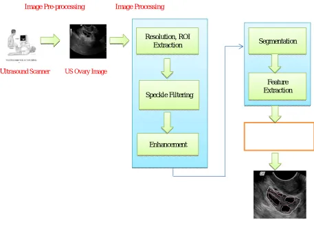
Image Pre-processing Image Processing
Ultrasound Scanner US Ovary Image
Fig. 1 Ultrasound Image Analysis
The ultrasound machine has transducer which places on body organ of interest by applying jelly on it. Transducer transmits high radio frequency (1 to 5 megahertz). These RF signals hits the body tissues, bones and displays binary image on machine monitor. Ultrasound diagnosis is safe, painless, less costly, requires less time and machine is portable and hence popular diagnostic tool in medical system [3]. Apart from this, images obtained are of low qualities and consists speckle noise, artificial borders. Therefore, these images required to process to get desired information. Fig. 1 shows the ultrasound image processing steps.The follicles are the regions of interest (ROIs) in an ovarian ultrasound image, which need to be detected by using image processing techniques. This is basically an object recognition problem. Thus, the basic image processing steps, namely, pre-processing, segmentation, feature extraction and classification, applies on US images. Image segmentation is one of the earliest and most important stages of image processing and plays an important role in both qualitative and quantitative analysis of medical ultrasound images. Segmentation is the process in which region of interest are extracted or distinguish from its background. Generally original image is divided into its subset till region of interest cannot be further divide.
Resolution, ROI Extraction
Speckle Filtering
Enhancement
Segmentation
Feature Extraction
Algorithm:
OSTU
OSTU Principle as the image segmentation standard. In this method, we can directly calculate the threshold without pretreatment to histogram. This algorithm is simple and is a remarkable method for selecting the threshold. Here’s the fundamental principle. The gray value of a grey-scale map is 0~255.The total number of pixels is defined as N,ni is the number of pixels which is gray value is i. By normalizing the histogram, the following equations could be obtained
,
Where Pi is the probability of pixel with grey value.the probability ϕ and mean value µ0 of the background can be calculated as
Genetic Algorithm:
A genetic algorithm repeatedly cycles through a loop which defines successive generations of objects:
1. Compare each object with the final goal to evaluate fitness. 2. Discard the objects that least fit the goal.
3. Use crossover to generate the next generation of objects. 4. Simply copy some objects--as is--to the next generation.
5. Add some objects with random mutations into the next generation.
Step 1.Initialize population with random individuals (candidate solutions)
Step 2.Evaluate (compute fitness of) all individuals
Step 3.WHILE not stop DO
Select genitors from parent population
Create offspring using variation operators on genitors Evaluate newborn offspring
Replace some parents by some offspring OD
At runtime beginning of a genetic algorithm, a large population of random chromosomes is created.
1. Test each chromosome to see how good it is at solving the problem at hand and assign a fitness score accordingly. The fitness score is a measure of how good that chromosome is at solving the problem to hand. 2. Select two members from the current population. The chance of being selected is proportional to the
chromosomes fitness. Roulette wheel selection is a commonly used method.
3. Dependent on the crossover rate crossover the bits from each chosen chromosome at a randomly chosen point. 4. Step through the chosen chromosomes bits and flip dependent on the mutation rate.
Step. 4Repeat step 2, 3, 4 until a new population of N members has been created.
A GA operates through a simple cycle of stages:
1. Creation of a "population" of strings, 2. Evaluation of each string,
3. Selection of best strings and
4. Genetic manipulation to create new population of strings
IV.EXPERIMENTALRESULT
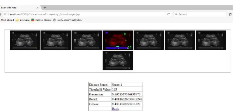
Fig. 3 Result- Colour transform Segmented USG images with disease stage, Threshold Value, Precision,
recall, Fitness
V.
Fig.5Result Analysis (a) Image Precesion (b) Image Recall (c) Image
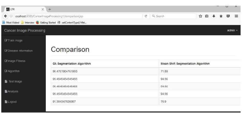
Fig. 6 Comparative Result
VI. CONCLUSION
We have implemented an automatic detection of ovarian cancer in its early stage. The system provides detail diagnosis of USG image by providing information as, size if image, no of follicles detected, precision, recall, fitness and no of pixcel in detected follicle. System provides comparative result with mean shift algorithm. From segmented images output obtained and image information, GA an evolutionary algorithm is proved as best method todetect ovarian cancer in its early stage.
VII. ACKNOWLEDGMENT
A large measure of any credit for the “Implementation of USG Ovarian Cancer Image Segmentation using Genetic Algorithm ” must go to my project guide Prof. Dr.Mr.B.D. Phulpagar.Associate Professor of PES’s Modern College of Engineering, Pune and our ME Coordinator Ms.Deipali V. Gore who with the author has assisted in the preparation of this paper. I admire their infinite patience and understanding that they guided us in field we had no previous experience. We are grateful to them for having faith in us
REFERENCES
[1] J. Alison Noble, Senior Member, IEEE, and DjamalBoukerrou,’Ultrasound Image Segmentation: A Survey’ , IEEE transactions on Medical Imaging, Vol. 25, No. 8, August 2006.
[2] Mohammad Talebi, AhamdAyatollahi, Ali Kermani,’Medical Ultrasound Image Segmentation Using Genetic Active Contour ’, J. Biomedical Science and Engineering, 2011, 4, 105-109.
[3] R.Jemila Rose 1 S.Allwin ,’Computerized Cancer Detection and Classification Using Ultrasound Images: A Survey’, International Journal of Engineering Research and Development, Volume 5, Issue 7 (January 2013), PP. 36-47
[4] Devesh D. Nawgaje, Rajendra D. Kanphade,’Hardware Implementation of Genetic Algorithm for Ovarian Cancer Image Segmentation’, International Journal of Soft Computing and Engineering (IJSCE) ISSN: 2231-2307, Volume-2, Issue-6, January 2013.
[5] Andrew Janowczyk, SharatChandran, Rajendra Singh, DimitraSasaroli, George Coukos, Michael D. Feldman, and AnantMadabhushi,’ High-Throughput Biomarker Segmentation on Ovarian Cancer Tissue Microarrays via Hierarchical Normalized Cuts ’, IEEE transactions on Biomedical Engineering, Vol.59, No 5, May 2012
[6] UjjwalMaulik, Senior Member, IEEE,’ Medical Image Segmentation Using Genetic Algorithm’, IEEE transactions on Biomedical Engineering, Vol.13, No 2, March 2009
[7] Jyothi R Tegnoor , ’Automated Ovarian Classification in Digital Ultrasound Images using SVM’, International Journal of Engineering Research & Technology (IJERT), Vol. 1 Issue 6, August - 2012 ISSN: 2278-0181.
[8] DivyaKaushik, Utkarsha Sing and ParidhiSinghal,’ Medical Image Segmentation using Genetic Algorithm’, International Journal of Computer Applications (0975 – 8887) Volume 81 – No 18, November 2013
[9] Christian Rem, Jagath C. Rajapaks, Khalil Razvi, ‘Ovarian Cancer Classification with Missing Data’, Proceedings of the 9th International Conference cm Neural Information Processing (ICONIP'OZ) , Vol.2
[10] Anju R.S, Radhakrishnan B, ’Detection of Ovary Cyst using Kirsch Template’, International Journal of Engineering Research and General Science Volume 3, Issue 4, Part-2, July-August, 2015
[11] ShilpaKalathiya, Prof. V.P.Patel, ’Implementation Of Otsu Method With Two Different Approaches’ International Journal of Software & Hardware Research in Engineering, ISSN No:2347-4890,Volume-2, Issue-e, February 2014.
[12] Ping-Sung Liao, Tse-Sheng Chen and Pau-ChooChunga, ’Fast Algorithm for Multilevel Thresholding’, Journal Of Information , Science and Engineering 17, 713-727 (2001).