Production of Australian normative data for an alternate scoring system for the Visual Reproduction subtest of the Wechsler Memory Scale-Revised
Helen Ann Jeges, Ba.App.Sci. (Hons)
Victoria University, School of Social Sciences and Psychology, Faculty of Arts, Education and Human Development, Melbourne, Australia
Research thesis submitted as partial fulfilment of the requirements for the degree of Doctor of Psychology (Clinical Neuropsychology)
ABSTRACT
DECLARATION
I, Helen Ann Jeges, declare that the Doctor of Psychology (Clinical Neuropsychology) thesis entitled ‘Production of Australian normative data for an alternate scoring system
for the Visual Reproduction subtest of the Wechsler Memory Scale-Revised’ is no more than 40,000 words in length including quotes and exclusive of tables, figures, appendices, references and footnotes. This thesis contains no material that has been submitted previously, in whole or in part, for the award of any other academic degree or diploma. Except where otherwise indicated, this thesis is my own work.
ACKNOWLEDGMENTS
Firstly, I would like to thank my principal supervisor Associate Professor Gerard Kennedy, for his ongoing support, assistance, and encouragement; without which, this work would not have been completed.
I would also like to express my gratitude to my initial supervisor, Dr Peter Dowling, who provided me with a source of inspiration and never-ending enthusiasm, and above all else, for constantly reassuring me that I could do it.
I also wish to thank Dr Arthur Shores and Dr Jane Carstairs, from the Department of Psychology, Macquarie University, Sydney, Australia, for providing me with access to the data collected through the Macquarie University Neuropsychological Normative Study (MUNNS).
I owe a great debt of thanks to my colleague, Dr Areti Plitas, Clinical Neuropsychologist, for generously sharing her time, expertise and encouragement.
Thanks also to Associate Professor Gavin Ivey for taking the time to review and comment on the research, prior to submission.
TABLE OF CONTENTS
Abstract……….. ...i
Declaration…………... ii
Acknowledgments ... iii
Table of Contents ...iv
Abbreviations ... viii
List of Tables ...ix
List of Figures ... x
INTRODUCTION 1. Memory ... 1
1.1 Memory systems ... 1
1.2 Taxonomy of memory ... 2
1.3 Models of memory ... 2
1.3.1 Multi-store memory model ... 2
1.3.2 Model of working memory ... 4
1.3.3 Summary of memory processes ... 4
1.4 The neuroanatomy of memory ... 4
1.4.1 Medial temporal lobes ... 5
1.4.2 Hippocampus ... 7
1.4.3 Parahippocampal region ... 8
1.4.4 Amygdala ... 9
1.4.5 Diencephalic structures ... 10
1.4.6 Prefrontal cortex ... 11
1.4.7 Striatum ... 11
1.4.8 Cerebellum ... 12
1.4.9 Parietal cortex ... 12
1.5 Neurophysiological processes of memory ... 13
1.6 Summary of neuroanatomical and neurophysiological memory processes14 1.7 Assessment of memory ... 15
1.7.1 Issues in the clinical assessment of memory ... 16
1.7.2 Approaches to the clinical assessment of memory ... 17
1.7.2.1 Assessment of non-verbal memory ... 18
1.8 Wechsler Memory Scales ... 20
1.8.1 The Wechsler Memory Scale ... 20
1.8.1.2 Administration, scoring and normative data ... 21
1.8.1.3 Clinical utility of the WMS ... 23
1.8.2 The Wechsler Memory Scale-Revised (WMS-R) ... 23
1.8.2.1 Structure and content ... 23
1.8.2.2 Administration, scoring and normative data ... 25
1.8.2.3 Clinical validity of the WMS-R ... 27
1.9 Visual Reproduction ... 29
1.9.1 Design ... 29
1.9.2 Clinical utility ... 30
1.9.3 Validity ... 30
1.9.4 Modifications ... 31
1.9.5 Scoring ... 34
1.9.6 Development of an alternate scoring system for the VR subtest of the WMS-R ... 35
1.9.6.1 Reliability and correlational analysis of the alternate scoring system ... 36
1.10 Rationale of the Current Study ... 37
1.11 Aims and Hypotheses ... 39
METHOD 2.1 Study 1 ... 40
2.1.1 The Macquarie University Neuropsychological Normative Study ... 40
2.1.1.1 Participants ... 40
2.1.1.2 Sample design and recruitment of participants ... 40
2.1.2 Procedure and measures ... 41
2.1.2.1 Background information questionnaire ... 41
2.1.2.2 Testing ... 41
2.1.2.3 Visual Reproduction subtest ... 41
2.1.3 Obtaining MUNNS data set ... 41
2.1.4 Protocols ... 42
2.1.5 The Alternate Scoring System ... 42
2.1.5.1 Refinement of the wording ... 42
2.1.5.2 Scoring ... 42
2.1.5.3 Scoring drift ... 42
2.1.5.4 Data entry ... 43
2.1.5.5 Intra-rater reliability ... 43
2.1.6 Design ... 44
2.1.6.1 Variables ... 44
2.2 Study 2 ... 44
2.2.1 Participants ... 44
2.2.1.1 Clinical group ... 45
2.2.1.2 Control Group ... 45
2.2.2 Materials ... 45
2.2.1.2 Visual Reproduction subtest (Wechsler Memory Scale-Revised) ... 45
2.1.6 Design ... 46
2.1.6.1 Variables ... 46
RESULTS 3.1 Statistical analysis ... 47
3.1.1 Data Analysis ... 47
3.2 Test development ... 47
3.2.1 Refinement of the Alternate Scoring System ... 47
3.3 Reliability ... 47
3.3.1 Intra-rater reliability ... 48
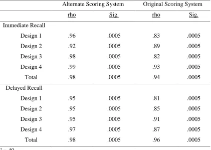
3.3.1.1 Correlational analysis ... 48
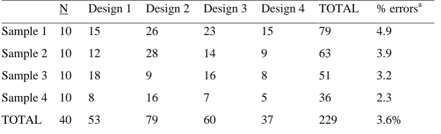
3.3.1.2 Scoring Agreement ... 49
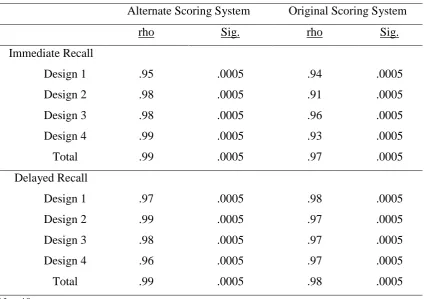
3.3.2 Inter-rater reliability ... 50
3.3.2.1 Correlational analysis ... 51
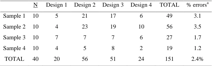
3.3.2.2 Scoring Agreement ... 52
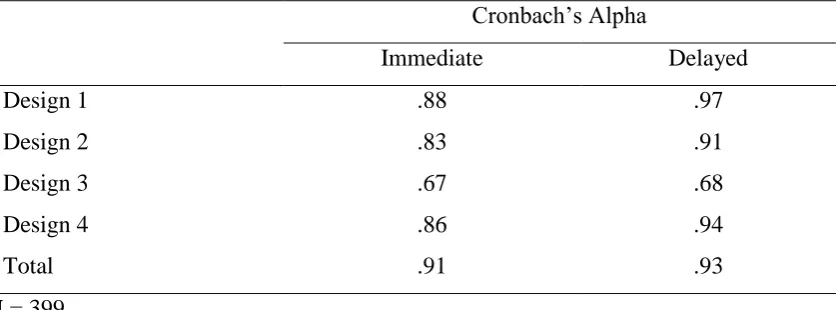
3.3.2 Internal Consistency ... 53
3.4 Normative data ... 54
3.4.1 Sample characteristics ... 54
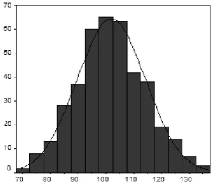
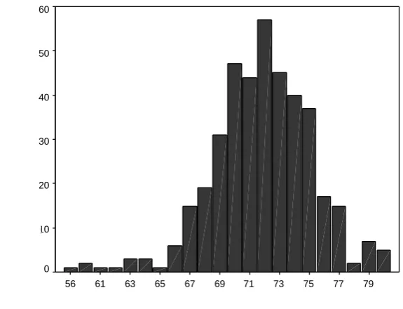
3.4.2 Sample intelligence quotient distribution ... 55
3.4.3 Scoring systems distributions ... 56
3.4.3.1 Distribution of scores for the original scoring system ... 56
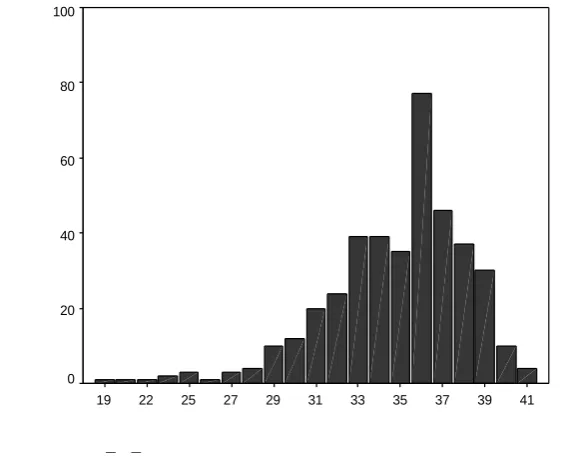
3.4.3.2 Distribution of scores for the alternate scoring system ... 57
3.4.3.3 Scaled scores for the Alternate Scoring System ... 60
3.5 Validity ... 61
3.5.1 Normality of the two scoring system distributions ... 61
3.5.2 Correlations within two scoring systems ... 61
3.5.4.1 Demographic Characteristics of the Study Sample ... 65
DISCUSSION
4.1 Refinement of the ASS ... 69
4.1.1 Intra-rater reliability ... 69
4.1.2 Inter-rater reliability ... 70
4.1.3 Internal consistency ... 72
4.2 Production of normative data ... 72
4.3 Further validation of ASS in comparison to OSS ... 73
4.3.1 Criterion Validity ... 74
4.3.2 Construct Validity ... 75
4.4 Problems and Limitations of the Study ... 76
4.5 Future Directions ... 77
4.6 Conclusion ... 78
REFERENCES ... 81
APPENDICES Appendix A. Stimulus cards from the WMS-R ... 104
Appendix B. Alternate Scoring System - Wording changes to scoring criteria ... 106
Appendix C. Alternate Scoring System - Refined scoring items for Design 1 ... 108
Appendix D. Alternate Scoring System - Refined scoring items for Design 2 ... 110
Appendix E. Alternate Scoring System - Refined scoring items for Design 3 ... 112
Appendix F. Alternate Scoring System - Refined scoring items for Design 4 ... 114
Appendix G. Intra-rater reliability analysis ... 116
Appendix H. Inter-rater reliability analysis ... 117
Appendix I. Descriptive Statistics for the Original and Alternate Scoring Systems ... 118
Appendix J. Percentile Ranks for Alternate Scoring System ... 119
Appendix K. Tests of Normality for the Original and Alternate Scoring Systems ... 123
Appendix L. Nonparametric Correlations ... 124
ABBREVIATIONS
AD Alzheimer's Disease
ASS Alternate Scoring System
BVMT Brief Visuospatial Memory Test
BVRT Benton Visual Retention Test
DLPFC Dorsolateral Prefrontal Cortex
ERP Evoked Response Potential
FM Figural Memory
fMRI Functional Magnetic Resonance Imaging
FSIQ Full Scale Intelligence Quotient
HD Huntington's Disease
LM Logical Memory
LTM Long-term Memory
LTP Long-term Potentiation
MEG Magnetoencephalography
MFD Memory for Designs Test
MQ Memory Quotient
MRI Magnetic Resonance Imaging
MS Multiple Sclerosis
MUNNS Macquarie University Neuropsychological Normative Study
NMDA N-methyl-D-aspartate
OSS Original Scoring System
PET Positron Emission Tomography
ROCFT The Rey-Osterrieth Complex Figure Test
STM Short-term Memory
TBI Traumatic Brain Injury
VePA Verbal Paired Associates
ViPA Visual Paired Associates
VMS Visual Memory Span
VR Visual Reproduction
WAIS-R Wechsler Adult Intelligence Scale-Revised
WMS Wechsler Memory Scale
LIST OF TABLES
Table 3.1 Correlations between scoring of Immediate and Delayed Recall on
Two Occasions……….. 50
Table 3.2 Intra-rater Reliability (number of non-identical item scores)……… 51
Table 3.3 Correlations between scoring of Immediate and Delayed Recall by Two Raters……… 52
Table 3.4 Inter-rater Reliability (number of non-identical item scores)………... 54
Table 3.5 Internal consistency of the Alternate Scoring System for immediate and delayed recall………. 55
Table 3.6 Sample Characteristics: Age Range... 55
Table 3.7 Sample Characteristics: Gender and Years of Education... 56
Table 3.8 WAIS-R Full Scale IQ Scores: Descriptive Statistics... 56
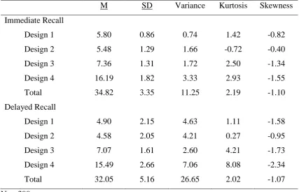
Table 3.9 Original Scoring System: Descriptive Statistics... 58
Table 3.10 Alternate Scoring System: Descriptive Statistics... 60
Table 3.11 Alternate Scoring System-Scaled Scores... 61
Table 3.12 Kolmogorov-Smirnov Normality of the Two Scoring System distributions... 62
Table 3.13 Spearman’s Rho Correlations Between the Original Scoring System Subtests... 63
Table 3.14 Spearman’s Rho Correlations Between the Alternate Scoring System Subtests... 63
Table 3.15 Correlations Between the Alternate and Original Scoring Systems... 65
Table 3.16 T-test Comparisons Between Groups for Age, Years of Education and Gender……… 66
LIST OF FIGURES
Figure 3.1 Frequency Distribution of the MUNNS sample WAIS-R Full Scale IQ... ...
Scores... .
56 Figure 3.2 Distribution of Original Scoring System-Immediate Recall Total
Scores... ...
57 Figure 3.3 Distribution of Original Scoring System-Delayed Recall Total
Scores... ...
57 Figure 3.4 Distribution of Alternate Scoring System-Immediate Recall Total
Scores... ...
59 Figure 3.5 Distribution of Alternate Scoring System-Delayed Recall Total
Scores... ...
59 Figure 3.6 Scatter plot of Immediate Recall Total Scores for Both Scoring
Systems... ...
64 Figure 3.7 Scatter plot of Immediate Recall Total Scores for Both Scoring
Systems... ...
INTRODUCTION
This introduction provides an overview of the concepts of memory and its assessment in the Australian context. Using a selective historical review of literature, this overview aims to detail the evolution of understanding about the assessment of memory and further, to identify limitations of existing memory assessments. The introduction concludes with the rationale for the production of Australian normative data for an alternate scoring system for the Visual Reproduction subtest of the Wechsler Memory Scale-Revised and subsequent hypotheses which this study aimed to answer.
1. Memory
Memory processes form an essential component of cognitive functioning and have been described, in the seminal work of Squire, as “the persistence of learning in a state that can be revealed at a later time” (1987, p.3). Learning broadly involves the acquisition of
new information, or knowledge, and is necessary for memory to occur; therefore, learning and memory are thought to represent the constant adaptations of neural connections to the environment (Eichenbaum, 2002).
1.1 Memory systems
The fundamental processes involved in memory function and the structure of memory have long been debated by researchers holding different views. Historically, the concept of distinct memory systems has been around since before the twentieth century (Squire, 2004). The following processes have generally been accepted to occur in the various theories that have been proposed about the nature of memory: (1) attention and orientation; (2) encoding, referring to the moving of information into a more permanent memory store through either acquisition, via a sensory buffer or consolidation into longer-term representation; and (3). retrieval, which involves moving information from memory store to ‘awareness’ in a conscious sense (Craik & Lockhart, 1972; Tulving &
1.2 Taxonomy of memory
Currently, the basic concepts of memory are stated to be that of ‘declarative’ (i.e., explicit) and ‘nondeclarative’ (i.e., implicit). Conscious, declarative memory can be
defined as the capacity to consciously recollect conscious information related to facts, locations, and personal information, in addition to information that has been processed in some manner. This is also known as episodic memory and it allows us to recall our past experiences (Tulving, 2002; Squire, 2009a; Squire, 2009b). Nondeclarative memory includes unconscious information such as skills, procedures, habits and within this memory; information is learned in the same way in which it was encountered (Squire, 1992; Tulving, 1985). There is agreement amongst researchers that the processes of memory are likely to be different for declarative and nondeclarative memory (Squire & Knowlton, 1994). Memory has also been classified into several groups or subtypes. Squire (1987) classifies memory subtypes by modality into the following five subtypes: verbal, including words and stories; visual, comprising of pictures, faces and also music; spatial and kinaesthetic, involving similar processes to visual memory; emotion and feelings, usually connected with other percepts; and multi-modal versus uni-multi-modal memory.
Memory is also often classified into the following temporal subtypes: sensory or registration memory, this lasts milliseconds to seconds and is longer for auditory than visual memory; short-term memory (STM), which lasts between seconds and hours and is related to working memory; and long-term memory (LTM), which can last for days, months and years (Atkinson & Shiffrin, 1968; Tulving, 1972). This distinction is somewhat similar to the distinction made between recent and remote memory (Deweer, Pillon, Pochon, & Dubois, 2001). These two memory subtypes make reference to memory that occurs in relation to time in the present. Some types of memory problems have a ‘temporal’ gradient, with this being more severe for recent memory
(Eichenbaum, 2002; Parkin, 1997; Squire, 2009a).
1.3 Models of memory
1.3.1 Multi-store memory model
In 1968 an influential multi-store model of human memory was proposed by Atkinson and Shiffrin. This model posited two distinct ‘memory stores’, which were: short-term
Short-term memory is also often referred to as ‘working memory’, and is the type of memory where information is temporarily stored within neural connections. The duration of STM was initially thought to be limited to eighteen to twenty seconds (Peterson & Peterson, 1959). However, more recently Cohen and Eichenbaum (1993) have argued that STM can endure for a period of up to several hours. The capacity of STM was famously reported by Miller (1956) in a paper where he demonstrated that about seven pieces, or ‘chunks’, (+/- two) of information could reside in STM at the same time. Once the information has entered STM, it fades or decays; a process expedited when new information enters the limited store. Unless rehearsed, the temporary store of information in STM is soon lost as attention is focussed in other directions. STM has been depicted as the ‘bottleneck’ of the human information
processing system because the capacity of STM limits the amount of information that can be held in mind simultaneously and also on the duration for which it can be held once attention is withdrawn (Baddeley, 1995). To retain information in STM, it is often encoded verbally, although other strategies may also be used such as visualisation. These strategies make it possible to ‘rehearse’ the information, thereby allowing it to
enter long-term memory (Parkin, 1997).
Long-Term Memory is a memory store in the human brain that is considered to be relatively permanent. The properties of LTM vary significantly from that of STM, its capacity is thought to be virtually unlimited, it can endure for a lifetime and humans do not appear to exhaust the capacity for storage of new information even after a full lifetime (Atkinson & Shiffrin, 1968). There is no clear consensus suggesting an accurate method of determining how long memories can be stored in LTM. Permanent losses of information in LTM are known to result from brain damage (Zola, 1997).
LTM has three basic processes; storage, deletion and retrieval. In the storage of LTM it appears that information is organised according to meaning and is associatively linked (Eichenbaum, 2002). There are two types of retrieval of information in the LTM process which are those of: recall and recognition. Recall reproduces the information from memory, while in recognition the information is presented providing the knowledge that the information has been known or seen before. Recognition memory is considered to be less complex than memory recall (Buffalo, Reber, & Squire, 1998).
as ‘iconic’ memory and another for auditory input referred to as ‘echoic’ memory.
(Atkinson & Shiffrin, 1968). These two memory systems share the characteristic of pure raw perceptual processing with no additional processing; but differ in capacity and duration. Iconic memory is purported to last approximately half to one second, whereas echoic memory was proposed to be more enduring lasting for four to five seconds (Baddeley 1994; Sperling, 1960).
1.3.2 Model of working memory
In 1974, Baddeley and Hitch modified the notion of STM by proposing a model of working memory that included the idea that two ‘slave systems’ serve long-term memory: ‘phonological loop’ and ‘visuospatial sketchpad’. The two systems were
argued to temporarily store information, as well as perform operations, such as rehearsal, that would maintain information and subsequently allow it to be transferred to LTM. The third component of the working memory model was the ‘central executive’
which provided an interface between the phonological loop, visuospatial sketchpad, and LTM. In addition to providing traditional frontal lobe functions the central executive allocated attention to different processes and carried out different activities, such as organisation. The working memory system has, more recently, been purported to serve a broader role in the temporary maintenance and manipulation of complex cognitive processes such as comprehension, learning and reasoning (Emilien et al., 2004; Lezak, Howieson, & Loring, 2004).
1.3.3 Summary of memory processes
When reviewing evolution of the understanding of memory processes, the literature indicates that essential elements of memory systems include: attention, encoding, storage and retrieval. Conceptually, memory has been classified both in a declarative or nondeclarative dichotomy, and segregated by temporal subtype, such as, STM and LTM. Cognitive models of memory function, including: the multi-store model of human memory (Atkinson & Shiffrin, 1968) and the model of working memory (Baddeley & Hitch, 1974) can be better understood with an exploration of the neuroanatomy and neurophysiological correlates of memory.
1.4 The neuroanatomy of memory
widely distributed among the connections that link the cells of a memory system and include the same neurons that are involved in sensation and perception. The brain can utilise the same cortical area for both processing of sensory information and storage of memories (Eichenbaum, 2002).
Studies investigating the neural and structural bases of typical memory function in the years before the emergence of modern neuroimaging techniques were limited mainly to the examination of brain damaged patients (Squire, 1986; Squire, 2009b), derived from animal models (Mishkin, 1978; 1982) and from ablation studies in animals (Damasio, Graff Radford, Eslinger, Damasio, & Kassell, 1985; Zola-Morgan, Squire, Amaral, & Suzuki, 1989. The availability of modern neuroimaging technology, such as Magnetic Resonance Imaging (MRI), Positron Emission Tomography (PET), functional Magnetic Resonance Imaging (fMRI), Event Related Potentials (ERP) and Magnetoencephalography (MEG), has substantially contributed to knowledge regarding the structural and functional aspects of typical memory function (Mayes & Montaldi, 2001; Squire, Amaral, & Press, 1990). The cumulative wealth of this contemporary research clearly indicates that, although no single brain structure or cellular mechanism accounts for all learning, all cortical areas do not contribute equally to memory (Cabeza & Nyberg, 2000). The overwhelming consensus reached across numerous, methodologically sound studies, concludes that memory processes are mediated by multiple inter-related and connected neural systems (Squire, 2004).
Consistent evidence has been presented that particular parts of the brain are involved in diverse forms of memory and that several different aspects of learning and memory utilise distinct neural systems (Squire & Knowlton, 1994). For example, short-term memories are modality specific and consequently, involve a range of modality specific storage areas in the brain. Various components of brain systems, such as medial temporal lobe structures, are involved in storing particular types of information by means of synaptic mechanisms. Although distinct neural systems subserve varying forms of memory function, interconnectedness remains a prime feature through the connections between cortical areas, as evidenced in a series of fMRI studies (Figueiredo et.al. 2008; Alessio et al, 2011).
1.4.1 Medial temporal lobes
(Mishkin, 1978). Using an animal model of memory impairment in monkeys, Mishkin (1982) was able to describe a detailed, human model of limbic memory function. His series of neuroanatomical studies, combined with behavioural observations assisted in the identification of the key anatomical components of the medial temporal lobe that were proposed to support and subserve declarative memory function (Mishkin, Malamut, Bachevalier, 1984).
The role of the medial temporal lobe in declarative memory formation has been outlined by Cohen and Squire (1980) and although there appears to be a division of labour within the medial temporal lobe, the available data do not support simple dichotomies, such as nondeclarative versus declarative memory. More recently, Cohen and Eichenbaum (1993) proposed a theory of relational memory which argues that as sensory information enters the medial temporal lobe and is processed; it leads to the storage of memories in a manner that relates all of the things happening at the time the memory was stored with the memory itself.
It has also been confirmed that a group of interconnected limbic structures in the medial temporal lobe play a critical role in the consolidation of declarative memory and recognition memory (Squire & Zola-Morgan, 1991; Clark & Squire, 2010). Anatomically related structures of the medial temporal lobe are principally concerned with memory and operate within the neocortex to establish and maintain long-term memory (Squire, Stark, & Clark, 2004). Lesions in this area typically result in amnesia of varying severity.
patient H.M. reproduced similar features of memory impairment in monkeys (Zola-Morgan & Squire, 1993).
1.4.2 Hippocampus
Of all medial temporal lobe structures, interest in the critical role of the hippocampus in memory dates from the earliest studies of memory function in brain damaged patients (Adeyemo, 2002; Squire, 2009b). The primary function of the hippocampus is to encode information into STM, which also necessarily involves working memory processes. It is also involved in retrieval of information from long-term memory (de Haan, Mishkin, Baldeweg, & Vargha-Khadem, 2006).
As expected, given the predilection of the medial temporal lobe for the processing and storage of declarative memory; memory deficits following hippocampal lesions are not global, but are much more specific, predominantly involving declarative or declarative memory (Eichenbaum, 2004). The evidence from H.M and animal models strongly implicated the hippocampal complex (including the CA fields, the dentate gyrus, and the subicular complex) as vital structures for the acquisition of new episodic and semantic memories (Mishkin, 1982; Tulving & Markowitsch, 1998; Broadbent, Clark, Zola & Squire, 2002; Deweer et.al, 2001; Lee, Yip, & Jones-Gotman, 2002). Even partial damage to the hippocampus has been found to produce substantial memory impairment in humans and monkeys (Zola-Morgan & Squire, 1993; Mishkin, Vargha-Khadem, & Gadian, 1998; Winocur, Moscovitch, Caruana, & Binns, 2005).
Results from a study of patient R.B., who had circumscribed memory impairment as a result of a hypoxic brain injury, revealed that a bilateral lesion involving the CA1 field of the hippocampus was sufficient to produce severe anterograde amnesia (Zola-Morgan, Squire, & Amaral, 1986), although concurrent damage to the entorhinal, perirhinal and parahippocampal cortices is reported produce a more severe amnesia (Mishkin, 1978; Squire, 1986; Zola, 1997). Lesions that extend to the entorhinal and perirhinal cortices in association with the hippocampus are implicated in a severe retrograde amnesia extending several decades (Emilien et al, 2004; Rempel-Clower, Zola, Squire, & Amaral, 1996; Baxter & Murray, 2001; Corkin, Amaral, Gonzalez, Johnson, & Hyman, 1997; Zola-Morgan et al, 1986; Markowitsch, 2000).
The fornix is a bundle of output axons that link the hippocampus to the thalamus and hypothalamus. Lesions to the fornix have been shown not to affect procedural learning, but do affect declarative memory formation. Furthermore, lesions to the fornix have not been shown to produce long lasting memory impairment in monkeys (Zola-Morgan et al., 1989).
Neuroimaging studies also consistently indicate a reduction in hippocampal size in those with clinically significant memory impairments (Squire, Amaral, & Press, 1990; Markowitsch, 2003; Mayes & Montaldi, 1999). Quantitative MRI studies have indicated the role of hippocampal atrophy in memory dysfunction in Alzheimer's disease (Deweer et al., 1995) and following traumatic brain injury (Bigler et al., 1996). In addition to the hippocampus, other specific temporolimbic structures implicated in memory function include the parahippocampal regions and the amygdala (Kelley et al., 1998; McGaugh et al., 1993; Tranel & Damasio, 1995; Squire & Zola-Morgan, 1991; Zola-Morgan & Squire, 1993).
1.4.3 Parahippocampal region
temporal. Only highly pre-processed sensory information reaches the medial temporal lobe structures (Kelley et al., 1998). The parahippocampal region of the brain plays a crucial role, in conjunction with other close structures in the brain, in functions such as learning, memory, emotions, and more multifaceted behavioural processes (Squire & Knowlton, 1994). The parahippocampal cortex has been identified as important for declarative memory (Squire & Shimamura, 1996; Zola-Morgan et al., 1989; Miller, Lai, & Munoz, 1998). Furthermore, recent research has implicated that damage to the parahippocampal area of the brain can directly contribute to degenerative processes, such as Alzheimer’s, a disease known for its severe memory impairment (Pantel, Kratz, Essig, & Schröder, 2003; Scharfman, Witter, & Schwarcz, 2000) and that parahippocampal atrophy might have potential as a predictor of Alzheimer’s disease
(Echávarri et al., 2011).
The rhinal cortex, which is an area of the temporal lobe just lateral to the hippocampus and amygdala, can be further divided into the entorhinal cortex and perirhinal cortices. The rhinal cortex is purported to be involved in recognition processes. Severe recognition memory deficits result from damage restricted to the perirhinal cortex (Burwell, Witter, & Amaral, 1995; Buffalo et al., 1998; Squire, 1992). Recent studies made possible by improved developments in genetic and physiological techniques have allowed researchers to explore the connectivity of the entorhinal cortex. Specifically, the separate contributions of the direct (temporoammonic) pathway from the entorhinal cortex to the CA1 subfield have been described in increasing detail, along with the indirect (trisynaptic) pathway from the entorhinal cortex to the CA1 subfield via the dentate gyrus and CA3 subfield (Bakker, Kirwan, Miller, & Stark, 2008; Nakashiba, Young, McHugh, Buhl, & Tonegawa, 2008). As the entorhinal cortex is a major source of projections to the hippocampus and the dentate gyrus, the anterograde amnesia becomes more severe when these cortical regions are damaged as well (Zola-Morgan & Squire, 1993; Zola, 1998; Zola-Morgan et al, 1989; Graham et al, 2000; Nadel, 1994).
1.4.4 Amygdala
It is generally accepted that the connections between the amygdala and the frontal cortical regions of the brain are involved in regulating emotion and in directing emotion-related behaviours. It subserves a basic associative learning function in emotional memory and memory for emotional responses. The amygdala also serves as a potent aide for the long-term retention of emotional events and has been implicated in playing a potentially central role in the development of conditioned fear and influencing the behavioural response to a neutral stimulus from previous experience (Davis, 2006; LeDoux, 2000; McGaugh et al., 1993). It is currently generally thought that the essential plasticity supporting the fear response develops directly in the amygdala. Recent fMRI evidence has reported gender differences in the lateralisation of amygdala function for emotionally influenced memory (Cahill et al., 2004). There has been no research to date that supports its involvement in declarative memory or amnesia (Butters, 1984; Zola-Morgan et al., 1989).
1.4.5 Diencephalic structures
1.4.6 Prefrontal cortex
Lesions of the frontal lobes may also result in reduced memory functioning but not amnesias (Squire & Butters, 1992). The prefrontal cortex has been proposed to subserve what was initially termed short-term memory and, in later elaborations, working memory (Goldman-Rakic, 1995; Fuster, 2008). Working memory function is generally considered to be a subsection of declarative memory. Eichenbaum (2004) reports lesion studies indicating deficits in working memory for problem solving and planning of behaviour. The prefrontal cortex has also been identified as broadly important for processes and strategies involved in monitoring, organising, and using memory (Busch et al., 2005; Squire, 2009b).
Preliminary research has suggested that different regions of the frontal lobes may be involved in different aspects of memory functioning. Studies have identified different regions of the prefrontal cortex as playing a crucial role during remote memory recall (Frankland & Bontempi, 2005). Of specific interest, has been the role of the dorsolateral prefrontal cortex (DLPFC) in episodic memory (Kapur et al., 1995); and the recent findings that retrieval is mediated within the right side of the DLPFC, while encoding is mediated on the left (Kelley et al., 1998). The DLPFC may also be involved in recalling frequency and recency; that is, how often an event has happened and how long ago an event occurred (Anderson, Damasio & Tranel, 1993; Smith & Milner, 1988). More recently, studies have also implicated the prefrontal cortex in priming and other nondeclarative memory processes (Fletcher & Henson, 2001), in addition to a role in prospective memory (McDaniel & Einstein, 2007).
1.4.7 Striatum
The striatum, comprised of the caudate nucleus and the putamen, has been shown to be vital in nondeclarative memory formation of skills and habits (Prado-Alcala et al, 2003; Mishkin et al., 1984). This memory function, known as procedural memory, centres around memory of behaviours. Lesions of the striatum have been shown to affect procedural learning and, more recently, working memory function (Lewis, Dove, Robbins, Barker & Owen, 2004). A key feature of the structure of the striatum is its connectivity. It takes in highly processed sensory information and sends out signals involved in motor responses. Pathological disruption to striatal function causes significant impairment in memory function, in such disease processes as, Huntington’s (Brandt, Shpritz, Munro, Marsh & Rosenblatt, 2005) and Parkinson’s diseases
1.4.8 Cerebellum
The cerebellum is the main component in a subsystem that contributes to the execution of movement details, and to the acquisition of conditioned reflexes and body adjustments to changing environmental inputs. It receives input from restricted sensory and motor areas within the cortex and has links to the brain stem, spinal cord and thalamus (Desmond & Fiez, 1998). The cerebellum is thought to be involved in the acquisition and retrieval of nondeclarative memory function, which includes basic associative learning related to skeletal musculature; although knowledge of the precise role of these areas in memory functioning remains limited (Desgranges et al., 1998; Squire, Knowlton, & Musen, 1993; Thompson, 1986; Tranel & Damasio, 1995). Aspects of cerebellar function have also been implicated in procedural and episodic memory. This system is relatively independent and damage to these areas appears to have no effect on the functioning of the declarative memory system of the medial temporal lobe (Cohen & Squire, 1980). Damage to the cerebellum has also been reported to give rise to cognitive disturbances and cerebellar lesions inhibit the learning and acquisition of new complex skills (Appollonio, Grafman, Schwartz, Massaquoi & Hallett, 1993).
1.4.9 Parietal cortex
The parietal lobe plays an important role in tertiary processing, integrating visual and auditory sensory input. Although inputs of the various sensory systems are often reported as being independent, we experience sensory events as a single perceptual experience. The ability of the human mind to recognize concurrent sensory signals as a single percept is known as ‘cross-modal matching’. This occurs with any combination of visual and auditory stimuli; in each case the ‘matching’ is purported to take place in the areas of the tertiary cortical regions where the inputs overlap (Jones & Connolly, 1970). The majority of this process of analysis is carried out by the posterior parietal cortex, which has also been reported to be heavily involved with visuospatial processing. The ‘two-streams hypothesis’, initially put forward by Mishkin and
Ungerleider (1982) is a popular, but debated, model of visual processing. They proposed that as information leaves the occipital cortex, it diverges into two ‘streams’: the ventral stream, also known as the ‘what pathway’, which terminates in the temporal
dissociation between perception and action processing of visual information in the posterior parietal cortex and instead suggested the possible existence of a more unitary processing stream (Cardoso-Leite & Gorea, 2010).
The role of the parietal cortex in memory function is not as well documented as the medial temporal lobe. Modality specific areas within the parietal cortex are argued to subserve working memory function (Squire & Zola-Morgan, 1991). For example, the lateral intraparietal area is actively engaged in visual working memory. The hypothesis of modality specific areas involved in working memory processes has also been supported by clinical observation that there are distinct auditory and visual working memory deficits in patients with schizophrenia (Huguelet, Zanello & Nicastro, 2000).
Functional neuroimaging studies, mostly using fMRI, have clearly showed activity in the posterior parietal cortex during memory retrieval (Todd & Marois, 2004). However, it has been argued that the observed pattern of parietal activation during memory retrieval reflects the region’s role in directing attention during perception (Cabeza,
Ciaramelli, Olson & Moscovitch, 2008). More research is needed to understand the interacting roles of attention and memory in the superior and inferior parietal cortices.
1.5 Neurophysiological processes of memory
Contemporary neuroscience philosophy embraces the neuronal doctrine, according to which, memory processes can be understood on the basis of cerebral neurophysiological processes. Just as no single brain structure accounts for all learning; so too, no single cellular mechanism accounts for all memory function. Although considerable debate continues around the molecular processes of memory, research points to changes in neurotransmitter release from neurons, fluctuations in hormone levels, and cortical protein synthesis (Kandel, 2004).
(Bliss & Lomo, 1973). LTP has been demonstrated in the CA3 and CA1 areas of the hippocampus, the dentate gyri and areas of the neocortex, all of which have been shown to be involved in normal memory function (Shepherd, 1994). Changes in N-methyl-D-aspartate (NMDA) receptor activity are believed to underlie LTP. NMDA receptors are the predominant molecular tool for controlling synaptic plasticity and, also, memory function (Panatier et al., 2006). NMDA receptor activation regulates local synaptic protein synthesis required for long-term changes in synaptic strength. Calcium flux through NMDA receptors is thought be crucial in synaptic plasticity, a cellular mechanism for learning and memory (Li & Tsien, 2009). Hippocampal NMDA receptors, in particular, have been comprehensively studied because the importance of this region in memory has been well established. NMDA receptors in the hippocampus have been demonstrated to be necessary for spatial memory function (Tsien, Huerta & Tonegawa, 1996).
The identification of differing processes for both STM and LTM have been proposed. STM involves modification of existing proteins via a second messenger, including closing calcium channels and increasing membrane potential. In contrast, LTM involves creation of new proteins, which produce more enduring channel closures and membrane potential changes (Suzuki & Eichenbaum, 2000).
1.7 Assessment of memory
The importance of accurately evaluating adult memory functioning has long been acknowledged as an essential and integral part in the framework of any comprehensive neuropsychological examination (Erikson & Scott, 1977). Assessment of memory can assist in the identification of any deficits in this vital cognitive function, diagnosis of an underlying disorder or cause of memory problems, measurement of the severity and extent of dysfunction, contribute to treatment and management, help determine whether a deficit is organic or functional in origin, monitor changes in functioning over time, and help gauge the success of rehabilitation strategies (Mayes, 1986; Eslinger, 2002; Delis, 1989; Lezak, et al, 2004; Gfeller, Meldrum & Jacobi, 1995; Squire, 1986). Detailed knowledge of the integrity of memory functioning also contributes to decisions regarding surgery options (Butters, Delis & Lucas, 1995). The evaluation of memory for research purposes is important for establishing the neuropsychological profiles of specific clinical populations with varied neuropathological conditions, and also for appraising theoretical conceptualisations of memory (Howieson & Lezak, 2004; Wilson, 2004). This longstanding recognition of the necessity of memory assessment has led to a proliferation of specific assessment approaches and tools. Over time, the tools for the assessment of memory have been developed, modified and refined; however, the majority of assessments have not been based on conceptual models, but rather on clinical values that have then been either confirmed or refuted (Butters, Delis & Lucas, 1995).
The type of memory assessment varies with the age and condition of the individual being assessed. A functional memory assessment generally tests the individual’s ability
1.7.1 Issues in the clinical assessment of memory
Memory problems are a common sequel of neurological trauma and disease and are often reported in association with affective disorders (Eichenbaum, 2002). Among patients referred for neuropsychological assessment, disturbed memory function is one of the most common presenting complaints (Caplan & Caffery, 1996). Disruption of memory function, such that occurs in amnesic disorders and dementia syndromes can have devastating effects given the importance of learning and memory in most aspects of life (Squire, 2009a; Troester, 1998). Despite this, many memory problems do not represent a complete loss of memory (Squire, 1987). Indeed, in some conditions memory for certain kinds of information can be intact, while memory for other types of information can be impaired (Mayes, 1995; Wilson, 2004). In addition, memory difficulties can occur secondary to impairment in other cognitive functions, such as attention and concentration (Reid & Kelly, 1993). There are similarities in memory functioning between some forms of amnesia and dementia; however differences can be observed, consequently, memory assessment procedures need to be adequate to address these different patterns of memory impairment (Butters, Delis, & Lucas, 1995; Papanicolaou, 2006).
Assessment of memory function also needs to allow for the memory changes that occur as a normal part of aging. Indeed, the pattern of memory decline in elderly people has been shown to be variable (Bornstein & Chelune, 1989; Fahle & Daum, 1997). Petersen, Smith, Kokmen, Ivnik and Tangalos (1992) demonstrated that while acquisition performance decreased with age, delayed recall remained stable across age when the amount of material initially learned was controlled for. Therefore, normative data for geriatric populations is necessary to control for the observable pattern of normal decline with increasing age (Ivnik et al., 1992). Clearly the clinical assessment of memory in elderly populations requires this factor to be taken into consideration.
result in visual memory deficits, although the findings are not as uniform (Chelune & Bornstein, 1988; Squire & Butters, 1992; Squire, 1986; Jones-Gotman, 1986; Naugle, Chelune, Cheek, Luders & Awad, 1993). The inconsistencies in the findings with regard to visual memory are thought to be partly explained by methodological issues and the challenges in developing a true measure of non-verbal memory. However, recent functional magnetic resonance imaging (fMRI) studies have indicated bilateral representation of visual memory function. Using patients with unilateral mesial temporal lobe epilepsy (MTLE) and matched controls it has been demonstrated that a complex network of connections in the parietal, temporal, and frontal lobes of the left hemisphere were activated in verbal memory processing. Results for visual memory indicated a more diffuse and bilateral representation both in controls and in patients with MTLE (Figueiredo et al., 2008; Alessio et al., 2011).
Additionally, memory function can be impacted by lesions anywhere in the brain. For example, language difficulties can impact verbal memory and parietal lobe lesions can impact visual memory (Mayes, 1986). Furthermore, most visual memory tests include materials that can be verbalized to a certain extent. The problem is that so many subsets of factors exist within the memory process that assessment of memory must be careful in grouping individuals based solely on a simplistic diagnoses (Jurden, Franzen, Callahan & Ledbetter, 1996; Russell, 1975). The clinical assessment of memory must take a multifaceted view of a diverse set of factors that help form the individual memory of each human being (Eslinger, 2002).
1.7.2 Approaches to the clinical assessment of memory
Traditionally, approaches to the assessment of memory function and test development have largely focused on have covered short-term and long-term memory techniques, and declarative memory, including episodic and semantic memory (Lezak, et al, 2004). The majority of memory testing approaches have focused on episodic memory, presumably because firstly, episodic memory function is most often affected by neurological lesions and secondly, it is thought to be particularly relevant for everyday adaptive function (Mayes, 1986).
measures of recall or recognition of word or picture lists, paired associates, story recall, and include most of the common tests of memory that are performed poorly by amnesic patients (Butters, Delis & Lucas, 1995; Loring & Papanicolaou, 1987).
Implicit memory involves unconscious changes in performance of a task as influenced by some previous experience. Implicit tests of memory involve indirect measures such as changes in the speed of performance or biases in choices made during performance of a task that can be solved with the information at hand (Squire, 1986). Examples of implicit memory tests include the full variety of assessments of motor, perceptual, and cognitive skills, habits, conditioning, and repetition priming at which amnesic patients usually succeed (Mayes, 1986). Notably none of these tests requires the subjects to be aware of their memory, or to ‘remember’, a specific event or fact. (Delis, 1989; Squire
& Shimamura, 1996).
1.7.2.1 Assessment of non-verbal memory
The assessment of declarative memory has been further refined to include tasks that reflect the distinction of verbal and non-verbal memory abilities (Smith, Malec, & Ivnik, 1992). The majority of tests, normative data and research into memory functioning have focused predominately on verbal memory function, specifically short-term verbal memory functioning. The assessment of non-verbal memory function, although vital, in neuropsychological assessment, has received comparatively little attention (Heilbronner, 1992). There is a well reported dissociation between verbal and non-verbal memory performance in various clinical groups, so any comprehensive assessment of memory function needs to address both verbal and non-verbal abilities (Parkin, 1997; Lee et al, 2002).
Measures of non-verbal memory have typically included: tests of immediate visual span; serial recall of visuospatial information; recognition of non-verbal information, such as, faces; recall, recognition and reproduction of abstract designs (Moye, 1997; Lezak et al, 2004). Immediate visual span measures, such as the Corsi blocks (Milner, 1971) and the spatial span subtest (Wechsler, 1987), involve the repetition of a particular sequence of taps in the same manner as the sequence was presented to evaluate immediate memory for visuospatial information.
reproduction of an abstract geometric figure or figures, followed by a delayed recall after a predetermined time period. The most common memory tests for figural reproduction include: Rey Complex Figure Test (Rey, 1964; Meyers & Meyers, 1995), Benton Visual Retention Test (Benton, 1992), Brief Visuospatial Memory Test (Benedict & Groninger, 1995) and the Visual Reproduction subtest of the Wechsler Memory Scale and its revisions (Wechsler, 1945; Wechsler 1987).
One of the earliest visual memory measures was the Rey-Osterrieth Complex Figure Test (ROCFT: Rey, 1964) which required an individual to recall and reproduce a complex design after a brief presentation. However, the ROCFT was designed to assess the integrity of different functions which, although including visual memory, also evaluated perception, visuospatial abilities, attention, planning and executive functions. Benton developed the Benton Visual Retention Test (BVRT) in 1974. The BVRT assessed only immediate and short-term memory, and did not allow for the assessment of long term memory function. The BVRT was widely used and included three alternate test forms. However, the evidence supporting the inter-form reliability was generally weak.
In 1995, Benedict and Groninger published the Brief Visuospatial Memory Test (BVMT). The test was produced with six alternate forms to address the lack of a multi-form visuospatial memory test with established equivalence. The test required subjects to immediately draw visually presented abstract designs after a 10-second exposure, followed by a 25-minute delayed recall. The authors reported satisfactory equivalence of the six alternate forms, but noted that the incorporation of learning trials and delayed recognition measures would improve the clinical utility of the test. Arguably one of the most well-known and well used measures of nonverbal memory has been the Visual Reproduction subtest from the Wechsler Memory Scale, which will be covered in greater detail in section 1.9.
Keefover & Rankin, 1994; Loring, 1989; Moye, 1997; Bowden, Ritter, Carstairs, Shores, Pead, Greeley et al, 2001; Fastenau, 1996; Gfeller et al., 1995). A limited number of standardised measures have included recognition procedures to provide information on the relative contributions of encoding, storage and retrieval on visual memory performance (Glosser et al, 1989; Meyers & Meyers, 1995).
One of the other main difficulties in using a figural reproduction tests to assess visual memory has been the unwanted potential for verbally-mediated memory process involvement (Heilbronner, 1992; Moye, 1997). The difficulty of developing a visual memory task that is not confounded by verbal mnemonics has resulted in the artefact of the test not exclusively addressing non-verbal memory functions. The fact that the majority of scoring criteria are entirely verbal has exacerbated the potential for verbal encoding. Nevertheless, in the absence of a ‘perfect’ measure of non-verbal memory,
figural reproduction tests have been used extensively (Heilbronner, 1992). In a more recent attempt to examine the construct of nonverbal memory, as assessed by figural reproduction tests no significant difference was demonstrated in performance between surgery candidates with right or left temporal lobe epilepsy (Barr et al., 1997). These findings once again highlighted the major inadequacies of contemporary figural reproduction tests and introduced uncertainty about the processes of nonverbal memory being exclusively mediated by right hemisphere functions.
A review of these select memory assessment tools has demonstrated the complex nature of memory assessment, particularly for non-verbal memory processes, and emphasised the need for development of a more sensitive and comprehensive memory assessment tool for use in a range of clinical scenarios. In the next section the development of one the most commonly used memory test batteries in clinical settings, the Wechsler Memory Scales, is discussed.
1.8 Wechsler Memory Scales 1.8.1 The Wechsler Memory Scale
of a substantive component of the early research into clinical memory assessment (Heilbronner, 1992). In spite of its extensive use in clinical practice and neuropsychological research, the WMS received widespread criticism on both theoretical and psychometric grounds, with regards to its normative data, reliability, factor structure, construct validity, scoring criteria, conceptual aspects and clinical utility (Erikson & Scott, 1977; Prigatano, 1978).
1.8.1.1 Structure and content
The WMS consisted of seven subtests: Personal and Current Information, Orientation, Mental Control, Logical Memory (LM), Memory Span, Visual Reproduction (VR), and Associate Learning; the origins of which were argued to be strongly influenced by Wechsler’s military psychological testing experiences (Boake, 2002). Critics identified
areas of weakness in the composition of the WMS including: a high reliance on short-term verbal memory, with only one test of visual memory; testing only free recall, with no recognition or cued recall; no focus on the role of learning; and the inclusion of tests that addressed functions other than memory, such as attention and concentration (Chelune & Bornstein, 1988; Delis, 1989; Erikson & Scott, 1977; Prigatano, 1978).
For the purposes of repeated memory assessments an alternative form (Form II) of the WMS was also developed for subsequent assessments. Form II was closely matched to Form I with the same seven subtests, but with changes to subtest content. Wechsler reported a satisfactory degree of equivalence between the two forms (1945). However, Bloom (1959) demonstrated that the two forms were not entirely comparable, and that in Form I the Associate Learning test was easier and that the Visual Reproduction subtest was easier on Form II. No empirical data were provided to support the claim that the WMS was a reliable measure. No information regarding the test-retest reliability of the WMS or the internal consistency of the subtests of either Form I or Form II were reported in the WMS Manual (Prigatano, 1978).
1.8.1.2 Administration, scoring and normative data
Subtests were scored on the basis of single point for a correct response and no points for an incorrect response. Subtest scores were then added together with an age correction factor. A global ‘memory quotient’ score (MQ) was then derived via summation of the
scores of verbal and visual memory, including the scores on the measures of orientation, attention and concentration (Wechsler, 1945). This conceptualisation of human memory as a unitary construct was at odds with research findings (Lezak et al., 2004) and limited the sensitivity of the WMS as a diagnostic instrument capable of identifying discrete memory dysfunction (Parkin & Leng, 1993). Furthermore, the dichotic factor structure of the WMS was later determined to be very weak (Larrabee, Kane & Schuck, 1983), although Wechsler’s assumption that the MQ would be directly comparable with performance on intellectual ability measures was summarily supported (Ryan, Rosenberg & Heilbronner, 1984).
Standardisation of the WMS was reported to be based on a normative sample of 200 ‘normal’ subjects aged 25-50 years of age. However the manual reported data for two
groups of participants: 50 aged 20-29 years and 48 aged 40-49 years (Wechsler, 1945). Either way, the standardization sample was universally described as inadequate and disparaged for failing to report the gender percentage of the sample or any performance differences between sexes (Loring & Papanicolaou, 1987). The truncated age range of the normative sample precluded its applicability and clinical utility in aging populations, in spite of the elderly frequently requiring memory assessment (Prigatano, 1978).
Various revisions to the administration were proposed in response to the well-recognised limitations of the WMS (Mitrushina, Boone, Razani, & D’Elia, 2005). Endeavours to improve the administration of the WMS were proposed firstly by Russell (1975) and later by Milberg, Hebben and Kaplan (1986). These modifications to the original WMS endeavoured to broaden the recall options to better evaluate both STM and LTM. Both versions provided delayed recall trials for the prose memory test, Logical Memory (LM), the graphomotor memory test, Visual Reproduction (VR), and the word-pair learning task, Associate Learning. Russell (1975) also provided enhanced scoring procedures for the LM subtest and included recognition, copy and perceptual match trials for the VR subtest. Russell (1975) also introduced the concept of calculating ‘saving scores’ (i.e., percent retention scores) for both the LM and VR
Although the widening of the recall options was welcomed by clinicians, the lack of clinical data restricted the usefulness of these revisions. In addition to modifications of the administration and scoring, improved collections of normative data were also established in response to the identified inadequacy of the standardization sample. Normative data were published for adolescents and older adults of various population groups (See Mitrushina et. al., 2005 for a detailed review).
1.8.1.3 Clinical utility of the WMS
The general consensus reached about the WMS was that it was a sensitive test of short-term verbal memory, limiting its clinical utility to identifying amnestic disturbances associated with left temporal lobe impairment and dysfunction in its medial hippocampal connections (Prigatano, 1978). Recommendations for improvement included complete re-standardisation. Furthermore, the importance of an Australian standardization sample to enhance clinical utility in an Australian population was put forward by Ivison (1977). Despite its numerous limitations, the WMS was widely used in clinical practice and generated a substantial body of research.
1.8.2 The Wechsler Memory Scale-Revised (WMS-R)
Before his death in 1981, Wechsler initiated an extensive revision and re-standardisation of the WMS. As a result, after four decades of widespread use and spirited debate, the WMS was superseded in 1987 by its formal revision: the Wechsler Memory Scale-Revised (WMS-R: Wechsler, 1987). The revised version of the WMS aimed to better assist the clinical evaluation of memory functions and memory disorders (Wechsler, 1987). Major improvements were inclusion of measures of delayed recall, more specific administration guidelines, greater ranges in score, and more detailed scoring criteria. The revision was generally considered to be a ‘purer’ measure of memory that yielded results that were more consistent with other memory instruments than the original version (Petersen et al., 1992).
1.8.2.1 Structure and content
Span, renamed as Digit Span. Three new measures were included in the WMS-R: Figural Memory, Visual Paired Associates and Visual Memory Span, which brought the number of subtests in the WMS-R to nine in total.
Story A was retained in the LM subtest story with some slight alterations to the content. A new story was developed to replace story B in an attempt to achieve better equivalency between stories A and B. In the VR subtest card A and card B were retained with two newly developed designs replacing cards C and D. Two of the easier word pairs were dropped from the VePA subtest in an attempt to shorten it. However, A further three trials were added so that learning could be examined in greater detail. Procedures for delayed recall were also developed for the LM, VR and VePA measures. Minor changes to the other retained subtests included removal of the speed bonus on the Mental Control subtest and the addition of more trials of a shorter digit sequence on the Digit Span subtest.
The original WMS received significant criticisms for principally being a test of immediate verbal memory, with only one subtest purporting to assess non-verbal memory. To provide a more comprehensive assessment of visual memory ability three non-verbal memory subtests were developed: Figural Memory (FM), Visual Memory Span (VMS) and Visual Paired Associates (ViPA). The FM subtest was reported to measure memory for figural material and required an individual to identify target patterns from a set of designs. The ViPA subtest was intended to be an analogue to the VePA subtest and required the individual to recall the association between line drawings and colours. The VMS was conceived as an analogue to the verbal Digit Span subtest and involved copying an increasing sequence of squares being tapped in a predetermined order (Loring, 1989).
with which the task could be verbally encoded (Loring, Lee, Martin & Meador, 1989; Wong & Gilpin, 1993).
Further shortcomings of the structure and content of the WMS-R included: a procedure to assess cued recall in the LM subtest but not the VR subtest, making specific performances between the VR and LM subtests difficult to compare; and the absence of an alternative form for subsequent administration (Lezak et al., 2004; Loring, 1989). The inclusion of only limited recognition procedures resulted in difficulty differentiating the role of recall and recognition in poor performances (Butters et al., 1988; Troester et al., 1993). However, several authors have subsequently developed their own recognition options for the LM and VR subtests, improving the clinical utility (Fastenau, 1996; Gass, 1995).
1.8.2.2 Administration, scoring and normative data
In light of the criticisms of the original WMS, five composite indices: Attention/Concentration, General Memory, Verbal Memory, Visual Memory, and Delayed Recall, replaced the single MQ score from the WMS (Loring, 1989). Initial results of confirmatory factor analytic studies were varied. Jurden, Franzen, Callahan, and Ledbetter (1996) demonstrated satisfactory factorial equivalence of the WMS-R between the original standardization sample and substance abusing inpatients, who were reported to have diffuse neuropsychological pathology. However, many authors reported that the factor structure of the WMS-R co-varied according to the population group and with age and years of education (Bornstein & Chelune, 1988; Loring et al., 1989; Wilhelm & Johnstone, 1995). Further significant modifications incorporated into the WMS-R included: more specific administration guidelines; a full revision of scoring procedures for several subtests, including more detailed scoring criteria; a greater range in possible scores; and normative data for different age levels from 16 to 74 years (Reid & Kelly, 1993; Williams et al., 1998).
subtests: LM and VR in a mixed clinical population, which was consistent with the findings in Wechsler’s normative sample.
It is a widely recognised fact that psychological tests assessing cognitive function require periodic revision of their content. Revisions of the original WMS have maintained its wide acceptance and also addressed the recognised importance of updating test standardisation (Flynn, 1998). However, to procure an accurate interpretation of test results, the requisition of reliable normative data has also been identified (Mitrushina et al., 2005; Spreen & Strauss, 1998). The WMS-R normative sample size of 316, although reasonable in comparison with the Wechsler Adult Intelligence Scale-Revised (WAIS-R), had several limitations. Firstly, the norms for the age ranges 18 to 19 and 25 to 34 years-of-age were statistically estimated, by interpolation from adjacent age groups, as opposed to being derived empirically. This method of statistical estimation has been shown to produce inaccurate normative data (Mittenberg, Burton, Darrow & Thompson, 1992) and was further criticised for being based on a relatively small standardization sample (Loring, 1989; Mitrushina et al., 2005). Secondly, details of individual normative data were not available for several of the subtests. Percentile norms were provided for the immediate and delayed components of the LM and VR subtests and for the forward and backward trials of the Digit Span and Visual Memory Span subtests. However, the lack of easily comparable scaled scores between measures precluded the clinical utility of depicting various profiles of clinical populations (Wilhelm & Johnstone, 1995). In addition, the WMS-R normative data were provided only to the age of 74, limiting the evaluation of memory problems as a result of degenerative disorders in later life. This inadequacy was addressed in 1992 by Ivnik et al., who published normative data for individuals aged 56 to 94 years on the WMS-R subtests. Subsequent normative studies were conducted to address the identified inadequacies of the original standardisation sample, but were predominately based on North American populations (Mitrushina et al., 2005). The importance of establishing local normative data to accurately assess memory functioning within specific populations has also been emphasised (Ivnik et al., 1992; Levin et. al., 1987; Mittenberg et al., 1992; Walker, Batchelor, & Shores, 2009). Indeed Holdnack, Lissner, Bowden and McCarthy (2004) reported that there have been concerns surrounding the uptake of the Wechsler Memory Scales in Australia due to the absence of local norms.
commonly used neuropsychological assessment measures, including the WMS-R (Carstairs & Shores, 2000). The published normative data for an Australian population aged 18 to 34 years provided an advantage to utilising the WMS-R for this cohort in Australia. The MUNNS normative data were published to provide national standards against which the test performances of brain-injured Australian patients could be directly compared. Analysis of the MUNNS data suggested that US norms were not identical to Australian norms and that results needed to account for gender and demographic variables (Shores & Carstairs, 2000).
1.8.2.3 Clinical validity of the WMS-R
The wide acceptance and use of the WMS-R generated prolific research around its clinical validity, particularly within diverse clinical populations, such as subjects with: unilateral brain lesions, amnesia, Alzheimer's disease (AD), Huntington's Disease (HD), Multiple Sclerosis (MS), alcoholism, schizophrenia, TBI define and closed head injury. Encouragingly the results of this research were almost uniformly positive. Butters et al. (1988) examined the clinical validity and sensitivity of the WMS-R in the differentiation of amnesic patients from those with dementia. Sixteen amnestic patients, 20 patients with AD, 24 patients with HD and 28 normal control subjects were administered the WMS-R. The authors reported that amnestic patients could be distinguished from patients with cortical and subcortical dementias, and control subjects on the basis of the differences between the two main WMS-R Indices; General Memory and Delayed Memory.
The use of the Wechsler Memory Scale-Revised to detect and characterize memory deficits in MS was investigated by Fischer (1988). A sample of 45 patients with MS and 25 normal controls were administered the WMS-R. As a group, the patients with MS performed significantly worse compared with the normal controls on all five WMS-R indexes. The authors concluded that the WMS-R demonstrated satisfactory clinical validity and sensitivity in the detection of memory impairment in MS. They also reported that the degree of impairment was not related to demographic, disease characteristics, medication status, or mental illness, thus providing further evidence of the overall satisfactory clinical validity of the use of the WMS-R in the MS population.
similarly to the patients with MS. Furthermore, the performance of 20 subjects with closed head injury on the WMS-R was compared with matched controls. The subjects in the closed head injury group also performed worse than controls on all five WMS-R indices (Reid & Kelly, 1993). In addition to demonstrating the sound clinical validity of the WMS-R in their population of alcoholics, Ryan and Lewis (1988) also provided evidence of ecological validity by reporting that observed memory status was strongly associated with WMS-R index scores. This finding was supported by Reid and Kelly (1993) who reported that patients with a closed head injury who performed worse on the WMS-R also received poorer ratings on an independent assessment of everyday memory.
The application of the WMS-R to a psychiatric sample was investigated by Gold, Randolph, Carpenter, Goldberg and Weinberger (1992). They examined the performance of 45 patients with schizophrenia on the WMS-R whose results indicated memory impairment when compared with the WMS-R normative sample. The researchers concluded that the findings supported the validity of the clinical use of the WMS-R to detect memory impairment in patients with schizophrenia