Indispensable CBR1 in Major Depressive
Disorder Therapy by Functionalized Solid
Lipid Nanoparticles
Xiaolie He
a,b,c, Li Yang
a, Zhaojie Wang
a, Guoxin Jing
a, Ruiqi Huang
a,b,c, Ziping Xu
a,
Shilong Wang
a,c,*, Rongrong Zhu
a,b,c*
a Division of Spine, Department of Orthopedics, Tongji Hospital affiliated to Tongji University School of Medicine, School of Life Science and Technology, Tongji University Shanghai, PR China
b Key Laboratory of spine and spinal cord injury repair and regeneration (Tongji University), Ministry of Education
c Research Center for Translational Medicine at East Hospital, Tongji University, Shanghai, PR China
Corresponding Author
Shilong Wang and Rongrong Zhu Shilong Wang
Address: Division of Spine, Department of Orthopedics, Tongji Hospital affiliated to Tongji University School of Medicine, School of Life Science and Technology, Tongji University Shanghai, PR China
Research Center for Translational Medicine at East Hospital, Tongji University, Shanghai, PR China Tel.: +86 21 65982595
E-mail: [email protected] Rongrong Zhu:
Address: Division of Spine, Department of Orthopedics, Tongji Hospital affiliated to Tongji University School of Medicine, School of Life Science and Technology, Tongji University Shanghai, PR China
Key Laboratory of spine and spinal cord injury repair and regeneration (Tongji University), Ministry of Education
Research Center for Translational Medicine at East Hospital, Tongji University, Shanghai, PR China Tel.: +86 21 65982595
Email: [email protected]
Abstract
Nanoparticles offer available tools for MDD research. In this assay, we applied CBR1 (cannabinoid receptor 1) knockout (CB1-/-) mice to study whether functionalized solid lipid
nanoparticles loading with curcumin and dexanabinol (Cur/SLNs-HU-211) exhibited anti-depressant outcomes through CBR1. Wild-type (CB1+/+) animals together with CBR1 knockout
(CB1-/-) animals received daily injections of Corticosterone (CORT) for 3 weeks to obtain MDD
mice model, and then the therapeutic action of Cur/SLNs-HU-211 were evaluated, respectively. Our work show that Cur/SLNs-HU-211 nanoparticles in the existence of CBR1 facilitate an efficient motor function improvement in CORT-induced MDD mice model. Cur/SLNs-HU-211 nanoparticles alleviated symptoms on CB1+/+ MDD mice and resulted in dopamine and
norepinephrine recovery following CORT-induced neurotoxicity. In conclusion, the possible mechanisms underlying the antidepressant effect of Cur/SLNs-HU-211 might be the induction of CB1 expression and downstream RASGEF1C and Egr1 expression, together with a significantly upregulation of neuron-specific genes in CB1+/+ mice only. In conclusion, CBR1 is necessary
during the process of antidepressant activities of Cur/SLNs-HU-211 nanoparticles. This study confirms that Cur/SLNs-HU-211 nanoparticles based CBR1 in vivo targeting would be a potentially feasible and safe way to motivate future therapeutic strategies of Major Depressive Disorder.
Introduction
Major Depressive Disorder (MDD) is a common mood disturbance that is characterized primarily by a wider loss of interest for at least two weeks[1]. Selective serotonin reuptake inhibitors are frequently-used drugs, nevertheless, long-term uptake of SSRIs may induce high toxicity negating its beneficial effects [2]. Ongoing studies focus on developing new antidepressants so as to effectively improve Major Depressive Disorder.
CBR1 primarily located in the peripheral and central nervous system. The endocannabinoid (eCB) system related to CBR1 are involved in human emotion regulation, especially depression occurrence [3]. At the preclinical level, the misregulation in eCB pathway is associated with a depression -like phenotype [4]. It has been found that there was a significantly lower mRNA expression level of CB1 receptor in the blood of patients suffered from major depressive disorder [5]. CBR1 agonists can enhance central neurotransmitters delivery and facilitate hippocampal neurogenesis, which share mechanisms with other antidepressants [6]. According to previous studies, cannabidiol, which refers to a potent CBR1 antagonist, as well as the central CB1 receptor agonist HU-210 reveal a antidepressant-like behavior in the rodent forced swim models dose-dependently , similar to that of an antidepressant drug [7]. Activated CB1 receptor would prevent the depressive symptom patterns of chronic mild stress rat models on emotional learning[8].Another research have revealed that ligands of CB1 receptor can strengthen the antidepressant effects of Bioactive metals [9]. Nanoparticles especially solid lipid nanoparticles has shown great potential in treating MDD [10,11], according to our previously research, functionalized solid lipid nanoparticles (Cur/SLNs-HU-211) could targeting CBR1 and may regarded as a potential antidepressant [12].Therefore, in this study, we applied Corticosterone (CORT) induced MDD mice model to more precisely understand the role Cur/SLNs-HU-211 played in CB1+/+ and CB1-/- animal, respectively.
Methods
Preparation of Cur/SLNs-HU-211 nanoparticles and CBR1 knockout mice
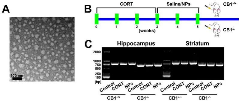
Cur/SLNs-HU-211 were composited according to a method as described previously [13]. Shape of nanoparticles was pictured with a TEM.
CBR1 knockout mice were obtained from BIORAY LABORATORIES Inc (Shanghai, China) and raised in the Laboratory Animal Centre of Tongji University. These experiments were authorized by the institutional research ethics committee of Tongji University, and strictly followed the National Institutes of Health Guide for the Use and Care of mice.
Mice were randomly assigned to six groups: (1) Normal mice (CB1+/+) received saline; (2) Normal mice (CB1+/+) injected with Corticosterone (CORT) only; (3) Normal mice (CB1+/+) received CORT and Cur/SLNs-HU-211 nanoparticles (NPs); (4) CBR1 knockout mice (CB1-/-) received saline; (5) CBR1 knockout mice (CB1 -/-) received with CORT only; (6-/-) CBR1 knockout mice (CB1+/+) received CORT and Cur/SLNs-HU-211 nanoparticles.
Model of depression was obtained with injection of CORT (40 mg/kg) for 21 days [14]. 14, 15 Then NPs were given for 2 weeks at a curcumin. Mice genotype were identified by using Direct Mouse Genotyping Kit bought from APExBIO Technology (Houston, USA), mice hippocampus and striatum of different group were used to confirm the genotype of CB1+/+ and CB1-/- mice.
Behavioral tests
To assess the impact on motor function, the mice were distributed to the Forced Swimming Test (FST) and the Pole test. 16-18 For FST, mice were well dropped into a beaker within water, all recorded. In the final 6 min of 8 min, float time were calculated. In the pole test, mice start from the top of the pole (0.8 cm in diameter; 60 cm in height) and the time of climbing down the pole was counted, as described before. All experiments were done 3 times and double blind.
DA and NE detection
Mice hippocampus and striatum were collected.Samples were immersed in 400 μL PBS and homogenized, mixing with 800 μL of acetonitrile, and then centrifuged. 1920 μl samples were automatically injected onto HPLC. The mobile phase used were 10% methanol and 90% 0.01 M KH2PO4 (pH 3.5) at a flow velosity of 500 μL/min. At 280 nm wavelength, the concentrations of dopamine (DA) were measured. For norepinephrine (NE), 60% methanol and 40% 0.05 M CH3COONa and a detection wavelength of 275 nm were applied. The protein concentrations in each areas of brain were quantified.
qRT-PCR
qRT–PCR was performed according to the standard protocols.
Western blot
Western blot was executed with a standard protocol. Antibodies specific to NeuN (ab104225, abcam), MAP2 (ab11267, abcam), Tuj1 (ab78078, abcam), CB1 (ab23703, abcam), RASGEF1C (abs139329, absin), MEK (ab32091, abcam), p-MEK (ab214445, abcam), ERK (ab184699, abcam), p-ERK (ab32538, abcam), Egr1
(ab133695, abcam) and β-actin (ab8227, abcam) were used.
Immunofluorescence
Immunofluorescence analysis was performed using standard protocols. Antibodies specific to NeuN (1:500,
ab104225, abcam), appropriate fluorescently tagged secondary antibodies and DAPI were used.
RNA-seq
We collected hippocampus of six groups and carried out RNA sequencing, total RNA was extracted with TRIZol Reagent (Takara). RNA sequencing was performed by BGI., Shenzhen, China.
Exprimental results were presentedas means ± standard deviation (SD). One-way ANOVA was performed for each analysis, and P<0.01 was statisticallysignificant.
Results
Preparation of nanoparticles and mice
We initially observed Cur/SLNs-HU-211 nanoparticles via a TEM (Fig. 1A), TEM analysis confirmed that our synthesis was successful and nanoparticles was in spherical shape. As shown by the schematic (Fig. 1B), normal mice (CB1+/+) and CBR1 knockout mice (CB1-/-) were given CORT and Cur/SLNs-HU-211 nanoparticles, CORT were used to modeling MDD, further experiments were carried out to test whether Cur/SLNs-HU-211 nanoparticles benefit CORT treated CB1+/+ and CB1-/- mice, respectively. Six group of mice hippocampus and striatum were collected and genotype of each mice were identified,PCR amplification was performed on a thermal cycler, yielding a 804-bp fragment from homozygous CB1+/+ mice and a 628-bp fragment from homozygous CB1-/- mice (Fig. 1 C).
Cur/SLNs-HU-211 promote motor function recovery from MDD only for CB1+/+ mice
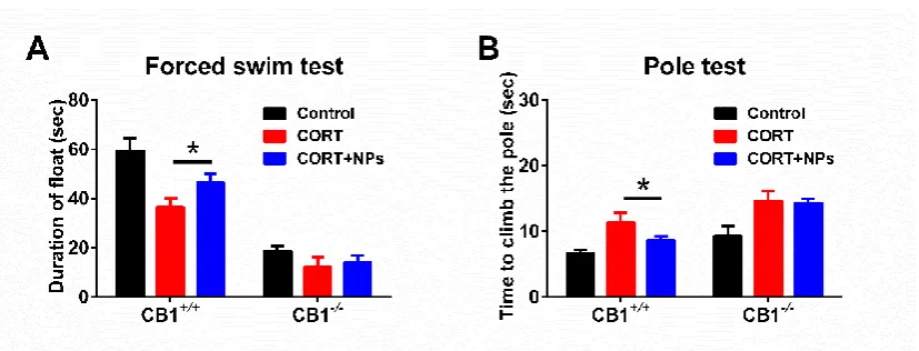
Mice behavioral tests can be a useful tool for evaluating the curative effect of drugs. In our study, we applied FST and Pole Test to evaluate mice motor function improvement. FST results (Fig. 2A) suggested that Cur/SLNs-HU-211 could greatly improve CORT treated CB1+/+ mice float time in water from 36.5 sec to 46.5 sec, however no such improvement has occurred in CB1-/- mice.
In order to measure bradykinesia,we implemented the Pole Test, results (Fig. 2B) shown that the CORT group mice took longer to climb down the pole compared with the control. Treatment of the nanoparticles group (8.7 sec) reduced climbing times compared with CORT group (11.3 sec) in CB1+/+ mice, which means they are much faster. For CB1-/- mice, there were no significant difference between CORT and nanoparticles group.
The results show that Cur/SLNs-HU-211 nanoparticles can just effectively reversed CORT-induced motor deficits in CB1+/+ mice not CB1-/- mice.
Cur/SLNs-HU-211 increases DA and NE level in MDD CB1+/+ mice
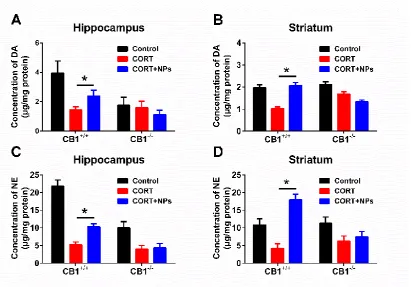
Harvest mice hippocampus and striatum for HPLC analysis, DA and NE level were detected. As shown in Fig. 3A,3B, there was a reduction in DA levels after CORT administration. for CB1-/- mice, nanoparticles treated animal exhibited no increase compared with CORT group mice in DA level. For CB1+/+ mice, nanoparticles could remarkably enhance the DA release of CORT treated mice both in hippocampus and striatum.
The NE level were also measured, the results (Fig. 3C,3D), CORT induced significantly decrease of NE both in hippocampus and striatum, and this decrease was reversed when given with Cur/SLNs-HU-211 in CB1+/+ mice. When it comes to CB1-/- MDD mice, Cur/SLNs-HU-211 induced no significantly higher NE level.
This recovery property of Cur/SLNs-HU-211 on CB1+/+ MDD mice in DA and NE levels indicated that Cur/SLNs-HU-211 could significantly elevate the levels of monoamine neurotransmitter in MDD mice, and those recovery would not happen when knock out CBR1.
qRT-PCR
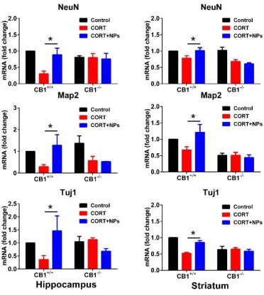
To study the impact of Cur/SLNs-HU-211, we analyzed the expression of neuronal-specific genes (NeuN, Map2, Tuj1). It could be seen from qRT-PCR that the mRNA levels of NeuN, Map2 and Tuj1 in hippocampus and striatum (Fig. 4) were reduce drastically in MDD mice following CORT injection compared to control group. After the administration of Cur/SLNs-HU-211, the levels of NeuN, Map2 and Tuj1 mRNA relative to the CB1+/+ MDD mice
increase significantly. And for CB1-/- MDD mice, Cur/SLNs-HU-211 lose this function and could not promote the
increase of these mRNA level. The results were in accordance with that of behavioral test and neurotransmitter detection. qRT-PCR results demonstrated that Cur/SLNs-HU-211 could only benefit CB1+/+ MDD mice by
Western blot
The expressions of NeuN, MAP2 and Tuj1 were also determined by western blot assay.Consistent with the result of qRT-PCR, protein expression levels of the mature DA neuronal marker NeuN, MAP2 and Tuj1were significantly increased in Cur/SLNs-HU-211 administrated CB1+/+ MDD mice (Fig. 5A). In Cur/SLNs-HU-211 treated CB1-/- MDD mice, there were no distinction as compare to CB1-/- MDD mice. Both the western blot bands
and densitometry quantification analysis (Fig. 5B) verified that in hippocampus and striatum. Immunofluorescence
To analysis neuronal survival, we applied NeuN as a Neuronal Biomarker, as shown (Fig. 6A). Compared to the CB1+/+ and CB1-/- Control group, CORT led to remarkable neuronal cell death and few NeuN positive cells were
observered in the hippocampus and striatum of mice. However, there were more neurons observed in CB1+/+ MDD
mice following treatment of Cur/SLNs-HU-211, but less NeuN positive cells were detected in CB1-/- MDD mice.
We quantify the intensity of NeuN fluorescence and found that the results (Fig. 6B) were consistent with qRT-PCR andWestern blot.
RNA-seq
To explore the underlying molecular mechanisms in more details, we employed transcriptome sequencing to examine the differential gene expression aiming to reveal in vivo working system.
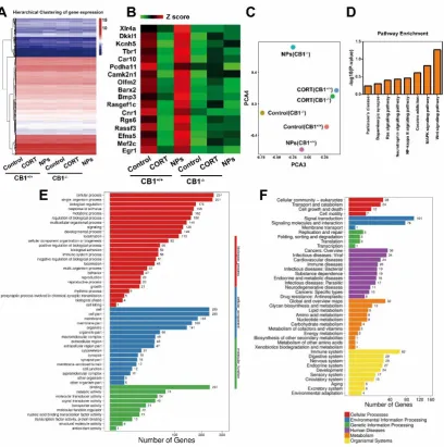
A heat map presenting mRNA expression profiles of all samples was shown (Fig. 7A). There were 505 marked differential expressed genes between Cur/SLNs-HU-211 and MDD CB1+/+ group, we listed the most relevant 17
genes (Fig. 7B), these genes are mostly neurodevelopment concerned.
2D principal component analysis (2D PCA) for all of the samples of all genes clearly separates NPs (CB1-/-)
group the farthest from Control (CB1+/+) group, NPs (CB1+/+) group the closest (Fig. 7C). We recognized 8 related
KEGG pathways from Cur/SLNs-HU-211 treated CB1+/+ group versus MDD CB1+/+ group (Fig. 7D). GO analysis
revealed that NPs (CB1+/+) group compared with CORT (CB1+/+) group differ most in biological process and cellular
component (Fig. 7E). KEGG results suggested that NPs (CB1+/+) group compared with CORT (CB1+/+) group may
have big difference in human diseases and organismal systems. Molecular mechanisms
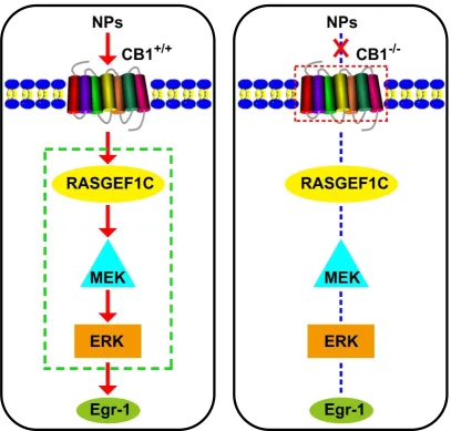
Among the 9 pathways with significant differences, we focused on MAPK pathway [15,16],and then we examined these genes by qRT-PCR (Fig.8A) and western blot assay (Fig.8B), quantification of western blot was also carried out (Fig.8C). The results of qRT-PCR (Fig.8A) showed that in nanaparticles treated CB1+/+ mice, the CB1,
Rasgef1c and Egr1 mRNA expression were increased compared with CB1+/+ MDD mice, and for CB1-/- mice the
expression of these genes was low and nanaparticles treated mice show no difference with MDD mice. We further investigated protein expression with western blot (Fig.8B, 8C), CB1, RASGEF1C, Egr1, together with phosphorylation of MEK and ERK were up-regulated in Cur/SLNs-HU-211 treated CB1+/+ MDD mice as compared
to CB1+/+ MDD mice, no significantly change observed in CB1-/- mice. The total protein of MEK and ERK were
almost in a same level in all group.
Discussion
CORT is a commonly used inducer of depression-like symptoms in mice. CORT model could be used to explore the underlying mechanisms of MDD and to verify novel chemical compounds for their neuroprotective effects
[17,18].Our research analysed the neuroprotective and MDD rehabilitation effects of nanaparticles in CORT
induced MDD CB1+/+ and CB1-/- mice.
Nanoparticles can influence neuron survival, neurotransmitters release, apoptosis and behavioral response
trigger gene-transcription networks crucial for the viability of cell, and Cur/SLNs-HU-211 mainly act through targeting CBR1. These findings allowed us to investigate whether Cur/SLNs-HU-211 can only improve the MDD condition in the presence of CBR1. So, we knock out CBR1 in C57BL/6 mice and compared the function of Cur/SLNs-HU-211 in MDD CB1+/+ and CB1-/- mice.
The in vivo test indicated that treatment of Cur/SLNs-HU-211 remarkably improved motor function recovery in CORT treated CB1+/+ mice, however, no such effects has shown in CB1-/- mice. Neurotransmitters like DA and
NE in hippocampus and striatum were also evaluated after Cur/SLNs-HU-211 treated for CB1+/+ mice only. In our
study, qRT-PCR and western blots assay all suggested that CORT-induced MDD mice exhibited a reduction of CB1 three weeks after receiving CORT, which indicate that CBR1 are essential for protection against CORT toxicity. In CB1+/+ mice hippocampus and striatum, we found that administration of nanoparticles could increase the expression
of NeuN, MAP2 and Tuj1, however, there were no significantly changes between Cur/SLNs-HU-211 and CORT administrated CB1-/- mice. Immunofluorescence analysis also verified that NeuN positive cells will increased in
Cur/SLNs-HU-211 treated CB1+/+ MDD mice hippocampus and striatum, these results not apply to CB1-/- mice. All
of this revealed that CBR1 is a must for Cur/SLNs-HU-211 exerted its neuroprotection and antidepressants effects. To better understand the mechanism involved in, we carried out RNA-seq, results suggested that MAPK signal pathway may be a key pathway, we applied qRT-PCR and western blot to verify. We found that the MEK/ERK signaling pathway was involved in and the neuroprotective effects of Cur/SLNs-HU-211was regulated by an enhanced expression of RASGEF1C/Egr1. Importantly, MEK/ERK signaling pathway could only activated in CB1+/+ MDD mice. Dueling to the knockout of CBR1, Cur/SLNs-HU-211 could not show its neuroprotective effects
in CB1-/- MDD mice for the pathway was blocked and thus there were no significantly difference compared with
CORT treated CB1-/- mice.
Taken together (Fig.9), our findings suggest that CBR1 is a necessary for Cur/SLNs-HU-211 to exerted its neuroprotection and antidepressants effects, and Cur/SLNs-HU-211 nanoparticles may provide a novel medicinal strategy for MDD and other neurodegenerative diseases.
Acknowledgments
This work was financially supported by the National Natural Science Foundation of China (Grant No. 81873994, 81671105, 31727801,31770923, 31700735, 31570849), the National key research and development program (Grant No. 2016YFA0100800), the Fundamental Research Funds for the Central Universities, and the Funds for International Cooperation and Exchange of the National Natural Science Foundation of China (Grant No. 8182010801).
Disclosure Statement
Reference